Gold Nanoparticles as Drug Delivery Agents. Detoxifying the Chemotherapeutic Drug Cisplatin
116
0
0
Texto completo
(2) Universitat Autònoma de Barcelona Departament de Bioquímica i Biologia Molecular. Gold Nanoparticles as Drug Delivery Agents. Detoxifying the Chemotherapeutic Drug Cisplatin. Joan Comenge Farré Inorganic Nanoparticles Group Institut Català de Nanotecnologia. Maig 2013.
(3) Gold Nanoparticles as Drug Delivery Agents. Detoxifying the Chemotherapeutic Drug Cisplatin. Memòria presentada per. Joan Comenge Farré. Per optar al grau de. Doctor per la Universitat Autònoma de Barcelona Programa de Doctorat en Bioquímica, Biologia Molecular i Biomedicina. Tesi realitzada sota la direcció del Dr. Víctor Franco Puntes, Inorganic Nanoparticles Group, Institut Català de Nanotecnologia, i amb la tutoria de la Dra. Ester Boix Borràs, Departament de Bioquímica i Biologia Molecular, Universitat Autònoma de Barcelona.. Cerdanyola del Vallès, Maig 2013.
(4)
(5)
(6)
(7) Prologue In this thesis dissertation, the use of gold nanoparticles as agents for delivery of cisplatin is discussed. The book has been organized following a logic sequence that starts by the synthesis of gold nanoparticles, followed by their functionalization, first with surfactants and then with the drug to finally analyze the performance of these drug delivery vehicles in in vitro and in in vivo experiments. Every chapter has been structured so to allow its individual lecture. Although aspects applicable to other drug delivery systems such as the colloidal and chemical stability in physiological media, the drug release profile, etc. are discussed here using our system as an example, this work does not pretend to be a general discussion about the state of the art in nanomedicine and/or drug delivery, but a contribution to these growing areas. However, I encourage the lecture of some of the very interesting references provided here to achieve a deeper understanding in specific aspects. This multidisciplinary work could have not been possible without the contribution and advice of a great number of people. Thus, I feel the obligation to acknowledge all those that directly contribute to this work especially all the members of the Inorganic Nanoparticles Group, led by Prof. Víctor Puntes, for their invaluable help and knowledge, but also Prof. Fernando Domínguez and Carmen Sotelo from school of medicine at Santiago de Compostela, prof. Franciso Romero from Molecular Science Institute at València and Dr. Óscar Gallego and Prof. Agustí Barnadas from the Santa Creu I Sant Pau Hospital at Barcelona for their active contribution and discussions about this work. Last but not least, my gratitude to all the members of the Radiation Biology Group lead by Prof. Kevin Prise in the Centre of Cancer Research and Cell Biology at Belfast for receiving me and teaching me the little I know about this field. Also, a special mention goes to Nanotargeting S.L. for trusting in this project. It is impossible to extend here my gratitude to all of those that indirectly contribute to the work so I will let you know one by one personally. Just be patient. i.
(8) Chapter 1 Introduction. Many of the conventional therapies can be improved through the use of drug delivery systems (DDS). They are designed mainly to modify the pharmacokinetics and biodistribution of small molecular drugs. This is of special importance in the case of anticancer therapies in which a widespread distribution of small molecular chemotherapeutic drugs is often limiting treatments due to severe side effects that make impossible to reach the full benefits of the therapy. In this context, nanotechnology emerges as a disruptive technology to design carriers that improve the delivery of drugs to their target organs.. 1.1 Engineering AuNPs as scaffolds for drug delivery Although a plethora of nanoparticles (NPs) including polymeric NPs, liposomes, dendrimers, metallic NPs, etc. have been used to design nanocarriers for drug delivery, 1, 2 we will focus the discussion on the use of gold nanoparticles (AuNPs). The synthesis of AuNPs have been first described by J. Turkevich at 1954,3 but it was not until last years that advances on the engineering of these NPs allowed researchers to control the size, shape, and functionalization of these nanocarriers and thus starting using them as scaffolds for drug delivery. Thus, new synthetic protocols make possible to achieve biocompatible spherical AuNPs from 5 to 200 nm with a monodispersity lower than 10 %.4 The control over the size is a key point in the use of AuNPs in biomedicine since it influences important biological properties such as. interaction with. proteins,. biodistribution and clearance rate. The variety of sizes of AuNPs that can be easily obtained facilitated a better understanding of the role played by this property in 1.
(9) Chapter 1. important parameters for cancer treatment with NPs such as accumulation in tumors, blood half-life, or penetration. From a simplistic point of view, one could say that the final fate of the NPs is strongly influenced by their size since smallest AuNPs (<5 nm, core + surfactant) are rapidly cleared up by the kidneys and the the largest ones (>100 nm) are removed easily from the circulation by the immune system.5 Sizes in between showed the best accumulation and penetration in tumors; at this range of sizes other parameters such as the presence of stealthing agents or different surface charge should be tuned case by case to achieve the desired behavior. In addition, shape of the AuNPs is also important. Spherical NPs are usually the chosen option when they wanted to be used just as carriers due to the simple synthesis and easy functionalization. However, other shapes such as nanorods or nanoboxes have been used too. These advanced AuNPs have special physicochemical properties that make them appealing to be used not only as vehicles but also as effectors by themselves. They present a surface plasmon resonance (SPR) in the near infrared (NIR) region, which make them suitable to be used in photothermal therapy in which NIR light is absorbed by NPs, delivering toxic amounts of heat.6 In this type of therapy, working with wavelengths that can penetrate deep into tissue such as IR is required and only non-isotropic AuNPs present a SPR at this region. Regarding functionality, chemistry on gold planar surfaces has been extensively studied to obtain molecular Self-Assembled Monolayers (SAMs).7 Thus, this technology has been easily adapted to functionalize AuNPs in solution. However, some considerations need to be taken into account for NPs: the curvature radius may play a role in the determination of important properties of the molecules conjugated to small NPs such as apparent pka 8 and the coverage density9 of the attached ligands amongst others. Also, AuNPs need to be stabilized during the synthesis to avoid aggregation by means of conjugation to a surfactant. Thus synthetic protocols in which the surfactant is weakly bound to the NPs (e.g. sodium citrate), and therefore can be easily exchanged by other ligands, are recommended for further functionalization of these AuNPs.4 Note that physiological conditions (high ionic strength) are harsh for citrate-capped AuNPs. Conjugation may also result in destabilization of NPs if special care is not taken.10 AuNPs show affinity to different chemical groups such as phosphates, carboxylic acids, ammines and specially to thiols. Thus, one can found in the literature AuNPs conjugated to a great variety of molecules including alkanethiols, polymers, proteins, amino acids, and drugs amongst others.11 Interestingly, the control over the conjugation facilitates engineering multifunctional AuNPs. Hence, it would be possible to create a nanocarrier that contains, for example, a molecule to stealth the DDS from the immune system, a ligand 2.
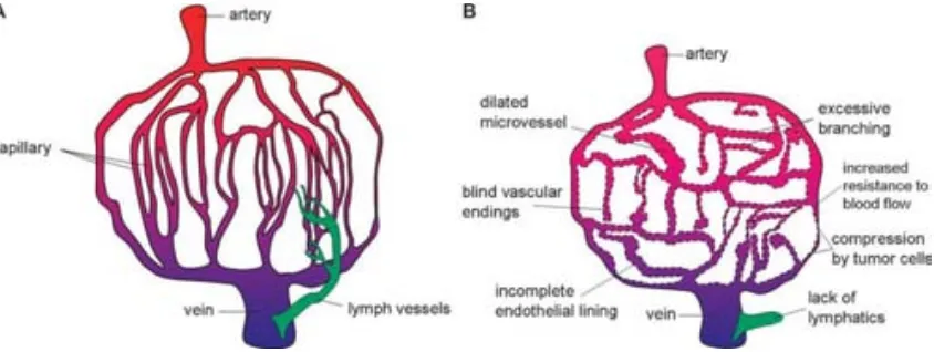
(10) Introduction. that promotes uptake by specific cells, and the drug which in its turn can be attached via a responsive link which release the drug only after a specific response such as a pH drop. Last but not least, the greater monodispersity of AuNPs compared to other metallic or polymeric NPs is another advantage of using this type of NPs. 1.2 Nanoparticles for anticancer therapy: Advances and challenges of a new therapy NPs are perfect candidates to be used in anticancer therapy since they showed passive accumulation in tumors due to the Enhanced Permeation and Retention effect (EPR). This effect first reported by Matsumura and Maeda12 exploits the ability of NPs to permeate through the leaky tumor vessels (with fenestrations between 100 and 800 nm 13, 14) and to be accumulated into the tumor due to the lack of a functional lymphatic system (Figure 1.1). Long-time circulating NPs maximize the EPR effect since the changes to permeate through the tumor vessels are greater. Related to this, the size of NPs plays an important role in determine tumor accumulation and distribution: 60nm AuNPs showed a greater accumulation than 20 nm AuNPs likely due to a slower clearance. However, larger AuNPs are accumulated in the perivascular region of the tumor failing to penetrate deeper, whilst 20 nm AuNPs were still found significantly at 50 m from the blood vessel.14. Figure 1.1. Scheme showing the differences of the vascular system of normal tissue (A) and tumor (B). Leaky vessels and non-functional lymphatic system are responsible for the passive accumulation of NPs into tumors. Adapted from reference 15.15. The intrinsic property of NPs to be passively accumulated into tumors has been exploited to deliver a great variety of drugs.1 Some groups proposed to improve further this accumulation by attaching a ligand recognized by receptors overexpressed in tumor cells. 3.
(11) Chapter 1. These include epidermal growth factor (EGF)16, folate17 and transferrin18 amongst other. However, there is some controversy regarding the efficiency of this approach.19 It is not clear that active targeting favors the accumulation in tumor, since physicochemical properties of NPs govern the pharmacokinetics properties of DDS. Thus, size and surface charge and composition (e.g. presence of stealthing agents or not) of NPs seems to play a more important role in determining final distribution of DDS20 and ligands influence only in interactions directly onto the cell once the cell is reached (e.g. a greater uptake) Once the NPs are localized in the tumor, different strategies have been used to release the drug. Normally, the strategies are based on the different physicochemical properties found in the cytoplasm compared to the extracellular environment (e.g. blood). For example, NPs are known to be internalized via endocytic pathways, in which a pH drop is produced.21 Thus, a pH-sensitive link between drug and NPs ensures non-specific release of drug during the circulation through the body.22 A controlled release can be also achieved by the reduction of an oxidized inactive prodrug in the cytoplasm, releasing the drug in its active form.23 Also one can take advantage of specific enzymes to cleave the link between NP and drug.24 The metallic core of AuNPs conjugates can also act as an effector in thermotherapy as explained above. In addition, AuNPs have been recently proposed as radiosensitizers due to the induction of an Auger cascade after an ionizing event localized in the vicinity of the AuNP surface. These special properties open up the possibility to strategies that use AuNPs as carrier and effector. Thus the use of AuNPs as carriers for drug delivery might provide several advantages including: . Increased blood half-life of the small drug. . Accumulation in the tumor via the EPR effect. . Reduced toxicity in normal tissue. . Controlled release of the drug, delivering high payloads directly to the site of action. . Possibility of multiple functionalization which allow combined therapy and/or tuning blood half-life, immune responses toward the vehicle, tumor uptake, etc.. . Increase the solubility of poorly soluble drugs in plasma. . Protection of the drug from immune system and from agents present in the plasma that might deactivate the drug. 4.
(12) Introduction. . Promote a double effect by acting as carriers and as effectors by themselves. However, the use of AuNPs for drug delivery is not without problems and some aspects should be considered: Classical syntheses of AuNPs render concentrations of AuNPs in the range of 10 12-1013 NP/mL,25 thus AuNPs are typically synthesized at the nanomolar scale. Even though high densities of loading have been achieved,26,. 27. concentrations of drugs onto NPs as. synthesized will rarely be higher than 10 micromolar. Taking into account that there is a limitation of volume that can be injected into the body (e.g. 10 mL/Kg for intravenous injection in mice), the therapeutic dose might not be reached unless NPs had been previously concentrated. This concentration step is not always straightforward because concentration of AuNPs could lead to aggregation if special care is not taken (e.g. concentration of AuNPs without any surfactant increase the chances of aggregation). Note that colloids are intrinsically out of equilibrium and they tend to minimize surface energy when this is possible either via functionalization or aggregation. The need of concentration of AuNPs to reach therapeutic doses implies that a single labscale synthesis might not be enough to achieve the amount of NPs needed to perform in vivo experiments. Thus, not only monodispersity should be guaranteed to control size effects, but also reproducibility batch to batch. In addition, robust synthesis protocols will also facilitate the scale up of the production, which is essential for the transference of the technology to the industry.28 The special properties of AuNPs are lost if they aggregate. In too many cases the loss of colloidal stability is behind the lack of (or unexpected) biological behaviors. 20 Therefore, one has to ensure the maintenance of colloidal stability along the process (synthesis – storage – exposure/treatment). It is common to assay the stability of AuNPs in the storage conditions (e.g. in aquose media) even if they are different from the working conditions. However, biological fluids such as cell culture media or blood are complex mixtures of salts, proteins, sugars, etc. that may promote destabilization of AuNPs, and therefore stability should be assayed in this media. The same consideration is valid for the stability of the link with the drug. For example, cisplatin adsorbed on AuNPs is not released in aquose media, whilst it is unspecifically quickly released in cell culture media. On the other hand, cisplatin linked via coordination bond is stable in both media and only released after a pH drop.26. 5.
(13) Chapter 1. The choose of appropriate models to determine the potential therapeutic benefits is also not trivial, specially for new technologies in which the amount of literature is limited. In vitro simple test such as those assessed in monolayer cell cultures do not consider important factors such as tumor vascularity, penetration and other differential properties given by the tumor microenvironment.15 Consequently, in vitro results do not necessarily reflect the behavior of the assayed DDS in a real case. In this context, 3D cell cultures have been proposed as models to study the behavior of drugs in the particular environment of solid tumors due to the possibility to have an extracellular matrix and different regions of tumor (e.g. hypoxic cells in the intern areas). Since the advantages of using DDS are achieved mainly by modification on the pharmacokinetic properties of the drug, in vivo models are preferred. However, there is a great variety of tumor models including xenografted, orthotopic, and spontaneously and induced autochthonous tumor models.29 This models show variations between them such as different size of porous in vessels or different immunological responses which may result in different efficiency of the same DDS depending on the model that has been used. Finally, treatments with AuNPs are still very few and not beyond clinical trials. Thus, there is a lack of knowledge about regulatory aspects. Possible adverse effects of NPs such as acute toxicity and longtime accumulation should not be underestimated and should be studied case by case. There is a controversy regarding the in vivo toxicity of NPs and the parameters that play a role in the NPs-induced toxicity.30 Some works have found no toxic signals after AuNPs administration31 or small alterations in the biochemical markers due to metabolization of the AuNPs or indicating a temporal inflammation.32 On the other hand, it is believed that the disfunction of major organs can be related to the presence of NPs at the site of abnormalities. For that reason, there are a lot of studies regarding toxicity in spleen and liver, which are generally accepted to be the organs with the highest accumulation of NPs. The liver toxicity, when found, seems to be associated to an hyperplasia of Kupffer cells that induce an acute inflammation with neutrophils influx. 33 This acute inflammation is a transient response due to the insult of AuNPs, however apoptosis and necrosis of hepatocytes as well as accumulation of AuNPs could be related to toxic effects.34 Regarding the spleen, macrophages of the periphery seem to be involved in the uptake of NPs by this organ. Thus, leading to a temporal inflammation of the spleen, however in other cases a loss of weight has been observed after intravenous administration.35 White pulp aberration has been also observed.36 Any general trend cannot be extracted from the current literature, but it is clear that there are some parameters that have to be taken into account before using AuNPs as a vehicle for medical applications: i. Size of the vehicle, since it will determine the clearance rate and the 6.
(14) Introduction. biodistribution. 36;. ii. Surface composition, it has been seen that by changing the surface. composition of AuNPs the toxicological profile might be different37; iii. Dose and administration route obviously will play a role in the potential toxicity. Therefore for every AuNP-conjugate, a toxicology study must be done to prove their usability in medical applications.. 1.3 Cisplatin as anticancer drug A paradigmatic case of anticancer drugs are platinum compounds. Cisplatin or cisdiamminedichloroplatinum(II), [PtCl2(NH3)2], was originally synthesized in 1845, but not until 1970 was its antitumor activity established.38 Today cisplatin is used to treat various types of cancers (i.e., non-small-cell lung cancer, ovarian cancer, germ cell tumors, osteosarcomas, etc), with a cure rate as high as 90% in testicular cancer. 39 After both passive and active cellular uptake, the platinum complex reacts in vivo to form adducts with DNA, which ultimately trigger apoptosis.40 However, chronic cisplatin usage results in resistance by several possible mechanisms including increased interactions with metallothioneins and glutathione, which deactivate the drug, as well as increased DNA repair and/or cisplatin efflux.41 To counteract resistance and its rapid renal clearance, which lowers the efficiency of cisplatin significantly, very high systemic doses of cisplatin should be administered. Unfortunately, such high dose of cisplatin results in severe systemic toxicity and poor patient wellfare, including nausea/vomiting, renal toxicity, gastrointestinal toxicity, peripheral neuropathy, asthenia, and ototoxicity, thus limiting its clinical use.41, 42 Of all the toxicities induced by cisplatin, nephrotoxicity is considered to be the dose-limiting factor.40 Such side effects make it impossible to achieve the full benefit of the treatment in a large number of patients.43 In humans, the cisplatin treatment generally involves series of intravenous injections administered every 3-4 weeks at a dose of 50-120 mg/m2 (1.2 – 2.7 mg/Kg). In addition to the undesired side effects, there is also a loss of drug activity in the body associated with poor circulation and poor delivery to the tumor, as well as deactivation mechanisms that irreversibly alter the chemistry of these molecules before reaching the tumor cells.42 Since its discovery, many attempts to find derivatives has been carried out looking for both reduced side effects and modified body distribution (in order to target different organs), rather than improving cisplatin efficacy.41,. 44. Here, second-generation platinum drugs as carboplatin and oxaliplatin. represent an improvement in some cancer treatments, as lung and colorectal respectively, although the limitations observed for cisplatin have not been entirely overcome40, 41.. 7.
(15) Chapter 1. 1.4 Carrying cisplatin Hence, recent efforts have been focused on targeting the tumor by using drug delivery systems to avoid the organs to which cisplatin is toxic. As the kidney is responsible for filtration and removal from the blood of molecules smaller than 50 KDa, which corresponds to molecular sizes of around 6 nm in diameter, any larger delivery vehicle will divert the drug away from the kidney.45 In addition, more than 90 % of the administrated cisplatin is known to bind irreversible to albumin, which deactivates the drug.46 Thus, a properly designed nanocarrier will not only transport the cisplatin to the tumor, but also protect it against plasma deactivation. In this context, approaches based on the encapsulation and transport of cisplatin have emerged recently. Sterically stabilized polymeric nanoparticles, having excellent stability in plasma, a much longer circulation time, better efficacy, and lower toxicity than free cisplatin have been reported.47-50 As the case. of. lipid. capsules51. or. polymers,. as. in. Prolindac®,. a. 22. kDa. hydroxypropylmethacrilamide copolymer as a backbone and then a glycine chelator linker which is pH sensitive.52 Other examples include soluble CNTs,53 carbon nanohorns,54 and Fe3O4 NPs.55 Similarly, CytImmune corp. is developing AuNPs as carrier for TNF-a and doxorubicin. In a close related work, Ren et al.56-58 reported the adsorption of commercial cisplatin to gold colloids via ionic interactions. In the case of adsorption via ionic interactions on the surface of the nanomaterials, a non-controlled rapid liberation of the drug is observed as soon as the conjugates are dispersed in highly ionic media as serum.26. 1.5 References (1) Davis, M. E.; Chen, Z.; Shin, D. M., Nanoparticle therapeutics: an emerging treatment modality for cancer. Nat Rev Drug Discov 2008, 7, 771-782. (2) Allen, T. M.; Cullis, P. R., Drug Delivery Systems: Entering the Mainstream. Science 2004, 303, 1818-1822. (3) Turkevich, J.; Garton, G.; Stevenson, P. C., The Color of Colloidal Gold. Journal of Colloid Science 1954, 9, S26-S35. (4) Synthesis of Citrate-Stabilized Gold Nanoparticles of up to 200 nm: Size Focusing versus Ostwald Ripening. Langmuir 2011, 27, 11098-11105. (5) Sperling, R. A.; Casals, E.; Comenge, J.; Bastus, N. G.; Puntes, V. F., Inorganic Engineered Nanoparticles and Their Impact on the Immune Response. Current Drug Metabolism 2009, 10, 895-904. (6) Jain, P. K.; El-Sayed, I. H.; El-Sayed, M. A., Au nanoparticles target cancer. Nano Today 2007, 2, 18-29. (7) Love, J. C.; Estroff, L. A.; Kriebel, J. K.; Nuzzo, R. G.; Whitesides, G. M., Self-Assembled Monolayers of Thiolates on Metals as a Form of Nanotechnology. Chemical Reviews 2005, 105, 1103-1170. 8.
(16) Introduction. (8) Wang, D.; Nap, R. J.; Lagzi, I. n.; Kowalczyk, B.; Han, S.; Grzybowski, B. A.; Szleifer, I., H W p ’ v R Apparent pKa of the Coating Ligands. J. Am. Chem. Soc. 2011, 133, 2192-2197. (9) Xia, X.; Yang, M.; Wang, Y.; Zheng, Y.; Li, Q.; Chen, J.; Xia, Y., Quantifying the Coverage Density of Poly(ethylene glycol) Chains on the Surface of Gold Nanostructures. ACS Nano 2011. (10) Ojea-JimeÌ•nez, I.; Puntes, V., Instability of Cationic Gold Nanoparticle Bioconjugates: The Role of Citrate Ions. J. Am. Chem. Soc. 2009, 131, 13320-13327. (11) Eugenii, K.; Itamar, W., Integrated Nanoparticle-Biomolecule Hybrid Systems: Synthesis, Properties, and Applications. Angewandte Chemie International Edition 2004, 43, 6042-6108. (12) Matsumura, Y.; Maeda, H., A New Concept for Macromolecular Therapeutics in Cancer Chemotherapy: Mechanism of Tumoritropic Accumulation of Proteins and the Antitumor Agent Smancs. Cancer Res 1986, 46, 6387-6392. (13) Vasir, J. K.; Reddy, M. K.; Labhasetwar, V. D., Nanosystems in drug targeting: Opportunities and challenges. Current Nanoscience 2005, 1, 47-64. (14) Perrault, S. D.; Walkey, C.; Jennings, T.; Fischer, H. C.; Chan, W. C. W., Mediating Tumor Targeting Efficiency of Nanoparticles Through Design. Nano Letters 2009, 9, 1909-1915. (15) Tredan, O.; Galmarini, C. M.; Patel, K.; Tannock, I. F., Drug Resistance and the Solid Tumor Microenvironment. J. Natl. Cancer Inst. 2007, 99, 1441-1454. (16) Tseng, C.-L.; Su, W.-Y.; Yen, K.-C.; Yang, K.-C.; Lin, F.-H., The use of biotinylated-EGFmodified gelatin nanoparticle carrier to enhance cisplatin accumulation in cancerous lungs via inhalation. Biomaterials 2009, 30, 3476-3485. (17) Hilgenbrink, A. R.; Low, P. S., Folate receptor-mediated drug targeting: From therapeutics to diagnostics. Journal of Pharmaceutical Sciences 2005, 94, 2135-2146. (18) Choi, C. H. J.; Alabi, C. A.; Webster, P.; Davis, M. E., Mechanism of active targeting in solid tumors with transferrin-containing gold nanoparticles. Proceedings of the National Academy of Sciences 107, 1235-1240. (19) Pirollo, K. F.; Chang, E. H., Does a targeting ligand influence nanoparticle tumor localization or uptake? Trends in Biotechnology 2008, 26, 552-558. (20) Ruenraroengsak, P.; Cook, J. M.; Florence, A. T., Nanosystem drug targeting: Facing up to complex realities. Journal of Controlled Release 2010, 141, 265-276. (21) Nel, A. E.; Madler, L.; Velegol, D.; Xia, T.; Hoek, E. M. V.; Somasundaran, P.; Klaessig, F.; Castranova, V.; Thompson, M., Understanding biophysicochemical interactions at the nano-bio interface. Nat Mater 2009, 8, 543-557. (22) Comenge, J.; Sotelo, C.; Romero, F.; Gallego, O.; Barnadas, A.; Parada, T. G.-C.; Domínguez, F.; Puntes, V. F., Detoxifying Antitumoral Drugs via Nanoconjugation: The Case of Gold Nanoparticles and Cisplatin. PLoS ONE 2012, 7, e47562. (23) Dhar, S.; Gu, F. X.; Langer, R.; Farokhzad, O. C.; Lippard, S. J., Targeted delivery of cisplatin to prostate cancer cells by aptamer functionalized Pt(IV) prodrug- L A– E nanoparticles. Proceedings of the National Academy of Sciences 2008, 105, 17356-17361. (24) de la Rica, R.; Aili, D.; Stevens, M. M., Enzyme-responsive nanoparticles for drug release and diagnostics. Advanced Drug Delivery Reviews 2012, 64, 967-978. (25) Daniel, M. C.; Astruc, D., Gold nanoparticles: Assembly, supramolecular chemistry, quantum-size-related properties, and applications toward biology, catalysis, and nanotechnology. Chemical Reviews 2004, 104, 293-346. (26) Comenge, J.; Romero, F. M.; Sotelo, C.; Dominguez, F.; Puntes, V., Exploring the binding of Pt drugs to gold nanoparticles for controlled passive release of cisplatin. Journal of controlled release : official journal of the Controlled Release Society 2010, 148, e31-2. (27) Craig, G. E.; Brown, S. D.; Lamprou, D. A.; Graham, D.; Wheate, N. J., Cisplatin-Tethered Gold Nanoparticles That Exhibit Enhanced Reproducibility, Drug Loading, and Stability: a Step Closer to Pharmaceutical Approval? Inorganic Chemistry 2012, 51, 3490-3497. 9.
(17) Chapter 1. (28) Wheate, N. J., Nanoparticles: the future for platinum drugs or a research red herring? Nanomedicine 2012, 7, 1285-1287. (29) Talmadge, J. E.; Singh, R. K.; Fidler, I. J.; Raz, A., Murine Models to Evaluate Novel and Conventional Therapeutic Strategies for Cancer. The American Journal of Pathology 2007, 170, 793-804. (30) Khlebtsov, N.; Dykman, L., Biodistribution and toxicity of engineered gold nanoparticles: a review of in vitro and in vivo studies. Chemical Society Reviews 40, 1647-1671. (31) Lasagna-Reeves, C.; Gonzalez-Romero, D.; Barria, M. A.; Olmedo, I.; Clos, A.; Sadagopa Ramanujam, V. M.; Urayama, A.; Vergara, L.; Kogan, M. J.; Soto, C., Bioaccumulation and toxicity of gold nanoparticles after repeated administration in mice. Biochemical and Biophysical Research Communications 2010, 393, 649-655. (32) Size-dependent in vivo toxicity of PEG-coated gold nanoparticles Journal: International Journal of Nanomedicine Volume: 6 Issue: 1 Pages: 2071-2081 Date: 20 September 2011 Short Title: Size-dependent in vivo toxicity of PEG coated gold nanoparticles. (33) Cho, W.-S.; Cho, M.; Jeong, J.; Choi, M.; Cho, H.-Y.; Han, B. S.; Kim, S. H.; Kim, H. O.; Lim, Y. T.; Chung, B. H.; Jeong, J., Acute toxicity and pharmacokinetics of 13 nm-sized PEGcoated gold nanoparticles. Toxicology and Applied Pharmacology 2009, 236, 16-24. (34) Abdelhalim, M.; Jarrar, B., Gold nanoparticles administration induced prominent inflammatory, central vein intima disruption, fatty change and Kupffer cells hyperplasia. Lipids in Health and Disease 2011, 10, 133. (35) Zhang, X.-D.; Wu, H.-Y.; Wu, D.; Wang, Y.-Y.; Chang, J.-H.; Zhai, Z.-B.; Meng, A.-M.; Liu, P.-X.; Zhang, L.-A.; Fan, F.-Y., Toxicologic effects of gold nanoparticles in vivo by different administration routes. International Journal of Nanomedicine 2010, 5, 771-781. (36) Chen, Y.-S.; Hung, Y.-C.; Liau, I.; Huang, G., Assessment of the In Vivo Toxicity of Gold Nanoparticles. Nanoscale Research Letters 2009, 4, 858 - 864. (37) Simpson, C. A.; Huffman, B. J.; Gerdon, A. E.; Cliffel, D. E., Unexpected Toxicity of Monolayer Protected Gold Clusters Eliminated by PEG-Thiol Place Exchange Reactions. Chemical Research in Toxicology 2010, 23, 1608-1616. (38) Rosenberg, B.; Vancamp, L.; Trosko, J. E.; Mansour, V. H., Platinum Compounds: a New Class of Potent Antitumour Agents. Nature 1969, 222, 385-386. (39) Wang, D.; Lippard, S. J., Cellular processing of platinum anticancer drugs. Nat Rev Drug Discov 2005, 4, 307-320. (40) Jung, Y.; Lippard, S. J., Direct Cellular Responses to Platinum-Induced DNA Damage. Chemical Reviews 2007, 107, 1387-1407. (41) Kelland, L., The resurgence of platinum-based cancer chemotherapy. Nat Rev Cancer 2007, 7, 573-584. (42) Reedijk, J., New clues for platinum antitumor chemistry: Kinetically controlled metal binding to DNA. Proceedings of the National Academy of Sciences of the United States of America 2003, 100, 3611-3616. (43) Oliver, T. G.; Mercer, K. L.; Sayles, L. C.; Burke, J. R.; Mendus, D.; Lovejoy, K. S.; Cheng, M.-H.; Subramanian, A.; Mu, D.; Powers, S.; Crowley, D.; Bronson, R. T.; Whittaker, C. A.; Bhutkar, A.; Lippard, S. J.; Golub, T.; Thomale, J.; Jacks, T.; Sweet-Cordero, E. A., Chronic cisplatin treatment promotes enhanced damage repair and tumor progression in a mouse model of lung cancer. Genes & Development 24, 837-852. (44) Graham, M. A.; Lockwood, G. F.; Greenslade, D.; Brienza, S.; Bayssas, M.; Gamelin, E., Clinical Pharmacokinetics of Oxaliplatin: A Critical Review. Clinical Cancer Research 2000, 6, 1205-1218. (45) Soo Choi, H.; Liu, W.; Misra, P.; Tanaka, E.; Zimmer, J. P.; Itty Ipe, B.; Bawendi, M. G.; Frangioni, J. V., Renal clearance of quantum dots. Nat Biotech 2007, 25, 1165-1170. (46) DeConti, R. C.; Toftness, B. R.; Lange, R. C.; Creasey, W. A., Clinical and Pharmacological Studies with cis-Diamminedichloroplatinum(II). Cancer Research 1973, 33, 1310-1315. 10.
(18) Introduction. (47) Dhar, S.; Kolishetti, N.; Lippard, S. J.; Farokhzad, O. C., Targeted delivery of a cisplatin prodrug for safer and more effective prostate cancer therapy in vivo. Proceedings of the National Academy of Sciences 2011, 108, 1850-1855. (48) Farokhzad, O. C.; Cheng, J.; Teply, B. A.; Sherifi, I.; Jon, S.; Kantoff, P. W.; Richie, J. P.; Langer, R., Targeted nanoparticle-aptamer bioconjugates for cancer chemotherapy in vivo. Proceedings of the National Academy of Sciences 2006, 103, 6315-6320. (49) Moreno, D.; Zalba, S.; Navarro, I.; Tros de Ilarduya, C.; Garrido, M. J., Pharmacodynamics of cisplatin-loaded PLGA nanoparticles administered to tumor-bearing mice. European journal of pharmaceutics and biopharmaceutics : official journal of Arbeitsgemeinschaft fur Pharmazeutische Verfahrenstechnik e.V 2010, 74, 265-74. (50) Cheng, L.; Jin, C.; Lv, W.; Ding, Q.; Han, X., Developing a Highly Stable PLGA-mPEG Nanoparticle Loaded with Cisplatin for Chemotherapy of Ovarian Cancer. PLoS ONE 2011, 6, e25433. (51) Burger, K. N. J.; Staffhorst, R. W. H. M.; de Vijlder, H. C.; Velinova, M. J.; Bomans, P. H.; Frederik, P. M.; de Kruijff, B., Nanocapsules: lipid-coated aggregates of cisplatin with high cytotoxicity. Nat Med 2002, 8, 81-84. (52) [http://www.accesspharma.com/prolindac-content.shtml. (accessed on 2013-04-25) (53) Feazell, R. P.; Nakayama-Ratchford, N.; Dai, H.; Lippard, S. J., Soluble Single-Walled Carbon Nanotubes as Longboat Delivery Systems for Platinum(IV) Anticancer Drug Design. J. Am. Chem. Soc. 2007, 129, 8438-8439. (54) Ajima, K.; Murakami, T.; Mizoguchi, Y.; Tsuchida, K.; Ichihashi, T.; Iijima, S.; Yudasaka, M., Enhancement of In Vivo Anticancer Effects of Cisplatin by Incorporation Inside Single-Wall Carbon Nanohorns. ACS Nano 2008, 2, 2057-2064. (55) Cheng, K.; Peng, S.; Xu, C.; Sun, S., Porous Hollow Fe3O4 Nanoparticles for Targeted Delivery and Controlled Release of Cisplatin. J. Am. Chem. Soc. 2009, 131, 10637-10644. (56) Huang, X.-L.; Zhang, B.; Ren, L.; Ye, S.-F.; Sun, L.-P.; Zhang, Q.-Q.; Tan, M.-C.; Chow, G.M., In vivo toxic studies and biodistribution of near infrared sensitive Au-Au2S nanoparticles as potential drug delivery carriers. Journal of Materials Science-Materials in Medicine 2008, 19, 2581-2588. (57) Ren, L.; Chow, G. M., Synthesis of nir-sensitive Au-Au2S nanocolloids for drug delivery. Materials Science and Engineering: C 2003, 23, 113-116. (58) Ren, L.; Huang, X.-L.; Zhang, B.; Sun, L.-P.; Zhang, Q.-Q.; Tan, M.-C.; Chow, G.-M., Cisplatin-loaded Au-Au2S nanoparticles for potential cancer therapy: Cytotoxicity, in vitro carcinogenicity, and cellular uptake. Journal of Biomedical Materials Research Part A 2008, 85A, 787-796.. 11.
(19) Chapter 1. 12.
(20) Introduction. 13.
(21) Chapter 2 Synthesis of Biocompatible Gold Nanoparticles with Control of the Size 2.1 Introduction When using AuNPs for biological applications, special care has to be taken in the synthesis step since the size and size distribution play an important role in some biological responses such as biodistribution, time of circulation, and immune response among other. Moreover, the nanoparticles surface should be ready for further chemical modifications in order to finally link the drug. The most common approach in the synthesis of AuNPs was introduced by Turkevich in 1951 and involved citrate reduction of a gold salt to produce 20 nm Au NPs with a relatively narrow size distribution.1. ,2. This methodology, as well as subsequent. modifications,3 is based on supersaturation of the monomer in solution which induces homogenous nucleation followed by growth of these nuclei without additional nucleation events.4 This “burst nucleation” is a necessary condition to obtain highly uniform nanoparticles.5 Nevertheless, the range of Au NPs sizes available to obtain using these methods is small and goes from 7 nm to 25 nm. Frens proposed to decrease the citrate / HAuCl4 ratio in order to increase the nanoparticle size up to 150 nm.6 However, it has been recently demonstrated that this approach do not follow the “burst nucleation” mechanism and consequently monodispersity becomes poor, and also the spherical shape of the Au NPs is partially lost.7 To overcome these limitations, a great number of synthetic protocols to achieve better control over the size and shape of nanoparticles have been developed during the last years.5 Amongst them, the seeding-mediated strategies based on the temporal separation of nucleation and growth processes are considered very efficient 13.
(22) Chapter 2. methods to obtain monodisperse NPs.8-11 In this strategy, small particles are synthesized first and later used as seeds (nucleation centers) to grow larger NPs. For example, Murphy and coworkers proposed the use of a weak reducing agent (ascorbic acid), which reduces Au3+ to Au+ and prevented the total reduction to Au0 unless seeds are present.12 By using this methodology additional nucleation that would lead to polidispersity is avoided. However, the use of CTAB as capping agent restricts the possibilities of further functionalization since their replacement by thiols is difficult to achieve.13 This condition is especially important in nanomedicine, where the ability to render a biological functionality to inorganic nanostructures is one of the cornerstones of this emerging field.14 In this context, citrate-stabilized AuNPs appear as unique candidates since the loosely bound capping layer provided by the sodium citrate can easily be exchanged by thiolated molecules that pseudo-covalently bind (~45 kcal/mol) to the gold surface.15 However, the control of the size is complicated since the energy needed to form Au nuclei is not much higher than the needed to grow the NPs and therefore to separate nucleation from growth is not trivial. Obviously, the formation of new nuclei during growing stages would lead to a broadening of the size distribution and should be avoided in any seed growth approach. In this chapter, a kinetically controlled seeded growth methodology to synthesize monodisperse citrate-stabilized gold nanoparticles from 8 to 200 nm will be discussed. By using this methodology, AuNPs of a desired size can be obtained with excellent control of the size (the relative standard deviation is not higher than 15 %) and secondary populations of particles are avoided.. 2.2 Experiminetal Synthesis of Gold Nanoparticles. Synthesis of Au seeds. A solution of 2.2 mM sodium citrate in Milli-Q water (150 mL) was heated with a heating mantle in a 250 mL three-necked round-bottomed flask for 15 min under vigorous stirring. A condenser was utilized to prevent the evaporation of the solvent. 5 minutes after boiling had commenced, 1 mL of HAuCl4 (25 mM) was injected. The color of the solution changed from yellow to bluish gray and then to red. The resulting particles (∼10 nm, ∼3 x 10¹² NPs/mL) are coated with negatively charged citrate ions and hence are well suspended in H2O. Seeded growth of AuNPs of up to 30 nm in diameter. Immediately after the synthesis of the Au seeds and in the same vessel, the reaction was cooled until the temperature of the 14.
(23) Synthesis of Biocompatible AuNPs. solution reached 90 ⁰C. Then, 1 mL of sodium citrate (60 mM) and 1 mL of a HAuCl4 solution (25 mM) were sequentially injected (time delay ∼2 min). After 30 min, aliquots of 2 mL were extracted for further characterization by transmission electron microscopy (TEM) and UV-Vis spectroscopy. By repeating this process (sequential addition of 1 mL of 60 mM sodium citrate and 1 mL of 25 mM HAuCl4), up to 14 generations of gold particles of progressively larger sizes were grown (further details in Scheme 2.1). The concentration of each generation of NPs was approximately the same as the original seed particles (∼3 x 10¹² NPs/mL). Seeded growth of AuNPs of up to 180 nm in diameter. Immediately after the synthesis of the Au seeds and in the same reaction vessel, the reaction was cooled until the temperature of the solution reached 90 ⁰C. Then, 1 mL of a HAuCl4 solution (25 mM) was injected. After 30 min, the reaction was finished. This process was repeated twice. After that, the sample was diluted by extracting 55 mL of sample and adding 53 mL of MQwater and 2 mL of 60mM sodium citrate (further details in Scheme 2.2). This solution was then used as a seed solution, and the process was repeated again. By changing the volume extracted in each growth step, it is possible to tune the seed particle concentration. UV-Vis Spectroscopy. UV-visible spectra were acquired with a Shimadzu UV-2400 spectrophotometer. AuNPs solution (1 mL) was placed in a cell, and spectral analysis was performed in the 300 to 800 nm range at room temperature. Transmission Electron Microscopy. Gold nanoparticles were visualized using 80 keV TEM (JEOL 1010, Japan). Ten microliter droplets of the sample were drop casted onto a piece of ultrathin Formvar-coated 200-mesh copper grid (Ted-pella, Inc.) and left to dry in air. TEM images of the prepared colloidal Au NPs were used for the size distribution measurements. For each sample, the size of at least 100 particles was measured and the average size and the standard distribution were obtained. ImageJ was used to perform the image analysis. ImageJ is open source free software, and the macro used here was developed by Dr. Ralph Sperling and can be found in Google docs free of charge at: https://docs.google.com/document/edit?id=1zNkzP5Ntan62CeRhABFzc2ZMcH8ZsUsC64 3M4f_YjDg. 15.
(24) Chapter 2. 2.3 Results and discussion 2.3.1 Step by step seeding growth approach It is important to understand the mechanism of AuNPs synthesis before introducing any variation of traditional methodologies to develop improved synthetic routes. Although several methods have been proposed for the bottom-up synthesis, the most popular one is the reduction of gold tetrachloroaurate (HAuCl4) by sodium citrate.1,. 3. When reaction. conditions are properly adjusted, a narrow size distribution is achieved. In those cases the size of AuNPs is controlled in a way similar to the concept introduced by LaMer of “burst nucleation”,16 in which many nuclei are generated at the same time, concentration of monomer dramatically decreases, and then they start to grow without any additional nucleation.4 Because all particles nucleate almost simultaneously, their growth histories are nearly the same, resulting in monodisperse nanoparticles. Thus, the mechanism can be divided into two main stages: nucleation and growth of these nuclei. Hence, it can be assumed that to prepare highly uniform nanoparticles it is necessary to induce a single nucleation event and to prevent additional nucleation during subsequent growth steps. In our first approach, we used previously synthesized AuNPs as nucleation centres for further growth after successive additions of gold salt (see scheme 2.1 and experimental section). The growth of the seeds is thermodynamically favored respect to the formation of new nuclei; additionally in previous results we observed a decrease of the nucleation rate when decreasing the temperature reaction. For those reasons, we expected that by decreasing the temperature 10 ⁰C, the nucleation rate will be drastically decreased and hence the growth will be even more favored minimizing the possibility of new nucleation events.. Scheme 2.1: Seeded Growth synthesis of Au NPs by subsequent injections of Au salt.. 16.
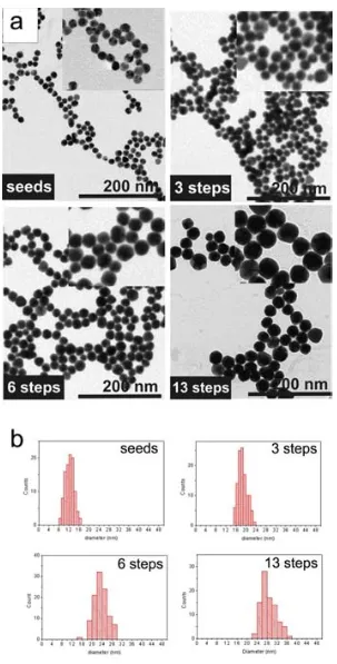
(25) Synthesis of Biocompatible AuNPs. The success in the growth of the AuNPs by this methodology is observed in the Transmission Electron Microscopy (TEM) images of relevant samples (figure 2.1). The initial seeds were 12.4 ± 1.7 nm, and they were growth up to 30.5 ± 3.9 nm after 13 generations (a generation is the AuNPs resulting from a growth step). No secondary populations are observed in any case, which indicates that no additional nucleation events were occurred. Moreover, a narrow size distribution was maintained all over the subsequent generations.. 17.
(26) Chapter 2. Figure 2.1. TEM characterization of relevant samples. (a) Representative TEM images of seeds, and AuNPs after 3, 6, and 13 growing steps. (b) The histograms showed the size distribution for these AuNPs. Mean sizes values are 12.4 ± 1.7 nm for the seeds, 18.7 ± 1.8 nm for the 3 rd generation of AuNPs, 23.5 ± 2.5 nm for the 6th, and finally 30.5 ± 3.9 for the 13th . 18.
(27) Synthesis of Biocompatible AuNPs. Additionally, the growth process was monitored by UV-VIS spectroscopy (Figure 2.2). The 6.3 nm red-shift of the SPR peak as well as the increase in the intensity (from 0.336 to 4.52) is consistent with the Mie theory in which the max as well as the intensity of the SPR increase with the volume of the AuNP.3, 17 This is related to an increase of the extinction coefficient as AuNPs size increases.18 Note that the first generations are not very sensitive to changes on the max. This deviation for smaller NPs has been reported elsewhere18.. Figure 2.2. UV-VIS monitorization of the growing steps. A red-shift of the SPR peak and a progressive increase of the Absorbance were observed. This is in agreement with the Mie theory in which the extinction coefficient increases with the volume of the metallic NP.. The pH control is an important point here since each growth step involves the reduction of HAuCl4 by citrate.7 Protons are formed in the first stage of this reaction19,20: C6H5O7 3- C5H4O52- + CO2 + 2e- + H+ AuCl4- + 2e- AuCl2- + 2 ClThis step is followed by the disproportionation of aurous species to gold atoms. This later step does not affect the pH values. To compensate the decrease of pH, an extra amount of sodium citrate is needed in every growth step in order to buffer the solution. In fact, a parallel experiment was carried out without the addition of this extra-amount of sodium citrate and the pH after each generation was measured (Figure 2.3). It is observed a constant decrease of 0.3 units of pH after each generation of NPs achieving a value of 3.14 after 14 generations. At this point, the AuNPs precipitated. This can be explained by considering the pKa values of sodium citrate (6.4, 4.8 and 3.2). Since at pH 3.14 all the carboxylic groups from citrate are protonated, the electrostatic interactions cannot maintain particles stable in solution. Thus, the amount of sodium citrate added in each 19.
(28) Chapter 2. growth step (1mL, 60mM) was the experimentally determined in order to keep a constant pH ~7 during the whole growth process.. 7. y= -0.30 x + 7.39. 2. r =0.99 pKa3 = 6.4. pH value. 6 5. pKa2 = 4.8. 4 pKa1 = 3.2. 3 2 g2. g4. g6. g8. g10. g12. g14. Figure 2.3 pH values after each growing step and pKa values of sodium citrate (6.4, 4.8 and 3.2). When solution pH is lower than pKa1 value for sodium citrate, all its carboxylic groups are protonated and hence electrostatic interactions cannot keep particles stable in solution, leading to immediate irreversible aggregation.. 2.3.2 Growth after dilution of the seeds Considering that all the gold is consumed by the reaction (confirmed by ICP-MS), that the particles are spherical and finally that no secondary nucleation events take place; it is possible to estimate the final size of the AuNPs by a simple approach based on the known amount of Au added and on the weight of a AuNP. Weight of a NP = weight of a seed +. VNP · ρAu = (Vseed ·. ρAu ) +. weight Au added nº seeds. weight Au added nº seeds. (4/3 rNP3) · ρAu= (4/3 rseed3) · ρseed +. 20. weight Au added nº seeds.
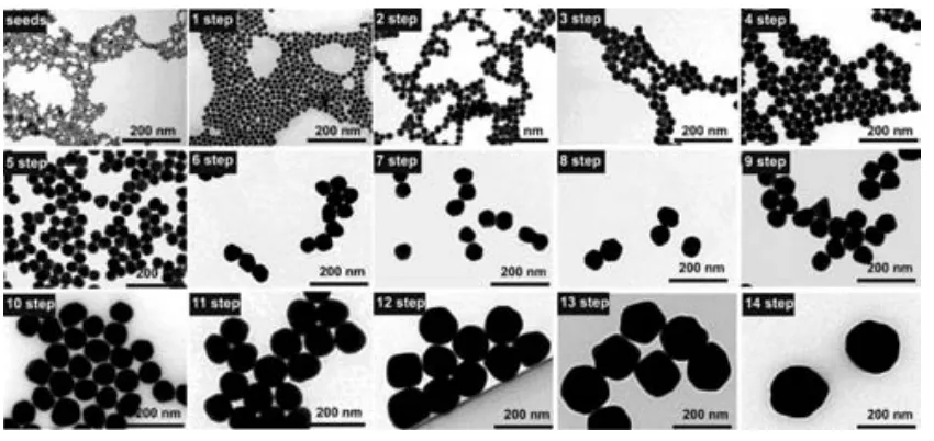
(29) Synthesis of Biocompatible AuNPs. rNP3 = 3/4. weight Au added 3 Au nº seeds + rseed (equation 2.1). Where VNP is the volume of a NP, is. ρAu gold density, rNP is the radius of the NP.. According to this theoretical approach, the amount of gold needed to obtain 50 nm AuNPs from 10 nm seeds at 3.2 x 10¹² NP/mL would be the equivalent of 125 consecutive injections. One could think on the addition of a higher amount of gold in each injection. Unfortunately, this would not be possible because the ratio seeds to gold monomer should be kept between a certain range to avoid secondary nucleation (vide infra). Thus, the previous growth protocol was modified by the addition of a dilution step every three additions of HAuCl4 as shown in scheme 2.2. The advantage of this approach is that one can synthesize large AuNPs in a few steps and without compromising the stability and the monodispersity of the NPs. AuNPs were grown up to 14 growing steps and all the generations analyzed by TEM and UV-VIS. Representative images of every step are shown in figure 2.4. It is observed that the monodispersity is maintained in all the samples as well as the absence of secondary populations. AuNPs were grown from the initial seeds (8.4 ± 1.0 nm) to the 14th generation (180.5 ± 10.7 nm). We decided to stop the synthesis here, but, in principle, there is no reason against keep growing these AuNPs. Although, the NPs are rather faceted and thus not perfectly spherical, the growth is very uniform, avoiding formation of elongated NPs such as the ones obtained by the Frens approach.6. 21.
(30) Chapter 2. Scheme 2.2 Seeded growth synthesis of AuNPs up to 180 nm. Dilution steps were introduced in this approach to facilitate the growth to larger sizes.. Figure 2.4 Representative TEM images of every growth step. Monodispersity and absence of secondary populations are maintained all over the process. The morphology of the AuNPs is quasispherical in all cases avoiding the formation of elongated AuNPs.. 22.
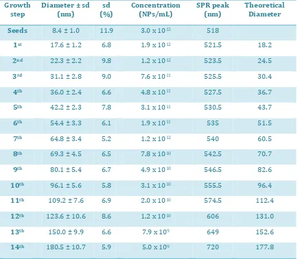
(31) Synthesis of Biocompatible AuNPs. A summary of sizes and size distributions, as well as SPR peak position is provided in table 2.1. Obviously, the final concentration of AuNPs depends on the dilution factor applied (95/150 in our case). As expected, a detailed analysis of the SPR position revealed a linear behavior due to the dependence of the peak position on the volume of the NPs.18 This tendency is however disrupted for AuNPs larger than 100 nm, in which appears a quadrupolar component of the SPR. Note that standard deviation is never higher than 10 % of mean value except for the seeds. Note also that the actual size of the AuNPs is quite similar to the theoretical size calculated as explained above (equation 2.1). Thus the presumptions made to calculate the formula (sphericity, all the added gold is consumed, and no secondary populations) are likely accomplished.. Growth step. Diameter ± sd (nm). sd (%). Concentration (NPs/mL). SPR peak (nm). Theoretical Diameter. Seeds. 8.4 ± 1.0. 11.9. 3.0 x 10¹². 518. 1st. 17.6 ± 1.2. 6.8. 1.9 x 10¹². 521.5. 18.2. 2nd. 22.3 ± 2.2. 9.8. 1.2 x 10¹². 523.5. 24.5. 3rd. 31.1 ± 2.8. 9.0. 7.6 x 10¹¹. 525.5. 30.4. 4th. 36.0 ± 2.4. 6.6. 4.8 x 10¹¹. 527.5. 36.7. 5th. 42.2 ± 2.3. 7.8. 3.1 x 10¹¹. 530.5. 43.7. 6th. 54.4 ± 3.3. 6.1. 1.9 x 10¹¹. 535. 51.5. 7th. 64.8 ± 3.4. 5.2. 1.2 x 10¹². 540. 60.5. 8th. 69.3 ± 4.5. 6.5. 7.8 x 10¹⁰. 542.5. 70.7. 9th. 80.1 ± 5.4. 6.7. 4.9 x 10¹⁰. 546.5. 82.6. 10th. 96.1 ± 5.6. 5.8. 3.1 x 10¹⁰. 555.5. 96.4. 11th. 109.2 ± 7.6. 6.9. 2.0 x 10¹⁰. 574.5. 112.4. 12th. 123.6 ± 10.6. 8.6. 1.2 x 10¹⁰. 606. 131.0. 13th. 150.0 ± 9.9. 6.6. 7.9 x 10⁹. 649. 152.6. 14th. 180.5 ± 10.7. 5.9. 5.0 x 10⁹. 720. 177.8. Table 2.1 Summary of sizes, concentration and SPR peak position of AuNPs after 14 consecutive growing steps. The theoretical size is also shown.. 23.
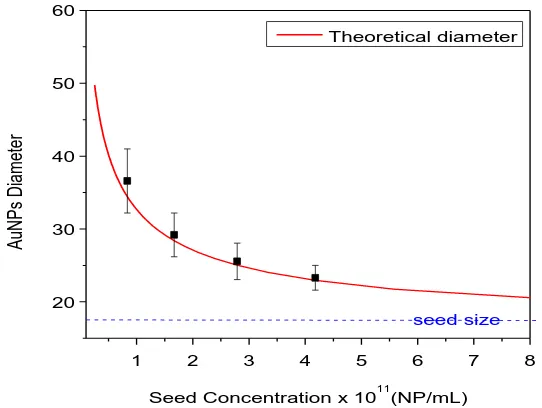
(32) Chapter 2. 2.3.3 Variation of seed and gold salt concentrations From the results above, it is observed that the final size of the nanoparticles will depend on the amount of seeds and gold added in solution. Taking into account these two parameters, it seems possible to obtain AuNPs of a desired size. In this section, we explore how these parameters affect the resultant AuNPs. Firstly, the effect of seed concentration on the size of AuNPs after a growing step was studied using 17.0 ± 1.7 nm seeds at concentrations ranging from 4.18 x 10¹¹ NP/mL to 8.36 x 10¹⁰ NP/mL. The final volume (75 mL) and the amount of gold precursor added (0.5 mL, 25 mM HAuCl4) were the same in all cases. Size and size distribution were analyzed by TEM imaging and they are showed in figure 2.5 (black squares). As expected, the higher is the concentration of seeds, the smaller is the growth of the resultant NPs. Hence, 23.3 ± 1.7 nm AuNPs were obtained at the highest concentration (4.18 x 10¹¹ NP/mL), whilst 36.6 ± 4.4 nm AuNPs were obtained when 5 times less seeds were added. It is also observed the fitting of the empirical values with the ones predicted using the equation 2.1. Also note the greater increase in the size for high dilutions of the seeds. Thus, one could think that a high dilution of seeds could lead to large AuNPs. However, it should be taken into account that new nucleation events must be avoided to get a single population of AuNPs with narrow size distribution. This cannot be ensured when very few AuNPs are present in solution. In fact, at the lower concentration assayed here a small percentage of a secondary population of AuNPs appeared. Therefore the addition of subsequent growth steps seems to be the appropriate strategy to get larger AuNPs. Additionally, it is shown here the great control of the process and the ability to get AuNPs of a desired size. Obviously, it is necessary to know previously both the size and the concentration of the seeds to make the required calculations.. 24.
(33) Synthesis of Biocompatible AuNPs. 60 Theoretical diameter. AuNPs Diameter. 50. 40. 30. 20 seed size 1. 2. 3. 4. 5. 6. 7. 8. 11. Seed Concentration x 10 (NP/mL). Figure 2.5 Theoretical and empirical AuNPs diameter in function of the concentration of seeds. The final size depends on the concentration of seeds added. It is observed here how the empirical values match satisfactorily the theoretical value.. The other component of this system that can be modified is the amount of HAuCl4 added in a growth step. We followed a similar strategy to analyze the role played by the precursor: The concentration of 17.0 ± 1.7 nm seeds was kept constant (1.71 x 10¹¹ NP/mL), whilst the concentration of HAuCl4 was varied from 0.022 mM to 0.334 mM. The size of the resultant AuNPs, as measured by TEM, as well as the predicted value are shown in table 2.2. Obviously, the size of the AuNPs increased as the amount of gold precursor increased. Here, the predicted size calculated from equation 2.1 also matches the empirical value. However, a secondary population of small AuNPs was clearly observed when the highest concentration of HAuCl4 was assayed. Thus, new nucleation of NPs was not avoided in the case of the highest HAuCl4 concentration and consequently the monodispersity of this sample was lost. This can be also observed analyzing the SPR peak shift: The peak shifts to higher values as long as the amount of gold added increases, but at 0.334 mM the peak shifts to a smaller wavelength indicating the deficient growth of the AuNPs. Since part of the Au added was consumed by the formation of new AuNPs, which in addition competed with the initial seeds for the remaining Au, the resultant AuNPs are polydisperse with an unpredictable size. Hence, it is important to add the precursor in successive steps instead of a single one which would increase the concentration of precursor too much causing new nucleation. 25.
(34) Chapter 2. HAuCl4 (mM). Size (nm). Theoretical size (nm). 0.022 0.083 0.167 0.334. 19.5 ± 2.1 24.7 ± 2.1 28.6 ± 2.7 xxx. 19.3 23.9 28.2 34.2. SPR peak (nm) 519.9 522.1 524.1 522.2. Table 2.2 Size of AuNPs depending on the amount of gold added.. Summarizing, it has been demonstrated here the possibility to obtain AuNPs of a desired size just by adjusting the amounts of seeds and HAuCl4 according to a simple theoretical approach. The less seeds and the higher amount of gold added, the larger are the resultant AuNPs. However, the variation of both components should be in a certain range to avoid new nucleation events and therefore the formation of a secondary population of AuNPs. Thus, a range of sizes can be obtained in a single step by adjusting these two parameters, but a subsequent synthesis step should be introduced if larger AuNPs are desired. For example, starting with 17 nm seeds it is easy to obtain AuNPs à la carte from 18 to 35 nm, but if larger AuNPs are desired, an additional synthesis step (normally implying dilution of the AuNPs) will be required.. 2.4 Conclusions The control of the size and morphology of NPs are key points in the synthesis of inorganic NPs. In the field of biomedicine, and specifically in the use of AuNPs as scaffolds for drug delivery, achieving a great control over the synthesis is recommended since some important properties such as biodistribution are strongly influenced by the size of the scaffold rather than by the composition. In fact, size effects are paramount at the nanoscale. Here it has been demonstrated that the synthesis of AuNPs is very well controlled in terms of morphology, size and size distribution. Our approach is based on a seeding growth synthesis due to the fact that the growth of AuNPs is to some extend thermodynamically favored respect to the formation of Au nuclei. To even favor more the growth, the temperature was decreased to 90 ⁰C instead of the 100 ⁰C required for the synthesis of new AuNPs. AuNPs up to 30 nm can be synthesized by successive growing steps of HAuCl 4. 26.
(35) Synthesis of Biocompatible AuNPs. The pH control has been shown to be crucial to maintain colloidal stability, since protons are formed in every growth step. To obtain AuNPs up to 180 nm, dilution steps were introduced to minimize the consumption of Au. In addition, it has been shown the possibility to nicely control the final size of AuNPs by adjusting the amounts of either seeds or gold precursor. The separation of nucleation and growth is the main factor that determines the success of the synthesis and therefore must be always accomplished if monodispersity is wanted.. 2.5 References (1) Turkevich, J.; Garton, G.; Stevenson, P. C., The Color of Colloidal Gold. Journal of Colloid Science 1954, 9, S26-S35. (2) Daniel, M. C.; Astruc, D., Gold nanoparticles: Assembly, supramolecular chemistry, quantum-size-related properties, and applications toward biology, catalysis, and nanotechnology. Chemical Reviews 2004, 104, 293-346. (3) Kimling, J.; Maier, M.; Okenve, B.; Kotaidis, V.; Ballot, H.; Plech, A., Turkevich Method for Gold Nanoparticle Synthesis Revisited. The Journal of Physical Chemistry B 2006, 110, 15700-15707. (4) Polte, J.; Ahner, T. T.; Delissen, F.; Sokolov, S.; Emmerling, F.; Thünemann, A. F.; Kraehnert, R., Mechanism of Gold Nanoparticle Formation in the Classical Citrate Synthesis Method Derived from Coupled In Situ XANES and SAXS Evaluation. J. Am. Chem. Soc. 2010, 132, 1296-1301. (5) Park, J.; Joo, J.; Kwon, S. G.; Jang, Y.; Hyeon, T., Synthesis of monodisperse spherical nanocrystals. Angewandte Chemie-International Edition 2007, 46, 4630-4660. (6) Frens, G., Controlled Nucleation for Regulation of Particle-Size in Monodisperse Gold Suspensions. Nature-Physical Science 1973, 241, 20-22. (7) Ji, X. H.; Song, X. N.; Li, J.; Bai, Y. B.; Yang, W. S.; Peng, X. G., Size control of gold nanocrystals in citrate reduction: The third role of citrate. J. Am. Chem. Soc. 2007, 129, 13939-13948. (8) Perrault, S. D.; Chan, W. C. W., Synthesis and Surface Modification of Highly Monodispersed, Spherical Gold Nanoparticles of 50−200 nm. Journal of the American Chemical Society 2009, 131, 17042-17043. (9) Bigall, N. C.; Halrtling, T.; Klose, M.; Simon, P.; Eng, L. M.; Eychmuller, A., Monodisperse Platinum Nanospheres with Adjustable Diameters from 10 to 100 nm: Synthesis and Distinct Optical Properties. Nano Letters 2008, 8, 4588-4592. (10) Jana, N. R.; Gearheart, L.; Murphy, C. J., Evidence for seed-mediated nucleation in the chemical reduction of gold salts to gold nanoparticles. Chemistry of Materials 2001, 13, 2313-2322. (11) astus, N. G. Comenge, . untes, . c., inetically Controlled Seeded Growth Synthesis of Citrate-Stabilized Gold Nanoparticles of up to 200 nm: Size Focusing versus Ostwald Ripening. Langmuir 2011, 27, 11098-11105. (12) Jana, N. R.; Gearheart, L.; Murphy, C. J., Seeding growth for size control of 5-40 nm diameter gold nanoparticles. Langmuir 2001, 17, 6782-6786. (13) Leonov, A. P.; Zheng, J.; Clogston, J. D.; Stern, S. T.; Patri, A. K.; Wei, A., Detoxification of Gold Nanorods by Treatment with Polystyrenesulfonate. Acs Nano 2008, 2, 2481-2488. (14) Eugenii, K.; Itamar, W., Integrated Nanoparticle-Biomolecule Hybrid Systems: Synthesis, Properties, and Applications. Angewandte Chemie International Edition 2004, 43, 6042-6108. 27.
(36) Chapter 2. (15) Sellers, H.; Ulman, A.; Shnidman, Y.; Eilers, J. E., Structure and binding of alkanethiolates on gold and silver surfaces: implications for self-assembled monolayers. J. Am. Chem. Soc. 1993, 115, 9389-9401. (16) LaMer, V. K.; Dinegar, R. H., Theory, Production and Mechanism of Formation of Monodispersed Hydrosols. Journal of the American Chemical Society 1950, 72, 4847-4854. (17) Haiss, W.; Thanh, N. T. K.; Aveyard, J.; Fernig, D. G., Determination of Size and Concentration of Gold Nanoparticles from U − is Spectra. Analytical Chemistry 2007, 79, 4215-4221. (18) Njoki, P. N.; Lim, I. I. S.; Mott, D.; Park, H.-Y.; Khan, B.; Mishra, S.; Sujakumar, R.; Luo, J.; Zhong, C.-J., Size Correlation of Optical and Spectroscopic Properties for Gold Nanoparticles. The Journal of Physical Chemistry C 2007, 111, 14664-14669. (19) Kumar, S.; Gandhi, K. S.; Kumar, R., Modeling of Formation of Gold Nanoparticles by Citrate Method†. Industrial & Engineering Chemistry Research 2007, 46, 31283136. (20) Young, J.; Lewinski, N.; Langsner, R.; Kennedy, L.; Satyanarayan, A.; Nammalvar, V.; Lin, A.; Drezek, R., Size-controlled synthesis of monodispersed gold nanoparticles via carbon monoxide gas reduction. Nanoscale Research Letters 2011, 6, 1-11.. 28.
(37) Chapter 3 Conjugation of AuNPs to Achieve Stable and Functional Scaffolds for Drug Delivery. 3.1 Introduction The world of NPs and the world of cells and biomolecules are quite different and often incompatible. Functionalization of NPs surface is our tool to interact at the nano-bio interface.1 Thus, engineering properly the AuNPs surface is a crucial step to determine the final fate of the AuNPs since it will provide the functionality of interest and it will determine important properties such as surface charge and stability. A great variety of chemical groups such as thiols, amines, phosphates or carboxylic acids have affinity to the AuNPs surface.2 This versatility allows the functionalization with different molecules such as organic molecules, proteins, DNA, polymers, etc.3 Among all the possible links, the SH-Au bond is the most widely exploited due to its pseudo-covalent character (≈45 Kcal/mol)4, this high bond strength allow the exchange with other ligands which bind Au more weakly such as citrate, amines, or phosphates. In addition, the high strength makes the bond very stable over the time. Thus, the use of thiolated molecules is a common strategy to functionalize AuNPs. Examples of this strategy includes conjugation with alkanethiols,5 thiolated DNA,6 proteins or peptides containing a superficial cysteine residue,7 and polymers with a terminal SH group.8 Alkanethiolates, such as 11mercaptoundecanoic acid (MUA), have been largely used to obtain self-assembled monolayers (SAM) onto Au surfaces9 due to the presence of a thiol and a hydrophobic chain which allow a very compact packing of the layer. In addition, a terminal group gives the desired functionality and/or charge to the AuNPs. This is of special importance since. 29.
(38) Chapter 3. some of their biological properties such as potential toxicity or cell uptake are dramatically influenced by the surface charge given by this layer.10 Moreover, the presence of terminal groups allows further conjugation steps. For example, EDC coupling or other click chemistry processes between the monolayer and a molecule of interest have been largely studied.11 Other strategies of functionalization are based on a non-specific absorption of molecules onto the AuNP surface. These include unspecific absorption of protein as in the case of the well-known protein corona12, 13, or weak interactions with chemical groups such as phosphates from a DNA strand or OH groups from polymers such as PEG, amongst others.1 In this case the link with the AuNPs is weaker than it was for thiolated molecules. Interestingly, by mixing AuNPs and surfactant molecules, AuNPs conjugates with a specific functionality can be properly inserted in the biological machinery. However, it is worth noting here that minute differences on the resulting conjugate, often ignored, might have a dramatic impact in the biological response of the system exposed to some determined AuNPs.14,. 15. Taking into account that nanoparticles for biomedical applications are. reaching a high level of complexity, the fine control and understanding of the conjugate composition is required to not halt the development of new products. Thus, factors such as conformational changes of the coating molecules or small variations of classic behavior induced by the nanoscale have to be considered before using the nanoparticle: For example, the curvature radius of the AuNPs modifies the pKa of attached alkanethiolates,16 thus two AuNPs with the very same composition might be stable or not at a specific pH depending on the size of the AuNPs. Also, a change on the conformation of components of the monolayer (e.g. PEG), rather than in the composition, modifies dramatically the biological properties of the conjugate.15 In this chapter, we carefully examine the conjugation of two widely used molecules (MUA and SH-PEG) to the surface of AuNPs and the design of mixed layers whose properties can be tuned depending on the composition of the layer and the conjugate architecture (global conformation of the ligands). We also propose methods and principles to control the conjugation and to characterize (physically, chemically and biologically) the resulting conjugates and their evolution in physiological conditions. Also, a study to determine how the layer composition influences the protein binding is showed. Note that the first step on tuning NP-cell interactions is to control this protein binding. This is of special relevance, since these are the conjugates that will be used as scaffolds for drug delivery in following chapters.. 30.
(39) Conjugation of AuNPs to Achieve Functional Scaffolds for Drug Delivery. 3.2 Experimental MUA Self-Assembled Monolayer on AuNPs. Different concentrations of MUA from 0 to 60 M were added to 20 nm AuNPs (21.3 ± 3.3 nm, 5.0 x10¹¹ NP/mL) and 40 nm AuNPs (39.3 ± 4.3 nm, 9.0 x10¹⁰ NP/mL) solution by adding corresponding volumes of 1mM or 5 mM MUA aqueous solution. Differences of volume were compensated by addition of miliQ water. The conjugation was gently stirred for 21 hours (an aliquote was taken and immediately measured at 30 minutes as well). UV-VIS spectra from 300 to 800 nm were taken for every sample at 30 minutes and 21 hours. SH-PEG Self-Assembled Monolayer on AuNPs: Different concentrations of SH-PEG (3.4 KDa) from 0 to 15 mM were added to 20 nm AuNPs (21.3 ± 3.3 nm, 5.0 x10¹¹ NP/mL) by adding corresponding volumes of 1 mM SH-PEG aqueous solution. Differences of volume were compensated by addition of miliQ water. The conjugation was gently stirred overnight. UV-VIS spectra from 300 to 800 nm were taken for every sample. Mixed layer-AuNPs obtained by co-conjugation. The different SH-PEG / MUA ratios were prepared by mixing the corresponding volumes of 1mM (or 0.3 mM, or 0.1 mM) solution of each component. 500 L of these mixtures were subsequently added to 4 mL of AuNPs as synthesized (14.6 ± 1.5 nm, 4.2 x 10¹² NP/mL) and gently stirred overnight. The SPR bands of the resulting conjugates were analyzed by UV-VIS spectroscopy at the range of 300 to 800 nm. The hydrodynamic diameter was analyzed by DLS. Mixed layer-AuNPs obtained by ligand exchange. An aqueous solution of SH-PEG (125 L, 0.3 mM) was added to 1 mL citrate-stabilized AuNPs (15.2 ± 2.0 nm, 3.7 x 10¹² NP/mL). The conjugation was allowed to run for 6 hours. Then, the excess of SH-PEG was removed by 2 centrifugation cycles (18000 rcf, 30 min) and resuspension of AuNPs in fresh water. Finally, different amounts of MUA (0 to 80 M) were added to the solution and the agitation was maintained overnight. The resulting AuNPs were analyzed by UV-VIS spectroscopy and DLS. Quantification of the SH-PEG-FITC loaded on the AuNPs by fluorescence. The excess of SH-PEG-FITC and MUA was removed by 3 centrifugation steps (30000 rcf, 30 min). After the last washing step, AuNPs were suspended in the same volume of an aqueous NaCN solution (100 mM) and stirred for 6h at 37 ºC. The total dissolution of AuNPs was proved by the disappearance of the SPR. All the samples were buffered using CAPS (25 mM, pH 10.6). Known concentrations of SH-PEG-FITC under the same conditions were used to do the calibration curve. 100 L of standards and samples were loaded in a 9631.
(40) Chapter 3. well plate and the fluorescence spectra (ex=494, em=521) was taken. The concentration of SH-PEG-FITC in the samples was calculated from the calibration curve. Quantification of the SH-PEG-FITC by absorbance. AuNPs were removed from the sample by 3 centrifugation steps (30000 g, 30 min). Centrifugation conditions were more aggressive here to ensure that all the NPs were removed. 25 L of CAPS buffer (200 M, pH 10.6) were aded to 975 L of the resulting supernatant. To prepare the standards the same procedure than the explained above to functionalize the AuNPs was followed but using sodium citrate 2.2 mM instead of the AuNPs solution. The absorbance of standards and samples were measured under the same conditions. The amount of SH-PEG-FITC loaded on the AuNPs was calculated substracting the concentration of the sample from the concentration of the standard. Stability in physiological and acidic media. AuNPs functionalized with MUA and SH-PEG were concentrated to 2.4 x 10¹⁴ NP/mL by centrifugation (18000 rcf, 14 min.) of 1 mL of AuNPs and resuspension of the pellet in a final volume of 20 l. Then these 20 l were added to 180 l of DMEM supplemented with 10 % FBS and softly mixed during 30 minutes. To assay the stability in acidic media, 1 mL of previously functionalized AuNPs were brought to pH 2.6 by adding 2.5 L of a 0.2 M glycine/HCl buffer. The stability in both cases was evaluated by analyzing the broadening and the red-shift of the SPR band. Protein adsorption. 1.3 mM solution of bovine serum albumin was prepared by diluting 182 mg in 2.1 mL of 5 mM Hepes buffer at pH 7.5. Then 7.7 L of this solution was added to 1 mL of previously functionalized AuNPs and softly mixed overnight. The changes of the hydrodynamic diameter were analyzed by DLS and the shift of the SPR band by UV-VIS. Gel electrophoresis.AuNPs were functionalized with relevant ratios of SH-PEG / MUA (0, 0.3, 0.6, 0.74, 0.88, 0.96, and 1) as explained above. Then the samples were splitted in two parts. 1 mL was incubated with 7.7 L of a 1.3 mM BSA solution in 5 mM Hepes buffer (pH 7.5) overnight and the other part was kept without any modification. Samples were loaded into the wells of a 1% agarose gel.The electrophoresis was run under 60 V in a sodium borate buffer (30 mM, pH 8.5).. 32.
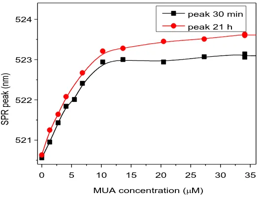
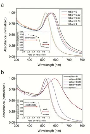
(41) Conjugation of AuNPs to Achieve Functional Scaffolds for Drug Delivery. 3.3 Results and discussion 3.3.1 MUA Self-Assembled Monolayers onto AuNPs. The choice of MUA to study the formation of SAM onto AuNPs is not trivial. The presence of a terminal thiol group, the relatively long (11 C) hydrocarbon chain, and the carboxylic acid at the other side of the molecule make it a model candidate to form highly stable SAMs. In addition, the presence of the carboxylic groups will allow a further step of cisplatin loading via a coordination bond (see chapter 4). To understand the formation of MUA SAMs onto AuNPs, different amounts of MUA were added to a solution of AuNPs (21.3 ± 3.3 nm, 5.0 x10¹¹ NP/mL) and the shift of the SPR analyzed by UV-VIS. The position of the SPR of the AuNPs after MUA addition is determined by changes in the local refractive index around the NP due to changes in the density of the monolayer which is determined by the concentration of ligand on the surface.17, 18 Thus, the denser is the layer, the higher is the shift. This is observed in figure 3.1 in which the position of the peak is represented in function of the concentration of MUA added. The SPR varies following a sigmoidal curve, typical of processes involving adsorption on a surface at constant temperatures (e.g. Langmuir isotherms).19 From this curve, the saturation point, or the point at which adding more MUA causes no change in the SPR peak is determined at approximately 15 M for the analyzed AuNPs. Taking into account the footprint of MUA in gold, which is reported to be 0.21 nm 2,4 it can be easily calculated that the amount of MUA needed to completely cover these AuNPs would be 7.25 M. Thus, at least a 2-fold excess of MUA is needed to be added in this case to ensure a saturation of the monolayer. This excess of ligand is low compared to unspecific and weaker bindings such as the proteins absorbed onto AuNPs, in which excess as high as 5 to 10 times the theoretical amount are required to saturate the surface.20, 21 Interestingly, there is a slight increase of the peak observed after 21h. This fact, which has been also described in SAMs on Au planar surfaces,22 it is attributed to an internal slow reorganization of the monolayer, to maximize the surface coverage, and consequently minimize the free surface energy. The differences between 30 minutes and 21 hours are greater for higher MUA excess than for low concentrations. At concentrations much lower than saturation, most of the added MUA is rapidly attached to the highly available surface and therefore the final state (or a very close state) is rapidly reached. On the other side, since the surface is densely occupied at high concentrations of MUA at short times, there is a need of reorganization of the layer before more MUA could be attached. This reorganization is a slow process which is not completed after the first 30 minutes, but 33.
(42) Chapter 3. hours. From a practical point of view, this suggests that conjugation of MUA on AuNPs should be carried out overnight to ensure a complete formation of the monolayer.. peak 30 min. 524. peak 21 h. SPR peak (nm). 523. 522. 521. 0. 5. 10. 15. 20. 25. 30. 35. MUA concentration (M). Figure 3.1 Shift of SPR in function of MUA added in solution. The formation of the monolayer was monitored by UV-VIS 30 minutes and 21 hours after MUA addition. The common saturation of a surface behavior is observed. The slight differences between times are due to a slow reorganization of the monolayer.. To evaluate whether the size of AuNPs has an influence on the formation of the MUA SAM, 40 nm AuNPs (39.3 ± 4.3 nm, 9.0 x10¹⁰ NP/mL) were also functionalized with MUA. The conjugation was run 21 hours under the same conditions than assayed before. In figure 3.2 the variation of peak in function of concentration of MUA added is represented for both 20 nm and 40 nm AuNPs. Note that the concentration is normalized by the surface area of the different AuNPs and the peak shift is normalized from 0 to 1 in order to compare both samples. It is observed that the conjugation of MUA is rather independent of the AuNPs size in this range of sizes, showing an almost identical behavior. The shift of the SPR as a function of the concentration of added MUA fits quite well the Langmuir isotherm (Figure 3.2 b). This simple approach for adsorption onto surfaces is based on four assumptions: i. All the surface sites have the same adsorption energy for the adsorbate. ii. The adsorption at one site does not affect the energy of adsorption of neighboring sites. iii. All adsorption occurs through the same mechanism. iv. At the maximum adsorption, only a monolayer is formed. The experimental points fit quite well this theoretical approach. The constant of Langmuir 34.
Figure
+7
Documento similar
In the preparation of this report, the Venice Commission has relied on the comments of its rapporteurs; its recently adopted Report on Respect for Democracy, Human Rights and the Rule
On the other hand, in terms of control in wind farms, the authors of [5] mention vari- ous wind measurement methods which allow measurements to be made before and after the creation
Very remarkably, we observed that cisPtI 2 , in contrast to expec- tations and to the current opinion, is on the whole more cyto- toxic than cisplatin in three out of four
On the other hand, SEM and XPS clearly indicated that the enhancement of the activity and selectivity to benzaldehyde was associated with the concentration of the
The secondary objectives were as follows: to quantify the effect of adding the study drug on central blood pressure and vascular compliance using the augmentation index and pulse
the effect of NO donors was only abolished by the adenosine A 1 receptors antagonist; 2) in tail arteries, noradrenaline release was increased by NO donors and it was reduced by
After achieving an effective abrogation of ATG5 at the protein level in both cell lines (Figure 3D), viability was evaluated, showing almost no effect in H460 cells, while
The effect of the sorption process on the HDC performance has been further supported by the results obtained using isopropanol as solvent where sorption was
