Amyloid-β oligomers reduces glutamatergic transmission and inhibits synaptic plasticity with underlying mechanisms suggestting akap150 modulates diverse synaptic functions
140
0
0
Texto completo
(2)
(3) AMYLOID-β OLIGOMERS REDUCES GLUTAMATERGIC TRANSMISSION AND INHIBITS SYNAPTIC PLASTICITY WITH UNDERLYING MECHANISMS SUGGESTTING AKAP150 MODULATES DIVERSE SYNAPTIC FUNCTIONS. Memoria de tesis doctoral presentada por Wenwen Cheng para optar al grado de Doctora en Bioquímica y Biología Molecular por la Universitat Autònoma de Barcelona.. Este trabajo ha sido realizado bajo la dirección del Doctor José Rodriguez Alvarez en la Facultat de Medicina del Departament de Bioquímica i Biologia Molecular y en el Institut de Neurociències de la Universitat Autònoma de Barcelona.. El director,. El doctorando,. Dr. José Rodriguez Alvarez. Wenwen Cheng. Bellaterra, 2014.
(4) “Al sac i ben lligat” “Qui no s’arrisca no pisca”.
(5)
(6) Acknowledgements 1.
(7) First I would like to express my deepest gratitude to my supervisor Dr. José Rodriguez Alvarez for giving me the opportunity to do my PhD studies in his lab. He has created an excellent environment in the lab and has shared his insights as I hurdle all the obstacles in the completion this research work. His professionalism has been invaluable for my development. I want to thank him for his inspiring guidance, continued support and encouragement throughout my PhD and finally, lots of thanks for the critical and proof-reading of this thesis. To my present and past lab members who in one way or another contributed and extended their assistance in this work. Special thanks to Alfredo Minano-Molina and Rut Fadó Andrés for their collaboration and their huge contribution to my work and for many funny times. Many thanks to the past lab members for making a friendly working atmosphere. I am indebted to many people who have helped me technically during my PhD, including Nuria Barba at the Microscopy Service and special thanks to Cristina, Suzana for the productive work and pleasant company during the long hours spent in the primary cell culture room. To every investigator and group leader of our department. In particular, I would like to thank all the members of the research groups. Especial thanks to Guillem Sanchez, Virginia Jimenez, Dolores Siedlecki for their help and many useful recommendations. Especially thanks to Meng Chen and Ping Sun. Thanks to all the former as well as current PhD students of the department for their kindness, understanding and hospitability. Thanks a lot! I would also like to extend my thanks to the administrative staff of the Institut de Neurociències & Departament de Bioquímica i Biologia Molecular. Thanks for facilitating those administrative tasks which were always ambiguous for me. During these years I have been supported by the China Scholarship Counsel without which this thesis would not have been possible. Throughout the entire process of my PhD studies in UAB I received a lot of support from many people around me who I cannot mention but I express my sincere gratefulness. I would also like to thank my thesis committee for their precious time. Bofei Wei deserves my deepest gratitude for her help and the opportunity which she gave to me at the very beginning of my PhD studies. Finally, I wish to thank my family for their support and encouragement throughout my study.. Moltes gràcies !. 2.
(8) 3.
(9) TABLE OF CONTENTS. 4.
(10) Table of contents Acknowledgements ................................................................................................................... 1 TABLE OF CONTENTS ................................................................................................................. 4 SUMMARY ................................................................................................................................. 9 ABBREVIATIONS....................................................................................................................... 13 INTRODUCTION ....................................................................................................................... 15 1. GLUTAMATE ........................................................................................................................ 17 1.1. GLUTAMATE RECEPTORS.............................................................................................. 17 1.2. GLUTAMATE RECEPTOR TYPES ..................................................................................... 17 1.3. STRUCTURE, MECHANISM AND FUNCTION ................................................................. 19 1.3.1. IONOTROPIC .......................................................................................................... 19 1.3.2. AMPA RECEPTORS ................................................................................................. 19 1.3.3. NMDA RECEPTOR .................................................................................................. 21 1.3.4. METABOTROPIC..................................................................................................... 23 2. SYNAPTIC PLASTICITY AND RECEPTOR TRAFFICKING .......................................................... 24 2. 1. SYNAPTIC PLASTICITY................................................................................................... 24 2.2. AMPARs TRAFFICKING .................................................................................................. 25 3. LONG TERM DEPRESSION .................................................................................................... 30 4. SOLUBLE PROTEIN OLIGOMERS IN NEURODEGENERATION: FROM THE ALZHEIMER’S AMYLOID β-PEPTIDE ................................................................................................................ 34 4.1 THE DISCOVERY OF THE FIRST AD CAUSATIVE GENE .................................................... 34 4.2 THE REGULATED INTRAMEMBRANE PROTEOLYSIS OF APP .......................................... 35 4.3. PROCESSING OF AMYLOID PRECURSOR PROTEIN ........................................................ 36 4.4. OLIGOMERS INDUCE SYNAPTIC DYSFUNCTION............................................................ 36 4.5. ADDLS INDUCE MEMORY LOSS .................................................................................... 38 4.6. OLIGOMERIC ASSEMBLY CHARACTERISTICS................................................................. 38 5. A- KINASE-ANCHORING PROTEINS ...................................................................................... 39 6. MICRORNAS AT THE SYNAPSE ............................................................................................. 42 6.1. REGULATION OF MIRNA BY NEURONAL ACTIVITY ....................................................... 44 6.2. MIRNAS IN NEUROLOGICAL DISORDERS ...................................................................... 45 AIM .......................................................................................................................................... 49 METHODS ................................................................................................................................ 52 1. EXPERIMENTAL ANIMAL MODEL ......................................................................................... 53 2. PRIMARY CELL CULTURES .................................................................................................... 53. 5.
(11) 3. Aβ OLIGOMER PREPARATION.............................................................................................. 55 4. CELL STIMULATION AND LYSATE PREPARATION ................................................................. 55 5. SURFACE BIOTINAYLATION.................................................................................................. 56 6. REVERSE TRANSCRIPT PCR .................................................................................................. 57 6.1. RNA extraction.............................................................................................................. 57 6.2. RNA concentration determination ............................................................................... 57 6.3. RNA quality assessment ............................................................................................... 57 6.4. Reverse transcription ................................................................................................... 57 6.5. Semiquantitative PCR ................................................................................................... 58 7. REAL-TIME POLYMERASE CHAIN REACTION (RT-PCR)......................................................... 59 7.1. RNA isolation and first strand synthesis ....................................................................... 59 7.2. Real-time PCR Protocol................................................................................................. 59 7.3. TaqMan Analysis........................................................................................................... 60 8. WESTERN BLOT ANALYSIS ................................................................................................... 61 8.1. GENERATION OF PROTEIN EXTRACTS FROM CELLS ..................................................... 61 8.2. SODIUM DODECYL SULFATE-POLYACRYLAMIDE GEL ELECTROPHORESIS .................... 62 8.2.1. SDS-PAGE gel preparation ..................................................................................... 62 8.2.2. Running a SDS-PAGE gel ........................................................................................ 62 8.3. TRANSFER OF PROTEINS TO NITROCELLULOSE MEMBRANES ..................................... 62 8.3.1. Antibody detection ................................................................................................ 64 9. LENTIVIRUS AND CONSTRUCT PREPARATION ..................................................................... 65 10. IMMUNOCYTOCHEMISTRY AND IMAGE ANALYSIS ........................................................... 65 10.1 Immunofluorescence staining for AKAP150 and PSD 95............................................. 65 10.2. Immunofluorescence staining for AMPARs ................................................................ 65 11. PRODUCTION OF HIGH-TITER HIV-1-BASED VECTOR STOCKS BY TRANSIENT TRANSFECTION OF HEK 293T CELLS ........................................................................................ 67 11.1. Prepare 293T cells for transfection ............................................................................ 67 11.2. Co-transfect plasmids encoding vector components ................................................. 67 11.3. Harvest and concentrate vector stocks ...................................................................... 68 11.4 REAGENTS AND SOLUTIONS ........................................................................................ 68 11.5. TITRATION OF LENTIVIRAL VECTORS .......................................................................... 69 12. STATISTICAL ANALYSIS....................................................................................................... 70 RESULTS ................................................................................................................................... 72. 6.
(12) 1. THE ENDOCYTOSIS OF AMPARS AND THE CHANGES IN THE LEVELS OF AKAP150 INDUCED BY NMDA AND AβO ................................................................................................................. 74 2. NMDA AND Aβo-INDUCED DOWN-REGULATION OF AKAP150 DEPENDS ON NMDAR, PP2B AND PROTEASOME ACTIVITIES, WHILE Aβo-INDUCED DOWN-REGULATION OF AKAP150 DEPENDS ON NMDAR ACTIVITY .............................................................................................. 77 3. CHEMICAL LTP WAS UNABLE TO RESCUE THE REDUCTION OF AKAP150 INDUCED BY NMDA (cLTD) OR Aβo .............................................................................................................. 83 4. OVEREXPRESSION OF AKAP150 BLOCKED BOTH THE NMDA AND AβO INDUCED GLUA1 ENDOCYTOSIS .......................................................................................................................... 87 5. ROLE OF AKAP150 IN NMDAR-TRIGGERED AMPAR ENDOCYTOSIS. KNOCK-DOWN OF AKAP150 INDUCED CHANGES OF AMPARS LEVELS. ................................................................ 89 6. LONG TERM EXPOSURE OF NMDA OR Aβo INDUCED A REDUCTION OF AMPARs AND miRNAS .................................................................................................................................... 92 DISCUSSION ............................................................................................................................. 96 1. AβO INDUCED ABERRATIONS IN SYNAPSE COMPOSITION PROVIDES A MOLECULAR BASIS FOR LOSS OF CONNECTIVITY IN ALZHEIMER’S DISEASE .......................................................... 98 1.1. MEMORY LOSS IS A DISRUPTION OF SYNAPTIC PLASTICITY: THE OLIGOMER HYPOTHESIS ......................................................................................................................... 98 1.2. OLIGOMERS ACCUMULATION ...................................................................................... 99 2. THE RELATIONSHIP BETWEEN AβO AND LONG TERM DEPRESSION OR LONG TERM POTENTIATION ...................................................................................................................... 100 3. THE ROLE OF AMPARS AND THEIR RELATIONSHIPS WITH AKAP150 IN AβO OR NMDATRIGGERED AMPARS ENDOCYTOSIS ..................................................................................... 102 4. THE ROLE OF MICRORNAS IN NEUROGENESIS AND NEUROPLASTICITY MECHANISMS ... 107 5. MIRNAS AND PATHOLOGICALLY STRESS INDUCED CHANGES .......................................... 109 CONCLUSIONS ....................................................................................................................... 112 REFERENCE LIST ..................................................................................................................... 116 APPENDIX .............................................................................................................................. 135. 7.
(13) 8.
(14) SUMMARY 9.
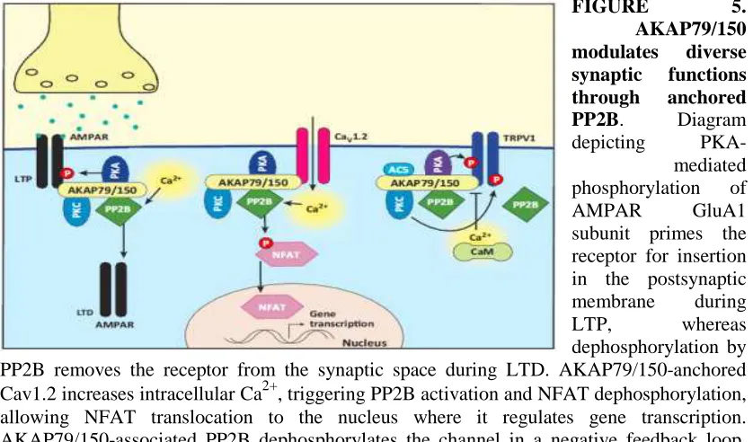
(15) Beta amyloid (Aβ), a peptide generated from the amyloid precursor protein (APP) by neurons, is widely believed to underlie the pathophysiology of Alzheimer’s disease (AD). Aβ is directly involved in the modulation of synaptic function and pathological levels of Aβ inhibit synaptic plasticity. The soluble forms of Aβ affect negatively the process of synaptic transmission involving the loss of synaptic receptors by mechanisms still unknown. The major ionotropic glutamate receptors involved in excitatory. synaptic. transmission. are. alpha-amino-3-hydroxy-5-methyl-4-. isoxazolepropionic acid receptors (AMPARs) and N-methyl D-aspartate receptors (NMDARs). AMPARs are best studied for their rapid trafficking into and out of the synapse by cycling between intracellular stores and the cell surface during synaptic potentiation and depression, respectively. There is an increasing body of evidence to show that Aβ molecules, especially soluble Aβ oligomers (Aβo), exert a negative impact on glutamate receptor trafficking in central synapses, leading to synaptic deficits. Our studies shown acute applications of soluble Aβo and NMDA promoted AMPARs endocytosis in mature mouse cortical primary culture.. However, the precise cellular mechanisms underlying Aβ effects on glutamate receptors remain to be elucidated. A-kinase anchoring proteins (AKAPs), which anchor receptors and signaling proteins to physiological substrates, especially as the AMPAR-Scaffold proteins-Cytoskeleton, AKAP150, in particular, plays a critical role in synaptic plasticity and the stabilization of AMPA (AMPARs) and NMDA (NMDARs) receptors at synapses, although the role of regulating AMPAR trafficking is unclear until now. In our current study, exposure of cultured cortical neurons to both Aβo and NMDA reduces AKAP150 protein levels. It is now recognized that the rate and spatial precision of phosphorylation and dephosphorylation reactions in cells are constrained through the anchoring of kinases and phosphatases near their targets by scaffold proteins. In particular, subcellular targeting by AKAP79/150 of the kinase PKA, phosphatase calcineurin (CaN; also known as PP2B and PPP3), and other enzymes promote highly localized signaling events at the postsynaptic membrane of neuronal dendritic spines (note: AKAP150 is the rodent orthologous of human AKAP79). Importantly, AKAP79/150, PKA, CaN, and CaV1.2 exhibit an enrichment and co-localization in dendritic spines of hippocampal neurons. By using NMDAR, AMPAR, CaN, proteasome inhibitors, we. 10.
(16) studied the mechanism by which Aβo and NMDA-induced decreases in AKAP150 levels. Our results indicated the reduction of AKAP150 requires NMDAR, CaN and proteasome activities. Moreover, the reducing effect of Aβo and NMDA on AMPAR levels was mimicked by knockdown of AKAP150 and blocked by overexpression of AKAP150.. Aβ can reduce long-term potentiation (LTP), a form of synaptic plasticity that is closely associated with learning and memory and can facilitate long-term depression (LTD), an opposing form of synaptic plasticity. LTP and LTD involve postsynaptic phosphorylation and glutamate receptor trafficking. The observation that Aβ reduces LTP and facilitates LTD is suggestive of a role for Aβ in regulating trafficking of glutamate receptors and postsynaptic phosphorylation, but this has not been previously examined. In our research, neither the Aβo nor the NMDA-induced reduction of AKAP150 could be rescued by chemical LTP (cLTP).. Aβ has a strong inhibitory effect on the induction of LTP. In addition, we have also shown that Aβo inhibits cLTP induction in vitro, extending previous report on inhibition of LTP in CA1 in vivo. When solubilized in aqueous buffers, Aβ assembles into a variety of structures, including low N-oligomers, ADDLs, protofibrils, and fibrils. The solutions of synthetic Aβ used in this study contain a mixture of these different assemblies. Moreover, the finding that Aβ inhibits AMPARs exocytosis on overexpression of AKAP150, but not unoverexpressed, could be partially rescued by cLTP in vitro confirms that AKAP150 plays an important role of AMPARs trafficking.. Aβo could be involved in the deficit of dendritic transport of mRNAs and their translation. It has been observed that different experimental models of AD have lower number of neurons with dendrites, indicating that Aβ can regulate synaptic structure and function. The miRNAs are small non-coding RNAs that act as post-transcriptional regulators of gene expression. In early AD, which presents dystrophic neurites and synaptic failure, there is an alteration in the levels of certain miRNAs. Long-term exposure of Aβo demonstrated that miR125a and miR132 were both reduced while long-term exposure of NMDA induced the decrease of miR181a. Experiments with 11.
(17) inhibitors indicated that CaN and Ca2+ may be involved in the modulation of mir181a by long-term exposure of NMDA.. 12.
(18) ABBREVIATIONS 13.
(19) AD. Alzheimer disease. AMPA. α-amino-3-hydroxy-5-methylisoxazole-4-propionic acid. AMPAR. AMPA receptor. NMDA. N-methyl-D-aspartic acid. NMDAR. NMDA receptor. Aβo. Amyloid β oligomer. ADDL. Amyloid beta derived diffusible ligands. AKAP150. A-kinase anchoring protein 150. LTD. long term depression. LTP. long term potentiation. APP. amyloid precursor protein. ACSF. artificial cerebrospinal fluid. F’R. forskolin plus rolipram. BAPTA. 2-bis (2-aminophenoxy) ethane-N’, N’, N’, N’- tetraacetic acid. tetrakis cLTP. chemical LTP. CaMKII. calcium/ calmodulin-dependent protein kinase II. PKA. protein kinase A. PKC. protein kinase C. PP2B. protein phosphatase 2B. PSD. postsynaptic density. EPSCs. excitatory postsynaptic currents. Less frequently used abbreviations are defined upon their first use in the text.. 14.
(20) INTRODUCTION 15.
(21) Neurons are fundamental building blocks with a complex morphology that represent an essential functional unit within the nervous system. AD is the most common neurodegenerative disease and the leading cause of dementia in the elderly. Accumulating evidence supports Aβo as the leading candidate for the causative agent in AD and synapses as the primary site of Aβo action. However, the molecular and cellular mechanisms by which Aβo cause synaptic dysfunction and cognitive impairments remain poorly understood. Although the underlying mechanisms are unclearly, some research have shown Aβo reduces glutamatergic transmission and inhibits synaptic plasticity, meanwhile, Aβo induced long-term depression (LTD) removes surface and synaptic glutamate receptors (Beattie et al., 2000; Lee et al., 2002; Luscher et al., 1999). Furthermore, application of synthetic Aβo results in surface removal of NMDA and GluR1 receptors in dissociated cultured neurons (Almeida et al., 2005; Snyder et al., 2005). Helen et al., 2006 shown that Aβo employs signalling pathways of LTD to drive endocytosis of synaptic AMPA receptors (AMPARs). Qinwen et al., 2005 also demonstrated that Aβ-mediated inhibition of LTP arises from activation of mGluR5 and stimulation of three kinases, JNKI, Cdk5, and p38 MAP kinase. Synaptic removal of AMPARs is necessary and sufficient to produce loss of dendritic spines and synaptic NMDA responses. Through association with the synaptic scaffolding MAGUK (membrane-associated guanylate kinase) proteins, AKAP79/150 and its associated enzymes (PKA, protein kinase C, and PP2B) are directed to both NMDA- and AMPA-type glutamate receptors (Colledge et al., 2000). The identification of an interaction between AKAP79/150 and the MAGUK synaptic scaffolding proteins, SAP97 and PSD-95, linked AKAP79/150 to synaptic AMPA receptors and provided a mechanism by which AKAP79/150 may regulate postsynaptic excitatory transmission (Colledge et al., 2000). A series of molecular, biochemical and electrophysiological experiments established that AKAP79/150-anchored PKA phosphorylates Ser-845 on GluR1 resulting in alteration of channel activity (Tavalin et al., 2002) and that the PKA site is selectively dephosphorylated during LTD (Kameyama et al., 1998; Lee et al., 2000). AKAPdirected kinase/phosphatase complexes have been implicated in the regulation of AMPA receptor surface expression, a process thought to contribute to LTP and LTD.. 16.
(22) 1. GLUTAMATE Glutamate is the most prominent neurotransmitter in the body, and it is the main excitatory neurotransmitter (Petroff et al., 2002), being present in over 50% of nervous tissue. Glutamate was initially discovered to be a neurotransmitter in insect studies in the early 1960s. Glutamate is also used by the brain to synthesise GABA (γAminobutyric acid), the main inhibitory neurotransmitter of the mammalian central nervous system, which plays a role in regulating neuronal excitability throughout the nervous system and is also directly responsible for the regulation of muscle tone in humans (Watanabe et al., 2002).. 1.1. GLUTAMATE RECEPTORS. Mammalian glutamate receptors are classified based on their pharmacology. However, glutamate receptors in other organisms have different pharmacology, and therefore these classifications do not hold. One of the major functions of glutamate receptors appears to be the modulation of synaptic plasticity, a property of the brain thought to be vital for memory and learning. Both metabotropic and ionotropic glutamate receptors have been shown to have an effect on synaptic plasticity (Debanne et al., 2003). An increase or decrease in the number of ionotropic glutamate receptors on a postsynaptic cell may lead to long-term potentiation or long-term depression of that cell, respectively (Pérez et al., 2006; Asztély et al., 1996). Additionally, metabotropic glutamate receptors may modulate synaptic plasticity by regulating postsynaptic protein synthesis through second messenger systems (Weiler et al., 1993). Research shows that glutamate receptors are present in CNS glial cells as well as neurons (Teichberg et al., 1991). These glutamate receptors are suggested to play a role in modulating gene expression in glial cells, both during the proliferation and differentiation of glial precursor cells in brain development and in mature glial cells (Steinhäuser et al., 1996).. 1.2. GLUTAMATE RECEPTOR TYPES Glutamate receptors can be divided into two groups according to the mechanism by which their activation gives rise to a postsynaptic current (Palmada et al., 17.
(23) 1996). Ionotropic glutamate receptors (iGluRs) form the ion channel pore that activates when glutamate binds to the receptor. Metabotropic glutamate receptors (mGluRs) indirectly activate ion channels on the plasma membrane through a signaling cascade that involves G proteins. Ionotropic receptors tend to be quicker in relaying information, but metabotropic ones are associated with a more prolonged stimulus. This is due to the usage of many different messengers to carry out the signal, but since there is a cascade, an activation of a G-protein can lead to multiple activations. Glutamate receptors are usually not specifically geared towards glutamate exclusively as the ligand and sometimes even require another agonist. Of the many specific subtypes of glutamate receptors, it is customary to refer to primary subtypes by a chemical that binds to it more selectively than glutamate. The research, however, is ongoing, as subtypes are identified and chemical affinities measured.. FIGURE 1. Glutamate (Glu) is the major excitatory neurotransmitter in the central nervous system (CNS). It also acts to modulate synaptic transmission both pre- and post-synaptically. These actions are mediated via a large range of ionotropic (iGluR, NMDA, AMPA, and kainate), and metabotropic (mGluR, group I, II, III) receptors. iGluR are directly coupled to cation channels, and their activation evokes fast synaptic events which may lead to longer-term changes in excitability. mGluR exist as eight subtypes of G-protein coupled receptor: Group I (mGluR1 and 5) are excitatory and are believed to act mainly via Gαq/phospholipase C. Group II (mGluR2 and 3), and III (mGluR4, 6, 7, and 8) inhibit adenylate cyclase and alter cation currents via Gαi and Gβγ subunits respectively. Figure is from Blackshaw et al., 2011.. Due to the diversity of glutamate receptors, their subunits are encoded by numerous gene families. Sequence similarities between mammals show a common evolutionary origin for many mGluR and all iGluR genes (Ohashi et al., 2002). Conservation of reading frames and splice sites of GluR genes between chimpanzees and humans are complete, suggesting no gross structural changes after humans diverged from the human-chimpanzee common ancestor. However, there is a possibility that two human-specific. "fixed". amino. acid. substitutions,. D71G in GRIN3A and R727H in GRIN3B, are specifically associated with human. 18.
(24) brain function (Goto et al., 2009).. 1.3. STRUCTURE, MECHANISM AND FUNCTION. Glutamate receptors exist primarily in the central nervous system. These receptors can be found on the dendrites of postsynaptic cells and bind to glutamate released into the synaptic. cleft. by. presynaptic. cells.. They. are. also. present. on. both astrocytes and oligodendrocytes (Dingledine et al., 1999). Glutamate binds to the extracellular portion of the receptor and provokes a different response depending on the type of receptors.. 1.3.1. IONOTROPIC. Ionotropic glutamate receptors, by definition, are ligand-gated cation channels that allow the flow of K+, Na+ and sometimes Ca2+ in response to glutamate binding. Upon binding, the agonist will stimulate the receptor, an ion channel, allowing ion flow and causing excitatory postsynaptic current (EPSC). This current is depolarizing and, if enough glutamate receptors are activated, may trigger an action potential in the postsynaptic neuron. All produce excitatory postsynaptic current, but the speed and duration of the current is different for each type. NMDA receptors have an internal binding site for an Mg2+ ion, creating a voltage-dependent block, which is removed by outward flow of positive current (Johnson et al., 1990), Since the block must be removed by outward current flow, NMDA receptors rely on the EPSC produced by AMPA receptors to open. NMDA receptors are permeable to Ca2+ (Crowder et al., 1987), which is an important cation in the nervous system (Gover et al., 2009) and has been linked to gene regulation (Barbado et al., 2009). The flow of Ca2+ through NMDA receptors is thought to cause both LTP (of synapse efficacy) and LTD by transducing signaling cascades and regulating gene expression.. 1.3.2. AMPA RECEPTORS. AMPARs are ionotropic glutamate receptors that mediate rapid excitatory transmission in the mammalian brain. They are heterotetrameric cation channels 19.
(25) comprised of a combinatorial assembly of four subunits, GluR1-GluR4 (GluRA-D) (Hollmann et al., 1994). Regulated trafficking of AMPARs has emerged as an important mechanism that underlies the activity-dependent modification of synaptic strength. The GluA2 subunit is uniquely edited at the mRNA level, where a glutamine codon is switched to arginine (Lomeli et al., 1994), which confers channel resistance to calcium. Therefore, AMPAR complexes lacking GluA2 are permeable to sodium and calcium ions resulting in higher conductance. In addition, calcium permeable AMPARs (CP-AMPARs) exhibit inward rectification in current–voltage plots that differs from the linear rectification of predominate AMPARs due to voltagedependent block by intracellular polyamines (Donevan and Rogawski, 1995). GluA2 lacking AMPARs can be formed through GluA1 homomers or combinations of GluA1, 3, and 4.. Delivery of AMPARs to the postsynaptic membrane leads to LTP, whereas removal of these receptors leads to LTD (Barry et al., 2002; Bredt et al., 2003; Malinow et al., 2002). Both of these forms of synaptic plasticity are influenced by NMDAR activity (Bliss et al., 1993). Regulation of AMPAR synaptic insertion is determined by the receptor subunit composition. While synaptic activity drives GluR1-containing receptors to the synapse, thus enhancing transmission, AMPARs lacking GluR1, such as GluR2/3 heteromers, constitutively cycle in and out of the synapse, in an activityindependent manner, entering and leaving sites initially occupied by GluR1containing receptors. This distinction in subunit trafficking is determined by the subunit intracellular CTDs (Passafaro et al., 2001).. Changes in synaptic strength are believed to be the basis of learning and memory. These alterations in neuronal communication are controlled in part by the insertion or removal of AMPA receptors (AMPARs) from the postsynaptic membrane of excitatory synapses. Regulation of AMPAR trafficking to and from the plasma membrane can occur through the C-terminal tails of AMPARs, either through interactions with other proteins or changes in phosphorylation (Malinow et al., 2002; Derkach et al., 2007). When surface AMPARs are internalized, subunit-specific interactions with other proteins and changes in their phosphorylation status allow for. 20.
(26) their recycling and functional insertion into the plasma membrane (Hirling et al., 2009). Alternatively, AMPARs have been shown to traffic to the lysosome by a distinct pathway, although the mechanisms regulating this process are not well understood (Ehlers et al., 2000; Kessels et al., 2009).. 1.3.3. NMDA RECEPTOR The N-methyl-D-aspartate receptor (also known as the NMDA receptor or NMDAR) is. the. predominant. molecular. device. for. controlling synaptic. plasticity and memory function. Activation of NMDRs results in the opening of an ion channel that is nonselective to cations with an equilibrium potential near 0 mV. A property of the NMDA receptor is its voltage-dependent activation, a result of ion channel blocked by extracellular Mg2+ & Zn2+ ions. This allows the flow of Na+ and Ca2+ ions into the cell and K+ out of the cell to be voltage-dependent (Dingledine et al., 1999; Liu et al., 2000; Cull et al., 2001; Paoletti et al., 2007). The predominant hypothesis for the mechanisms of NMDAR involvement is that the activation of NMDAR elevates intracellular calcium concentrations ([Ca2+]) that trigger various intracellular signaling systems. Cytosolic calcium is an important mediator of neuronal signal transduction, participating in diverse biochemical reactions that elicit changes in synaptic function, metabolic rate, and gene transcription. Up-regulation of NMDA receptors or high extracellular glutamate concentration can result in an excessive entry of calcium, triggering a series of cytoplasmic and nuclear processes such as loss of mitochondrial membrane potential, which ultimately results in neuronal cell death. Moreover, calcium flux through NMDARs is thought to be critical in synaptic plasticity, a cellular mechanism for learning and memory. The NMDA receptor is distinct in two ways: first, it is both ligand-gated and voltagedependent; second, it requires co-activation by two ligands: glutamate and either Dserine or glycine (Kleckner et al., 1988).. The NMDA receptor forms a heterotetramer between two GluN1 and two GluN2 subunits (the subunits were previously denoted as NR1 and NR2), two obligatory NR1 subunits and two regionally localized NR2 subunits. A related gene family of NR3 A and B subunits have an inhibitory effect on receptor activity. Multiple 21.
(27) receptor isoforms with distinct brain distributions and functional properties arise by selective splicing of the NR1 transcripts and differential expression of the NR2 subunits. There are eight variants of the NR1 subunit produced by alternative splicing of GRIN1 (Stephenson et al., 2006): NR1-1a, NR1-1b; NR1-1a is the most abundantly expressed form. NR1-2a, NR1-2b; NR1-3a, NR1-3b; NR1-4a, NR1-4b; GluN2: NR2 subunit in vertebrates and invertebrates. While a single NR2 subunit is found in invertebrate organisms, four distinct isoforms of the NR2 subunit are expressed in vertebrates and are referred to with the nomenclature NR2A through D (coded by GRIN2A, GRIN2B, GRIN2C, GRIN2D). Each NR2 subunit has a different intracellular C-terminal domain that can interact with different sets of signaling molecules (Ryan et al., 2009). Unlike NR1 subunits, NR2 subunits are expressed differentially across various cell types and control the electrophysiological properties of the NMDA receptor. One particular subunit, NR2B, is mainly present in immature neurons and in extrasynaptic locations, and contains the binding-site for the selective inhibitor ifenprodil. Whereas NR2B is predominant in the early postnatal brain, the number of NR2A subunits grows, and eventually NR2A subunits outnumber NR2B. This is called NR2B-NR2A developmental switch, and is notable because of the different kinetics each NR2 subunit lends to the receptor (Liu et al., 2004). For instance, greater ratios of the NR2B subunit leads to NMDA receptors which remain open longer compared to those with more NR2A. This may in part account for greater memory abilities in the immediate postnatal period compared to late in life, which is the principle behind genetically-altered 'doogie mice'. The NR2B and NR2A subunits also have differential roles in mediating excitotoxic neuronal death (Liu et al., 2007). The developmental switch in subunit composition is thought to explain the developmental changes in NMDA neurotoxicity (Zhou et al., 2006). Disruption of the gene for NR2B in mice causes perinatal lethality, whereas the disruption of NR2A gene produces viable mice, although with impaired hippocampal plasticity (Sprengel et al., 1998). One study suggests that Reelin may play a role in the NMDA receptor maturation by increasing the NR2B subunit mobility (Groc et al., 2007).. Each receptor subunit has modular design and each structural module also represents a functional unit: 1) The extracellular domain contains two globular structures: a modulatory domain and a ligand-binding domain. NR1 subunits bind the co-agonist glycine and NR2 subunits bind the neurotransmitter glutamate. 2) The agonist-binding 22.
(28) module links to a membrane domain, which consists of three trans-membrane segments and a re-entrant loop reminiscent of the selectivity filter of potassium channels. The membrane domain contributes residues to the channel pore and is responsible for the receptor's high-unitary conductance, high-calcium permeability, and voltage-dependent magnesium block. Each subunit has an extensive cytoplasmic domain, which contain residues that can be directly modified by a series of protein kinases and protein phosphatases, as well as residues that interact with a large number of structural, adaptor, and scaffolding proteins.. Recently, Lee et al., 2014 presented X-ray crystal structures of the Xenopus laevis GluN1–GluN2B NMDA receptor with the allosteric inhibitor, Ro25-6981, partial agonists and the ion channel blocker, MK-801. Receptor subunits are arranged in a 12-1-2 fashion, demonstrating extensive interactions between the amino terminal and ligand-binding domains. The transmembrane domains harbour a closed-blocked ion channel, a pyramidal central vestibule lined by residues implicated in binding ion channel blockers and magnesium, and a twofold symmetric arrangement of ion channel pore loops. These structures provide new insights into the architecture, allosteric coupling and ion channel function of NMDA receptors. The glycine-binding modules of the NR1 and NR3 subunits and the glutamate-binding module of the NR2A subunit have been expressed as soluble proteins, and their three-dimensional structure has been solved at atomic resolution by x-ray crystallography. This has revealed a common fold with amino acid-binding bacterial proteins and with the glutamate-binding module of AMPA-receptors and kainate-receptors.. 1.3.4. METABOTROPIC. Metabotropic glutamate receptors, which belong to subfamily C of G protein-coupled receptors are divided into three groups, with a total of eight subtypes (in mammals; this is not necessarily the case for most organisms). The mGluRs are composed of three distinct regions: the extracellular region, the transmembrane region, and the intracellular region (Muto et al., 2007). The extracellular region is composed of a venus flytrap (VFT) module that binds glutamate (Pin et al., 2002), and a cysteine-. 23.
(29) rich domain that is thought to play a role in transmitting the conformational change induced by ligand binding from in the VFT module to the transmembrane region. The transmembrane region consists of seven transmembrane domains and connects the extracellular region to the intracellular region where G protein coupling occurs. Glutamate binding to the extracellular region of an mGluR causes G proteins bound to the intracellular region to be phosphorylated, affecting multiple biochemical pathways and ion channels in the cell (Platt et al., 2007). Because of this, mGluRs can both increase or decrease the exitability of the postsynaptic cell, thereby causing a wide range of physiological effects.. 2. SYNAPTIC PLASTICITY AND RECEPTOR TRAFFICKING. 2. 1. SYNAPTIC PLASTICITY. The most remarkable property of synapses is not that they convey information from one neuron to another, but that they can readily alter the efficiency with which they do this. This property, known as synaptic plasticity, is the basis of information storage in the brain. It enables us to store and use vast amounts of information in the form of learnt behaviors and conscious memories. During the past 40 years, one form of synaptic plasticity, known as LTP has consolidated its status as the pre-eminent synaptic model for investigating the molecular basis of memory (Bliss et al., 1993). LTP has been most extensively studied in the hippocampus, although it is also seen at many synapses throughout the CNS. It is characterized by a long-lasting increase in synaptic strength that is caused by a brief period of coordinated neuronal activity. Although LTP is persistent, the increase in synaptic strength can be reversed by different patterns of neuronal activity by a process known as depotentiation. These same patterns of neuronal activity can, under certain circumstances, lead to LTD of synaptic transmission from the basal (non-potentiated) state, and this de novo LTD can be reversed by a process of de-depression. Bi-directional and reversible alterations in synaptic efficiency make possible the dynamic storage of vast amounts of neurally encoded information. GluRs and GABA (-aminobutyric acid) receptors (GABARs) mediate most synaptic transmission in the vertebrate CNS and are pivotal. 24.
(30) in the induction, expression and/or modulation of synaptic plasticity. NMDA receptors (NMDARs) are the main triggers for the induction of LTP and LTD, and their activation is tightly regulated by the activation of GABAA and GABAB receptors. Both kainate receptors and mGluRs can trigger the induction of NMDAR independent forms of LTP and LTD. AMPARs mediate the synaptic response that is most commonly studied during LTP and LTD experiments, and are believed to be an important target of modulation during synaptic plasticity. In recent years, glutamate and GABA receptors have been shown to move rapidly around the neuron so as to alter the number and composition of receptors that are available to respond to released neurotransmitters. Receptor trafficking involves the intracellular movement of receptors from sites of synthesis to the plasma membrane, where they function, and then to sites of degradation. The receptors are inserted into and removed from the plasma membrane by exocytosis and endocytosis respectively, and diffuse laterally within the plasma membrane. 2.2. AMPARs TRAFFICKING It is generally agreed that postsynaptic changes in AMPAR function make an important contribution to the expression of LTP. Broadly speaking, LTP could involve the modulation of AMPARs that are already expressed at the synapse and/or the rapid recruitment of new AMPARs to the synapse to alter the number present (Benke et al., 1998). In the latter mechanism, the new receptors could have either the same properties as the existing population or different ones (Liu et al., 2000; Palmer et al., 2004). Whatever mechanism is employed for LTP, LTD could be its mirror image, or might evoke mechanistically distinct processes to reduce synaptic strength. Studies of AMPAR trafficking in synaptic plasticity have focused on the idea that an alteration in AMPAR number is one of the expression mechanisms for LTP and LTD. Early evidence for this was provided when it was shown that LTP involves the rapid ‘unsilencing’ of ‘silent’ synapses (Malinow et al., 2002) - synapses that show NMDAR- but not AMPAR-mediated responses. Further work showed that LTD could involve the ‘re-silencing’ of synapses (Luthi et al., 1999). The mechanistic explanation for silent synapses and the locus of their unsilencing have been hotly debated, but one idea involves the rapid recruitment of AMPARs to the plasma membrane of synapses that previously lacked AMPARs. Consistent with this idea, 25.
(31) antibodies that bind AMPARs on living neurons have revealed that spines with AMPARs on their surface can be found adjacent to spines in which AMPARs can only be detected intracellularly (Richmond et al., 1996). Over the past few years, much information has been obtained concerning the trafficking of AMPARs, from their synthesis and dendritic trafficking to their membrane insertion and removal (Bredt et al., 2003). In this study, we focus on the rapid movement of AMPARs that might be associated with LTD induced by either Aβo or NMDA stimulations. Recently, two broad approaches have been using to visualize AMPARs in living neurons to investigate how their movement relates to synaptic plasticity. One approach uses recombinant AMPAR subunits that contain a fluorescent reporter, such as green fluorescent protein (GFP). LTP was found to be associated with the delivery of GFP–GluR1 to spines in organotypic slice cultures (Shi et al., 1999). As the use of imaging techniques could not determine whether these constructs were inserted into the plasma membrane during LTP, mutated receptors were used that had a distinct electrophysiological signature. Alterations in the rectification properties of the synaptic currents showed that recombinant AMPARs were delivered to the synapse (Hayashi et al., 2000). The development of pH-sensitive GFP-AMPAR constructs means that researchers can now directly observe the insertion and removal of recombinant receptors into and from the membrane, because the pH of the lumen of intracellular vesicles differs from that of the extracellular environment (Ashby et al., 2004). This new approach is likely to facilitate greater understanding of many aspects of AMPAR trafficking in synaptic plasticity. In the second method, antibodies that recognize extracellular epitopes of native AMPAR subunits were used in conjunction with protocols that induce NMDAR dependent LTP and LTD in dissociated cultured neurons; the alteration in native AMPAR distributions was determined using fluorescently conjugated secondary antibodies. This approach has been used to show that AMPARs are internalized during NMDAR-dependent LTD (Beattie et al, 2000; Carroll et al, 2001) and inserted into the membrane during NMDAR dependent LTP (Lu et al., 2001; Pickard et al., 2001). LTD induced by activation of mGluRs might also involve the internalization of AMPARs (Snyder et al., 2001; Xiao et al., 2001).The use of antibodies is a less dynamic and flexible approach than the imaging of recombinant receptors but provides essential information about how native receptors behave during synaptic plasticity.. 26.
(32) FIGURE 2. Synaptic plasticity in the hippocampus and glutamate receptor subtypes. a. schematic diagram of the rodent hippocampal slice preparation, showing the main excitatory pathways (AC, associational/commissural; MF, mossy fibre; PP, perforant path; SC, Schaffer collateral). Typical electrode placements for studying synaptic plasticity at Schaffer collateral–commissural synapses are indicated. The traces are field excitatory postsynaptic potentials (EPSPs) recorded before (1) and during (2) LTP (long-term potentiation). b. Timecourse plots showing alterations in field EPSP (rising slope normalized to baseline) against time, during LTP (100-Hz stimulation, 1 s, baseline intensity) or after the induction of de novo LTD (long-term depression) (1-Hz stimulation, 15 min, baseline intensity). The black bar represents the time of the stimulus and the numbers (1 and 2) indicate the time points illustrated in (a). c. KA, kainate receptor subunit. Figure is from Richmond, et al., 1996.. 27.
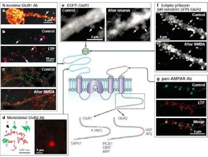
(33) FIGURE 3. Rapid trafficking of AMPARs during synaptic plasticity. Centre: schematic diagram of an AMPA (-amino-3-hydroxy-5-methyl-4-isoxazole-propionic acid) receptor (AMPAR) subunit showing the domain structure, protein–protein interaction sites, GFP (green fluorescent protein) insertion sites and sites of epitopes for various antibodies used for imaging AMPAR trafficking. a. Superimposed image showing the localization of surface GluR1 (glutamate receptor 1) subunits using an N-terminal antibody (Ab) on a living hippocampal neuron (yellow) and the localization of total GluR1 using a C-terminal antibody after permeabilization (red). b. Induction of LTP (long-term potentiation) using glycine increases surface GluR1 in cultured hippocampal neurons. Surface GluR1 labelled using an N-terminal GluR1 antibody under nonpermeabilizing conditions in control (upper) and glycine-treated (lower) cultures. c. NMDA (N-methyl-D-aspartate)-induced internalization of AMPARs in cultured hippocampal neurons. Internalized GluR1 labelled with an N-terminal GluR1 antibody using an acid-stripping protocol (red) and co-localized with transferrin (green) in control (upper) and NMDA-treated (lower) cultures. d. AMPARs are mobile in the plasma membrane and in spines. Single-molecule tracking traces for individual GluR2 subunits (left), and an example of the signal from a single molecule (right). e. Redistribution of GFP-tagged GluR1 to spines after LTP induction. Arrows indicate individual spine locations. f. pH-sensitive GFP tagged GluR2 shows the removal of AMPARs from spines after NMDA treatment. g. Sequential labelling of surface AMPARs using pan-AMPAR antibodies shows an increase in surface AMPAR puncta (red) after LTP induction. ABP, AMPAR binding protein; AP2, adaptor complex; GRIP, glutamate receptor-interacting protein; NSF, N-ethylmaleimide-sensitive factor; PICK1, protein interacting with C-kinase; SAP97, synapse-associated protein 97; 4.1N/G, BANDS 4.1N AND 4.1G. Figure is from Ashby et al., 2004.. More precise measurements using single molecule fluorescence microscopy have revealed that both immobile receptors and mobile AMPARs are contained in spines 28.
(34) (Tardin et al., 2003). Stimulation with L-glutamate increases the rate of diffusion of synaptic AMPARs, decreases the proportion of immobile receptors and increases the proportion of receptors in the area around synapses. This indicates that lateral movement of AMPARs in the plasma membrane might be a mechanism for the regulation of AMPAR number during synaptic plasticity. In support of this, there is evidence that, after exocytosis, AMPARs are diffusely distributed along dendrites before accumulating at synaptic sites (Passafaro et al., 2001). The lateral diffusion model also predicts that a component of LTD is due to the rapid lateral diffusion of AMPARs from synaptic to extrasynaptic sites where they are then internalized. In agreement with this, endocytic zones close to the postsynaptic density have been identified. Moreover, AMPARs tagged with pH-sensitive GFP have been used to show that NMDAR activation leads to the rapid internalization of AMPARs from extrasynaptic sites, which precedes the loss of AMPARs from synapses (Ashby et al., 2004).. FIGURE 4. Proteins that associate with the C termini of AMPAR (-amino-3-hydroxy-5methyl-4-isoxazole-propionic acid receptor) (a) or GABAAR (type A -aminobutyric acid receptor) (b) subunits. Multi-domain scaffolding proteins: the multi-PDZ DOMAINcontaining proteins SAP97 (synapse-associated protein 97) for GluR1 (glutamate receptor 1) and GRIP (glutamate receptorinteracting protein)/ABP (AMPAR binding protein) for GluR2 and 3 (a), and the trimeric scaffolding protein gephyrin for -subunits of GABAARs (b). NSF (N-ethylmaleimide-sensitive factor): direct interaction with GluR2 and 3 (a), and association with -subunits through its interaction with GABARAP (GABAR associated protein) (b). AP2 (adaptor complex): interaction with GluR2 and 3 subunits (a), and with the - and -subunits (b). AKAPs (A-kinase-anchoring proteins): AKAP150 (AKAP79) associates with the GluR1 subunit through an interaction with SAP97 (a), and AKAP150 directly interacts with the subunit (b). Proteins that interact with PKC (protein kinase C) (PKC chaperones): PICK1 (protein interacting with C-kinase) binds directly to GluR2 and 3 (a), and RACK1 (receptor for activated C-kinase) binds to -subunits (b). Cytoskeleton-interacting proteins: bands 4.1N 29.
(35) and 4.1G interact with GluR1 (a), GABARAP interacts with - subunits, and PLIC1 (protein that links IAP to the cytoskeleton) interacts with -subunits (b). Motor proteins: KIF5 (kinesin family member 5) associates with GluR2 and 3 through its interaction with GRIP, myosin-VI associates with GluR1 through its interaction with SAP97 (a), and GRIF1, a hypothesized motor protein, interacts directly with -subunits (b). UBIQUITINATION pathway proteins: the E3 ligase Mdm2 interacts with PSD-95 (postsynaptic density protein 95), which associates with AMPARs through its interaction with TARPs (transmembrane AMPAR regulatory proteins) (a), and PLIC1, which regulates ubiquitination, interacts directly with -subunits (b). PKA, protein kinase A; PP2B, protein phosphatase 2B. Figure is from Collingridge G. et,. al., 2004. So, a general model is emerging in which synaptic plasticity involves both regulated exocytosis and endocytosis of AMPARs at extrasynaptic sites and their regulated lateral diffusion into and out of the synapse, perhaps involving a pool of juxtasynaptic AMPARs that are available for rapid recruitment. What mechanisms drive AMPARs into and out of synapses during LTP and LTD through exocytosis, endocytosis and lateral diffusion? Various protein kinases have been implicated in the induction of NMDAR-dependent LTP; these include protein kinase C (PKC), calcium/ calmodulin-dependent protein kinase II (CaMKII), protein kinase A (PKA), p42/44 mitogen-activated protein kinase (MAPK) and phosphatidylinositol 3-kinase (PI3K).Conversely, protein phosphatases, in particular protein phosphatase 1 (PP1) and protein phosphatase 2B (PP2B), have been implicated in NMDAR-dependent LTD. It is likely that the insertion and removal of AMPARs involves many kinases and phosphatases and that some of these are involved in aspects of LTP and LTD other than AMPAR trafficking. Our discussion of the role of kinases is therefore limited to those studies that directly address the trafficking of AMPARs during synaptic plasticity. There is evidence that CaMKII PKA and PI3K (Man et al., 2003; Esteban et al., 2003) are involved in LTP, and that protein phosphatases and PKC are involved in different forms of LTD. 3. LONG TERM DEPRESSION. Although genetic and pharmacological studies have suggested that NMDAR-LTD of AMPAR-mediated synaptic transmission involves the activation of specific NMDAR subtypes (Collingridge et al., 2004), there is considerable flexibility - for example, GluN2B-containing NMDARs are required for LTD, but only under certain circumstances (Wong et al., 2007; Morishita et al., 2007). It is therefore likely that various NMDAR subtypes can trigger LTD and that the actual subtype(s) that are 30.
(36) involved depend on various factors, such as the induction protocol that is employed, expression levels (which vary according to brain region and developmental stage (Yashiro et al., 2008) and environmental conditions (for example, access to a running wheel) (Vasuta et al., 2007). In addition, in young adult animals proBDNF (the precursor of brain-derived neurotrophic factor (BDNF)), acting via neurotrophin receptor P75 (p75NTR), increases GluN2B expression and so enables a GluN2Bsensitive form of NMDAR-LTD to occur (Woo et al., 2005). Furthermore, hippocampal NMDAR-LTD that is induced in adult animals by blocking l-glutamate uptake may also be dependent on GluN2B activation. De novo NMDAR-LTD in CA1 is pronounced early in development, but is more difficult to induce in brain slices from adult animals (Yang et al., 2005) or in intact rodent hippocampi in vivo (Staubli et al., 1997). However, hippocampal LTD can be induced using certain protocols (Staubli et al., 1997) and is facilitated by exposing an animal to novelty or mild stress (Kemp et al., 2007). The effects of stress can be mimicked by blocking l-glutamate uptake (Yang et al., 2005). As the CNS develops, glutamate transporter mechanisms may limit activation of the NMDARs that would otherwise trigger LTD. Some studies have also shown strain differences, with LTD being more easily induced in Wistar and Sprague - Dawley strains than in hooded rat strains (Manahan et al, 2000a; Manahan et al, 2000b). However, there is also evidence that NMDAR-LTD is important for certain forms of learning and memory in the adult animal in the absence of stress. NMDAR-LTD (A). In NMDAR-LTD (A), Ca2+ that enters through NMDARs binds to calmodulin to activate PP2B (also known as calcineurin), which dephosphorylates inhibitor-1 and this leads to the activation of protein phosphatase 1 (PP1) (Mulkey et al., 1993). PP1 then dephosphorylates its substrate(s), including ser845 on the AMPAR subunit GluA1 and this leads to LTD. In addition, Ca2+ entry triggers Ca2+ release from intracellular stores (Nishiyama et al., 2000) and this may serve to activate Ca2+ sensitive enzymes that are located further away from the postsynaptic density (PSD), where endocytosis may occur. The first clue to the molecular mechanism that drives the endocytosis of AMPARs during NMDAR-LTD was the observation that disruption of an interaction between GluA2 and Nethylmaleimide-sensitive factor (NSF; an ATPase involved in membrane fusion events) causes AMPAR internalisation, mimicking NMDAR-LTD. Later, it was shown that clathrin-mediated endocytosis was involved in this process and that the clathrin adaptor protein AP2 also binds to the NSF site on the GluA2 subunit. This 31.
(37) suggests that AMPARs are stabilised on the membrane by NSF and that AP2 replaces NSF to initiate AMPAR endocytosis during NMDAR-LTD. A potential mechanism for the triggering of this exchange involves hippocalcin, a member of the neuronal calcium sensor (NCS) family. On sensing small rises in Ca2+ hippocalcin translocates to the plasma membrane, where it forms a complex with AP2 and GluA2 that may initiate clathrin-mediated AMPAR endocytosis (Palmer et al., 2005). In addition, the small GTPase Rab5 has been implicated in LTD (Brown et al., 2005). Other steps in clathrin-mediated endocytosis that underlies NMDAR-LTD are also beginning to be determined - for example, calcyon, a protein that regulates clathrin assembly is involved in NMDAR-LTD in the hippocampus (Davidson et al., 2009). Protein interacting with C kinase 1 (PICK1) is another protein that binds directly to GluA2 and has been implicated in NMDAR-LTD. PICK1 is a low-affinity Ca2+ sensor that can also bind protein kinase Cα (PKCα) and that can sense, and perhaps induce, membrane curvature. PICK1 competes with the scaffolding proteins AMPAR binding protein (ABP) and glutamate receptor interacting protein (GRIP) for binding to the carboxy-terminal (C-terminal) region of GluA2 and it also promotes internalization of GluA2-containing AMPARs. It was originally proposed that during NMDAR-LTD PICK1 may promote the synaptic removal of AMPARs by inducing the PKCαmediated phosphorylation of ser880 of GluA2 to dissociate AMPARs from ABP– GRIP. However, most of the experimental data do not support this hypothesis. NMDA-induced internalization of AMPARs is not dependent on the phosphorylation status of ser880 or on PICK1 (Lin et al., 2007) and PKC inhibitors do not affect NMDAR-LTD (Peineau et al., 2009; Oliet et al., 1997). Experiments that use inhibitors of the GluA2–PICK1 interaction have yielded conflicting results. One study acutely applied an interfering peptide and reported no effect on NMDAR LTD, whereas a subsequent report described a partial inhibition. A small-molecule inhibitor of the GluA2–PICK1 interaction was also found to partially inhibit NMDAR-LTD (Thorsen et al., 2010). These partial effects are in contrast with the abolition of NMDAR-LTD that results from chronic manipulation of PICK1 (Terashima et al., 2008). Thus, the presence of PICK1 may be necessary for NMDAR-LTD in the longterm but it probably plays only a minor part, if any, in the release of AMPARs from their synaptic tethers. A more important role of ABP–GRIP may be to anchor AMPARs at non-synaptic sites (intracellular or extra-synaptic sites on the plasma membrane), as recently confirmed by paired-cell recording experiments (Emond et 32.
(38) al., 2010). NMDAR-LTD becomes unstable if the ability of AMPARs to bind ABPGRIP is impaired. This implies that by retaining AMPARs at non-synaptic sites this scaffolding molecule is crucial for the expression of this form of LTD. PICK1 may enable the disassociation of AMPARs from ABP–GRIP at these non-synaptic sites, thereby enabling de-depression (re-potentiation) of synaptic transmission. Another key function of PICK1 in NMDAR-LTD may be to enable actin depolymerization through an interaction with F-actin and the actin-related protein 2/3 (Arp2/3 complex), and through this process to modify neuronal architecture (Rocca et al., 2008). NMDAR-LTD (A) is classically assumed to require the activation of phosphatases. However, studies using kinase inhibitors have implicated various serine/ threonine (Ser/Thr) protein kinases in this process as well. These include PKA (Brandon et al., 1995), cyclin-dependent kinase 5 (Ohshima et al., 2005), P38 mitogen-activated protein kinase (p38MAPK) (Zhu et al., 2005) and glycogen synthase kinase-3 (GSK3) (Peineau et al., 2007). As with all inhibitor studies, potential off-target effects should be taken into account and this is particularly important for protein kinases because the mammalian genome encodes over 500 protein kinases. A role for GSK3 in NMDAR-LTD is supported by the effects of six different GSK3 inhibitors including lithium, which may exert some of its therapeutic actions via this mechanism. A direct link between the protein phosphatase cascade and GSK3 was observed during NMDAR-LTD; PP1 dephosphorylates GSK3β and its upstream inhibitor Akt and these actions result in activation of GSK3β (Peineau et al, 2007). An additional mechanism of GSK3 activation may occur via caspase-3 (Li et al, 2010). This protease is activated during NMDAR-LTD through a cascade involving cytochrome c and caspase-9 and is able to cleave Akt, thus removing its tonic inhibition of GSK3. The finding that both caspases and GSK3β, an enzyme that is deregulated in patients with Alzheimer’s disease, are involved in NMDAR-LTD raises the possibility that the neurodegeneration that underlies Alzheimer’s disease and related dementias may be caused at least in part by pathological activation of this form of LTD. NMDAR-LTD is associated with tyrosine phosphorylation of GluA2 and this suggests that protein tyrosine kinases (PTKs) are also involved. PTKs of the sarcoma (Src) family phosphorylate GluA2 at a tyrosine residue in a tyrosine-rich region of the C-terminal tail of GluA2 and this is thought to be required for AMPAR endocytosis. Consistent with this idea, a peptide that mimics this tyrosine-rich region has been found to block NMDAR-LTD (Ahmadian et al., 2004). What are the targets 33.
(39) of enzymes that are activated during NMDAR-LTD and underlie an alteration in the synaptic expression of AMPARs? A major target seems to be PSD95, which positions calcineurin near the mouth of the NMDAR channel through an interaction with Akinase anchor protein (AKAP)-150 (Bhattacharyya et al., 2009) and which is dephosphorylated on ser295 during LTD to enable the removal of PSD95 from the synapse and thereby permit AMPAR endocytosis (Kim et al., 2007). These mechanisms occur rapidly after NMDAR LTD is triggered. However, protein synthesis is required for LTD to be sustained as inhibitors of translation cause a recovery of synaptic transmission in a few hours. How these newly synthesized proteins sustain LTD for longer periods of time is not known but regulators of gene transcription that may be involved in NMDAR-LTD are starting to be investigated (Wu et al., 2010).. 4. SOLUBLE PROTEIN OLIGOMERS IN NEURODEGENERATION: FROM THE ALZHEIMER’S AMYLOID β-PEPTIDE. 4.1 THE DISCOVERY OF THE FIRST AD CAUSATIVE GENE The AD causative gene encodes -amyloid precursor protein (APP), which is a singletransmembrane, receptor-like protein that is expressed ubiquitously in neural and nonneural cells. After the Bavarian psychiatrist Alois Alzheimer presented his first clinicpathological case in Tübingen on November 3, 1906 (Alzheimer A 1907), it became clear that amyloid plaques in the cerebral cortex (were invariably associated with the disease that now bears his name. In the 1980s, biochemists focused on the isolation of the amyloid to identify its principal component. Glenner and Wong purified microvascular amyloid deposits from the meninges of AD brains and provided a partial sequence of an ~4-kDa subunit protein that they named amyloid -protein. Shortly thereafter, Masters, Beyreuther and co-workers identified the same protein as the subunit of amyloid plaque cores that were isolated from post-mortem AD cortices (Masters et al., 1985). Around the same time, the microtubule-associated protein tau was identified as the main constituent of the hallmark neurofibrillary tangles that accumulate inside many neurons and their processes in brains (Grundke et al., 1986; Kosik et al., 1986). Tau is a highly soluble cytoplasmic protein that binds to tubulin 34.
(40) during its polymerization into microtubules in neurons and thereby stabilizes these important cytoskeletal organelles. Glenner also showed that the amyloid deposits that occur in the brain vessels of young adults with Down syndrome were composed of AD. Since 1969, it had been known that middle-aged patients with Down syndrome develop the amyloid plaques and neurofibrillary tangles that are typical of AD (Olson et al., 1969). On this basis, Glenner assumed that the gene ultimately found to encode might be causative of AD cases. The subsequent cloning of APP, which encodes a large, type 1 membrane glycoprotein, by Beyreuther and co-workers in 1987, and its localization to the long arm of chromosome, was consistent with this hypothesis (Kang et al., 1987). These biochemical findings pointed strongly to the APP gene as a site which geneticists should search for AD-causing mutations. The first such mutation was discovered in a family with hereditary cerebrovascular amyloidosis with multiple haemorrhages (Levy et al., 1990). Shortly thereafter, a distinct APP missense mutation was found in a family with early-onset AD (Goate et al., 1991), and then additional mutations in other families were detected (Chartier et al., 1991). These and other findings led to the formal proposal of a hypothesis of disease in which excessive accumulation and deposition could trigger a complex downstream cascade that resulted in the symptoms of AD (Hardy et al., 1992). In its most recent iteration (Hardy et al., 2002), the amyloid (or Aβ) hypothesis states that the gradual accumulation and aggregation of this small hydrophobic peptide initiates a slow but deadly cascade that leads to synaptic alterations, microglial and astrocytic activation, the modification of the normally soluble tau protein into oligomers and then into insoluble paired helical filaments, and progressive neuronal loss associated with multiple neurotransmitter deficiencies and cognitive failure. 4.2 THE REGULATED INTRAMEMBRANE PROTEOLYSIS OF APP Initially, the mechanism by which the partially intra-membrane region could be liberated as a free peptide from its precursor was enigmatic and was considered to require some pre-existing membrane injury. It was assumed that the hydrophobic interior of the membrane bilayer would need to be damaged to allow access of a protease and water to effect cleavage. But this concept was disproven when was unexpectedly discovered to be produced normally by the intramembranous proteolysis of APP throughout life and APP was found to circulate in extracellular fluids, 35.
(41) including cerebrospinal fluid (CSF) and plasma (Busciglio et al., 1993). This discovery opened up the dynamic study of A, which heretofore had only been obtained through painstaking isolation from post-mortem human brain. As predicted by the amyloid hypothesis, all AD-causing APP mutations that have been identified so far occur either within or flanking the region of this large polypeptide. Accordingly, the mutations that flank the region increase the production of the highly amyloidogenic Aβ 42 isoform, whereas the mutations within the region enhance the oligomerization of the peptide (Haass et al., 2004).. 4.3. PROCESSING OF AMYLOID PRECURSOR PROTEIN Cleavage by α-secretase interior to the β-amyloid peptide (Aβ) sequence initiates nonamyloidogenic processing. A large amyloid precursor protein (sAPPα) ectodomain is released, leaving behind an 83-residue carboxy-terminal fragment. C83 is then digested by γ-secretase, liberating extracellular p3 and the amyloid intracellular domain (AICD). A myloidogenic processing is initiated by β-secretase beta-site amyloid precursor protein–cleaving enzyme 1 (BACE-1), releasing a shortened sAPPα. The retained C99 is also a γ-secretase substrate, generating Aβ and AICD. γ-Secretase cleavage occurs within the cell membrane in a unique process termed “regulated intramembranous proteolysis.” sAPPα and sAPPβ are secreted APP fragments after α-secretase and β-secretase cleavages, respectively. AICD is a short tail (approximately 50 amino acids) that is released into the cytoplasm after progressive ε-to-γ cleavages by γ-secretase. AICD is targeted to the nucleus, signaling transcription activation. Lipid rafts are tightly packed membrane micro-environments enriched in sphingomylelin, cholesterol, and glycophosphatidylinositol (GPI)– anchored proteins. Soluble Aβ is prone to aggregation. Activation of NMDAR led to rapid internalization of cell surface (Querfurth and LaFerla, 2010).. 4.4. OLIGOMERS INDUCE SYNAPTIC DYSFUNCTION Research on AD seeks to answer a central question: what causes the onset of a subtle, intermittent impairment of hippocampal neuronal function and, therefore, episodic memory? However, perhaps the most persistent argument against the amyloid hypothesis as summarized earlier is that many apparently healthy older humans have 36.
(42) substantial amounts of amyloid in their limbic and association cortices upon postmortem examination. These deposits are overwhelmingly of the diffuse type - they are not composed of amyloid fibrils and they have little or none of the surrounding neuritic and glial cytopathology found in mature neuritic plaques (Dickson et al., 1997). Furthermore, reports of weak quantitative correlations between manual microscopic counts of amyloid plaques in post-mortem brain sections and the extent of cognitive symptoms measured pre-mortem are fraught with methodological challenges. Counting spherical plaques in two-dimensional cross sections provides an imprecise measure of amounts and misses small and heterogeneous Aβ-assembly forms. Last, the cognitive testing done before the patient’s death has often been done with simple, insensitive mental status screens. The advent of specific enzyme-linkedimmunosorbent assays (ELISAs) coupled with western blotting and mass spectrometry has now enabled a more precise and comprehensive assessment of quality and quantity. Such studies indicate that biochemically measured levels of soluble Aβ, including soluble oligomers, correlate much better with the presence and degreeof cognitive deficits than do simple plaque counts (McLean et al., 1999). This evidence, coupled with the fact that large (~20-120-m diameter) fibrillar plaques present much less surface area to neuronal membranes than do a multitude of small oligomers that can diffuse into synaptic clefts, indicates that such soluble assembly forms are better candidates for inducing neuronal and/or synaptic dysfunction than plaques, per se. Importantly, the idea that large aggregates of a disease causing protein can actually be inert or even protective to neurons has been supported by work on other protein folding disorders. For example, in cell-culture studies of HD, less cell death has been observed when large aggregates of polyglutamine-rich huntingtin protein are present in the cells than when only soluble huntingtin is present without these inclusions (Schaffar et al., 2004). Analogous findings have been reported in a mouse model of spinocerebellar ataxia in which the polyglutamine-rich forms of the ataxin-1 protein are expressed (Cummings et al., 1999). However, it must also be pointed out that large plaques of fibrillar in AD brains typically show surrounding dystrophic neurites, indicating that insoluble aggregates might contribute to neuronal injury. Indeed, fibrillar deposits have been associated with local synaptic abnormalities and even with the breakage of neuronal processes (Tsai et al., 2004). The problem is that large, insoluble protein aggregates are likely to be intimately surrounded by a number of smaller, more diffusible, assemblies (for example, 37.
(43) oligomers). So, it becomes difficult to ascertain whether the large aggregates are directly inducing local neuronal injury and dysfunction. At the current stage of research, one should not conclude that either large, insoluble deposits or small, soluble oligomers represent the sole neurotoxic entity; indeed, a continuous dynamic exchange between these forms might well be detrimental. Nevertheless, we hypothesize that diffusible oligomers have the principal role, particularly during the earliest, even pre-symptomatic, stages of the AD process. 4.5. ADDLS INDUCE MEMORY LOSS An intensively studied electrophysiological correlate of learning and memory is LTP. Repetitive, high frequency electrical stimulation of certain synaptic circuits, for example the CA3-Schaefer collateral-CA1 pathway in the mammalian hippocampus, can induce a prolonged potentiation of synapse firing (LTP) that is referred to as inducing synaptic plasticity. There is now considerable evidence that ADDLs of synthetic human and soluble, low-number (low-n) oligomers of naturally secreted human ADDLs can all inhibit the maintenance of hippocampal LTP. In the case of the cell-derived oligomers, this inhibition occurs at low- to sub-nanomolar concentrations that are similar to those that can be found in human CSF. This effect has been shown by both in vivo micro injection in living rats and by treatment of hippocampal slices (Walsh et al., 2002). The effects of the natural oligomers on LTP are specifically neutralized by anti-antibodies in vivo, either through active vaccination or passive infusion (Klyubin et al., 2005). The same oligomers have been shown to interfere rapidly and reversibly with the memory of a learned behaviour in wake, behaving rats (Cleary et al., 2005). Taken together, these various results provide compelling evidence that decreased hippocampal LTP and altered memory function can be directly attributed to an isolated, biochemically defined, assembly form of human A (with low-n soluble oligomers probably ranging from dimers to dodecamers), in the absence of amyloid fibrils or PFs.. 4.6. OLIGOMERIC ASSEMBLY CHARACTERISTICS. The biochemical mechanism by which soluble oligomers bind to synaptic plasma membranes and interfere with the complex system of receptor and/or channel proteins 38.
Figure


+7


Documento similar
Synchronization of two synaptically coupled neurons with memory and synaptic delay is studied using the Rulkov map, one of the simplest neuron models which displays specific
To test this hypothesis we evaluated the effects of chondroitin sulphate (CS) on excitatory synaptic transmission, cellular excitability, and neuronal plasticity using
In fact, a form of Hebbian learning which is extremely sensitive to the relative timing of pre and post synaptic firing patterns, known as STDP (Spike Timing-Dependent
Our genome-wide transcriptome analyses revealed significant changes in transcriptional pathways related to neurotransmission, synaptic plasticity, learning/memory, and
The strength of excitatory synaptic transmission depends partly on the number of AMPA receptors (AMPARs) at the postsynaptic surface and, thus, can be modulated by membrane
This very preliminary result, along with our results pointing to the fact that dynein functions dependent on dynactin were not needed either for basal transmission
(BIOS Scientific Publishers).. Allosteric Activation of PTEN Phosphatase by Phosphatidylinositol 4,5-Bisphosphate. The Phosphoinositide 3-Kinase Pathway. Structural plasticity
Early intervention with intravenous (IV) metoprolol before reperfusion (METOCARD-CNIC [Effect of Meto- prolol in Cardioprotection During an Acute Myocardial Infarction] trial)
