Original Paper
Ann Nutr Metab 2008;53:117–121 DOI: 10.1159/000170886
Effect of the TNF
␣
-308 G/A Polymorphism on the
Changes Produced by Atorvastatin in Bone Mineral
Density in Patients with Acute Coronary Syndrome
José Luis Pérez-Castrillón
a, d
Gemma Vega
a
Laura Abad
a
Alberto Sanz-Cantalapiedra
a
Manuel Gonzalez Sagredo
b, d
Daniel de Luis
b, d
Antonio Duenas-Laita
c
a Department of Internal Medicine, b Institute of Endocrinology and Nutrition Research Support Unit,
Rio Hortega University Hospital, Faculty of Medicine, c Clinical Pharmacology Service, Rio Hortega University
Hospital, and d RD-056/0013 RETICEF, Valladolid , Spain
and/or hip) was 33% for the G/G genotype and 35% for the G/A genotype, with no statistically significant differences between groups. There was a statistically significant increase in bone mineral density (BMD) in the lumbar spine (1.107 8
0.32 vs. 1.129 8 0.23; p = 0.0001) in patients with the G/G genotype. No changes were observed in patients with the G/A genotype. Conclusion: In patients with acute coronary syndrome, atorvastatin increases lumbar spine BMD solely in patients with the G/G genotype of the TNF ␣ -308 G/A poly-morphism. Copyright © 2008 S. Karger AG, Basel
Introduction
The financial and social costs of atherosclerosis and osteoporosis are determined by the consequences of these clinically silent diseases, i.e. vascular disease and frac-tures. The relationship between these disorders has not been clearly established although clinical studies have shown an association [1, 2] with subjects with reduced bone mass or fracture presenting increased global mor-tality, especially cardiovascular mortality. Marcovitz et al. [3] recently reported that bone mass loss in nonverte-bral sites is a predictive factor for coronary disease, with an odds ratio superior to traditional risk factors. Other
Key Words
TNF ␣ ⴢ 308 G/A polymorphism ⴢ Bone mineral density ⴢ Atorvastatin
Abstract
Aims: To evaluate the effect of atorvastatin on bone mass and markers of bone remodeling in patients with acute cor-onary syndrome depending on the tumor necrosis factor- ␣ (TNF ␣)-308 G/A polymorphism. Methods: Sixty-two pa-tients with acute coronary syndrome (35 males and 27 fe-males), average age 60 8 10 years, were included. Patients
were given low (10–20 mg) and high doses (40–80 mg) ator-vastatin according to their baseline levels of cholesterol and triglycerides and their index of vascular risk. Patients were studied during hospital admission (baseline) and at 12 months of follow-up. Cholesterol, triglycerides, total calci-um, phosphorus, magnesicalci-um, osteocalcin and urinary de-oxypyridinoline were determined in all patients at baseline and at 12 months of follow-up. Densitometric studies were conducted in the lumbar spine (L 2 –L 4 ), femoral neck and
trochanter using an X-ray densitometer. The TNF ␣ -308 G/A polymorphism was determined by the polymerase chain re-action. Results: Forty-five patients were homozygous for G/G (72.5%) and 17 were heterozygous for G/A (27.5%). The prevalence of osteoporosis (T score ^ 2.5 in the lumbar spine
Received: June 25, 2007 Accepted: May 13, 2008
Published online: November 10, 2008
José Luis Pérez-Castrillón Hospital Río Hortega © 2008 S. Karger AG, Basel
studies have shown an association between osteoporosis and coronary calcification, subrogate markers of athero-sclerosis and predictors of future cardiovascular events [4, 5] . Barengolts et al. [6] , using electron beam computed tomography, found an inverse relationship between bone mass and coronary calcification. Other authors have not confirmed these data and suggest that age may be the nexus, since both pathologies predominate in the
elder-ly [7] .
Atherosclerosis and osteoporosis share etiopathogenic mechanisms modulated by the effect of various inflam-matory mediators, with proinflaminflam-matory cytokines be-ing key elements. Inflammation is implicated both in the formation of an atheroma plaque and its rupture, which causes acute coronary syndrome [8] . Inflammatory cyto-kines play an important role in the imbalance between bone formation and resorption that leads to the reduced bone mass seen in osteoporosis [9] . In addition, drugs like statins are effective in both diseases: they diminish the number of vascular events in patients with atherosclero-sis by reducing the atheroma plaque and increasing bone mass in patients with high levels of cholesterol [10] .
Tumor necrosis factor- ␣ (TNF ␣ ) is a cytokine that plays a key role in the inflammatory cascade which has been implicated in coronary disease [11] . It is also impli-cated in the etiopathogenesis of osteoporosis by stimulat-ing bone resorption, either directly by increasstimulat-ing the dif-ferentiation of osteoclasts from their precursors [12] or indirectly by stimulating the production of other cyto-kines (IL-11, IL-6) [13] . Likewise, it may inhibit bone formation by blocking the wingless signaling pathway (Wnt) and increasing levels of Dickkopf-1 (DKK-1) [14] . The gene that codifies this cytokine is located on chro-mosome 6 (p21.1-p21.3), with various polymorphisms be-ing described. One is located at position 308 and results from the substitution of alanine (A) for guanine (G). It is located in the promoter region of the gene and is a func-tional polymorphism [15] . Blood cells of individuals with allele A express more TNF ␣ in vitro after stimulation with lipopolysaccharides than cells of individuals with allele G [16] . It is not clear whether the TNF ␣ promoter 308 A/G polymorphism has a functional significance; however, there may be a small but significant effect, with the A allele being associated with higher levels of TNF transcription [17] .
The objective of this study was to evaluate the effect of a statin, atorvastatin, on bone mass and markers of bone remodeling in patients with acute coronary syndrome depending on the TNF ␣ -308 G/A polymorphism.
Material and Methods
Subjects
Patients with acute coronary syndrome (acute myocardial in-farction or unstable angina) diagnosed according to European So-ciety of Cardiology criteria were included. During a hospital stay, a medical history was obtained using a standard questionnaire. Exclusion criteria were chronic alcohol abuse, neoplasia, chronic renal insufficiency, hyper- and hypocalcemia, hyperparathyroid-ism and the use of drugs modifying bone mineral density (BMD) (calcium, vitamin D, estrogens, calcitonin, bis phosphonates, fluo-rine). Patients were given low (10–20 mg) or high doses (40–80 mg) of atorvastatin according to their index of vascular risk, but there was no adjustment for body weight. Based on the presence of one or no cardiovascular risk factors (smoking, hypertension, diabe-tes, family history or low HDL cholesterol), the patients were clas-sified in the low- or high-risk group [18] . Patients were studied during their stay in hospital and at 12 months of follow-up. The study was approved by the hospital ethics committee, and written informed consent was obtained from all participants.
Measurements
Blood samples were obtained after 8 h fasting. Cholesterol, triglycerides, total calcium, phosphorus, magnesium, and alka-line phosphatase were measured using a Hitachi 917 autoanalyzer (Tokyo, Japan). Osteocalcin was measured by immunoassay (Im-mulite DPC, Los Angeles, Calif., USA) with a 6.7% interassay co-efficient of variation (CV). Urinary deoxypyridinoline levels were determined by immunoassay after 24 h (Immulite DPC, Dipesa, Los Angeles, Calif., USA). The results were expressed with respect to creatinine excretion with a 14% interassay CV.
Densitometric studies were conducted in the lumbar spine
(L 2 –L 4 ), femoral neck and trochanter using an X-ray densitometer
(DXA, Lunar Corporation, Madison, Wisc., USA). BMD was
ex-pressed in g/cm 2 and as peak bone mass percentage in normal
subjects (T-score), depending on the software used in the device.
Patients with a T-score ^ 2.5 were considered to be osteoporotic.
The precision of the method (CV) was determined to be 1.5% at the lumbar spine, femoral neck and trochanter.
Genotyping of G308A Gene Polymorphism
Oligonucleotide primers and probes were designed using the
Beacon Designer 4.0 (Premier Biosoft International 쏐 , Los Angeles,
Calif., USA). The polymerase chain reaction (PCR) was carried
out with 50 ng of genomic DNA, 0.5 l of each oligonucleotide
primer (primer forward: 5 ⴕ -CTG TCT GGA AGT TAG AAG GAA
AC-3 ⴕ ; primer reverse: 5 ⴕ -TGT GTG TAG GAC CCT GGA G-3 ⴕ ),
and 0.25 l of each probe (wild probe: 5 ⴕ -Fam-AAC CCC GTC
CTC ATG CCC-Tamra-3 ⴕ ; mutant probe: 5 ⴕ -Hex-ACC CCG TCT
TCA TGC CCC-Tamra-3 ⴕ ) in a 25- l final volume (Termociclador
iCycler IQ (Bio-Rad 쏐 ), Hercules, Calif., USA). DNA was
dena-tured at 95 ° C for 3 min, followed by 50 cycles of denaturation at
95 ° C for 15 s, and annealing at 59.3 ° C for 45 s. PCRs were run in
a 25- l final volume containing 12.5 l of IQTM Supermix
(Bio-Rad 쏐 ) with hot-start Taq DNA polymerase.
Statistical Analyses
The results are expressed as mean 8 standard deviation. All
variables and absolute and relative frequencies for qualitative variables. Means were compared using the paired t test and the Mann-Whitney nonparametric U test. Correlations between variables were made using Pearson’s r test and Spearman’s test. A multivariate logistic regression analysis was performed to evalu-ate the effects of osteocalcin, magnesium, parathormone and 308 G/A polymorphism on spine BMD. The statistical analysis used SPSS software (SPSS, Chicago, Ill., USA; Base 11.4 for Windows) and SAS (SAS Institute, Carg, N.C., USA; Version 8.2). All
statis-tical tests were two-tailed with p ! 0.05 considered to be
signifi-cant.
Results
Sixty-two patients (35 males and 27 females) with acute coronary syndrome (54 patients with acute myo-cardial infarction and 8 with unstable angina) with an average age 60 8 10 years were included. Patients were
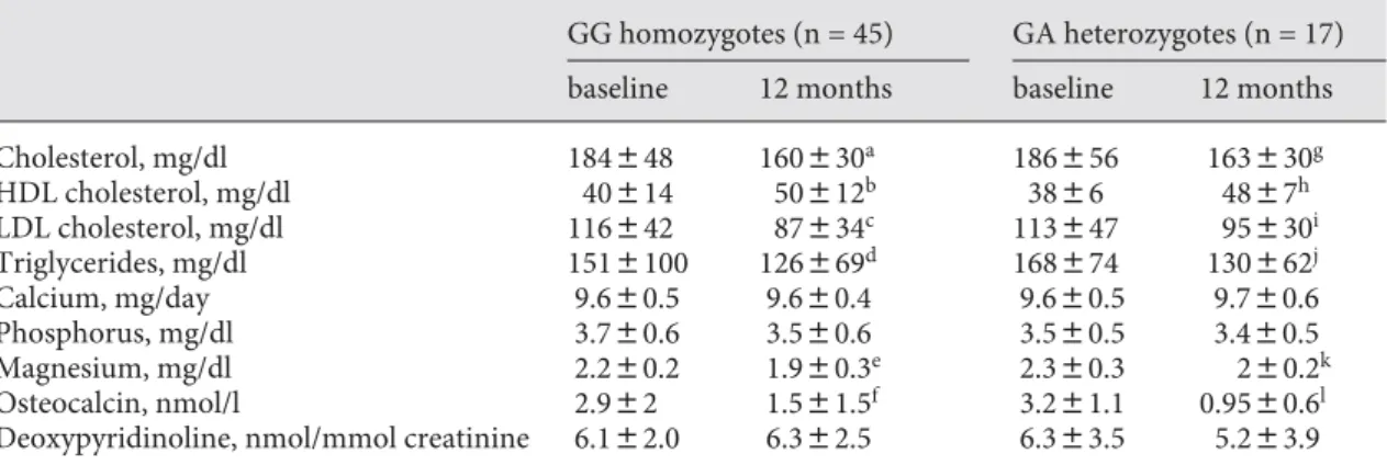
divided into two groups according to 308 G/A polymor-phism. Forty-five patients were homozygous for G/G (72.5%) and 17 heterozygous for G/A (27.5%). The preva-lence of osteoporosis (T score ^ 2.5 in the lumbar spine and/or hip) was 33% in the G/G genotype and 35% in the G/A genotype, with no statistically significant differenc-es between the two groups (p = 0.556). Baseline param-eters ( tables 1, 2 ) showed no differences between groups. Analysis of the response to atorvastatin showed reduced cholesterol and triglyceride levels in both groups. There was a similar reduction in osteocalcin and magnesium ( table 1 ).
There were differences in the response of bone mass to avortastatin according to genotype. In the lumbar spine (L2–L4) there was a statistically significant increase in BMD (1.107 8 0.32 vs. 1.129 8 0.23, p = 0.0001) in pa-tients with the G/G genotype, but not in those with the G/A genotype. No changes in BMD were found in either
Table 1. Analytical parameters at study entry (baseline) and 12 months after treatment with atorvastatin ac-cording to genotype
GG homozygotes (n = 45) GA heterozygotes (n = 17)
baseline 12 months baseline 12 months
Cholesterol, mg/dl 184848 160830a 186856 163830g
HDL cholesterol, mg/dl 40814 50812b 3886 4887h
LDL cholesterol, mg/dl 116842 87834c 113847 95830i
Triglycerides, mg/dl 1518100 126869d 168874 130862j
Calcium, mg/day 9.680.5 9.680.4 9.680.5 9.780.6
Phosphorus, mg/dl 3.780.6 3.580.6 3.580.5 3.480.5
Magnesium, mg/dl 2.280.2 1.980.3e 2.380.3 280.2k
Osteocalcin, nmol/l 2.982 1.581.5f 3.281.1 0.9580.6l
Deoxypyridinoline, nmol/mmol creatinine 6.182.0 6.382.5 6.383.5 5.283.9
a p = 0.001; b p = 0.001; c p = 0.0001; d p = 0.0001; e p = 0.001; f p = 0.0001; g p = 0.022; h p = 0.0001; i p = 0.0001;
j p = 0.022; k p = 0.022; l p = 0.001.
Table 2. Densitometric parameters at study entry (baseline) and at 12 months after treatment with atorvastatin according to genotype
GG homozygotes (n = 45) GA heterozygotes (n = 17)
baseline 12 months baseline 12 months
BMD L2–L4, g/cm2 1.10780.23 1.12980.21a 1.18080.24 1.18780.24
BMD femoral neck, g/cm2 0.90280.15 0.89780.15 0.97280.12 0.96680.13
BMD femoral trochanter, g/cm2 0.75780.22 0.76180.22 0.86380.14 0.81380.26
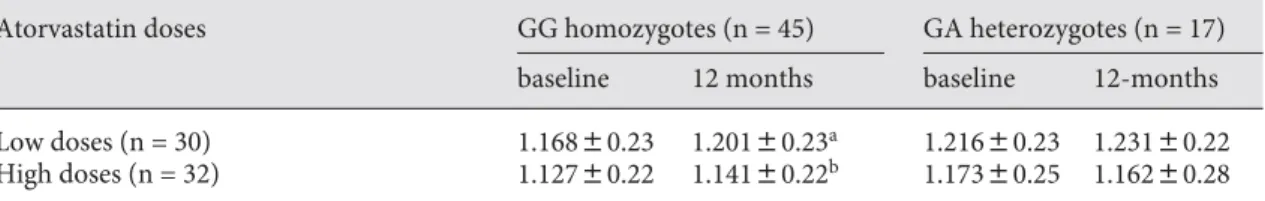
group in the femoral neck or trochanter ( table 2 ). There were no differences in the response of bone mass to ator-vastatin according to the drug doses ( table 3 ). When a multivariate logistic regression analysis was performed to evaluate the effects of osteocalcin, magnesium, para-thormone and the 308 G/A polymorphism on spine BMD, only the latter two were found to be significantly associ-ated with BMD ( table 4 ).
Discussion
Our results show that the TNF ␣ -308 G/A polymor-phism does not influence BMD in the lumbar spine and hip in patients with acute coronary syndrome. The inci-dence of osteoporosis was similar in the two genotypes analyzed. However, we found a different response to ator-vastatin according to genotype. Patients with the G/G genotype showed increased BMD in the lumbar spine in response to treatment while those with the G/A genotype did not. The genotype distribution of our patients was similar to the European distribution, with more than 70% having the G/G genotype, although different from Asia, where the AA genotype predominates [15, 19, 20] .
The reduction in serum magnesium observed in both groups is remarkable. Haenni et al. [21] demonstrated that the administration of simvastatin for 6 weeks caused
a statistically significant reduction in magnesium levels in a group of 23 diabetic patients. These results are com-parable to our findings. We observed a reduction in os-teocalcin, a turnover marker. Atorvastatin is anticatabol-ic and reduces bone remodeling.
High circulating levels of TNF ␣ have been associated with unstable angina and myocardial infarction and may predict a second infarction [22] . However, no significant association was found between the TNF ␣ -308 G/A poly-morphism and the incidence of coronary disease [15] or osteoporosis (BMD) either in Europeans or Asians [19, 20] . Nor does the polymorphism influence the peak bone mass although another polymorphism located in the pro-moter region of the gene, –863 CA [19] , does. Only Fon-tova et al. [23] , in a study on 104 postmenopausal women with osteoporosis and 51 without, found a higher bone mass in a group of patients with nonsevere osteoporosis and the G allele.
Few studies have evaluated the response to statins as a function of the TNF ␣ -308 G/A polymorphism. In the Li-poprotein and Coronary Atherosclerosis Study (LCAS), no association was found between the 308 G/A polymor-phism and the biochemical, angiographic and clinical re-sponse to fluvastatin [24] and no baseline differences were observed. No previous studies have evaluated the response of bone mass to statins according to the selected genotype. We found a favorable response in patients with the G/G genotype, comparable to the response observed in patients with rheumatoid arthritis treated with anti-TNF antibodies. The number of responders was greater in subjects with the G allele [25] . The worse response ob-tained by our patients with the G/A genotype may be due to the fact that these patients produce more TNF ␣ and that, possibly, atorvastatin cannot reduce it below a threshold level. Another possibility is that disease sever-ity is greater in these patients. However, this is unlikely as we found no baseline differences in BMD or the prev-alence of osteoporosis. The role of other polymorphisms
Table 3. BMD L2–L4 (g/cm2)at study entry (baseline) and 12 months after treatment with atorvastatin
accord-ing to genotype and atorvastatin doses
Atorvastatin doses GG homozygotes (n = 45) GA heterozygotes (n = 17)
baseline 12 months baseline 12-months
Low doses (n = 30) 1.16880.23 1.20180.23a 1.21680.23 1.23180.22
High doses (n = 32) 1.12780.22 1.14180.22b 1.17380.25 1.16280.28
a p = 0.002; b p = 0.035.
Table 4. Results of the multiple linear regression analysis predict-ing changes in spine BMD
 p
308 G/A polymorphism –0.392 0.011
Magnesium –0.008 0.954
Osteocalcin –0.081 0.579
close to the polymorphism we analyzed cannot be ex-cluded and should be further studied. The response was only observed in the lumbar spine and not in the hip. This may be because the hip bone is metabolically less active, with a poorer response to anticatabolic drugs, meaning that greater antiresorptive power would be needed and that the effect of atorvastatin is small.
In conclusion, in patients with acute coronary syn-drome, atorvastatin increases lumbar spine BMD only in patients with the G/G genotype of the TNF␣-308 G/A polymorphism.
The main limitation of our study is the sample size even though the population was uniform. Moreover, we have not measured the TNF levels. Another limitation is the absence of an objective method for assessing thera-peutic compliance. This was performed using the infor-mation provided by the patient at the last visit. In addi-tion, initial triglyceride and cholesterol levels were not too high. These facts can explain the absence of differ-ences between high and low drug doses.
References
1 Kado DM, Browner WS, Blackwell T, Gore R, Cummings SR: Rate of bone loss is associated with mortality in older women: a prospective
study. J Bone Miner Res 2000; 15: 1974–1980.
2 Bauer DC, Palermo L, Black D, Cauley JA: Quantitative ultrasound and mortality: a
prospective study. Osteoporos Int 2002; 13:
606–612.
3 Marcovitz PA, Tran HH, Franklin BA, O’Neill WW, Yerkey M, Boura J, Kleereko-per M, Dickinson CZ: Usefulness of bone mineral density to predict significant
coro-nary artery disease. Am J Cardiol 2005; 96:
1059–1063.
4 Vogt MT, Cauley JA, Kuller LH, Nevitt MC: Bone mineral density and blood flow to the lower extremities: the study of osteoporotic
fractures. J Bone Miner Res 1997; 12: 283–
289.
5 Tanko LB, Christiansen C, Cox DA, Geiger MJ, McNabb MA, Cummings SR: Relation-ship between osteoporosis and cardiovascu-lar disease in postmenopausal women. J
Bone Miner Res 2005; 20: 1912–1920.
6 Barengolts EI, Berman M, Kukreja SC, Kouznetsova T, Lin C, Chomka EV: Osteo-porosis and coronary atherosclerosis in as-ymptomatic postmenopausal women. Calcif
Tissue Int 1998; 62: 209–213.
7 Sinnott B, Syed I, Sevrukov A, Barengolts E: Coronary calcification and osteoporosis in men and postmenopausal women are inde-pendent processes associated with aging.
Calcif Tissue Int 2006; 78: 195–202.
8 Hansson GK: Inflammation, atherosclero-sis, and coronary artery disease. N Engl J
Med 2005; 352: 1685–1695.
9 Pacifici R: Estrogen, cytokines and patho-genesis of postmenopausal osteoporosis. J
Bone Miner Res 1996; 11: 1043–1051.
10 Pérez Castrillón JL, Abad L, Vega G, Sanz-Cantalapiedra A, Sanchez S, Hernandez G, Dueñas Laita A: Effects of statins on bone markers, bone mineral density and frac-tures. Possible role in osteoporosis
treat-ment. Curr Pharm Anal 2006; 2: 161–168.
11 Barath P, Fishbein MC, Cao J, Berenson J, Helfant RH, Forrester JS: Detection and lo-calization of tumor necrosis factor in human
atheroma. Am J Cardiol 1990; 65: 297–302.
12 Kobayashi K, Takahashi N, Jimi E, Udagawa N, Takami M, Kotake S, Nakagawa S, Kino-saki M, Yamaguchi K, Shima N, Yasuda H, Morinaga T, Higashio T, Martin TJ, Suda T: Tumor necrosis factor stimulates osteoclast differentiation by a mechanism independent of the ODF/RANKL-RANK interaction. J
Exp Med 2000; 191: 275–278.
13 Romas E, Martin TJ: Cytokines in the patho-genesis of osteoporosis. Osteoporos Int 1997; 7:S47–S53.
14 Goldring SR, Goldring MB: Eating bone or adding it: the Wnt pathway decides. Nat Med
2007; 13: 133–134.
15 Gander ML, Fischer JE, Maly FE, Von Känel R: Effect of the G-308A polymorphism of the
tumor necrosis factor (TNF)- ␣ gene
pro-moter site on plasma levels of TNF- ␣ and
C-reactive protein in smokers: a
cross-section-al study. BMC Cardiovasc Disord 2004; 4:
17–23.
16 Louis E, Franchimont D, Piron A, Gevaert Y, Schaaf-Lafontaine N, Roland S, Mathieu P, Malaise M, De Groote D, Louis R, Belaiche J: Tumor necrosis factor (TNF) gene
poly-morphism influences TNF- ␣ production in
lipopolysaccharide (LPS) stimulated whole blood cell culture in healthy humans. Clin
Exp Immunol 1998; 113: 401–406.
17 Bouma G, Crusius JB, Oudkerk Pool M, Kolkman JJ, Von Blomberg BM, Kostense PJ, Giphart MJ, Schreuder GM, Meuwissen SG, Pena AS: Secretion of tumor necrosis factor alpha and lymphotoxin alpha in relation to polymorphisms in the TNF genes and HLA-DR alleles. Relevance for inflammatory
bowel disease. Scand J Immunol 1996; 43:
456–463.
18 Grundy SM, Cleeman JI, Merz CN, Brewer HB, Clark LT, Hunninghake DB, Pasternak RC, Smith SC, Stone NJ: Implications of re-cent clinical trials for the National Choles-terol Education Program Adult Treatment
Panel III guidelines. Circulation 2004; 110:
227–239.
19 Wennberg P, Nordström P, Lorentzon R,
Ler-ner UH, Lorentzon M: TNF- ␣ gene
poly-morphism and plasma TNF- ␣ levels are
re-lated to lumbar spine bone area in healthy female caucasian adolescents. Eur J
Endocri-nol 2002; 146: 629–634.
20 Chen HY, Chen WC, Hsu CM, Tsai FJ, Tsai
CH: Tumor necrosis factor ␣ , CYP 17,
uroki-nase and interleukin 10 gene polymorphisms in postmenopausal women: correlation to bone mineral density and susceptibility to osteoporosis. Eur J Obstet Gynecol Reprod
Biol 2005; 122: 73–78.
21 Haenni A, Öhrvall M, Lithell H: Serum mag-nesium status during lipid-lowering drug treatment in non-insulin dependent diabetic
patients. Metabolism 2001; 50: 1147–1151.
22 Westerberg M, Bengtsson A, Ricksten A, Jeppsson A: Tumor necrosis factor gene polymorphisms and inflammatory response in artery coronary bypass grafting patients.
Scand Cardiovasc J 2004; 38: 312–317.
23 Fontova R, Gutierrez C, Vendrell J, Broch M, Vendrell I, Simon I, Fernandez-Real JM, Richart C: Bone mineral mass is associated with interleukin 1 receptor autoantigen and TNF-alpha gene polymorphism in post-menopausal Mediterranean women. J
Endo-crinol Invest 2002; 25: 684–690.
24 Elghannam H, Tavackoli S, Ferlic L, Gotto AM, Ballantyne CM, Marian AJ: A prospec-tive study of genetic markers of susceptibil-ity to infection and inflammation and the severity, progression and regression of coro-nary atherosclerosis and its response to
ther-apy. J Mol Med 2000; 78: 562–568.
25 Lee YH, Rho YH, Choi SJ, Ji JD, Song GG: Association of TNF-alpha-308G/A
polymor-phism with responsiveness to TNF- ␣
-block-ers in rheumatoid arthritis: a meta-analysis.