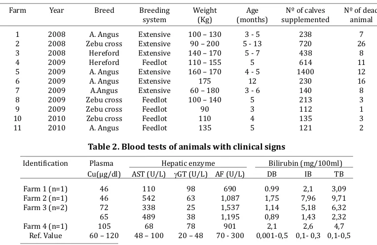
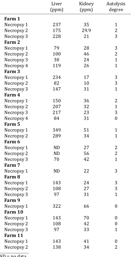
Renal cortex copper concentration in acute copper poisoning in calves
Texto completo
Figure
Documento similar
The values of the different variables in a square area in a certain moment as long as the value of the possibility of finding oil slicks in the following day is what is called a
On the other hand, the larvae from 18 ovipostions that were kept in the laboratory at ambient temperatu- re emerged between two and four days after they were collected in the
Nearly one million Central Americans sought shelter in North America as a consequence of the civil wars in Guatemala and El Salvador, where the acute polarisation
The problem analyzed in the previous sections was used as an introductory situation in one of the topics of mathematics for teachers in a training course for in-training
Accordingly, Chilean copper exports are determined by the Chilean peso-US$ exchange rate, the development of world copper prices in the stock exchanges (e.g., London Metal
The meeting of experts was organized and convened by the Spanish Academy of Sexology and Sexual Medicine (AESMES); the Spanish Association of Sexology Specialists (AEES); the
The interplay of poverty and climate change from the perspective of environmental justice and international governance.. We propose that development not merely be thought of as
How a universal, empirical craft- based activity has been turned into a globalized industry following government intervention in the housing market after World War I,
