PERMANYER
ORIGINAL ARTICLE Rev Inves Clin. 2018;70:82-7
Pulmonary Vasoreactivity
and Phenotypes in Pulmonary Arterial
Hypertension Associated to Connective
Tissue Diseases
José Luis Hernández-Oropeza
1*, Tatiana Sofía Rodríguez-Reyna
2, Diego Luis Carrillo-Pérez
3,
José de Jesús Rodríguez-Andoney
1, René Narváez-David
4, Yesenia Salado-Morales
1,
Eduardo Rivero-Sigarroa
5, Guillermo Domínguez-Cherit
5and Tomás Pulido-Zamudio
61Department of Cardiopulmonary Disease, 2Department of Immunology and Rheumatology, 3Department of Internal Medicine, 4Department of Cardiology, and 5Department of Critical Care Medicine, Instituto Nacional de Ciencias Médicas y Nutrición Salvador Zubirán; and 6Department of Cardiopulmonary Disease, Instituto Nacional de Cardiología Ignacio Chávez, Mexico City, Mexico
Received for publication: 08-11-2017 Accepted for publication: 13-02-2018 doi: 10.24875/RIC.18002437
ABSTRACT
Background: Pulmonary arterial hypertension (PAH) is a fatal complication in patients with connective tissue disease (CTD). Objective: The objective of the study was to study the prognostic value of the acute pulmonary vasoreactivity test with inhaled iloprost and its association with clinical deterioration in a tertiary care academic medical center. Methods: We conducted a prospective study of patients with CTD and the diagnosis of PAH established by right heart catheterization. Patients were clas-sified into classic responders, partial responders, and non-responders. The association of the pulmonary response and clinical deterioration was analyzed. Results: We enrolled 25 patients (mean age of 47 ± 13.4 years); 88% were female The most frequent rheumatologic diagnosis was systemic lupus erythematosus, in 16 (64%) patients. Seventy-two percent of patients were classified as non-responders, and 28% were partial responders. Patients with a partial response had lower right atrial pressure values (5.1 ± 3.1 vs. 8.5 ± 3.2, p = 0.01) and greater systolic pulmonary arterial pressure (87.6 ± 8.1 vs. 72.4 ± 16.2, p = 0.02), compared with non-responders. Non-responders had a tendency for a shorter time to clinical deterioration than partial responders (17.8 vs. 41.1 months, p = 0.052). Conclusions: Patients with a partial response to the acute pulmonary vasodilator test with inhaled iloprost had a longer clinical deterioration-free period than non-responders. (REV INVES CLIN. 2018;70:82-7)
Key words: Pulmonary hypertension. Right heart catheterization. Connective tissue disease. Prognosis.
Corresponding author: *José Luis Hernández-Oropeza
Department of Cardiopulmonary Disease Instituto Nacional de Ciencias Médicas y Nutrición Salvador Zubirán
Definitions
Patients were considered to have PAH if their mean pulmonary arterial pressure (mPAP) was ≥ 25 mmHg, the pulmonary capillary pressure was < 15 mmHg, and pulmonary vascular resistance was > 3 Wood units, according to RHC results1,2. Patients with CTD were those cases with a clinical and immunological diagnosis of systemic lupus erythematosus (SLE), mixed CTD (MCTD), or systemic sclerosis (SS), all confirmed by the rheumatology department. Inhaled iloprost (5 mcg) was used in all cases to conduct the APVT during RHC1,9. A classic positive response was defined as the decrease of at least 10 mmHg in the mPAP to an absolute value of < 40 mmHg with an increased or unchanged cardiac output. A partial positive response referred to a decrease of at least 10 mmHg to an absolute mPAP > 40 mmHg with an increased or unchanged cardiac output. Non-re-sponders were those patients who fulfilled none of these criteria7.
In accordance with current definitions10, all patients were considered prevalent cases since both the CTD and the PAH had been diagnosed for over 6 months.
Clinical deterioration was defined as worsening of PAH (worsening symptoms and signs of right-sided heart failure), decrease in functional class, and de-crease of at least 15% in the distance covered in the 6-minute walk test (6MWT) (confirmed by a second test 2 weeks later), or death.
Statistical analysis
Descriptive statistics were used according to the vari-ables’ characteristics. Frequencies and percentages were used for categorical variables, and continuous variables were reported as means and standard de-viations, or medians, and interquartile ranges. The normality of the variables’ distribution was evaluated with the Kolmogorov–Smirnov and the Shapiro–Wilk tests. comparison of continous variables were per-formed with Student's t-test and Wilcoxon's ranked sum test and the comparison of categorical variables were performed with Chi-square and Fisher's exact tests. Survival was analyzed with the Log-rank test. p < 0.05 was considered statistically significant. Sta-tistical analysis was performed with the STATA sta-tistical package, version 11.
INTRODUCTION
Pulmonary arterial hypertension (PAH) is charac-terized by the progressive increase in pulmonary vascular resistance, leading to right ventricular dysfunction. Right heart catheterization (RHC) establishes the diagnosis of PAH and guides the most appropriate medical therapy according to the patient’s response to the acute pulmonary vasoreactivity test (APVT) with short-acting va-sodilators (nitric oxide, epoprostenol, adenosine, and iloprost)1,2.
PAH is present in 13-25% of patients with connective tissue disease (CTD), and it is a major cause of death in this sub-group3. The use of calcium channel block-ers is precluded in this population since these patients are considered non-responders in the long-term4-6.
Recent studies have evaluated the predictive useful-ness of the APVT in individuals with PAH in associa-tion with CTD (PAH-CTD), in partial responders with contoversial prognostic results7,8.
The aim of this study was to determine the prog-nostic value of the APVT with inhaled iloprost in patients with PAH-CTD, according to the response in the test. We have hypothesized that patients with some type of pulmonary vascular response would present fewer events compatible with clinical deterioration.
PATIENTS AND METHODS
Study population
We conducted a prospective study of patients with CTD in whom, as part of their standard medical care, RHC was required, and an APVT was not contraindi-cated. The study was performed in a tertiary care academic medical center in Mexico City, which is a referral center for CTD, between January 2010 and December 2015.
RESULTS
We included 25 patients throughout the study period, of whom 22 (88%) were female. 16 (64%) patients had a diagnosis of SLE, 6 (24%) had SS, and 3 (12%) had MCTD. In the SLE group, 13 (81%) patients were on prednisone therapy, 3 (12%) were treated with mycophenolate mofetil, and their average disease ac-tivity index according to the SLE disease acac-tivity index was 6.8 ± 3.9.
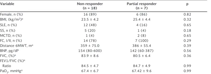
Demographic, clinical and laboratory characteristics are shown in table 1. None of the patients fulfilled classic responder criteria. 18 patients (72%) were considered non-responders and 7 (28%) had a partial positive response. The mean age of the partial positive response group was 40 ± 12.5 years versus 49.3 ± 13.2 years of the non-responder group.
Twenty-one (84%) patients were WHO functional Class I and II, and 4 (16%) were class III. Simple spi-rometry revealed a mildly restrictive pattern for both groups, and no interstitial involvement was observed by high-resolution tomography in any patient.
We detected the presence of anti-U1 ribonucleopro-tein (RNP) antibodies in 4 patients (16%), and 7 (28%) patients were receiving low-dose calcium channel blockers (CCBs) for the management of Raynaud’s phenomenon (nifedipine, 10 mg twice a day). By Cox
analysis, we found an association of 3 protective fac-tors against clinical deterioration: a partial pulmonary vascular response, with an hazard ratio (HR) of 0.36 [95% confidence interval (CI) 0.021-1.302]; pres-ence of anti-U1-RNP antibodies, HR 0.50 (95% CI 0.032-1.403); and the use of CCB, HR of 0.34 (95% CI 0.042-2.780).
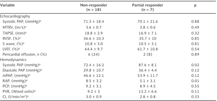
Hemodynamic characteristics of patients by echocar-diography and cardiac catheterization are shown in table 2. In partial responders, the percentage of change in peripheral vascular resistance and mPAP was 21%, with an unchanged cardiac output. Patients with a partial response had lower right atrial pressure values (5.1 ± 3.1 vs. 8.5 ± 3.2, p = 0.01) and greater systolic pulmonary arterial pressure (87.6 ± 8.1 vs. 72.4 ± 16.2, p = 0.02), compared with non-respond-ers. There were no statistically significant differences in other hemodynamic parameters.
All patients were administered supplemental oxygen as initial therapy (standard treatment in our center) and specific treatment with sildenafil monotherapy (25 mg PO every 8 h) after PAH confirmation by RHC.
The median time period to clinical deterioration of the 3 CTD sub-groups was 17.8 months (range, 1.3-58.5 months) in the non-responder group versus 41.1 months (range, 12.5-59.5 months) in the partial responder group (p = 0.052). Figure 1 shows the survival
Table 1. Demographic characteristics, stress test, and lung function test results
Variable Non-responder
(n = 18) Partial responder (n = 7) p
Female, n (%) 16 (89) 6 (86) 0.82
BMI, (kg/m2)a 23.5 ± 4.2 25.4 ± 4.4 0.32
SLE, n (%) 12 (48) 4 (16) 0.65
SS, n (%) 5 (20) 1 (4) 0.18
MCTD, n (%) 1 (4) 2 (8) 0.65
FC, I/II, n (%) 14 (78) 7 (100) 0.29
Distance 6MWT, ma 359 ± 75.0 386 ± 55.4 0.39
BNP, pg/dlb 154 (80-600) 142 (60-387) 0.56
FVC, (%)a 83.9 ± 8.6 80.5 ± 6.4 0.36
FEV1/FVC (%)a
Ratio 84.5 ± 4.7 84.7 ± 4.9 0.99
PaO2, mmHga 67.4 ± 6.7 67.42 ± 9.6 0.99
BMI: body mass index, SLE: systemic lupus erythematosus, SS: systemic sclerosis, MCTD: mixed connective tissue disease, FC: functional class, 6MWT: 6-minute walk test, BNP: brain natriuretic peptide, FVC: forced vital capacity, FEV1: forced expiratory volume in 1 second; PaO2: arterial oxygen pressure
analyses between groups. We found that the SLE sub-group did yield a statistically significant difference in terms of clinical deterioration between partial re-sponders and non-rere-sponders, using the Log-Rank test (p=0.044). Figure 2 shows the survival analysis between groups.
Throughout follow-up, two deaths (8%) were regis-tered, one per group. The other clinical deterioration events were worsening of PAH in 5 patients (20%); decrease in functional class in 18 (72%); and a distance
decrease in the 6MWT in 23 (92%), with an average decrease on the number of meters walked, were 38%.
DISCUSSION
This study showed that patients with PAH-CTD are partially responding to inhaled iloprost in the APVT, had a better prognosis in terms of remaining free of clinical deterioration for a longer time period than non-responders.
Table 2. Hemodynamic characteristics by echocardiography and cardiac catheterization
Variable Non-responder
(n = 18) Partial responder (n = 7) p
Echocardiography
Systolic PAP, (mmHg)a 71.3 ± 18.4 70.1 ± 21.6 0.88
MTRV, (m/s)a 3.6 ± 0.7 3.8 ± 0.6 0.49
TAPSE, (mm)a 18.8 ± 2.9 16.9 ± 7.1 0.32
RVSF, (%)a 36.6 ± 10.3 35.7 ± 10 0.85
S wave, (%)a 10.8 ± 3.0 10.5 ± 3.1 0.81
LVEF, (%)a 64.4 ± 9.7 61.7 ± 10.8 0.54
Pericardial effusion, n (%) 6 (24) 2 (8) 0.89
Hemodynamics
Systolic PAP (mmHg)a 72.4 ± 16.2 87.6 ± 8.1 0.02
Diastolic PAP (mmHg)a 29.8 ± 10.7 36.4 ± 4.4 0.12
mPAP, (mmHg)a 46.6 ± 12.1 53.9 ± 11.7 0.12
RAP, (mmHg)a 8.5 ± 3.2 5.1 ± 3.1 0.01
PCP, (mmHg)a 9.2 ± 3.1 8.9 ± 4.5 0.55
PVR, (Wood units)a 9.2 ± 5 13.2 ± 6.6 0.11
CI, (l/min/m2)a 3.0 ± 0.9 2.8 ± 0.8 0.33
PAP: pulmonary arterial pressure, MTRV: maximal tricuspid regurgitation velocity, TAPSE: tricuspid annular pulmonary systolic excursion, RVSF: right ventricular shortening fraction, LVEF: left ventricular ejection fraction, mPAP: mean pulmonary arterial pressure, RAP: right atrial pressure, PCP: pulmonary capillary pressure, PVR: pulmonary vascular resistance, CI: cardiac index
aExpressed as a mean ± standard deviation
0 20 40 60 80 100
0 20 40 60
Clinical det
erior
ation
Months
Non-responder Partial-responder
p = 0.052
Figure 1. Time to clinical deterioration (months), Subjects at risk, 3 CTD sub-groups (n = 25)
0 20 40 60 80 100
0 20 40 60
Clinical det
erior
ation
Months
Non-responder Partial-responder
p = 0.044
The definition of response in the APVT used for this study differs from that used in recent PAH diagnostic and treatment guidelines1,2. None of the patients in our cohort had a classic response in the test, a finding similar to that reported by Halliday et al.7 in a pro-spective analysis of 155 patients with group 1 PAH. In their study, 33% belonged in the PAH-CTD group, and only 16% of patients in this sub-group had a clas-sic positive response in the APVT with nitric oxide; however, only responders with idiopathic PAH had im-proved survival compared with non-responders. Unlike that study, we only evaluated clinical deterioration and not survival, in patients with some degree of pul-monary vascular response.
Although in earlier studies, the APVT was used to identify patients with better survival and those who were candidates to treatment with the less expensive albeit effective option, calcium channel blockers4,6, our study results suggest that the APVT can predict outcomes such as clinical deterioration.
Approximately, only 13% of patients with idiopathic PAH respond in the APVT5,7 and this group, at most 80% still respond to calcium antagonist drugs after 1 year of follow-up1,5. To date, there are no prospective studies on PAH-CTD that evaluate the prognostic val-ue of the APVT response in terms of other outcomes (different from survival), such as clinical deterioration.
Our results reflect that a partial positive response, defined as a decrease in mPAP of ≥ 10 mmHg but an absolute value of > 40 mmHg and no change in the cardiac output, confers a better prognosis in terms of clinical deterioration in this sub-group of patients. These findings are similar to those reported by Mal-hotra et al. [8] in a retrospective cohort of 80 pa-tients with group 1 PAH, in which 29% of cases had PAH-CTD, and an ≥ 12% decrease in mPAP with in-haled nitric oxide was an independent survival predic-tor when compared with non-responding patients, including those that were never treated with calcium channel blockers.
In other pulmonary hypertension groups, observing some degree of response in the APVT suggests a better prognosis. In a prospective study of 103 pa-tients with chronic thromboembolic pulmonary hy-pertension (group IV), Skoro-Sajer et al.11 demon-strated that decreasing the mPAP to > 10.4% during
the APVT with inhaled nitric oxide predicted long-term survival and longer lung transplant-free period in adult patients that underwent pulmonary endar-terectomy.
A possible explanation to why patients with a partial pulmonary vascular response had less severe pulmo-nary hypertension and higher cardiac output may be the presence of protective antibodies (anti-U1-RNP). Although their role in disease has not been estab-lished in cohort studies, these antibodies predict bet-ter long-bet-term survival in the three types of CTD12,13 and, perhaps, the pulmonary vascular response is an epiphenomenon of a phenotype associated to milder disease (less right ventricular remodeling).
Our study has several limitations such as the small patient number, a bias due to referral to a tertiary care hospital, and the convenience analysis of pa-tients in a single center. However, despite these ob-servations, there are no reports of prospective studies characterizing this specific sub-group of patients and results require validation by future studies.
Patients with a partial positive response in the acute pulmonary vasodilator test with inhaled ilo-prost had a better prognosis, remaining free of clinical deterioration during a longer time period, com-pared with non-responding patients.
ACKNOWLEDGMENTS
The authors gratefully acknowledge Deborah Alemán-Hoey, MD, for her assistance in the review of this manuscript. This study was funded by the Depart-ment of Cardiopulmonary Disease of the Instituto Nacional de Ciencias Médicas y Nutrición Salvador Zubirán, Mexico City, Mexico.
REFERENCES
1. Galiè N, Humbert M, Vachiery JL, et al. 2015 ESC/ERS guidelines for the diagnosis and treatment of pulmonary hypertension: the joint task force for the diagnosis and treatment of pulmonary hypertension of the European society of cardiology (ESC) and the European respiratory society (ERS): endorsed by: associa-tion for European paediatric and congenital cardiology (AEPC), international society for heart and lung transplantation (ISHLT). Eur Heart J. 2016;37:67-119.
3. Chung L, Liu J, Parsons L, et al. Characterization of connective tissue disease-associated pulmonary arterial hypertension from REVEAL: identifying systemic sclerosis as a unique phenotype. Chest. 2010;138:1383-94.
4. Rich S, Kaufmann E, Levy PS. The effect of high doses of calci-um-channel blockers on survival in primary pulmonary hyperten-sion. N Engl J Med. 1992;327:76-81.
5. Sitbon O, Humbert M, Jaïs X, et al. Long-term response to cal-cium channel blockers in idiopathic pulmonary arterial hyperten-sion. Circulation. 2005;111:3105-11.
6. Montani D, Savale L, Natali D, et al. Long-term response to calcium-channel blockers in non-idiopathic pulmonary arterial hypertension. Eur Heart J. 2010;31:1898-907.
7. Halliday SJ, Hemnes AR, Robbins IM, et al. Prognostic value of acute vasodilator response in pulmonary arterial hypertension: beyond the “classic” responders. J Heart Lung Transplant. 2015; 34:312-8.
8. Malhotra R, Hess D, Lewis GD, Bloch KD, Waxman AB, Semigran MJ. Vasoreactivity to inhaled nitric oxide with oxygen predicts
long-term survival in pulmonary arterial hypertension. Pulm Circ. 2011;1:250-8.
9. Jing ZC, Jiang X, Han ZY, et al. Iloprost for pulmonary vasodila-tor testing in idiopathic pulmonary arterial hypertension. Eur Respir J. 2009;33:1354-60.
10. Badesch DB, Raskob GE, Elliot CG, et al. Pulmonary arterial hy-pertension. Baseline characteristics from the REVEAL Registry. Chest. 2010;137:376-87.
11. Skoro-Sajer N, Hack N, Sadushi-Koliçi R, et al. Pulmonary vas-cular reactivity and prognosis in patients with chronic thrombo-embolic pulmonary hypertension: a pilot study. Circulation. 2009; 119:298-305.
12. Sobansky V, Giovannelli J, Lynch BM, et al. Characteristics and survival of anti-U1 RNP antibody-positive patients with connec-tive tissue disease-associated pulmonary arterial hypertension. Arthritis Rheumatol. 2016;68:484-93.