Otras secciones de este sitio:
☞ ☞ ☞ ☞
☞ Índice de este número
☞ ☞ ☞ ☞
☞ Más revistas
☞ ☞ ☞ ☞
☞ Búsqueda
Others sections in this web site:
☞ ☞ ☞ ☞
☞ Contents of this number ☞
☞ ☞ ☞
☞ More journals ☞
☞ ☞ ☞ ☞ Search Article:
Weight reduction and ursodeoxycholic acid in subjects with nonalcoholic fatty liver disease. A double-blind, placebo-controlled trial
Copyright © 2004: Mexican Association of Hepatology
ANNALS OF HEPATOLOGY
Number 3 July-September 2 0 0 4 Volume 3
Annals of Hepatology 3(3) 2004: 108-112
108
edigraphic.com
Annals of Hepatology 2004; 3(3): July-September: 108-112
Annals of Hepatology
Abstract
Objetive: Nonalcoholic fatty liver disease is an
increas-ingly recognized condition that may progress to end-stage liver disease. We investigated the effects of weight reduction and ursodeoxycholic acid adminis-tration in patients with this disease.
Research methods and procedures: A double-blind,
pla-cebo-controlled trial.
Twenty-seven women with a body mass index of >30
kg/m2 and willing to participate in the diet plan for six
weeks were studied were assigned to one of two treat-ment groups (ursodeoxycholic acid, n = 14: placebo n = 13). Both groups received a normal diet (1,200 kcal/d) plus 1200 mg/d of ursodeoxycholic acid or placebo. Hepatic steatosis, was assessed by abdominal ultra-sound. Fasting glucose, cholesterol, triglycerides, and aminotransferases levels were determined before and after treatment.
Results: Body mass index decreases significantly from
34.2 ± 4.2 kg/m2 and 33.3 ± 1.6 kg/m2 to 31.8 ± 4.5 kg/
m2 and 30.6 ± 2.6 kg/m2 in the ursodeoxycholic acid
and placebo groups, p < 0.001. The hepatic steatosis index decreased from 2.3 ± 0.7 to 1.0 ± 0.6 and 2.2 ± 0.7 to 1.1 ± 0.7 in the ursodeoxycholic acid and place-bo groups, p<0.003. Serum AST decreased significant-ly from 41.2 ± 5.6 to 34.5 ± 3.4 in the ursodeoxycholic acid group, p <0.001, and from 43.6 ± 4.2 to 35.3 ± 2.9 in the placebo group, p <0.001. Serum ALT decreased from 62.9 ± 6.5 to 44.0 ± 3.5 in the ursodeoxycholic acid group, p <0.001, and from 63.5 ± 4.5 to 44.0 ± 3.5 in the placebo group. We did not find any differences in all variables studied between groups. Conclusions:
Departments of Biomedical Research and Gastroenterology & Liver Unit. Medica Sur Clinic & Foundation. Mexico City, Mexico.
Address for correspondence:
Nahum Méndez-Sánchez, MD, PhD Departments of Biomedical Research, Gastroenterology & Liver Unit, Medica Sur Clinic & Foundation, Puente de Piedra 150, Col. Toriello Guerra, Mexico City, Mexico. Phone: (+525) 55606-6222, ext. 4215 Fax: (+525) 55666-4031 and 55606-1651; E-mail: [email protected]
Original article
Weight reduction and ursodeoxycholic
acid in subjects with nonalcoholic fatty
liver disease. A double-blind, placebo-controlled trial
Nahum Méndez-Sánchez; Verónica González; Norberto Chávez-Tapia; Martha H Ramos; Misael Uribe
The present study shows beneficial effect of weight re-duction, producing improvements in biochemical and imaging markers of liver disease.
Key words: Nonalcoholic fatty liver disease, obesity, weight reduction, ursodeoxycholic acid, women.
Introduction
Nonalcoholic fatty liver disease (NAFLD) is an in-creasingly recognized condition that may progress to end-stage liver disease. The pathological picture resembles that of alcohol-induced liver injury, but it occurs in pa-tients who do not abuse alcohol.1 Various terms have
been used to describe this entity, including fatty-liver hepatitis, nonalcoholic Laënnec’s disease, diabetes hepa-titis, alcohol-like liver disease, and nonalcoholic steato-hepatitis.2 Nonalcoholic fatty liver disease is becoming
the preferred term, and it refers to a wide spectrum of liv-er damage, ranging from simple steatosis to steatohepati-tis, advanced fibrosis, and cirrhosis. Steatohepatitis (non-alcoholic steatohepatitis) represents only a stage within the spectrum of NAFLD.3
The influence of obesity on liver physiology has been demonstrated in several studies and populations, the anal-yses showing the effects of weight reduction on biochem-ical indicators of liver function.4,5 Obesity, or indirectly,
body mass index (BMI), is a major risk factor for devel-opment of liver disease, and for NAFLD the prevalence increases 4.6-fold in obese people.3,6 Other risk factors
as-sociated with NAFLD are waist circumference, hyperin-sulinemia, hypertriglyceridemia and impaired glucose tolerance or type 2 diabetes.7,8 These risk factors have
also been examined in animal models. For example, stud-ies of genetically obese ob/ob mice and fa/fa rats have provided information about the pathogenesis of obesity-related fatty liver disease.3,9 The clinical implications of
N Méndez-Sánchez et al. Nonalcoholic fatty liver disease and weight reduction 109
edigraphic.com
is no specific treatment for NAFLD. We hypothesized that weight reduction in patients with NAFLD would be accompanied by a decrease in the degree of hepatic ste-atosis and a reduction of serum transaminase levels. Fur-thermore, it has been suggested that ursodeoxycholic acid (UDCA) the epimer of chenodeoxycholic acid and ap-pears to replace endogenous bile acids, some of which may be hepatotoxic, with the nonhepatotoxic UDCA This bile acid has also membrane stabilizing or cytoprotective effects as well as antiapoptotic actions.10 The aim of this
study was to investigate the effects of weight reduction and UDCA on patients with NAFLD.
Research Methods and Procedures
Subjects
We recruited subjects from our medical center in Mex-ico City (Medica Sur Clinic & Foundation). Obese wom-en were informed of the risk of NAFLD developing ac-cording to the degree of hepatic steatosis and were asked whether they would participate in a study of NAFLD treatment. The diagnosis of NAFLD was first suspected in those subjects who had had, for at least six months, ab-normal serum aminotransferase levels that were not relat-ed to other causes of liver disease, including the hepatitis B and C viruses, autoimmune disorders, alcohol, hemo-chromatosis, and sonographic findings compatible with hepatic steatosis. To be accepted into this trial, women had to (a) have a BMI of >30 kg/m2, (b) be between 20
and 60 years of age, (c) be willing to participate in the diet plan for six weeks, and (d) have normal serum potas-sium and calcium levels. Women of childbearing age had to have a negative serum pregnancy test. Women were excluded from the study if they had any one of the fol-lowing: (1) a history of hypothyroidism or Cushing syn-drome, (2) an eating disorder or other psychological problem that would interfere with participation in the diet program, (3) use of oral bile acid preparations, alumi-num-based antacids or lithium, and (4) long-term use of nonsteroidal anti-inflammatory agents (including aspirin) or antihyperlipidemic agents (including cholestyramine) within two weeks of entering the trial. Diuretic therapy had to have been discontinued at least one day before trial entry. Twenty-seven women who met all entry criteria and agreed to participate were enrolled in the trial. The study was approved by the Human Subjects Committee of the Medica Sur Clinic & Foundation, conforming to the ethi-cal guidelines of the 1975 Declaration of Helsinki, and written informed consent was obtained from all partici-pants before entry.
Study design
Women were assigned to one of two treatment groups in a double-blind fashion, with blocking for body weight
(BMI >30 kg/m2), according to a table of random
num-bers. One group received a normal diet (1200 kcal/d) plus 1,200 mg/d of UDCA (Laboratorios Farmasa, S.A. de C.V., Mexico); the second group received a normal diet (1200 kcal/d) plus a placebo. All capsules were identical in appearance and number. Treatment began on the day on which energy restriction began. Compliance was de-termined by counting unused capsules each week. Wom-en were treated for six weeks.
Weight-control program. The women underwent the
Department of Program Control’s standard history-taking and physical examination, as well as laboratory tests in-cluding a complete blood count, measurement of electro-lytes, liver function tests, measurement of fasting lipids, thyroid-function tests, and electrocardiography. They were placed on a food diet (1200 kcal/d) consisting of 20% fat, 60% carbohydrates, 20% protein, and 1 L of wa-ter daily. The composition of the diet was carbohydrates 195.6 g/d, total fat 26.4 g/d (saturated fat 7.6 g/d, polyun-saturated fat 2.3 g/d, monounpolyun-saturated fat 3.1 g/d, choles-terol 73.8 mg/d), fiber 30.2 g/d, iron 7.1 mg/d, sodium 873.2 mg/d, zinc 9.28 mg/d, and Vitamin C 54.8 mg/d.
Experimental procedures. Real-time ultrasonographic
studies were carried out before and after the experimental period. Women fasted overnight before each ultrasound was performed. The protocol used to evaluate the pattern of hepatic steatosis by ultrasound was graded as follows: (0) Normal, (1) Diffuse, homogeneous, (2) Geographic pattern: sharp demarcation between normal and fatty liver, not confined to a lobar distribution and no mass effect on hepatic vessels, (3) Focal: no mass effect, no vessel dis-placement, segmental or lobar wedge distribution, low tenuation, “earthquake” pattern (scattered irregular low at-tenuation lines), (4) Focal sparing: pseudotumor (anterior to right portal vein/gallbladder fossa, medial segment of left lobe, porta hepatis), glove pattern (high attenuation fin-ger-like interdigitations of spared parenchyma within a low attenuation background), simulating metastases.11 The
se-verity of hepatic fatty infiltration was graded as follows: grade 0, normal; grade 1, liver attenuation slightly less than spleen; grade 2, a more pronounced difference between liv-er and spleen, and intrahepatic vessels not seen or with slightly higher attenuation than liver; grade 3, markedly re-duced liver attenuation with sharp contrast between liver and intrahepatic vessels.
Analytical methods
110
edigraphic.com
sustraídode-m.e.d.i.g.r.a.p.h.i.c cihpargidemedodabor
:rop odarobale FDP
VC ed AS, cidemihparG
arap
acidémoiB arutaretiL :cihpargideM
Statistical analysis
Differences between the groups over time were evalu-ated by paired Student’s t-test.13 The differences were
considered significant when p < 0.05. Values in the text are means ± SD.
Results
Twenty-seven women were enrolled in the trial. Of these, three withdrew prematurely and were not included in the final analysis. The major reason for early termina-tion was voluntary withdrawal from the program (n = 2 (7.4%)). The percentage of women who voluntarily with-drew from the trial did not differ between the two treat-ment groups of subjects receiving UDCA, or placebo. Two women, one in each group, were excluded after be-coming pregnant during the trial (n = 2 (7.4%)). The clin-ical characteristics of subjects at the beginning of the diet trial are given in Table I. The initial BMIs were 34.2 ± 4.2 kg/m2 and 33.3 ± 1.6 kg/m2, respectively. BMI did not
differ between the two treatment groups before the start of the study.
Table II shows the weight loss in both groups. BMI de-creased significantly from 34.2 ± 4.2 kg/m2 and 33.3 ± 1.6
kg/m2 to 31.8 ± 4.5 kg/m2 and 30.6 ± 2.6 kg/m2 in the
UDCA and placebo groups, respectively, p < 0.001. Serum AST decreased significantly from 41.2 ± 5.6 to 34.5 ± 3.4 in the UDCA group, p <0.001, and from 43.6 ± 4.2 to 35.3 ± 2.9 in the placebo group, p <0.001. Serum ALT de-creased from 62.9 ± 6.5 to 44.0 ± 3.5 in the UDCA group, p <0.001, and from 63.5 ± 4.5 to 44.0 ± 3.5 in the placebo
group. We did not find any differences in those variables studied between groups. However, cholesterol and glucose decreased significantly only in the UDCA group. Serum triglycerides did not change in either group.
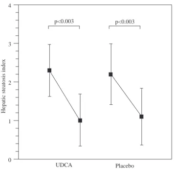
Figure 1 shows the hepatic steatosis index. There was a decrease from 2.3 ± 0.7 to 1.0 ± 0.6 and 2.2 ± 0.7 to 1.1 ± 0.7 in the UDCA and placebo groups, p<0.003 re-spectively.
Discussion
In this study, we analyzed the effects of weight reduc-tion with or without pharmacological treatment with UDCA. Currently there is no consensus regarding effec-tive therapy for NAFLD; attempts are being made to di-rect treatment toward avoiding or cordi-recting risk factors, including insulin resistance and decreasing hyperin-sulinemia, and using drugs with potential hepatoprotec-tive effects.14 Our data shows the beneficial effect of diet,
especially in body weight reduction, with a decrease of 8% and 7% in the placebo and UDCA groups. In addi-tion, we observed a decrease (16.2%) in the levels of as-partate aminotransferase in both groups, whereas the de-crease in alanine aminotransferase (ALT) levels was 30% in UDCA group versus 16.2% in the placebo group. No statistical effect on glucose and cholesterol levels was ob-served. Schaffner et al.2 showed that a weight reduction
of greater than or equal to 10% corrected abnormal hepat-ic test results, decreased hepatosplenomegaly, and re-solved some stigmata of liver disease, while minimal changes in BMI were important in liver function and for every 1% reduction in body weight, alanine aminotrans-ferase activity improved by 8.1%.5 The effects of weight
reduction on liver function have been demonstrated in several mammal models.16-18 In cats, the reduction of
25-30 per cent of body weight was associated with signifi-cant changes in plasma insulin, cholesterol, triglyceride, and serum glucose concentrations,19 and changes in the
metabolism of free fatty acids and hepatic fatty acid syn-thesis are involved in the development of hepatic lipido-sis.20 These data indicate the importance of weight
reduc-tion, especially considering that the cat model has similarities with NAFLD in humans.21 One of the most
Table I. Characteristics of patients at the start of the diet trial.
UDCA Placebo
(1200 mg/d)
n 14 13
Age, 39.7 ± 8 37.8 ± 8
Initial body weight (kg) 79.8 ± 12.1 84.3 ± 9.8 Initial body mass Index (kg/m2) 34.2 ± 4.2 33.3 ± 1.6
UDCA, ursodeoxycholic acid; * Values are given as mean ± SD
Table II. Characteristics of patients at the baseline and at six-weeks of the diet trial.
UDCA PLACEBO
n 12 11
Baseline Six-weeks Baseline Six-weeks
BMI (kg/m2) 34.2 ± 4.2 31.8 ± 4.5a 33.3 ± 1.6 30.6 ± 2.6a
AST (UI/L) 41.2 ± 5.6 34.5 ± 3.4a 43.6 ± 4.2 35.3 ± 2.9a
ALT (UI/L) 62.9 ± 6.5 44.0 ± 3.5a 63.5 ± 4.5 44.0 ± 3.5a
Glucose (mg/dL) 94.0± 6.7 83.3 ± 7.3 a 90.4 ± 12 88.2 ± 9.4c
Cholesterol (mg/dL) 196.1 ± 36.7 166.6 ± 21.4b 177.7 ± 29.3 165.0 ± 33.9c
N Méndez-Sánchez et al. Nonalcoholic fatty liver disease and weight reduction 111
edigraphic.com
important pathogenic mechanisms involved in NAFLD is insulin resistance. In the cat model, Biourge et al.22
showed that weight reduction was associated with a sig-nificant decrease in mean serum insulin concentration and the glucose disappearance coefficient. Similarly in hu-mans, weight reduction has major importance in insulin resistance. Case et al.23 observed that a moderate decrease
in weight (6.5%) induced by a very low calorie diet re-sulted in substantial reductions in glucose (17 mg/dL), triglycerides (94 mg/dL) and total cholesterol (37 mg/dL) after four weeks. According with those observations, we believe that the effects of weight reduction on human liv-er diseases with similar pathological substrates will have prognostic implications.24 Recently, Hickman et al.25
showed the effect of weight reduction in patients with chronic hepatitis C. They found a decrease in ALT levels and in stellate cell activation and, in some cases, regres-sion of hepatic fibrosis. Weight reduction is also accom-panied by diminution of hepatomegaly in obese women with liver steatosis.26 In subjects who underwent
gastro-plasty for morbid obesity, the weight reduction was asso-ciated with morphological liver changes, particularly in the severity of the steatosis seen in liver biopsies.27 In
ad-dition, when subjects with non-alcoholic steatohepatitis received a low energy diet for three months, a decrease in the degree of steatosis was observed by tomography.28
On the other hand, a recent study published by Lindor and colleagues29 shows that 2 years of therapy UDCA at a
dose of 13 to 15 mg/kg/d was not better than placebo for patients with NASH. These data confirm the previous ob-servation published by Laurin who found no significant changes in the histological grade of inflammation or
fi-brosis.30 Also Santos et al31 have recently reported that
UDCA is able to reduce serum levels of hepatic enzymes in patients with nonalcoholic fatty liver disease, but this effect is not related to modifications in liver fat content.
Finally, the main limitation of our study has been the lack of liver biopsies to assess the histology of the liver, which is the “gold standard” in the diagnosis of NAFLD. Morphologic changes evaluated by ultrasound should be considered with caution, as it is well known that the re-producibility of this technique is related to the operator.
In conclusion, the present results shown a beneficial effect of weight reduction on patients with NAFLD by improving the biochemical and imaging markers of liver disease. This improvement was independent of the quan-tity of weight reduction. A prospective study with a large population is necessary to determine the histopathology changes associated with weight reduction.
Acknowledgements
Grant support: This work was partly supported by the Medica Sur Clinic & Foundation.
This paper was presented in part at the Annual Meet-ing of the American Association for the Study of Liver Diseases in Boston, MA, USA, and published in its ab-stracts (Hepatology 2002;36:412A).
References
1. Ludwig J, Viggiano TR, McGill DB, Oh BJ. Nonalcoholic steatohepatitis: Mayo Clinic experiences with a hitherto unnamed disease. Mayo Clin Proc 1980; 55: 434-438.
2. Schaffner F, Thaler H. Nonalcoholic fatty liver disease. Prog Liver
Dis 1986; 8: 283-298.
3. Angulo P. Nonalcoholic fatty liver disease. N Engl J Med 2002; 346: 1221-1231.
4. Park HS, Kim MW, Shin ES. Effect of weight control on hepatic abnormalities in obese patients with fatty liver. J Korean Med Sci 1995; 10: 414-421.
5. Palmer M, Schaffner F. Effect of weight reduction on hepatic abnor-malities in overweight patients. Gastroenterology 1990; 99: 1408-1413. 6. Angulo P. Treatment of nonalcoholic fatty liver disease. Ann Hepatol
2001; 1: 12-19.
7. Nakao K, Nakata K, Ohtsubo N, Maeda M, Moriuchi T, Ichikawa T, Hamasaki K, Kato Y, Eguchi K, Yukawa K, Ishii N. Association be-tween nonalcoholic fatty liver, markers of obesity, and serum leptin level in young adults. Am J Gastroenterol 2002; 97: 1796-1801. 8. Scheen AJ, Luyckx FH. Obesity and liver disease. Best Pract Res
Clin Endocrinol Metab 2002; 16: 703-716.
9. Herbert T, Diehl AM. Cytokines in alcoholic and nonalcoholic steatohepatitis. N Engl J Med 2000;343:1467-1476.
10. Hofmann AF. Bile Acids: The Good, the Bad, and the Ugly. News
Physiol Sci 1999; 14: 24-29.
11. Saadeh S, Younossi ZM, Remer EM, Gramlich T, Ong JP, Hurley M, Mullen KD, Cooper JN, Sheridan MJ. The utility of radiological imaging in nonalcoholic fatty liver disease. Gastroenterology 2002; 123: 745-50.
12. Friedewald WT, Levy RI, Fredrickson DS. Estimation of the concen-tration of low density lipoprotein cholesterol in plasma without use of preparative ultracentrifugation. Clin Chem 1972; 18: 499-502.
Figure 1. Shows the hepatic steatosis index. There was a decrease
from 2.3 ± 0.7 to 1.0 ± 0.6 and 2.2 ± 0.7 to 1.1 ± 0.7 in the UDCA and placebo groups respectively.
Hepatic steatosis inde
x
4
3
2
1
0
UDCA Placebo
112
edigraphic.com
13. Winer BJ. Statistical principles in experimental design. 2nd ed. NewYork: McGraw-Hill; 1971.
14. Agrawal S, Bonkovsky HL. Management of nonalcoholic steatohepatitis: an analytic review. J Clin Gastroenterol 2002; 35: 253-261.
15. Fan J, Zhong L, Wang G, Tian L, Wu W, Li M. Influence of ursodeoxycholic acid on the therapeutic effects of low-calorie diet in obesity and hyperlipidemia rats with steatohepatitis. Zhonghua
Gan Zang Bing Za Zhi 2002; 10: 43-45.
16. Lukivskaya OY, Maskevich AA, Buko VU. Effect of ursodeoxycholic acid on prostaglandin metabolism and microsomal membranes in alcoholic fatty liver. Alcohol 2001; 25: 99-105.
17. Leclercq IA, Farrell GC, Field J, Bell DR, Gonzalez FJ, Robertson GR. CYP2E1 and CYP4A as microsomal catalysts of lipid perox-ides in murine nonalcoholic steatohepatitis. J Clin Invest 2000; 105: 1067-1075.
18. Simon E, del Puy Portillo M, Fernandez-Quintela A, Zulet MA, Martinez JA, Del Barrio AS. Responses to dietary macronutrient dis-tribution of overweight rats under restricted feeding. Ann Nutr Metab 2002; 46: 24-31.
19. Tornquist SJ, Cebra CK, Van Saun RJ, Smith BB, Mattoon JS. Meta-bolic changes and induction of hepatic lipidosis during feed restric-tion in llamas. Am J Vet Res 2001; 62: 1081-1087.
20. Szabo J, Ibrahim WH, Sunvold GD, Dickey KM, Rodgers JB, Toth IE, Boissonneault GA, Bruckner GG. Influence of dietary protein and lipid on weight loss in obese ovariohysterectomized cats. Am J
Vet Res 2000; 61: 559-565.
21. Ibrahim WH, Szabo J, Sunvold GD, Kelleher JK, Bruckner GG. Effect of dietary protein quality and fatty acid composition on plasma lipopro-tein concentrations and hepatic triglyceride fatty acid synthesis in obese cats undergoing rapid weight loss. Am J Vet Res 2000; 61: 566-572. 22. Brown B, Mauldin GE, Armstrong J, Moroff SD, Mauldin GN.
Meta-bolic and hormonal alterations in cats with hepatic lipidosis. J Vet
Intern Med 2000; 14: 20-26.
23. Biourge V, Nelson RW, Feldman EC, Willits NH, Morris JG, Rogers QR. Effect of weight gain and subsequent weight loss on glucose tolerance and insulin response in healthy cats. J Vet Intern Med 1997; 11: 86-91.
24. Case CC, Jones PH, Nelson K, O’Brian Smith E, Ballantyne CM. Impact of weight loss on the metabolic syndrome. Diabetes Obes
Metab 2002; 4: 407-414.
25. Reuben A. Long-term management of the liver transplant patient: dia-betes, hyperlipidemia, and obesity. Liver Transpl 2001; 7: S13-S21. 26. Hickman IJ, Clouston AD, Macdonald GA, et al. Effect of weight
reduction on liver histology and biochemistry in patients with chronic hepatitis C. Gut 2002; 51: 89-94.
27. Busetto L, Tregnaghi A, De Marchi F, et al. Liver volume and vis-ceral obesity in women with hepatic steatosis undergoing gastric band-ing. Obes Res 2002; 10: 408-411.
28. Luyckx FH, Desaive C, Thiry A, et al. Liver abnormalities in se-verely obese subjects: effect of drastic weight loss after gastroplasty.
Int J Obes Relat Metab Disord 1998 ;22: 222-226.
29. Lindor KD, Kowdley KV, Heathcote EJ, Harrison ME, Jorgensen R, Angulo P, Lymp JF, Burgart L, Colin P. Ursodeoxycholic acid for treatment of nonalcoholic steatohepatitis: results of a randomized trial. Hepatology 2004; 39: 770-8.
30. Laurin J, Lindor KD, Crippin JS, Gossard A, Gores GJ, Ludwig J, Rakela J, McGill DB. Ursodeoxycholic acid or clofibrate in the Marked increases in hepatic iron concentrations clearly treatment of non-alcohol-induced steatohepatitis: a pilot study. Hepatology 1996; 23: 1464-1467.
31. Santos VN, Lanzoni VP, Szejnfeld J, Shigueoka D, Parise ER. A ran-domized double-blind study of the short-time treatment of obese pa-tients with nonalcoholic fatty liver disease with ursodeoxycholic acid.