Hypothermia is better than ischemic preconditioning
for preventing early hepatic ischemia/reperfusion in rats
Larisse Longo,*,† Leila Xavier Sinigaglia-Fratta,*,† Giovana R. Weber,*,† Andrea Janz-Moreira,* Nélson A. Kretzmann,‡ Tomaz de J.M. Grezzana-Filho,§ Norma Possa-Marroni,* Carlos O. Corso,§ Carlos T. Schmidt-Cerski,|| Themis Reverbel-da-Silveira,*,† Mário R. Álvares-da-Silva,†,¶ Jorge L. dos-Santos*,†,**
* Experimental Laboratory of Hepatology and Gastroenterology, ‡ Gene Therapy Center, §Surgery Unit, ||Pathology Unit, ¶ Gastroenterology Unit, ** Pediatric Hepatology Unit, Hospital de Clínicas de Porto Alegre (HCPA), Porto Alegre, Brazil. † Graduate Program in Gastroenterology and Hepatology, Universidade Federal do Rio Grande do Sul (UFRGS), Porto Alegre, Brazil.
♦ All authors contributed equally to this study.
A B S T R A C T A B S T R A C T A B S T R A C T A B S T R A C T A B S T R A C T
Background. Background. Background. Background.
Background. Topical hypothermia (TH) and ischemic preconditioning (IPC) are used to decrease I/R injury. The efficacy of isolat-ed or combinisolat-ed use of TH and IPC in the liver regarding inflammation and cytoprotection in early ischemia/reperfusion (I/R) injury needs to be evaluated. Material and methods. Material and methods. Material and methods. Material and methods. Material and methods. Wistar rats underwent 70% liver ischemia for 90 min followed by 120 min of reperfusion. Livers of animals allocated in the sham, normothermic ischemia (NI), IPC, TH, and TH+IPC groups were collected for molecular analyses by ELISA and Western blot, aiming to compare proinflammatory, anti-inflammatory, and antioxidant profiles. Re- Re- Re- Re- Re-sults.
sults.sults. sults.
sults. Compared with NI, TH presented decreased tumor necrosis factor (TNF)-α, interleukin (IL)-1β, IL-6 and IL-12 concentra-tions and increased IL-10 levels. TH animals displayed lower inducible nitric oxide synthase (iNOS) and higher endothelial nitric oxide synthase (eNOS) expressions. NAD(P)H-quinone oxidoreductase-1(NQO1) expression was also lower with TH. Isolated IPC and NI were similar regarding all these markers. TH+IPC was associated with decreased IL-12 concentration and reduced iNOS and NQO1 expressions, similarly to isolated TH. Expression of Kelch-like ECH-associated protein (Keap)-1 was increased and expression of nuclear and cytosolic nuclear erythroid 2-related factor 2 (Nrf2) was decreased with TH+IPC vs. NI. Conclusion.Conclusion.Conclusion.Conclusion.Conclusion. TH was the most effective method of protection against early I/R injury. Isolated IPC entailed triggering of second-line antioxidant defense enzymes. Combined TH+IPC seemed to confer no additional advantage over isolated TH in relation to the inflammatory process, but had the advantage of completely avoid second-line antioxidant defense enzymes.
Key words. Key words. Key words. Key words.
Key words. Antioxidant. Inflammation. Liver. Pathophysiology.
January-February, Vol. 15 No. 1, 2016: 110-120
INTRODUCTION
The reversibility of ischemia-related liver dysfunction depends on the cause, intensity, and duration of the ischemic process.1,2 Paradoxically, even though
reper-fusion is essential for protecting the liver against irreversi-ble dysfunction, it is exactly during this process that most liver injury occurs.3,4 Hepatic ischemia/reperfusion (I/R)
injury results from normothermic ischemia (NI) associat-ed with surgical and clinical scenarios such as liver resec-tion, vascular procedures, or hypovolemic shock. In liver transplantation, in which the liver is submitted to both NI and hypothermic ischemia (during storage in a
preserva-tion solupreserva-tion), I/R injury accounts for 10-30% of primary graft dysfunction.2,4,5
Various mechanisms play a role in I/R.2,4,6 During
reperfusion, endothelial cell edema, vasoconstriction, leu-cocyte adhesion, and platelet aggregation in hepatic sinu-soids cause an uneven decrease in microvascular blood flow, producing scattered hypoxic regions.2,4 Kupffer cells
(KC) and neutrophils are thus activated and secrete in-flammatory mediators and reactive oxygen species (ROS), increasing tissue damage.7,8 Inflammatory mediators
relat-ed to I/R include, among several others, tumor necrosis factor-α (TNF-α), interleukin (IL)-1β, IL-6, and IL-12, while IL-10 acts as an anti-inflammatory cytokine.3,8,9 Ni-The Official Journal of the Mexican Association of Hepatology,
the Latin-American Association for Study of the Liver and the Canadian Association for the Study of the Liver
Manuscript received: Manuscript received: Manuscript received: Manuscript received:
tric oxide (NO), in addition to inducing vasodilation, produces reactive nitrogen species (RNS), contributing to oxidative stress.10,11 An imbalance between endothelial
ni-tric oxide synthase (eNOS) and inducible nini-tric oxide synthase (iNOS) promotes I/R injury.10-12 If I/R injury is
not soon halted, the liver will unleash the production of second-line antioxidant enzymes, including NAD(P)H quinone oxidoreductase-1 (NQO1), by regulating signal-ing molecules such as the nuclear erythroid 2-related fac-tor 2 (Nrf2).13-16 Activated Nrf2 is uncoupled from a
cytosolic complex with Kelch-like ECH-associated pro-tein (Keap)-1, and translocates to the nucleus, in order to affect I/R.6,15,17
Some therapeutic measures have been developed with the aim of protecting the liver from I/R injury, such as in-duction of topical hypothermia (TH) and ischemic pre-conditioning (IPC), which consists in the use of brief periods of ischemia interspersed with reperfusion inter-vals.18-21
Our group has previously evaluated the efficacy of TH in isolation or associated with IPC (TH+IPC) to amelio-rate early stage I/R injury in rats submitted to 90-min 70% liver ischemia followed by 120 min reperfusion.22 In that
study, all animals presented bile flow blockade during ischemia; however, bile flow was completely restored at 45 min of reperfusion in the groups submitted to TH alone and TH + IPC, but not in the animals submitted to isolated IPC or maintained in NI. This suggests that TH itself might ameliorate the I/R injury, with the possibility of an additive protective role IPC when the two measures were combined. The present study aimed to evaluate the behavior of pro- and anti-inflammatory cytokines, as well as oxidative stress and second-line antioxidant markers, in livers of the animals analyzed in that previous experiment, helping to define at the molecular level the mode of action and efficacy of each protective method.22
MATERIAL AND METHODS
Surgical procedure
This study evaluated liver samples obtained following euthanasia from animals (n = 32) submitted to I/R injury by Grezzana Filho, et al.22 Male Wistar rats, weighing
be-tween 200-250 g, underwent a 90 min 70% liver ischemia, including left and median lobes by clamping the hepatic artery and the portal vein and preserving the patency of bile ducts, followed by 120 min of reperfusion. Five groups were studied:
• Sham (n = 4). • NI (n = 7). • IPC (n = 7).
• TH (n = 7), and • TH + IPC (n = 7).
IPC consisted of consecutive 10-min periods of ischemia and reperfusion before the ischemic insult. TH was induced by the superfusion of cooled saline at 26 °C onto the ischemic lobes. All animals were euthanized im-mediately after the end of the experiment. Samples were immediately stored in liquid nitrogen and kept at -80°C.
Protein concentration assessment
Protein concentration in tissue homogenates was as-sessed in accordance with the technique described by Bradford.23 Samples were analyzed by spectrophotometry
at 595 nm and the obtained values were expressed in mg/ mL. These values were employed for the calculations of ELISA and Western blot assays. For each method, specific homogenates were prepared.
Enzyme linked immune sorbent assay (ELISA)
ELISA was used to assess concentrations of IL-1β (eBi-oscience 88-6010, San Diego, CA, USA), TNF-α (eBio-science 88-7340, San Diego, CA, USA), IL-6 (BioSource KRC0061, Camarillo, CA, USA), IL-12p70 (Bio-Source KRC2371, Camarillo, CA, USA), and IL-10 (Bio(Bio-Source KRC0102, Camarillo, CA, USA).
The liver tissue samples were homogenized in a PBS buffer containing a protease inhibitor mix/cocktail (Sigma P8340, Saint Louis, MO, USA) in ice and centrifuged at 4,000 rpm for 10 min at 4 °C. The supernatant was collect-ed for determining the concentration of the studicollect-ed mark-ers.
All analyses were done according to manufacturer in-structions. Absorbance was measured by a spectropho-tometer at 450 nm. Color intensity was directly proportional to cytokine concentration in the samples. Results were expressed in pg/mg of protein.
Cytoplasm and nuclear extracts preparation
Cytoplasm extracts for determining eNOS, iNOS, Nrf2, Keap1 and NQO1 protein expressions, as well as nuclear extracts for ascertaining nuclear Nrf2 protein ex-pressions, all by Western blot, were prepared from liver tissue fragments using a protocol adapted from Sadowiski & Gilman.24 Briefly, liver samples were homogenized in
added to this solution. Samples were centrifuged at 1,000 rpm during 10 min at 4°C. The supernatant was removed and stored at -80°C for Western blotting of the cytoplas-mic extract. The nuclear pellet present in the liver sedi-ment fraction was resuspended with 50 μL of hypotonic buffer B (1M HEPES pH 7.9, 0.5M NaF, 100mM Na3VO4, 4M NaCl, 100 mM glycerolphosphate Na, 0.5M EDTA-Na pH 7.5, 100 mM EGTA pH 7.5 and 1 mM DTT) and vortexed at 5 min intervals during 30 min. Nuclear extracts were then centrifuged at 12,000 rpm during 20 min at 4 °C and the supernatant was stored at -80 °C until the accom-plishment of the Western blot assay.
Western blot
Samples containing 100 μg of protein were separated by sodium dodecyl sulfate-polyacrylamide gel electro-phoresis (9-12% acrylamide) and transferred to polyvi-nylidene fluoride membranes. The membranes were then blocked with 5% nonfat dry milk in PBS containing 0.05% Tween 20(PBS-T) for 1 h at room temperature and probed overnight at 4°C with polyclonal anti-eNOS (SC 8311/ 130 kDa), Keap1 (SC 33569/ 70 kDa), anti-Nrf2 (SC 30915/ 57 kDa), monoclonal anti-iNOS (SC 7271/ 120 kDa), anti-NQO1 (SC 376023/ 31 kDa) and anti-β-actin (SC 8432/ 43 kDa) antibodies (Santa Cruz Biotechnology, Santa Cruz, CA, USA) at 1:200-1:1,000 dilution with PBS-T in 5% nonfat dry milk. After the membranes were washed with PBS-T and incubated for 1 h at room temperature with secondary HRP-conjugated antibody (Santa Cruz Biotechnology, Santa Cruz, CA, USA) at 1:5,000-1:10,000 dilution with PBS-T in 5% non-fat dry milk. Protein detection was performed via chemi-luminescence using a commercial ECL kit (WBKLS0050 Millipore Co., Billerica, MA, USA). The density of the specific bands was quantified with an L-Pix Chemi Mo-lecular Imaging densitometer. Results were expressed in arbitrary units (au).
Liver histology
Samples of liver tissue from every studied animal were formalin-fixed and embedded in paraffin, and five milim-eter sections were obtained and stained with hematoxylin and eosin. Slides were evaluated under light microscopy with a 400 x magnification. A semi-quantitative assessment of the liver histopathology alteration was done according to the scale developed by Suzuki, et al., which assigns a val-ue from 0 to 4 to observed morphologic alterations in rela-tion to sinusoidal congesrela-tion, neutrophil infiltrarela-tion and hepatocellular necrosis.25 The sum of these values was
calculated for each animal, and the scores were presented as a mean of each group.
The histologic evaluation was performed by one expert in Pathology, blinded in regards to the experimental group in which every single animal was included.
Statistics
The calculation of the necessary sample size was based on the preliminary analysis of the behavior of the pro-in-flammatory molecules TNF-α and IL-1β evaluated by the ELISA assay. We concluded that the number of 32 animals, previously used in the study by Grezzana Filho, et al. con-ferred a statistical power of 80% at 5% level significance to the present study.22
Normality of data distribution was evaluated using the Shapiro-Wilk test. Because distributions were parametric in the comparison between groups, ANOVA was used fol-lowed by the Tukey post-hoc test. P < 0.05 was considered statistically significant.
Ethics
The study was approved by the Ethics Committee at Hospital de Clínicas de Porto Alegre, which follows the Council for International Organization of Medical Sci-ences (CIOMS) ethical code for animal experimentation.
RESULTS
In this study, the NI group represented the unprotect-ed liver against I/R injury, while sham animals were em-ployed as normal controls. Animals submitted to the protective methods IPC, TH and TH + IPC were com-pared with both these groups.
Effect of protective methods on inflammatory cytokine behavior
values comparable to the sham group, while the IPC group presented IL-12 concentrations similar to those of NI (P = 0.411).
Effect of protective methods on anti-inflammatory cytokine IL-10
We evaluated the role of anti-inflammatory cytokine IL-10 in the protection against I/R injury. Results are de-scribed in figure 2. Liver concentrations of IL-10 were sig-nificantly different among the groups (P < 0.001), with the TH group presenting higher IL-10 concentrations vs. the NI group (P = 0.015). In the isolated IPC and TH + IPC groups, IL-10 levels were similar to those of the NI group (P = 0.988 and P = 0.981 respectively).
Effect of protective methods on nitric oxide synthase isoforms
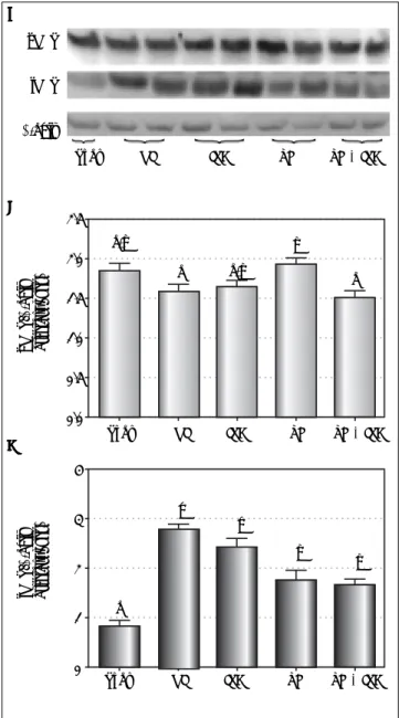
In addition to inflammation markers, we investigated the expression of nitric oxide synthases with each protec-tive method (Figures 3A-3C). eNOS expression differed
significantly among the groups (P = 0.006), with the TH group displaying higher expression in comparison with both the NI (P = 0.037) and TH + IPC (P = 0.006) groups. Regarding iNOS expression, significant differenc-es were also observed among the groups (P < 0.001). The sham group presented the lowest expression compared to NI (P < 0.001), IPC (P = 0.013), and TH + IPC (P = 0.029), while TH and TH + IPC presented a lower ex-pression in comparison with both NI (P = 0.001 and P < 0.001, respectively) and isolated IPC (P = 0.034 and P = 0.013 respectively).
Activation of the second-line antioxidant defenses by protective methods
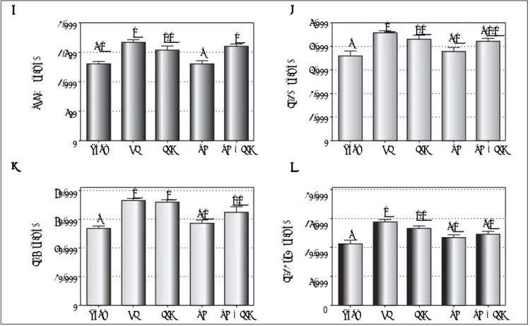
We tried to identify which protective method activated second-line antioxidant defenses. For that, the expression of NQO1 protein and the behavior of the Keap1-Nrf2 complex, including cytosolic and nuclear Nrf2 expres-sion, were analyzed (Figures 4A-4F). We observed differ-ences among groups in the expression of NQO1 (P = 0.001), Keap1 (P = 0.027), and cytosolic (P = 0.006) and
Figure 1. Figure 1.Figure 1.
Figure 1.Figure 1. Liver expression of TNF-α (AAAA), IL-1A β (BB), IL-6 (CBBB CCCC) and IL-12 p70 (DDDDD) after reperfusion (by ELISA assay). Data expressed as mean ± SEM. Different letters indicate a significant difference observed in the means of groups (P < 0.05). NI: normothermic ischemia. IPC: ischemic preconditioning. TH: topical hypothermia.
A AA A
A BBBBB
D D D D D C
C C C C
TNF-α
(pg/mg)
2,000
1,500
1,000
500
0
Sham NI IPC TH TH + IPC Sham NI IPC TH TH + IPC
a,b
c b,c
a c
a
a
a c
c
c c
b,c
b,c a,b
a,b
a,b a,b b,c
a,b,c
80,000
60,000
40,000
20,000
0
IL-6 (pg/mg)
5,000 4,000 3,000 2,000 1,000 0
IL-1
β
(pg/mg)
20,000
15,000
10,000
5,000
0
IL-12 p70 (pg/mg)
nuclear (P = 0.005) Nrf2. NQO1 expression was similar in the IPC and NI (P = 0.991) groups, and was higher in both these groups as compared to the sham (P = 0.022 and P = 0.042, respectively), TH (P = 0.009 and P = 0.020, spectively) and TH + IPC (P = 0.010 and P = 0.022, re-spectively) groups. Keap1 protein expression in TH + IPC group was higher than in NI (P = 0.035). The NI group displayed the highest cytosolic Nrf2 expression, with values similar to those observed in the isolated IPC group (P = 0.722), and the highest nuclear Nrf2 expres-sion, which was higher than that recorded in the sham (P = 0.004) and TH + IPC (P = 0.029) groups.
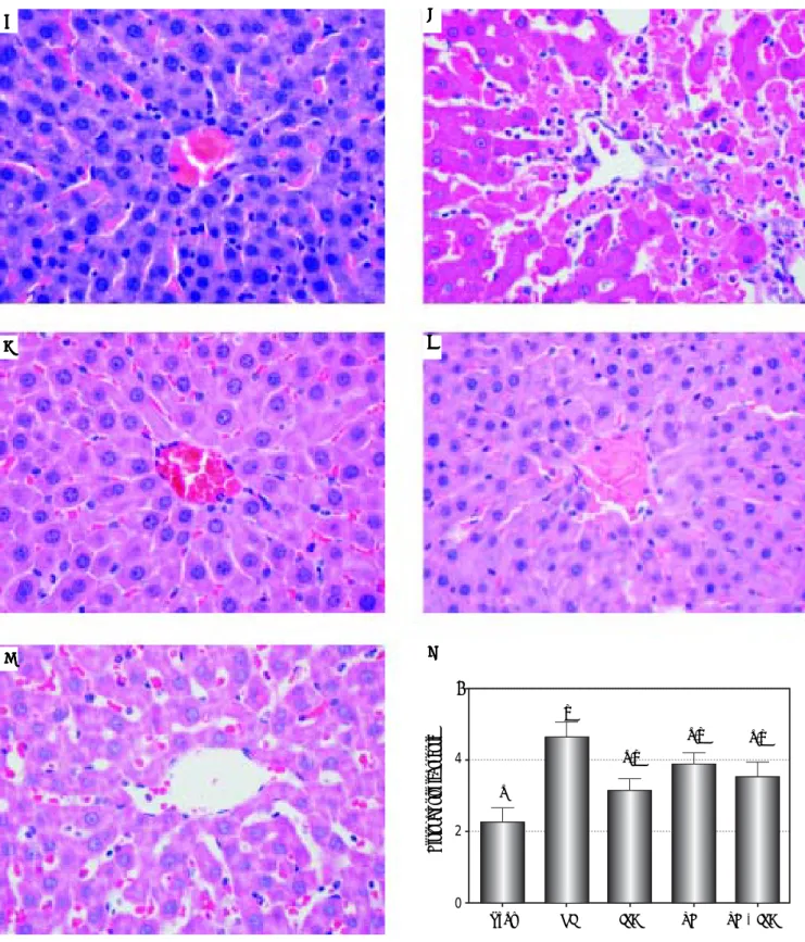
Effect of protective methods on histopathological score
The histopathology effect of the I/R injury after a 2-h reperfusion period was evaluated (Figures 5A-5F). The overall comparison among all the groups showed signifi-cant differences in relation to the histopathology score (P = 0.011). Regarding the differences between the specific groups, a significant difference was observed between the NI and sham groups (P = 0.012). The remaining groups presented only negligible alterations.
DISCUSSION
The mechanisms by which TH and IPC operate in I/R injury are not completely clear, but they are thought to play a crucial role in relieving inflammation and production of ROS and RNS following reperfusion.18,19,21,26 The
associa-tion of TH + IPC has been used in cardiac and retinal sur-geries.27-29 Regarding the liver, our group was the first to
report on the effectiveness of this synergistic approach
to prevent early I/R injury in a rat model.22 We analyzed the
protective effect of isolated or combined TH and IPC on bile flow and on the behavior of first-line antioxidant en-zymes, comparing these treatment groups with sham and NI groups. We observed that in animals submitted to TH and TH + IPC, bile flow was restored during reperfusion after bile flow blockade during ischemia. The present study was designed to continue that initial investigation. For that, we focused on the molecular mechanisms and effectiveness
Figure 3. Figure 3.Figure 3.
Figure 3.Figure 3. Liver expression of nitric oxide synthase isoforms. A.A.A.A.A. Wes-tern blot analysis. B.B.B.B.B. eNOS expression (densitometry analysis). C.C.C.C. iNOS ex-C. pression (densitometry analysis). Data expressed as mean ± SEM. Different letters indicate a significant difference observed in the means of groups (P < 0.05). NI: normothermic ischemia. IPC: ischemic preconditioning. TH: topical hypothermia.
2.5 2.0 1.5 1.0 0.5 0.0
Sham NI IPC TH TH + IPC
4
3
2
1
0
Sham NI IPC TH TH + IPC
Sham NI IPC TH TH + IPC
A AA AA
B BB BB
C C C C C
eNOS/
β
-actin
(arbitrary units)
iNOS/
β
-actin
(arbitrary units)
eNOS
iNOS
β-actin
a
c
c
b
b a,b
a a,b
b
a Figure 2.
Figure 2. Figure 2. Figure 2.
Figure 2. Liver expression of IL-10 after reperfusion (by ELISA assay). Data expressed as mean ± SEM. Different letters indicate a significant diffe-rence observed in the means of groups (P < 0.05). NI: normothermic ische-mia. IPC: ischemic preconditioning. TH: topical hypotherische-mia.
40,000
30,000
20,000
10,000
0
IL-10 (pg/mg)
Sham NI IPC TH TH + IPC
a
a,b b
c
A A A A
A BBBBB
C C C C
C DDDDD
F FF FF
Figure 4. Figure 4.Figure 4.
Figure 4.Figure 4. Liver expression of NQO1, Keap1 and Nrf2 (cytosolic and nuclear). A.A.A.A.A. Western blot analysis. B.B.B.B.B. NQO1 expression (densitometry analysis). C.
C.C.
C.C. Keap1 expression (densitometry analysis). D.D.D.D.D. Cytosolic Nrf2 expression (densitometry analysis). E.E.E.E. Western blot analysis. F.E. F.F.F.F. Nuclear Nrf2 (densitometry analysis). Data expressed as mean ± SEM. Different letters indicate a significant difference observed in the means of groups (P < 0.05). NI: normothermic ischemia. IPC: ischemic preconditioning. TH: topical hypothermia.
2.0
1.5
1.0
0.5
0.0
Nuclear Nrf2/
β
-actin
(arbitrary units)
Cytosolic Nrf2/
β
-actin
(arbitrary units)
2.0
1.5
1.0
0.5
0.0 0.8
0.6
0.4
0.2
0.0
NQO1/
β
-actin
(arbitrary units)
Sham NI IPC TH TH + IPC
Sham NI IPC TH TH + IPC
Sham NI IPC TH TH + IPC
a a
a
b b
a a a
b
a
a a,b
a,b b
a,b
E E E E E
Sham NI IPC TH TH + IPC
Nuclear Nrf2
β-actin NQO1
Keap 1 Cytosolic Nrf2
β-actin
Sham NI IPC TH TH + IPC
Sham NI IPC TH TH + IPC
Keap1/
β
-actin
(arbitrary units)
2.0
1.5
1.0
0.5
0.0
a a,b
a,b
a,b
of TH and IPC methods used alone or in combination for protecting against hepatic I/R injury, considering pro- and anti-inflammatory molecules as well as markers associated with the second-line antioxidant defense.22
Hepatic I/R injury can be classified into two stages in relation to reperfusion: an early phase, corresponding to the first 6 h after the start of reperfusion, and a late phase that lasts 48 h.5,12 In early I/R, activated KC secrete
pro-in-flammatory mediators, such as TNF-α, IL-1β, and IL-6, which intensify cell injury.3,7 In this study, the TH group
showed decreased hepatic concentrations of TNF-α and IL-1β in comparison with the NI group, and the concen-tration of these molecules in the TH group was similar to that found in sham animals.
In the presence of I/R injury, TNF-α and IL-1β have similar activity, exerting a stimulatory effect on the expres-sion of other inflammatory cytokines and chemokines, regulating free radical production, and helping neutrophil recruitment and adhesion to sinusoidal endothelial cells.30
The present findings agree with those obtained by other groups, showing that the use of hypothermia in hepatic I/R injury reduces inflammation by lowering TNF-α and IL-1β production.18,30,31 Mahmoud, et al. observed that
inhibi-tion of TNF-α induces decreased inflammation and oxidative stress, possibly providing a useful therapeutic approach to prevent I/R injury.8 In the present study,
TNF-α and IL-1β concentrations were similar in IPC and NI animals. Koneru, et al. did not observe advantages in using IPC during surgical procedures to decrease TNF-α
concentrations.32
In this study, hepatic IL-6 concentrations were de-creased in TH and sham groups in comparison with NI, while in the IPC group IL-6 concentrations were not dif-ferent from NI. IL-6 is an important marker of tissue damage and inflammation, and decreasing its expression minimizes I/R injury.33 The present findings suggest that
TH, and not IPC, has a protective role against hepatic in-flammation. Qi, et al. using an animal model of hepatocel-lular carcinoma, reported that the use of IPC induced decreases in TNF-α and IL-1β serum levels, associated with increased levels of IL-6 and higher signal transduc-tion and activatransduc-tion of transcriptransduc-tion 3 (STAT3) expres-sion.18 They proposed that the beneficial effects of IPC
might be cancelled out by the activation of the IL-6/ STAT3 pathway, thus accelerating carcinogenesis. Other studies, however, propose that increased IL-6 concentra-tions lead to STAT3 activation, inducing the expression of mitogenic and anti-apoptotic proteins capable of protect-ing against I/R injury and contributprotect-ing to hepatocellular regeneration.34,35
In the present study, TH, alone or associated with IPC, led to diminished hepatic IL-12 concentration, with levels similar to those of the sham group. IL-12 is a
pro-inflam-matory mediator involved in hepatic I/R injury, and animal models have revealed that exogenous application of this cytokine led to KC activation, expression of cell adhesion molecules, and neutrophil accumulation in liver paren-chyma. Conversely, mice treated with neutralizing anti-bodies for IL-12 presented decreased TNF-α and interferon-γ expression, causing reduced neutrophil re-cruitment to liver parenchyma.9,36 In the present study, the
IPC group presented the highest concentrations of IL-12, suggesting that IPC does not protect against the action of the pro-inflammatory mediators that worsen hepatic I/R injury.
In turn, IL-10 (previously called cytokine synthesis in-hibitory factor) plays an important anti-inflammatory role against I/R injury. In this study, not only was TH responsi-ble for decreasing the concentration of inflammatory cy-tokines, it also caused increased secretion of IL-10 in comparison with the NI group. IL-10 levels were similar in livers of IPC and NI animals. This finding differs from the report by Serafín, et al. who observed a protective ef-fect of IPC in livers of animals with fatty liver disease, as-sociated with increased levels of IL-10 and reduced inflammation.37 Some studies have reported blocked
nu-clear factor-κβ activity as a result of IL-10 secretion in I/R injury, and thus the expression of inflammatory mediators, protecting liver against cell damage.3,7,38
NO plays a key role in the regulation of liver blood flow in normal conditions, but the increased production of NO during I/R injury aggravates hepatic tissue damage owing to free radical production.10,12 NO is synthesized
through the action of NOS. eNOS leads to the produc-tion of physiological amounts of NO, while iNOS prompts copious and lasting NO synthesis, thus explain-ing iNOS involvement in several pathologic condi-tions.10,11 The present study showed higher eNOS
expression in the TH group compared to NI. NO synthe-sis attributable to the action of eNOS has a protective ef-fect against I/R injury, modulating vasodilation and inhibiting the production of inflammatory cytokines and free radicals by KC.12,39,40 In this study, higher iNOS
ex-pression was also observed in the IPC group as compared to the TH alone or TH+IPC group, but similar to the NI group. iNOS derived NO may be toxic or protective, de-pending on the type of aggression, on NO levels, and on the duration of iNOS expression.4,11 Our findings indicate
that TH provided protection against I/R injury in liver by promoting an increase in the levels of eNOS, while IPC, on the contrary, led to increased expression of iNOS, which may aggravate hepatocellular damage.
Figure 5. Figure 5.Figure 5.
Figure 5.Figure 5. Liver histology of the groups under study and comparison by the histopathological score. Slide images refer to sham (AAAAA), NI (BBBBB), IPC (CCCCC), TH (DDDD), TH+IPC (ED EEEE) groups. The NI group presented sinusoidal congestion, neutrophil infiltration and hepatocellular necrosis, alterations not found in the remai-ning groups. Magnifications: 400X. 5f- Comparison by the histopathological score. Data expressed as mean ± SEM. Different letters indicate a signifi-cant difference observed in the means of groups (P < 0.05). NI: normothermic ischemia. IPC: ischemic preconditioning. TH: topical hypothermia.
Histopathological score
6
4
2
0
Sham NI IPC TH TH + IPC
a
a,b a,b
b
a,b A
A A A
A BBBBB
C C C C
C DDDDD
E E E E
dismutase (SOD), catalase and glutathione peroxidase are classified as first-line antioxidant defenses against I/R inju-ry.41 Grezzana Filho, et al. showed that TH induced
in-creased levels of SOD in comparison with NI, IPC and TH+IPC.22 The increased levels of SOD induced by TH
are capable of abolishing the damage caused by I/R injury, precluding the use of second-line antioxidant defenses.
Second-line antioxidant defense enzymes are activated whenever those in the first line fail.17,41 Second-line
en-zymes synthesis is mediated through activation of the Nrf2-antioxidant response element signaling pathway.6,13
In normal conditions, Nrf2 is located in cytoplasm, asso-ciated with its repressor Keap1, both forming an inactive complex. Under stress condition, inducers such as ROS oxidize Keap1 cysteine residue, leading to dissociation of the inhibitory complex.15,17 Nrf2 uncouples from Keap1,
and translocates to the nucleus in order to promote the gene transcription of second-line antioxidant enzymes, such as NQO1.13-15 However, during a short time span
be-fore entering the nucleus, activated Nrf2 accumulates in cytoplasm.17
In this study, TH was effective in controlling the genera-tion of ROS, protecting against the cellular damage caused by reperfusion. The TH group had the lowest concentra-tions of inflammatory cytokines and the highest IL-10 lev-els, as well as a more favorable liver vasoactive response as assessed by iNOS and eNOS expression. These findings indicate lower production of ROS and RNS and an ade-quate restoration of the cell redox balance with TH. Re-duced oxidative stress prevents damage to lipids, proteins, and DNA.2,16 The use of hypothermia requires special care
in terms of hemodynamic monitoring, since it can trigger some deleterious effects such as edema, lactate accumula-tion and intracellular acidosis.21,27 Several methods of
hypo-thermia application have described, including the TH. Yamanaka, et al. studied patients with chronic liver diseases and hepatocellular carcinoma who underwent right-sided segmentectomy under normothermia or TH with tempera-tures ranging from 20 to 25 °C.42 Those authors showed that
the patients submitted to TH presented a reduced blood loss and tolerated a larger ischemia period in comparison with those who underwent the procedure under normoth-ermia. Beneficial effects of a moderate hypothermia have also been reported in studies that evaluated patients suffer-ing from acute liver failure.43
Conversely, NQO1 expression was higher in the IPC group in comparison with the TH and TH+IPC groups, suggesting that IPC failed to control oxidative stress through first-line antioxidant defense enzymes. Shokeir, et al. evaluated Nrf2 and NQO1 expressions in animals sub-mitted to the IPC approach against renal I/R injury.13 The
results obtained by those authors are similar to those of our study, with IPC animals presenting higher levels of
Nrf2 and NQO1 expression in comparison with the un-protected sample. Some studies, however, report a syner-gic effect of NQO1 and SOD, which protects cells against the generation of ROS and RNS.16,44 In the present study,
TH alone or in combination with IPC produced the low-est expression of Nrf2 in cytoplasm, indicating less Nrf2-Keap1 uncoupling and activation and suggesting that in this group activation of the second-line defense system was unnecessary. Isolated IPC, on the contrary, produced similar cytosolic Nrf2 expression to NI, indicating failure of the first antioxidant defense line. The combination of the two methods, TH and IPC, caused complete inhibi-tion of Keap1/Nrf2 uncoupling, preventing translocainhibi-tion of Nrf2 to the nucleus. This strong inhibition resulted in reduced expression of both NQO1 and nuclear Nrf2. These findings suggest that the association of IPC with TH may contribute an additional protective effect as com-pared to TH alone.
In this study, findings of significant histopathologic in-jury occurred only in the NI group in comparison with sham group. Dinant, et al. evaluated the protective effects of cooling by in situ hypothermic perfusion with a Ringer solution at 4 °C during 60-min of total vascular exclusion in a porcine liver I/R model.45 Those investigators showed
a significant increase of histopathologic injury in the nor-mothermic group, which presented increased liver cell vacuolization, neutrophil infiltration and parenchymal necrosis. The histopathologic alterations were not ob-served in the animals submitted to hypothermia. The his-topathologic alterations and bad outcome were not observed in the animals submitted to hypothermia. Nieu-wenhuijs, et al. evaluated the bile flow and histologic alter-ations in an experimental protocol of IPC, using 10-min ischemia and 10-min reperfusion periods followed by a 60-min reperfusion phase.46 Those authors observed that
the bile flow levels obtained at reperfusion were depend-ent on the length of the ischemia period. Significant his-topathologic alterations associated to the functional disturbance were absent since, according to the authors, necrosis and apoptosis occur only in the late phases of reperfusion.
One unexpected finding in this study was that isolated IPC was ineffective against I/R injury in the liver. Al-though most studies present evidences favoring the use of IPC against the I/R injury in the liver, some groups showed that this method offers only minor beneficial ef-fects, or is useless.47,48 Pasupathy, et al. reported that IPC
offers a protective effect against I/R injury in two distinct time points: in the early stage, comprising the first 3 h af-ter the organ reperfusion, and in the late stage, between 12 and 24 h. If IPC is applied in this last situation, the protec-tive effect can persist for up to 3 days.49 A putative
chosen in the design of this study can be beyond the limits of the protection affordable by IPC. Additionally, other groups obtained similar results to our findings.47,50
Pres-ently, our team is carrying out projects making use of the same experimental model, but applying a longer period of reperfusion, and assessing histologic and liver enzyme al-terations in the late phase of reperfusion (24 h).
In conclusion, using an animal model of early I/R inju-ry, we showed that TH alone was more effective than IPC alone to protect the liver against inflammation and oxida-tive stress, precluding the need to activate second-line antioxidant defenses. Isolated IPC did not have a protec-tive effect as indicated by the markers evaluated in this study.
ABBREVIATIONS
• eNOS: endothelial nitric oxide synthase.
• I/R: ischemia/reperfusion. • IL: interleukin.
• iNOS: inducible nitric oxide synthase.
• IPC: ischemic preconditioning. • KC: Kupffer cells.
• Keap1: Kelch-like ECH-associated protein-1.
• NI: normothermic ischemia. • NO: nitric oxide.
• NQO1: NAD•(P)H quinone oxidoreductase-1.
• Nrf2: nuclear erythroid 2-related factor 2.
• RNS: reactive nitrogen species. • ROS: reactive oxygen species.
• SOD: superoxide dismutase.
• TH: topical hypothermia. • TNF-ααααα: tumor necrosis factor-α.
GRANT SUPPORT
Fundo de Incentivo a Pesquisa e Eventos-Hospital de Clínicas de Porto Alegre (FIPE-HCPA) and Coordenação de Aperfeiçoamento de Pessoal de Nível Superior (CAPES).
REFERENCES
1. Knudsen AR, Kannerup AS, Grønbæk H, Dutoit SH, Nyen-gaard JR, Funch-Jensen P, Mortensen FV. Quantitative his-tological assessment of hepatic ischemia-reperfusion injuries following ischemic pre- and post-conditioning in the rat liver. J Surg Res 2013; 180: 11-20.
2. Ildefonso JA, Arias-Díaz J. Pathophysiology of liver ischemia-reperfusion injury. Cir Esp 2010; 87: 202-9. 3. Dinant S, Veteläinen RL, Florquin S, van Vliet AK, van Gulik
TM. IL-10 attenuates hepatic I/R injury and promotes hepato-cyte proliferation. J Surg Res 2007; 141: 176-82.
4. Glantzounis GK, Salacinski HJ, Yang W, Davidson BR, Sei-falian AM. The contemporary role of antioxidant therapy in
attenuating liver ischemia-reperfusion injury: a review. Liver Transpl 2005; 11: 1031-47.
5. Teoh NC. Hepatic ischemia reperfusion injury: Contemporary perspectives on pathogenic mechanisms and basis for hepatoprotection-The good, bad and deadly. J Gastroenter-ol HepatGastroenter-ol 2011; 26: 180-7.
6. Ke B, Shen XD, Zhang Y, Ji H, Gao F, Yue S, Kamo N, et al. KEAP1-NRF2 complex in ischemia-induced hepatocellular damage of mouse liver transplants. J Hepatol 2013; 59: 1200-7.
7. Kireev RA, Cuesta S, Ibarrola C, Bela T, Moreno Gonzalez E, Vara E, Tresguerres JA. Age-related differences in hepatic ischemia/reperfusion: gene activation, liver injury, and pro-tective effect of melatonin. J Surg Res 2012; 178: 922-34. 8. Mahmoud MF, El Shazly SM, Barakat W. Inhibition of TNF-α
protects against hepatic ischemia-reperfusion injury in rats via NF-κβ dependent pathway. Naunyn Schmiedebergs. Arch Pharmacol 2012; 385: 465-71.
9. Lentsch AB, Yoshidome H, Kato A, Warner RL, Cheadle WG, Ward PA, Edwards MJ. Requirement for interleukin-12 in the pathogenesis of warm hepatic ischemia/reperfusion injury in mice. Hepatology 1999; 30: 1448-53.
10. Miyake T, Yokoyama Y, Kokuryo T, Mizutani T, Imamura A, Nagino M. Endothelial nitric oxide synthase plays a main role in producing nitric oxide in the superacute phase of hepatic ischemia prior to the upregulation of inducible nitric oxide synthase. J Surg Res 2013; 183: 742-51.
11. Tsuchihashi S, Kaldas F, Chida N, Sudo Y, Tamura K, Zhai Y, Qiao B, et al. FK330, a novel inducible nitric oxide syn-thase inhibitor, prevents ischemia and reperfusion injury in rat liver transplantation. Am J Transplant 2006; 6: 2013-22. 12. Hines IN, Harada H, Flores S, Gao B, McCord JM, Grisham
MB. Endothelial nitric oxide synthase protects the post-ischemic liver: potential interactions with superoxide. Bi-omed Pharmacother 2005; 59: 183-9.
13. Shokeir AA, Hussein AM, Barakat N, Abdelaziz A, Elgarba M, Awadalla A. Activation of nuclear factor erythroid 2-related factor 2 (Nrf2) and Nrf-2-dependent genes by ischaemic pre-conditioning and post-conditioning: new adaptive endog-enous protective responses against renal ischaemia/reper-fusion injury. Acta Physiol (Oxf) 2013; 210: 342-53. 14. Kudoh K, Uchinami H, Yoshioka M, Seki E, Yamamoto Y. Nrf2
activation protects the liver from ischemia/reperfusion injury in mice. Ann Surg 2014; 260: 118-27.
15. Leonard MO, Kieran NE, Howell K, Burne MJ, Varadarajan R, Dhakshinamoorthy S, Porter AG, et al. Reoxygenation-spe-cific activation of the antioxidant transcription factor Nrf2 mediates cytoprotective gene expression in ischemia-reper-fusion injury. FASEB J 2006; 20: 2624-6.
16. Gang GT, Hwang JH, Kim YH, Noh JR, Kim KS, Jeong JY, Choi DE, et al. Protection of NAD(P)H:quinone oxidoreduct-ase 1 against renal ischemia/reperfusion injury in mice. Free Radic Biol Med 2014; 67: 139-49.
17. Morales P, Vargas R, Videla LA, Fernández V. Nrf2 activa-tion in the liver of rats subjected to a precondiactiva-tioning sub-chronic iron protocol. Food Funct 2014; 5: 243-50.
18. Qi Q, Bie P. Different roles of hepatic hypothermic ischemia and ischemic preconditioning in chemically induced hepato-carcinogenesis in rats. J Surg Res 2014; 189: 213-21. 19. Behrends M, Hirose R, Serkova NJ, Coatney JL, Bedolli M,
Yardi J, Park YH, et al. Mild hypothermia reduces the inflam-matory response and hepatic ischemia/reperfusion injury in rats. Liver Int 2006; 26: 734-41.
versus without ischemic preconditioning. Ann Surg 2003; 238: 843-50.
21. Choi S, Noh J, Hirose R, Ferell L, Bedolli M, Roberts JP, Nie-mann CU. Mild hypothermia provides significant protection against ischemia/reperfusion injury in livers of obese and lean rats. Ann Surg 2005; 241: 470-6.
22. Grezzana Filho TeJ, Mendonça TB, Gabiatti G, Rodrigues G, Marroni NA, Treis L, De Rossi SD, et al. Topical hepatic hy-pothermia plus ischemic preconditioning: analysis of bile flow and ischemic injuries after initial reperfusion in rats. Acta Cir Bras 2011; 26: 194-201.
23. Bradford MM. A rapid and sensitive method for the quantita-tion of microgram quantities of protein utilizing the principle of protein-dye binding. Anal Biochem 1976; 72: 248-54. 24. Sadowski HB, Gilman MZ. Cell-free activation of a
DNA-binding protein by epidermal growth factor. Nature 1993; 362: 79-83.
25. Suzuki S, Toledo-Pereyra LH, Rodriguez FJ, Cejalvo D. Neu-trophil infiltration as an important factor in liver ischemia and reperfusion injury. Modulating effects of FK506 and cy-closporine. Transplantation 1993; 55: 1265-72.
26. Eipel C, Glanemann M, Nuessler AK, Menger MD, Neuhaus P, Vollmar B. Ischemic preconditioning impairs liver regeneration in extended reduced-size livers. Ann Surg 2005; 241: 477-84. 27. Khaliulin I, Halestrap AP, Suleiman MS. Temperature precon-ditioning is optimal at 26 °C and confers additional protection to hypothermic cardioplegic ischemic arrest. Exp Biol Med (Maywood) 2011; 236: 736-45.
28. Salido EM, Dorfman D, Bordone M, Chianelli M, González Flei-tas MF, Rosenstein RE. Global and ocular hypothermic pre-conditioning protect the rat retina from ischemic damage. PLoS One 2013; 8: 61656.
29. An J, Camara AK, Rhodes SS, Riess ML, Stowe DF. Warm ischemic preconditioning improves mitochondrial redox bal-ance during and after mild hypothermic ischemia in guinea pig isolated hearts. Am J Physiol Heart Circ Physiol 2005; 288: H2620-H2627.
30. Kato A, Singh S, McLeish KR, Edwards MJ, Lentsch AB. Mechanisms of hypothermic protection against ischemic liver injury in mice. Am J Physiol Gastrointest Liver Physiol 2002; 282: G608-G616.
31. Henry SD, Nachber E, Tulipan J, Stone J, Bae C, Reznik L, Kato T, et al. Hypothermic machine preservation reduces molecular markers of ischemia/reperfusion injury in human liver transplantation. Am J Transplant 2012; 12: 2477-86. 32. Koneru B, Shareef A, Dikdan G, Desai K, Klein KM, Peng B,
Wachsberg RH, et al. The ischemic preconditioning paradox in deceased donor liver transplantation-evidence from a pro-spective randomized single blind clinical trial. Am J Trans-plant 2007; 7: 2788-96.
33. Cui LZ, Wang B, Chen LY, Zhou J. The effect of ischemic precondition to IL-6 on rat liver ischemia-reperfusion injury in transplantation. Asian Pac J Trop Med 2013; 6: 395-9. 34. Matsumoto T, O’Malley K, Efron PA, Burger C, McAuliffe PF,
Scumpia PO, Uchida T, et al. Interleukin-6 and STAT3 protect the liver from hepatic ischemia and reperfusion injury during ischemic preconditioning. Surgery 2006; 140: 793-802. 35. Camargo CA, Madden JF, Gao W, Selvan RS, Clavien PA.
In-terleukin-6 protects liver against warm ischemia/reperfusion injury and promotes hepatocyte proliferation in the rodent. Hepatology 1997; 26: 1513-20.
36. Myers KJ, Eppihimer MJ, Hall L, Wolitzky B. Interleukin-12-in-duced adhesion molecule expression in murine liver. Am J Pathol 1998; 152: 457-68.
37. Serafín A, Roselló-Catafau J, Prats N, Gelpí E, Rodés J, Per-alta C. Ischemic preconditioning affects interleukin release in
fatty livers of rats undergoing ischemia/reperfusion. Hepa-tology 2004; 39: 688-98.
38. Yoshidome H, Kato A, Edwards MJ, Lentsch AB. Interleukin-10 suppresses hepatic ischemia/reperfusion injury in mice: implications of a central role for nuclear factor kap-paB. Hepatology 1999; 30: 203-8.
39. Peralta C, Fernández L, Panés J, Prats N, Sans M, Piqué JM, Gelpí E, et al. Preconditioning protects against systemic dis-orders associated with hepatic ischemia-reperfusion through blockade of tumor necrosis factor-induced P-selec-tin up-regulation in the rat. Hepatology 2001; 33: 100-13. 40. Theruvath TP, Zhong Z, Currin RT, Ramshesh VK,
Lemas-ters JJ. Endothelial nitric oxide synthase protects transplant-ed mouse livers against storage/reperfusion injury: Role of vasodilatory and innate immunity pathways. Transplant Proc 2006; 38: 3351-7.
41. Ge M, Chi X, Zhang A, Luo G, Sun G, Xie H, Hei Z. Intestinal NF-E2-related factor-2 expression and antioxidant activity changes in rats undergoing orthotopic liver autotransplanta-tion. Oncol Lett 2013; 6: 1307-12.
42. Yamanaka N, Furukawa K, Tanaka T, Tanaka W, Yamanaka J, Imakita M, Okamoto E. Topical cooling-assisted hepatic segmentectomy for cirrhotic liver with hepatocellular carci-noma. J Am Coll Surg 1997; 184: 290-6.
43. Jalan R, Olde Damink SW, Deutz NE, Davies NA, Garden OJ, Madhavan KK, Hayes PC, et al. Moderate hypothermia pre-vents cerebral hyperemia and increase in intracranial pres-sure in patients undergoing liver transplantation for acute liver failure. Transplantation 2003; 75: 2034-9.
44. Siegel D, Gustafson DL, Dehn DL, Han JY, Boonchoong P, Ber-liner LJ, Ross D. NAD(P)H:quinone oxidoreductase 1: role as a superoxide scavenger. Mol Pharmacol 2004; 65: 1238-47. 45. Dinant S, van Veen SQ, Roseboom HJ, van Vliet AK, van
Gulik TM. Liver protection by hypothermic perfusion at differ-ent temperatures during total vascular exclusion. Liver Int 2006; 26: 486-93.
46. Nieuwenhuijs VB, de Bruijn MT, Schiesser M, Morphett A, Padbury RT, Barritt GJ. Ischemic preconditioning and intermit-tent ischemia preserve bile flow in a rat model of ischemia/ reperfusion injury. Dig Dis Sci 2007; 52: 3029-37.
47. Azoulay D, Lucidi V, Andreani P, Maggi U, Sebagh M, Ichai P, Lemoine A, et al. Ischemic preconditioning for major liver re-section under vascular exclusion of the liver preserving the caval flow: a randomized prospective study. J Am Coll Surg 2006; 202: 203-11.
48. Gurusamy KS, Kumar Y, Sharma D, Davidson BR. Ischemic preconditioning for liver transplantation. Cochrane Database Syst Ver 2008; 1: CD006315.
49. Pasupathy S, Homer-Vanniasinkam S. Ischemic precondi-tioning protects against ischemia/reperfusion injury: emerg-ing concepts. Eur J Vasc Endovasc Surg 2005; 29: 106-15. 50. Schiesser M, Wittert A, Nieuwenhuijs VB, Morphett A,
Pad-bury RT, Barritt GJ. Intermittent ischemia but not ischemic preconditioning is effective in restoring bile flow after ischemia reperfusion injury in the livers of aged rats. J Surg Res 2009; 152: 61-8.
Correspondence and reprint request:
Larisse Longo, BMedSci, MSc.
Laboratório Experimental de Hepatologia e Gastroenterologia, Centro de Pesquisa Experimental do Hospital de Clínicas de
Porto Alegre. Rua Ramiro Barcelos, 2350/ sala 12214. CEP 90035-003, Bairro Rio Branco, Porto Alegre, RS, Brazil.