A modified heterotopic auxiliary living donor liver
transplantation: report of a case
Kefeng Dou,*, Desheng Wang,*, Kaishan Tao,* Shuqiang Yue,* Zhenyu Ti,* Zhenshun Song,* Lin Li,* Yong He,* Xiaojuan Hou*
* Department of Hepatobiliary Surgery, Xijing Hospital, The Fourth Military Medical University, Xi’an, People’s Republic of China. These authors contributed equally to this work and should be recognized as co-first authors.
ABSTRACT
Liver transplantation is regarded as an effective treatment for Wilson’s disease (WD), and recently has been shown to improve not only hepatic but also neurologic manifestations. Conventional auxiliary liver transplantation for WD is orthotopic liver transplantation and heterotopic liver transplantation. But the conventional procedure could not avoid the problem of space, functional competition, hemodynamic vari-ation. Here we report a case of heterotopic auxiliary living-donor liver transplantation (HALDLT) to treat WD. We modified the operation to have a splenectomy, implant graft into the splenic fossa. The patient recovered well after the transplantation and has been symptom-free during a 5-year follow-up. This modi-fied operation is more safe and simple. HALDLT might be an effective treatment for WD patients with splenomegaly.
Key words. Left hepatectomy. Splenectomy. Wilson’s disease. Heterotopic liver transplantation. Splenic fossa.
Correspondence and reprint request: Kefeng Dou, M.D., Ph.D.
Department of Hepatobiliary Surgery, Xijing Hospital, The Fourth Military Medical University, West Changle Road 17, Xi’an, Shaanxi, 710032, China. Ph.: 86-29-84775255. Fax: 86-29-84775255
E-mail: [email protected]
Manuscript received: December 19, 2012. Manuscript accepted: October 06, 2013.
INTRODUCTION
Liver transplantation (LT) in the pediatric popu-lation has become a viable treatment method for children with end-stage liver disease.
Technical advances in the transplantation proce-dure such as auxiliary liver transplantation, reduced-size transplantation, split liver transplantation, and use of living donors for partial liver transplanta-tion,1,2 as well as modifications to
immunosuppres-sion and improvements in prophylaxis, have led to better survival rates in pediatric patients.
Wilson’s disease (WD) is an autosomal, recessive, inherited disorder of hepatic copper metabolism that results in the accumulation of copper in many or-gans and tissues. The hallmarks of the disease are the presence of liver disease, neurological symptoms and Kayser-Fleischer corneal rings.
LT is regarded as an effective treatment for WD, and recently has been shown to improve not only
hepatic but also neurologic manifestations.3-5 Stampfl,
et al. reported a successful heterotopic auxiliary partial liver transplantation in a patient (HAPLT) with Wilson’s disease presenting with fulminant hepatocellular failure. The advantages of HAPLT include avoidance of the anhepatic phase, limited surgical trauma, and less operating room time with elimination of recipient hepatectomy.6 Here we
report a case of heterotopic auxiliary living donor liver transplantation (HALDLT) to treat WD using a novel operation procedure and producing a good recovery.
CASE REPORT
In June 2007, a 12-year-old girl with Wilson’s dis-ease, presenting with manifestations including chronic liver failure, tremors and dyskinesia, under-went HALDLT in our unit. At the time of writing, the patient is alive and well with persistent normaliza-tion of graft funcnormaliza-tion for 5 years.
Before transplantation
movement disorder characterized by dysarthria, apraxia and a tremor-rigidity syndrome (also known as ‘juvenile Parkinsonism’).7 Her liver
dys-function gradually deteriorated into decompensated liver cirrhosis. Laboratory evaluations showed hy-poalbuminemia (32 g/L), leukopenia (2.1 x 10⁹ cells/
L), thrombocytopenia (39 x 109/L), and high
uncon-jugated bilirubin (23 μmol/L). Prothrombin time (PT) was 16.1 s.
The patient was 140 cm tall and 30.0 kg weight when she is on admission. Vital signs were within normal limits. Physical examination on admission showed splenomegaly and mild ascites. Abdominal ultrasonography and computed tomography revealed marked liver cirrhosis and splenomegaly.
Copper metabolism was evaluated in our labora-tory. The results showed a low plasma copper level (1.0 μg/dL) and a low plasma ceruloplasmin level (2.0 mg/dL). Her 24-h urine copper excretion was normal because she had been receiving continuous anti-copper therapy (D-penicillamine and zinc sulfate). Kayser-Fleischer (KF) rings were found by slit lamp examination. T2-weighted magnetic resonance imaging (MRI) revealed bilateral lucencies in the basal ganglia and thalami.
HALDLT was performed in June 2007. The donor was her mother who was heterozygous for the Wil-son genetic defect. She had no history suggestive of WD, and her serum copper and ceruloplasmin levels were 3.4 μg/dL and 20.2 mg/dL, respectively.
Donor surgical procedure
The donor was evaluated according to a previous-ly reported algorithm including donor liver biopsy and hepatic angiography.8 Histological examination
of the liver showed moderate infiltration by aggre-gates of lymphoid cells as well as mild piecemeal necrosis. Size calculations for this recipient and her liver graft were performed using 3-dimensional com-puterized tomographic imaging. Volumetric study estimated that the left lobe volume was 255.98 cm3.
Left hepatectomy (segment 2, 3 and 4) were performed with an ultrasonic dissector and bipolar electrocoagulation. Routine intraoperative cholangi-ography was performed before bile duct splitting. On the back table, the liver graft was flushed with Uni-versity of Wisconsin (UW) solution. The diameters of the left portal vein, left hepatic artery and vein of the graft were 6, 2, and 14 mm respectively. The graft to recipient body weight ratio was 0.77%.9
Recipient surgical procedure
The graft was performed using the following oper-ation procedure:
• Leave the native liver intact.
• Graft the implant into the splenic fossa.
Firstly, a complete splenectomy to make a room for the graft in the splenic fossa was performed. Be-fore the resection, the splenic vein and artery were identified since they passed above the body of pan-creas. Close to the hilum of the spleen, these two vessels were divided and ligated. Then, the tail of the pancreas was isolated, thereby creating a small recess anterior to the left renal vein. Following this, the left renal vein was divided and ligated. In order to retain the left gonadal vein and the central vein of the suprarenal gland, we isolated the left renal vein as closely to the vena cava as possible to allow application of a Satinsky vascular clamp. The diam-eters of the recipient’s splenic vein, splenic artery and left renal vein were carefully measured (7, 2, 13 mm respectively). Splenic venous pressure was 24 and 19 mmHg before and after spleentecomy.
Secondly, the graft was implanted heterotopically into the splenic fossa. The left hepatic vein of the graft and the free margin of the recipient left renal vein were brought together. A continuous running suture using a 4-0 Prolene stitch (Ethicon, Somerville, NJ) was placed between these two veins in an end-to-end fashion. Then, the graft portal vein was anastomosed with the recipient’s splenic vein in an
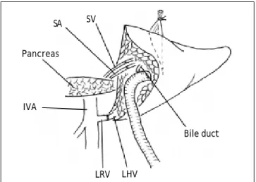
Figure 1. Schematic review of the surgical technique for
heterotopic auxiliary liver transplantation used in this case. SA: splenic artery. SV: splenic vein. IVA: inferior vena cava. LHV: left hepatic vein. LRV: left renal vein.
SA SV
Pancreas
IVA
LRV LHV
end-to-end fashion. Reconstruction of the left hepatic artery and splenic artery was performed with a corner-saving suture (9-0 silk suture thread, Ethicon), end-to-end anastomosis using loop magni-fication. Intraoperative Doppler ultrasound exami-nation showed that all vessels were patent. An anastomosis between the left bile duct and a Roux-en-Y jejunum loop restored the bile drainage (Figure 1). A thin polyethylene catheter was inserted through the choledochojejunostomy into the Roux-en-Y limb and attached to a collecting bag via a stab wound in the abdominal wall for bile production monitoring.
Immunosuppressive therapy
Post operation, the patient received well-described immunosuppressive protocol.10,11 Intravenous
meth-ylprednisolone (10 mg/kg) administered on the day of transplantation. It was continued postoperatively at dosages tapered from 10 mg/kg to 0.1 mg/kg at the end of the first month. A maintenance prednisone dose of 0.25 mg/kg/d was continued for 1 month, after which time steroid therapy was stopped. Tac-rolimus (FK506, Prograf, Astellas) was used for maintenance therapy. The maintenance dosage of tacrolimus was adjusted to maintain a level of 8 to 10 ng/mL during the first 2 months and 7 to 8 ng/mL thereafter.
Liver function and renal function monitor
After operation, blood samples were obtained from peripheral veins at 6:00 and 12:00 daily to measure the levels of serum aspartate aminotransferase, alanine transaminase, bilirubin, serum copper and ceruloplasmin. One week later, serum aspartate aminotransferase and alanine transaminase reached normal levels (Figure 2).
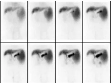
The graft and native liver were observed by
99mTC-Sodium phytate. The result showed that the
function of the graft was well (Figure 3).
Although the blood urea nitrogen (BUN) and catinine were not influenced by the ligation of left re-nal vein, the red blood cell in urine was observed after several days postoperation. Haematuria disap-peared as soon as the size of left kidney recovered to normal on the 7th day post operation.
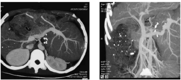
One month after HALDLT, the recipient’s serum copper and ceruloplasmin reached normal levels. Neurological symptoms also dramatically improved. CT showed that the graft had proliferated
signifi-Figure 2. Outline of the clinical course. Changes in serum
aspartate aminotransferase (AST) and alanine transaminase (ALT) levels are shown.
1 3 5 7 9 11 13 15
Day 180
160 140 120 100 80 60 40 20 0
IU/L
ALT AST
Figure 3. Liver function observed by nuclear medicine.
cantly at the same time (Figure 4). Two months lat-er, neurological symptoms became almost nor-mal. Liver biopsy in the heterograft eight months after transplantation showed the structure of he-patic lobule was normal with Local hehe-patic Sinus Di-lation, a few cell degeneration and necrosis could be found in central lobular area, as well as a few lym-phoid cells infiltration in portal area.
Doppler ultrasound
By using Doppler ultrasound, we were able to ob-serve a good spectrum of the waveform of the graft, which demonstrated that the graft was receiving a better blood supply according to the formula.12
native liver was so poor that sometimes it could not be detected.
At the time of writing, both the recipient and the donor were leading normal lives, and the recipient was free of neurological symptoms.
DISCUSSION
Since the first successful liver transplantation for WD in 1971,13 a number of reports in the literature
have considered WD as an excellent indication for transplantation, which can abrogate biochemical and clinical signs and offer long-term survival.14,15
Previous studies have never implanted the graft in the splenic fossa to solve the “space problem”. To date, there are few reports of living donor liver transplantation (LDLT) for WD. Asonuma, et al.16
described 11 pediatric (ages 6-16 yrs) cases of LDLT in which all liver grafts were obtained from parents who were one-haplotype matched and naturally het-erozygous for the Wilsonian genetic defect. They re-ported that all recipients had improved copper metabolism and remained free of recurrent WD after transplantation in their series. They concluded that the use of partial liver grafts obtained from asymp-tomatic heterozygote carriers of the WD genetic de-fect did not pose any difficulties regarding restoration of copper metabolism. Similar results re-ported by Wang, et al.17 also confirmed the validity
of partial liver grafts obtained from living haplo-type-matched donors.
Since the development of auxiliary partial ortho-topic liver transplantation (APOLT), controversies still remain, such as the “functional competition”
that results from the portal blood flow shared be-tween the graft and the native liver. To over-come this problem, and to optimize flow to the graft, some authors have banded or even ligated the native liver’s portal vein.18 In this case, portal blood flow
competition was not observed. We speculated that this was because the severe cirrhosis caused the blood flow to the graft to be more than that to native liver, which was able to be monitored by type-B ultrasound as soon as the graft was implanted. Schleimer, et al.19 reported that native liver
regener-ation seemed negatively influenced by a graft. These results do not support the hypothesis of functional portal flow competition between different livers. If portal inflow to the graft is decreased, some authors recommend step-by-step percutaneous em-bolization of the branches of the native right lateral portal vein, along with careful follow-up studies of the liver function.20
In this case, we supposed that the survival of the graft would be better if the pressure of the outflow drainage is low enough. The renal vein can provide a low outflow pressure because of its close proximity to the right atrium beside the hepatic vein. Notably, the right renal venous collateral circulation was weaker than that of the left. Choosing the right re-nal vein as the outflow drainage will damage the right renal function. Therefore, the left renal vein was chosen to be the outflow for the graft. Ligation of the left renal vein did not influence the renal function; in fact, the size of left renal was normal by the 7th day after operation.
In patients who have undergone LT, hypersplen-ism becomes problematic when persistent
neutrope-Figure 4. CT image of the recipient two months post operation. The graft has proliferated significantly. Hepatic angiography
nia and thrombocytopenia interfere with the ability to start or continue antiviral therapy or chemothera-py. Splenectomy is effective measure in liver transplant recipients for treatment of a variety of indications, including hypersplenism, prevention of gastroesophageal variceal hemorrhage, and for controlling portal pressure during small grafts in living donor liver transplantation. Splenectomy im-proved liver function in patients with liver cir-rho-sis, and could be a supportive and bridging therapy for patients waiting for liver transplantation.21
Re-cent advances in surgical techniques have enabled surgeons to perform splenectomy more safely and less invasively, but the procedure still has consider-able clinical outcomes. The major drawback of splenectomy is sepsis.
The potential risk for carcinogenicity of the rem-nant native liver is the one shortcoming of HAPLT for cirrhotic liver disease. This problem remains to be discussed. However, Kasahara’s report from the Kyoto group with more than 5 years follow-up showed no tumor development in the remnant native liver.18 Therefore, delayed native hepatectomy
after complete graft regeneration might not be necessary.
ABBREVIATIONS
• ALT: auxiliary liver transplantation.
• APOLT: auxiliary partial orthotopic liver
trans-plantation.
• HALDLT: heterotopic auxiliary living donor liver
transplantation.
• HAPLT: heterotopic auxiliary partial liver
transplantation in a patient.
• LDLT: living donor liver transplantation.
• LT: liver transplantation.
• WD: Wilson’s disease.
REFERENCES
1. Busuttil RW, Seu P, Millis JM, Olthoff KM, Hiatt JR, Milewicz A, Nuesse B, et al. Liver transplantation in children. Ann
Surg 1991; 213: 48-57.
2. Whitington PF, Balisteri WF. Liver transplantation in pedi-atrics: Indications, contraindications and pretransplant management. J Pediatr 1991; 118: 169-77.
3. Stracciari A, Tempestini A, Borghi A, Guarino M. Effect of liver transplantation on neurological manifestations in Wil-son disease. Arch Neuro 2000; l57: 384-6.
4. Chen CL, Chen YS, Lui CC, Hsu SP. Neurological improve-ment of Wilson’s disease after liver transplantation.
Transplant Proc 1997; 29: 497-8.
5. Mason AL, Marsh W, Alpers DH. Intractable neurological Wilson’s disease treated with orthotopic liver transplan-tation. Digest Dis Sci 1993; 38: 1746-50.
6. Stampfl DA, Muñoz SJ, Moritz MJ, Rubin R, Armenti VT, Jar-rell BE, Maddrey WC. Heterotopic liver transplantation for fulminant Wilson’s disease. Gastroenterology 1990; 99: 1834-6.
7. Ferenci P. Diagnosis and current therapy of Wilson’s dis-ease. Aliment Pharmacol Ther 2004; 19: 157-65.
8. Urata K, Kawasaki S, Matsunami H, Hashikura Y, Ikegami T, Ishizone S, Momose Y, et al. Calculation of child and adult standardliver volume for liver transplantation.
Hepa-tology 1995; 21: 1317-21.
9. Tanaka K, Uemoto S, Tokunaga Y, Fujita S, Sano K, Nishizawa T, Sawada H, et al. Surgical techniques and in-novations in living-related liver transplantation. Ann Surg 1993; 217: 82-91.
10. Haberal M, Dalgic A. New concepts in organ transplanta-tion. Transplant Proc 2004; 36: 1219-24.
11. Haberal M, Emiroglu R, Dalgiç A, Karakayli H, Moray G, Bilg-in N. The impact of cyclosporBilg-ine on the development of immunosuppressive therapy. Transplant Proc 2004, 36 (suppl.): 143S-147S.
12. Silva-Neto WDB, Cavarzan A, Herman P. Intra-operative of portal pressure and immediate results of surgical treat-ment of portal hypertension in schistosomotic patients submitted to esophagogastric devascularization with splenectomy. Arq Gastroenterol 2004; 41: 150-4. 13. DuBois RS, Giles G, Rogerson DO, Lilly J, Martineau G,
Hal-grimson CG, et al. Orthotopic liver transplantation for Wilson’s disease. Lancet 1971; 1: 505-8.
14. Schumacher G, Platz KP, Mueller AR, Neuhaus R, Stein-müller T, Bechstein WO, Becker M, et al. Liver transplan-tation: treatment of choice for hepatic and neurological manifestations of Wilson’s disease. Clin Transplantation 1997; 11: 217-24.
15. Emre S, Atillasoy EO, Ozdemir S, Schilsky M, Rathna Varma CV, Thung SN, Sternlieb I, et al. Orthotopic liver trans-plantation for Wilson’s disease. Transtrans-plantation 2001; 72: 1232-6.
16. Asonuma K, Inomata Y, Kasahara M, Uemoto S, Egawa H, Fujita S, Kiuchi T, et al. Living related liver transplanta-tion fromheterozygote genetic carriers to children with Wilson’s disease. Pediatr Transplant 1999: 3: 201-5. 17. Wang XH, Cheng F, Zhang F, Li XC, Qian JM, Kong LB, et
al. Copper metabolism after living related liver transplan-tation for Wilson’s disease. World J Gastroenterol 2003: 9: 2836-8.
18. Kasahara M, Takada Y, Egawa H, Fujimoto Y, Ogura Y, Ogawa K, Kozaki K, et al. Auxiliary partial orthotopic liv-ing donor liver transplantation: Kyoto University experi-ence. Am J Transpl 2005; 5: 558-65.
19. Schleimer K, Stippel DL, Kasper HU, Allwissner R, Yavuz-yasar S, Hölscher AH, Beckurts KT. Beckurts. Competition between native liver and graft in auxiliary liver transplan-tation in a rat model. Transplantransplan-tation Proceedings 2008, 40: 967-70.
20. Hasegawa K, Sugawara Y, Makuuchi M. Which is better to overcome the portal steal phenomenon in auxiliary partial orthotopic liver transplantation: inflow or outflow occlu-sion? Liver Transpl 2006; 12: 692-3.