Otras secciones de este sitio:
☞ ☞ ☞ ☞
☞ Índice de este número
☞ ☞ ☞ ☞
☞ Más revistas
☞ ☞ ☞ ☞
☞ Búsqueda
Others sections in this web site:
☞ ☞ ☞ ☞
☞ Contents of this number
☞ ☞ ☞ ☞
☞ More journals
☞ ☞ ☞ ☞ ☞ Search
Artículo:
Ketorolac pharmacokinetics in experimental cirrhosis by bile duct ligation in the rat
Copyright © 2003: Mexican Association of Hepatology
ANNALS OF HEPATOLOGY
Number 4 October - December 2003
Volume 2
L Rivera-Espinosa et al. Ketorolac pharmacokinetics in experimental cirrhosis by bile duct ligation in the rat 175
edigraphic.com
Annals of Hepatology 2003; 2(4): October-December: 175-181
Annals of Hepatology
Abstract
The purpose of the present work was to study the pharmacokinetics of ketorolac, a poorly metabolized drug, in experimental cirrhosis. Cirrhosis was induced by bile duct ligation (BDL) for four weeks in male Wistar rats. Ketorolac was given intravenously (1 mg/ kg ) or orally (3.2 mg/kg) to control (sham-operated) and BDL-rats. Determination of ketorolac in plasma was carried out by HPLC and estimation of pharma-cokinetic parameters was performed by non-compart-mental analysis. Indicators of liver damage and liver
1Sección Externa de Farmacología, Centro de Investigación y de
Estudios Avanzados del Instituto Politécnico Nacional, México, D.F., México.
2Facultad de Ciencias Químicas, Universidad Autónoma de San Luis
Potosí, México.
3Unidad de Investigación Médica en Farmacología. Centro Médico
Nacional Siglo XXI, IMSS.
Address for correspondence: Gilberto Castañeda-Hernández, PhD. Sección Externa de Farmacología
Centro de Investigación y de Estudios Avanzados del Instituto Politéc-nico Nacional
Apartado Postal 14-740 07000 México, D.F. México. Tel.: (5255) 5747-33-05 Fax: (5255) 5747-70-95 E-mail: [email protected]
Abbreviations
BDL bile duct ligation
HPLC high performance liquid chromatography
AUC area under the curve
t1/2 terminal half-life
Cmax peak concentration
tmax time to reachCmax
NSAID non-steroidal anti-inflammatory drug
Vd volume of distribution
Cl total drug clearance
F absolute bio-availability after the oral route
γ-GTP γ-glutamyltranspeptidase
ALT alanine aminotranspeptidase
Grants
This study was supported in part by grant 38940-M from Conacyt, Mexico. Liliana Rivera-Espinosa and Mónica Ordaz-Gallo were fellows of Conacyt.
Original Article
Ketorolac pharmacokinetics in experimental
cirrhosis by bile duct ligation in the rat
Liliana Rivera-Espinosa,PhD1 Pablo Muriel,PhD1 Mónica Ordaz Gallo,PhD1 José Pérez-Urizar,PhD2
Antonio Palma-Aguirre,PhD3 and Gilberto Castañeda-Hernández PhD1
fibrosis were significantly increased (p < 0.05) in BDL compared to control rats. Experimental cirrhosis did not induce any significant alteration in intravenous ketorolac pharmacokinetics. Volume of distribution, clearance, AUC and t1/2 were similar in BDL and con-trol animals. Notwithstanding, oral ketorolac bioavail-ability was significantly altered in BDL rats. AUC and Cmax were reduced, while tmax was prolonged, suggest-ing that both, the extent and the rate of ketorolac ab-sorption were decreased. Results show that liver cir-rhosis may result in significant pharmacokinetic alter-ations, even for poorly bio-transformed drugs, but that alterations may vary with the route of adminis-tration. In conclusion, uncritical generalizations on the effect of liver damage on drug kinetics should be avoided and systematic studies for every drug and ev-ery route of administration are thus recommended.
Key words: Ketorolac, pharmacokinetics, cirrhosis, liver damage, bile duct ligation.
Introduction
Ketorolac is a potent non-steroidal anti-inflammatory drug (NSAID) widely used for the treatment of moderate to severe pain, and it has been shown to be effective after administration by the oral, intramuscular and intravenous
routes.1,2 As do other NSAIDs, ketorolac produces its
ef-fect through the inhibition of prostaglandin synthesis.3
However, due to its high potency and efficacy, additional mechanisms of action have been proposed, including a
participation of opioid receptors,4 the stimulation of the
L-arginine-nitric oxide-cyclic GMP pathway,5 and
potas-sium channel opening.6 Using an experimental model
with a noxious stimulus of constant intensity,7 reported
that antinociceptive effect of ketorolac is directly related to its blood concentration. Notwithstanding, clinical stud-ies in postoperative pain have shown that there is no di-rect relation between ketorolac plasma concentrations and
its analgesic effect.8,9 The lack of direct correlation
place-Annals of Hepatology 2(4) 2003: 175-181
176
edigraphic.com
bo effect including a reduction in pain intensity during
the first hours of the postoperative period.8 It then appears
that, either directly or indirectly, the analgesic response to ketorolac depends on its pharmacokinetics. Therefore, any change in ketorolac absorption, distribution and/or elimination will likely have an impact on its pharmaco-logical response.
There are some concerns on ketorolac side effects. Ketorolac, as other NSAIDs, may produce
gastrointesti-nal side effects including peptic ulcer and bleeding.2,10
However, the main concern relates to its effect on
kid-ney function, particularly in hypovolemic patients.11
There are several reports on acute renal failure observed with ketorolac, even after a single dose
administra-tion.12,13 Life-threatening reactions have been reported
with doses of 60 mg or higher. Thus, the use of
ketoro-lac doses higher than 30 mg is not recommended.1,9
Ke-torolac then appears to be a drug with a relatively nar-row therapeutic index. Hence, alterations in its bioavail-ability may result in a lack of efficacy or in toxicity. Situations such as hepatic and renal impairment may
al-ter drug bioavailability.14 Despite of the wide use of
ke-torolac, the information available in the scientific litera-ture on its pharmacokinetics in liver damage is scarce, and it is limited to a single published abstract on which patients with alcoholic cirrhosis are compared with
young healthy individuals.15 According to this report in
patients with cirrhosis, ketorolac half-life was slight, but significantly prolonged after intra-muscular administra-tion, but there was no significant change in clearance. After oral administration, the time to reach the maxi-mum concentration was prolonged in patients with cir-rhosis, although no other oral bio-availability parameter was altered. Hence, it appears that the actual role of liv-er damage on ketorolac pharmacokinetics is not yet clear. Ketorolac is mainly eliminated by renal excretion
of the unchanged drug.2,16 Thus, it can be assumed that
liver damage will produce little change on its disposi-tion due to the small role played by bio-transformadisposi-tion. Liver damage, however, may alter pharmacokinetics if only by impairing drug metabolism. Drug distribution can be altered by changes in drug binding to plasma
pro-teins due to a reduced albumin production.17 Liver
dam-age also may result in alterations in blood flow in the
portal vein,18-20 which may modify the extent and rate of
drug absorption from the gastrointestinal tract.
Systematic studies on the role of hepatic diseases on drug kinetics are difficult to perform in humans due to the extremely high interindividual variability found in the clinical situation. Besides, experimental animal models have shown to be a suitable alternative for the character-ization of the pharmacokinetic alterations induced by
liv-er damage.17,21 Therefore, the purpose of this work was to
evaluate the effect of experimental liver cirrhosis on the pharmacokinetics of ketorolac after both, intravenous and oral administration.
Materials and methods
Materials. Ketorolac tromethamine was obtained from
Laboratorios Liomont S.A. (Mexico City). Sodium tol-metin was obtained as a sample from Cilag de México, S.A. (Mexico City). Acetonitrile, chromatographic grade, was purchased from Merck (Darmstadt, Germany). All other reagents used were of analytical grade.
Animals. Male Wistar rats (200-250 g) from our own
breeding facilities were used in this study. All animals re-ceived human care and the study complied with the insti-tution’s guidelines and the Mexican official regulation re-garding technical specifications for production, care and
use of laboratory animals (NOM-062-ZOO-1999).22
Ad-ditionally, the protocol followed the guidelines of the
Ca-nadian Council on Animal Care.23
Experimental cirrhosis. The cirrhosis was produced by
prolonged bile duct ligation (BDL) in the rat, which is a widely used model to simulate the condition of cirrhosis
in humans.24,25 Extrahepatic cholestasis was induced by
the double ligation and section of the common bile duct,
as described previously.24 Control sham-operated animals
were submitted to all surgical procedures, but without ob-struction of the bile duct. Twenty-eight days after sur-gery, liver damage was assessed by the plasma activities
of γ-glutamyltranspeptidase (γ-GTP)26 and alanine
ami-notransferase (ALT).27 Small liver sections fixed in
for-malin were used for Mallory trichromic staining for histo-logical examination under light microscopy. Collagen content, a marker of fibrosis, was estimated by the deter-mination of hydroxyproline in fresh liver samples, as
des-cribed previously.28
Study design. On the 28th day after BDL, food was
withheld but animals had free access to drinking water. Under light ether anesthesia, polyethylene catheters (a combination of a PE-10 and PE-50, i.d. 0.28 mm, o.d. 61 mm and i.d. 0.58 mm, o.d. 0.96 mm, respectively, Clay Adams, Parsippany, NJ, USA) were implanted into the caudal artery to collect blood samples. For animals which received the drug by the intravenous route, a polyethyl-ene catheter (PE-50) was implanted in a femoral vein and 1 mg/kg ketorolac was administered as a bolus dissolved in isotonic saline. For animals receiving the oral drug, 3.2. mg/kg ketorolac dissolved in saline was given by gavage. Ketorolac was administered after a 12 h fasting period. The used doses were selected as it has been shown that both, the intravenous and the oral doses yield
compa-rable analgesic effects in experimental pain.7
177
edigraphic.com
sustraídode-m.e.d.i.g.r.a.p.h.i.c cihpargidemedodabor
:rop odarobale FDP
VC ed AS, cidemihparG
arap
acidémoiB arutaretiL :cihpargideM
damage. Finally, animals were sacrificed by an excess of anesthesia and the liver was immediately removed to esti-mate hepatic collagen content.
Determination of ketorolac in plasma. Ketorolac
con-centration in plasma samples was determined using a high-performance liquid chromatographic method with
UV detection, as described by Flores-Murrieta et al.29
Briefly, 0.4 mL of 0.05 M potassium phosphate buffer (pH= 7.4) and 100 ng of sodium tolmetin (internal stan-dard) were added to 0.1 mL plasma samples. Samples were then acidified by the addition of 0.1 mL of 0.5 M solution of sodium acetate (pH=4) and extracted with 1 mL diethyl ether by vortex agitation during 1 min at max-imum speed. The two phases were separated by centrifu-gation at 10,000 rpm for 5 min. The organic layer was transferred to a clean conical glass tube and evaporated to dryness at 50 °C under a gentle nitrogen stream. The dry residue was redissolved in 0.1 mL of deionized water and 40 µL aliquots were injected into the chromatographic system. Analyses were performed using a Novapak C-18 column (150 x 3.9 mm I.D., particle size 4 µm, Waters Assoc., Milford, MA, USA) eluted with a mixture of ace-tonitrile and phosphoric acid 1 mM (pH=3) 43:57 v/v at a constant flow rate of 1.4 mL/min and at room tempera-ture. The effluent from the column was monitored at 313 nm. Retention times were 2.8 and 4.0 min for ketorolac and tolmetin, respectively.
Analysis of results. Individual plasma concentration
versus time plots were constructed. For the intravenous route, the area under the curve (AUC), the volume of dis-tribution (Vd), the total drug clearance (CL) and the
ter-minal half-life (t1/2) were determined. AUC was estimated
by the trapezoidal rule and extrapolated to infinity by multiplying the last detectable concentration by the time constant of the terminal concentration decay phase. Vd was obtained by dividing the dose by the extrapolated concentration corresponding to time zero. Clearance was estimated by dividing the dose by AUC. Half-life was es-timated from the slope obtained by linear regression of the terminal phase of semilogarithmic concentration ver-sus time plots. For the oral route, the peak concentration
(Cmax), the time to reach this peak (tmax), and AUC were
estimated. Cmax and tmax were determined graphically from
concentration versus time plots. AUC was estimated as above. All pharmacokinetic parameters were obtained by non-compartmental analysis using the Win NONLIN software program (Pharsight Corp., Mountain View, CA, USA). In estimation of absolute bioavailability after the
oral route (F) was calculated as: F = (AUCORAL /AUCIV)
(DOSEIV /DOSEORAL ) using mean values, since the intra-venous and oral experiments were performed in different
animals.14 Vd and CL were not estimated for the oral
route as F values for individual animals were not avail-able.
Results are presented as mean ± SEM. Comparisons between groups were performed using the student t test
for unpaired data. Differences were considered to achieve statistical significance when p < 0.05. Since intravenous and oral pharmacokinetics were determined in different animals, F was estimated from mean AUC values. Hence, no SEM values are provided and no statistical test was performed to compare the values of this parameter be-tween groups.
Results
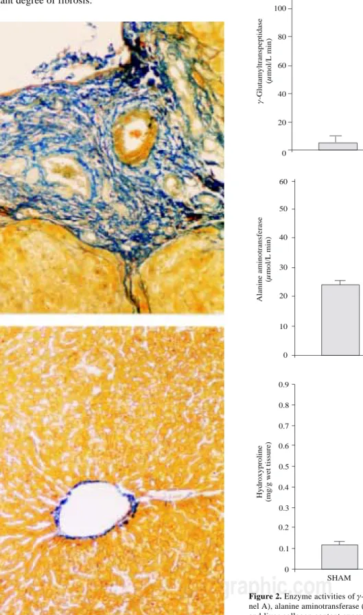
After four weeks, bile duct obstruction shown a pro-nounced liver damage. Fibrosis was evaluated by a histo-logical approach. Prolonged biliary obstruction was ac-companied by a marked increase in collagen disposition around the portal triad. The normal architecture was lost, extended necrotic areas were frequently observed and a marked ductular proliferation was present (Figure 1, right panel) with regard to sham operated animals (Figure 1,
left panel). Plasma γ-GTP augmented about 14-times
(Figure 2A) whereas ALT exhibited only a two-fold
in-crease (Figure 2B). Hepatic collagen content, estimated as the hydroxyproline accumulation, was about six times higher in DBL than in sham-operated rats (Figure 2C). All the differences in liver damage markers between BDL and sham-operated animals achieved statistical signifi-cance (p < 0.05). All of the bile-duct blocked animals ex-hibited ascites at the end of the fourth week.
Plasma ketorolac concentrations following intrave-nous and oral administration are depicted in figure 3. Pharmacokinetic parameters obtained by non-compart-mental analysis are shown in table I. BDL did not induce any significant change in ketorolac pharmacokinetics with regard to sham-operated animals when administered by the intravenous route. Notwithstanding, BDL was able
to alter oral ketorolac bioavailability. AUC and Cmax were
significantly reduced (p < 0.05) whereas tmax was
signifi-cantly prolonged (p < 0.05). As a result of the AUC re-duction after oral administration, without any change by the intravenous route, the absolute bioavailability (F), which was nearly complete in sham-operated animals, was reduced in BDL rats.
Discussion
Ketorolac pharmacokinetics were determined in sham-operated rats and in animals submitted to an irreversible obstruction of bile flow. This model is widely used, since it shows several analogies with the condition of
cholesta-sis and cirrhocholesta-sis in humans.24,25 At the end of the
four-week period of biliary obstruction, rats exhibited an es-tablished liver damage, reflected by the presence of ac-ites, by the increase of plasma enzymes activities, and fibrosis. The activity of γ-GTP increased about 14 times, while that of ALT augmented only about twice. This was expected, as γ-GTP is a marker of cholestasis, while ALT
accumu-Annals of Hepatology 2(4) 2003: 175-181
178
edigraphic.com
0 20 40 60 80 100
120 A
*
γ
µ
-Glutamyltranspeptidase
(
mol/L
min)
0 10 20 30 40 50 60
*
B
Alanine
a
minotransferase
(
mol/L
min)
µ
0.9
0 0.1 0.2 0.3 0.4 0.5 0.6 0.7 0.8
* C
SHAM BDL
Hydroxyproline (mg/g
w
et
tissure)
Figure 2. Enzyme activities of γ-glutamyl transpeptidase (γ-GTP; pa-nel A), alanine aminotransferase (ALT; papa-nel B) determined in plasma and liver collagen content express as hydroxyproline (panel C) from sham-operated rats (SHAM) and bile duct ligated rats (BDL) Each bar represents the mean value of experiments performed in duplicate as-says with samples from 12 animals ± SEM. (*) Statistically significan-tly different from the SHAM group, P < 0.05.
lation of hydroxyproline in the liver of animals with BDL compared to sham-operated controls, indicating an im-portant degree of fibrosis.
Figure 1. Mallory´s trichromic stains of liver sections from a sham
179
edigraphic.com
Ketorolac pharmacokinetics after intravenous admin-istration did not show any significant alteration in BDL animals with regard to sham-operated controls. These data suggest that BDL-induced cirrhosis does not modify ketorolac distribution and systemic elimination. Nonethe-less, liver damage produced a significant reduction in oral
ketorolac bioavailability. AUC and Cmax were
significant-ly reduced in about half while tmax was prolonged about
three times. These results indicate that BDL reduced both, the extent and rate of ketorolac absorption despite the fact that drug distribution and systemic clearance were not al-tered. These results were unexpected, as it is frequently assumed that liver damage increases drug bioavailability or produces no change. Increases in bioavailability could be due to a decreased hepatic first-pass effect of by the presence of porto-systemic shunts which allow a drug to bypass parenchymal liver tissue and thus reducing first
pass extraction.14 It should be noted, however, that most
pharmacokinetic studies showing bioavailability increas-es in liver damage have been performed for compounds which are cleared, at least partially, by hepatic metabo-lism and/or excretion. Poorly metabolized drugs with no biliary excretion, such as ketorolac, are seldom character-ized.
A reduction in oral drug bioavailability in liver damage can be explained by an impaired drug transfer from the gastrointestinal lumen to the splachnic circulation, by drug transfer to ascitis fluid before arrival to the systemic circu-lation or by a decreased hepatic first pass effect. In the case of oral ketorolac bioavailability in experimental cirrhosis, an alteration on hepatic first pass effect can be discarded. BDL did not result in a significant change in ketorolac total systemic clearance. Thus, it seems unlikely that hepatic biotransformation could play a significant role in the ob-served alterations in oral bioavailability. Furthermore, it has been reported that ketorolac is poorly metabolized,
be-ing mainly excreted as the unchanged drug.2,16 In
agree-ment with these data, we observed that, in sham-operated animals, ketorolac absolute bioavailability by the oral route was almost complete (F = 90%), indicating a negligible participation of first pass extraction.
The significantly reduced ketorolac absolute bioavail-ability by the oral route (from 90% to 48%) observed in experimental cirrhosis could be due to an impaired trans-fer from the gastrointestinal lumen to the splachnic circu-lation. As mentioned above, BDL rats exhibited an im-portant degree of liver fibrosis. Fibrosis is an imim-portant consequence of chronic liver diseases, which consists of deposition of connective tissue around the hepatic
sinuso-Table 1. Pharmacokinetic parameters of ketorolac observed after administration of a single 1 mg/kg intravenous dose and a single oral 3.2 mg/kg dose to
rats with experimental cirrhosis by bile duct ligation (BDL) and sham-operated controls.
Intravenous Oral
Parameter Sham BDL Sham BDL
AUC (µg.h/mL) 9.33 ± 2.70 9.00 ± 1.80 27.12 ± 3.72 14.11 ± 5.49*
Vd (L/kg) 0.36 ± 0.070 0.37 ± 0.09
CL (L/h.kg) 0.17 ± 0.04 0.16 ± 0.04
Half-life (h) 1.89 ± 0.41 2.16 ± 0.39
Cmax (µg/mL) 10.98 ± 3.70 5.04 ± 1.25*
tmax (h) 0.20 ± 0.02 0.62 ± 0.17*
F (%) 91 48
Data are presented as mean ± SEM of 6 animals. * Statistically significantly different from sham-operated animals (p< 0.05).
Time (h)
i.v. A
SHAM
BDL
0 2 4 6 8 10
0 2 4 6 8
p.o.
0 2 4 6 8
0 2 4 6 8 10
B
K
etorolac plasma concentration (
g/mL)
µ
Figure 3. Ketorolac plasma concentrations observed after
administra-tion of a single 1 mg/kg intravenous (i.v.) dose (panel A) and after ad-ministration of a single oral (p.o.) 3.2. mg/kg dose (panel B) to
sham-operated ( ) and bile duct ligated (DBL) rats (!) Data are presented
Annals of Hepatology 2(4) 2003: 175-181
180
edigraphic.com
ids. As a global consequence of collagen accumulation, liver blood flow resistance ensues, causing portal hyper-tension which, in turn may lead to stasis in the splachnic circulation and ascites.18,20
Ketorolac transference from the gastrointestinal lumen to the splachnic circulation likely occurs by passive diffu-sion, since it has been shown that its oral pharmacokinet-ics are linear in both, experimental animals and
hu-mans.31,32 Thus, the transference process should be
de-scribed according to Fick’s law of diffusion.33 In such
case, drug transference depends on the drug concentra-tion gradient established between gastrointestinal lumen and the splachnic circulation. If the drug is efficiently cleared from the capillaries irrigating the stomach and small intestine, capillary blood concentration will be maintained at near-zero values, favouring drug transfer-ence from the gastrointestinal lumen. In the case of stasis, drug clearance from the capillaries may be less efficient, thus blood concentration can rise to significant values im-pairing drug transference from the gastrointestinal lumen. It has been reported that rats with BDL, hepatic circulato-ry disturbance is caused by narrowing of peripheral portal vein branches and sinusoidal stenosis, with no perceptible
change in hepatic vein branches.19 Narrowing of
peripher-al portperipher-al vein branches can lead to a reduced splachnic blood flow affecting ketorolac absorption, without affect-ing systemic clearance and thus could be involved in the reduced oral ketorolac bioavailability in BDL rats.
Other possible explanation for a reduced oral ketorolac bioavailability is drug transference to ascites fluid, as rats with DBL exhibited ascites. Ascites is a consequence of portal hypertension due to the infiltration of plasma
through the walls of capillaries18 and thus it can be argued
that ketorolac had been sequestered in the ascites com-partment, impairing its transference to the systemic circu-lation. With the information available at present, it is not possible to establish whether reduced oral ketorolac bio-availability in experimental cirrhosis is the result of im-paired drug transfer from the gastrointestinal lumen to the splachnic circulation, or drug being sequestered in ascites fluid or whether both processes are the cause for it. Fur-ther research is required to elucidate this issue.
Despite the assumption that drug bioavailability is in-creased in liver damage, our results show that, at least in experimental BDL-induced cirrhosis, ketorolac bioavail-ability is significantly reduced when given by the oral route, being unchanged by the intravenous route. The present data demonstrate that liver damage can alter the pharmacokinetics of poorly metabolized compounds, and that these observations can be unexpected considering data from extensively metabolized drugs. Pharmacokinet-ic changes can also be due to alterations in drug absorp-tion, which have not been studied thoroughly. In fact,
Pages and colleagues15 observed a prolonged t
max for oral
ketorolac in patients with alcoholic cirrhosis while sys-temic clearance remained unchanged. These data suggest
that in these patients, as in our experimental animals, ke-torolac absorption from the gastrointestinal tract was al-tered despite a lack of significant changes in clearance. In summary, our results demonstrate that liver damage can alter pharmacokinetics even in absence of effects on drug metabolism. The overall effect of liver diseases on drug kinetics appears to be more complex than assumed. Hence, uncritical extrapolation of data should be avoided. Systematic pharmacokinetic studies in different types of liver damage should be performed to fully characterize this matter. The use of experimental models of liver dam-age, mimicking the different disease states found in the clinical situation, appears to be a suitable strategy for this purpose.
Acknowledgments
The authors express their gratitude to Ms. Patricia González for the preparation of the manuscript and fig-ures, and to Ms. Lourdes González, Mr. Mario Gil Moreno, Mr. Ramón Hernández, and Mr. Benjamín Sali-nas for their excellent technical assistance. This study was supported in part by grant 38940-M from Conacyt, Mexi-co. Liliana Rivera-Espinosa and Mónica Ordaz-Gallo were fellows of Conacyt..
References
1. Flores-Murrieta FJ, Granados-Soto V. Pharmacologic properties
of ketorolac tromethamine: a potent analgesic drug. Rev 1996; 2: 75-90.
2. Gillis JC, Brogden RN. Ketorolac. A reappraisal of its
pharmacody-namic and pharmacokinetic properties and therapeutic use in pain management. Drugs 1997; 139-188.
3. Rooks WH, Maloney PJ, Tolomonis AJ, Wallace MB, Schuler ME.
The analgesic and anti-inflammatory profile of (+)-5-benzoil-1,2-dihydro-3H-pyrrolo[1,2a]pyrrole-1-carboxylic acid (RS-37619).
Agents and Actions 1982; 12: 684-690.
4. Domer F. Characterization of the analgesic activity of ketorolac in
mice. Eur J Pharmacol 1990; 177: 127-135.
5. Granados-Soto V, Flores-Murrieta FJ, Castañeda-Hernández G,
López-Muñoz FJ. Evidence for the involvement of nitric oxide in the antinociceptive effect of ketorolac. Eur J Pharmacol 1995; 277: 281-284.
6. Lázaro-Ibañez GG, Torres-López JE, Granados-Soto V. Participation
of the nitric oxide-cyclic GMP-ATP-sensitive K+ channel pathway
in the antinociceptive action of ketorolac. Eur J Pharmacol 2001; 426: 41-46.
7. Granados-Soto V, López-Muñoz J, Hong E, Flores-Murrieta.
Rela-tionship between pharmacokinetics and the analgesic effect of ketorolac in the rat. J Pharmacol Exp Ther 1995; 272: 352-356.
8. Mandema JW, Stanski DR. Population pharmacodynamic model for
ketorolac analgesia. Clin Pharmacol Ther 1996; 60: 619-635.
9. Pérez-Urizar J, Granados-Soto V, Castañeda-Hernández G, Hong E,
González C, Martínez JL, Flores-Murrieta FJ. Analgesic efficacy and bioavailability of ketorolac in postoperative pain: a probability analy-sis. Arch Med Res 2000; 31: 191-196.
10. Estes LL, Fuhs DW, Heaton AH, Butwinick CS. Gastric ulcer perfo-ration associated with the use of injectable ketorolac. Ann
181
edigraphic.com
11. Kenny GNC. (1992) Potential renal, haematological and allergic adverse effects associated with nonsteroidal anti-inflammatory drugs.
Drugs 1992; 44: 5: 31-37.
12. Boras-Uber LA, Brackett NC. (1992) Ketorolac-induced acute renal failure. Am J Med 1992; 450-452.
13. Quan DJ, Kayser SR. Ketorolac induced acute renal failure follow-ing a sfollow-ingle dose. Clin Toxicol 1994; 32: 305-309.
14. Rowland M, Tozer TN. (1995) Clinical Pharmacokinetics: Concepts
and applications, 3rd ed., Williams & Wilkins, Media, PA. 1995;
248-266.
15. Pages LJ, Martínez JJ, Garg DC, Yee JP, Mroszczac ES, Renneke GA, Weidler DJ. Pharmacokinetics of ketorolac tromethamine in hepatically impaired versus young healthy subjects. J Clin Pharmacol 1987; 27: 724.
16. Brocks DR, Jamali F. Clinical pharmacokinetics of ketorolac tromethamine. Clin Pharmacokinet 1992; 23: 415-27.
17. Favari L, Castañeda-Hernández G, Hoyo-Vadillo C. Naproxen phar-macokinetics and pharmacodynamics in acute experimental hepati-tis. Arzneim Forsch 1993; 43: 675-679.
18. Groszmann RJ, Loureiro-Silva MR, Tsai MH. The biology of por-tal hypertension. In The liver biology and pathobiology. Edited by I.M. Arias JL, Boyer FV, Chisari N, Fausto D, Schachter DA. Shafritz. Lippincott Williams & Wilkins Publishers, Philadelphia, P.A. 2001; 679-719.
19. Shibayama Y, Nakata K. Haemodynamic alterations and their mor-phological basis in biliary obstruction. Liver 1992; 12: 175-178. 20. Van de Casteele M, Sagesser H, Zimmermann H, Reichen J.
Char-acterization of portal hypertension models by microspheres in anes-thetized rats: a comparison of liver flow. Pharmacol Ther 2001; 90: 35-43.
21. Castañeda-Hernández G, Favari L, Hoyo-Vadillo C. Relationship between naproxen plasma concentration and its antiinflammatory effect in experimental hepatitis. Arzneim Forsch 1995; 45: 585-589.
22. Norma Oficial Mexicana NOM-062-ZOO-1999.
23. Canadian Council on Animal Care. Guide to the care and use of experimental animals. Canadian Council of animal Care. Ottawa, Ont. 1993. http://www.ccac.ca/english/gui_pol/guides/english/
toc_v1.htm
24. Fernández-Martínez E, Morales-Ríos MS, Pérez-Álvarez V, Muriel P. Effects of thalidomide and 3-phtalimido-3-(3,4-dimethoxyphenyl)-propanamide on bile duct obstruction-induced cirrhosis in the rat.
Drug Dev Res 2001; 54: 209-218.
25. Kountaras J, Biling BH, Scheuer PJ. Prolonged bile duct obstruc-tion: a new experimental model for cirrhosis in the rat. Br J Exp
Pathol 1984; 65: 305-308.
26. Glossman M, Neville DM. Gamma-glutamyl transferase in kidney brush border membranes. Febs Lett 1972; 19: 340-344.
27. Reitman S, Frankel SA. A colorimetric method for the determina-tion of rum glutamic oxaloacetic pyruvic and glutamic pyruvic tran-saminases. Am J Clin Pathol 1957; 28: 56-63.
28. Muriel P. Nitric oxide protection of rat liver from lipid peroxidation, collagen accumulation, and liver damage induced by carbon tetra-chloride. Biochem Pharmacol 1998; 56: 773-779.
29. Flores-Murrieta FJ, Granados-Soto V, Hong E. Determination of ketorolac in blood and plasma samples by high-performance liquid chromatography. Boll Chim Farm 1994; 133: 588-591.
30. Rosen HR, Keeffe EB. Evaluation of abnormal liver enzymes, use of liver test, and the serology of viral hepatitis. In: Liver disease
Diag-nosis and management. Edited by B. Di Bisceglie. Churchill
Livingstone, New York, NY. 2000; 24-35.
31. Granados-Soto V, Flores-Murrieta FJ. Pharmacokinetics of oral ketorolac in the rat. Methods Find Exp Clin Pharmacol 1995; 17: 535-538. 32. Mroszczac EJ, Jung D, Yee J, Bynum L, Sevelius H, Massey I.
Ketorolac tromethamine pharmacokinetics and metabolism after in-travenous, intramuscular and oral administration in humans and ani-mals. Pharmacotherapy 1990; 10: 33S-39S.