Portal vein thrombosis:
What is new?
María del Carmen Manzano-Robleda,* Beatriz Barranco-Fragoso,** Misael Uribe,* Nahum Méndez-Sánchez*
* Liver Research Unit. Medica Sur Clinic & Foundation. Mexico City, Mexico.
** Department of Gastroenterology, National Medical Center “20 Noviembre”. Mexico City. Mexico.
ABSTRACT
Portal vein thrombosis (PVT) is one of the most common vascular disorders of the liver with significant mor-bidity and mortality. Large cohort studies have reported a global prevalence of 1%, but in some risk groups it can be up to 26%. Causes of PVT are cirrhosis, hepatobiliary malignancy, abdominal infectious or inflam-matory diseases, and myeloproliferative disorders. Most patients with PVT have a general risk factor. The natural history of PVT results in portal hypertension leading to splenomegaly and the formation of porto-systemic collateral blood vessels and esophageal, gastric, duodenal, and jejunal varices. Diagnosis of PVT is made by imaging, mainly Doppler ultrasonography. According to its time of development, localization, pathophysiology, and evolution, PVT should be classified in every patient. Some clinical features such as cirrhosis, hepatocellular carcinoma, and hepatic transplantation are areas of special interest and are dis-cussed in this review. The goal of treatment of acute PVT is to reconstruct the blocked veins. Endoscopic variceal ligation is safe and highly effective in patients with variceal bleeding caused by chronic PVT. In conclusion, PVT is the most common cause of vascular disease of the liver and its prevalence has being in-creasing, especially among patients with an underlying liver disease. All patients should be investigated for thrombophilic conditions, and in those with cirrhosis, anticoagulation prophylaxis should be considered.
Key words. Acute and chronic portal vein thromboses. Liver transplantation. Hepatocellular carcinoma. Coagulation. Anticoagulation. Cirrhosis.
Correspondence and reprint request: Prof. Nahum Méndez-Sánchez, M.D.,
Ph.D.
Liver Research Unit, Medica Sur Clinic & Foundation.
Puente de Piedra 150, Col. Toriello Guerra, Mexico City, Mexico. Telephone: +525-554247200 (4215); Fax: +525-55666-4031 E-mail: [email protected]
Manuscript received: December 4, 2014. Manuscript accepted: December 4, 2014.
January-February, Vol. 14 No. 1, 2015: 20-27
INTRODUCTION
Vascular diseases of the liver include a heteroge-neous group of disorders resulting from hepatic, vascular, and cardiovascular causes. The term “por-tal vein thrombosis” (PVT) refers to an obstruction in the trunk of the hepatic portal vein. It is impor-tant to describe the anatomy of the portal vein to understand how the thrombosis or its obstruction might occur. The portal vein accounts for 75% of the blood supply to the liver. It is an 8-cm wide, val-veless conduit originating from the confluence of the superior mesenteric and splenic veins posterior to the neck of the pancreas.
The importance of this vascular disease of the liver lies in its significant morbidity and mortality, which can occur without timely diagnosis or dis-ease-specific management, or even with an inappro-priate work-up.1 Its clinical presentation, prognosis, and management vary substantially according to etiology. For these reasons the cause must be inves-tigated for each patient. In this review, we will discuss only PVT because most of the other vascular disorders of the liver are so rare (Table 1).
Prevalence
Amitrano, et al. in a group of patients with cirrho-sis followed up prospectively during a similar time.3
Etiology
In a Swedish study of 254 autopsies, 28% had cir-rhosis, 23% a primary hepatobiliary malignancy, 44% secondary hepatobiliary malignancy, 10% ab-dominal infectious of inflammatory disease, and up to 3% a myeloproliferative disorder2(Figure 1). It has been reported that patients with cirrhosis and hepatic carcinoma have a high risk of PVT (odd ra-tio, OR, 17.1; 95% confidence interval, CI, 11.1-26.4). Is also important to note that the cause was not found in 14% of the patients.2,4
Risk factors
PVT is caused by a combination of local and gen-eral risk factors. Local risk factors can be identified in about 30% of patients, and a general risk factor in 70%. Local factors include cancers (any abdominal organ); focal inflammatory lesions (diverticulitis, appendicitis, pancreatitis, duodenal ulcer, cholecys-titis, Crohn’s disease, or cytomegalovirus hepatitis);
injury to the portal venous system (splenectomy, colectomy, gastrectomy, cholecystectomy, liver transplantation, or abdominal trauma); and cirrho-sis with preserved liver function with precipitating factors, or advanced disease without an obvious pre-cipitating factor. General risk factors for PVT include myeloproliferative disorders (40%), factor V Leiden mutation (32%), factor II mutation (40%), proteins C and S (26 and 30%), recent pregnancy (40%), antiphospholipid syndrome (19%), recent contraceptive use (12%), hyperhomocysteinemia (22%), and paroxysmal nocturnal hemoglobinuria (2%), among others.1
Genetics
Currently it is known that genetic mutations are important underlying factors that increase the pre-disposition to venous thromboses and thromboem-bolisms. In the next paragraphs we will discuss briefly some of the main mutations.
Factor V (FV) is a protein involved in blood coag-ulation that acts as a cofactor in transforming pro-thrombin into pro-thrombin, leading to fibrin formation. The C.1691G>A mutation results in arginine at po-sition 506 being replaced with glutamine, leading to the occurrence of Factor V Leiden (FVL), which in-creases the tendency to thrombosis.5
A G20210A mutation in the prothrombin gene (PTM) and the increase in mRNA production caused by transitions between guanine and adenine nucle-otides at the 20210 position lead to an increase in prothrombin level and in the risk of thromboembolic disease.6
Methylene tetrahydrofolate reductase (MTHFR) plays a role as a transmethylation enzyme catalyz-ing methionine synthesis in DNA. Therefore
muta-Table 1. Vascular diseases of the liver.
Budd-Chiari syndrome
Sinusoidal obstruction syndrome (veno-occlusive disease) Portal vein thrombosis (PVT)
Ischemic hepatitis Congestive hepatopathy Peliosis hepatis
Hepatic artery aneurysm Hepatic artery atherosclerosis Congenital vascular malformations Radiation-induced liver disease
Figure 1. of PVT showing local causes such as cirrhosis, inflammatory diseases, malignancy, and infections, and systemic caus-es, both congenital and acquired.
Etiology of PVT
Local Systemic
Cirrhosis Infection Congenital Acquired
tions to the gene encoding MTHFR reduce methyla-tion synthesis in DNA and cause hypercoagulamethyla-tion, which progresses together with plasma homo-cysteine volume. Polymorphisms in this gene are found in regions C677T and A1298C.6
Plasminogen Activator Inhibitor-Type 1 (PAI-1) is a specific plasminogen activator inhibitor that is re-leased from endothelial cells, hepatocytes, and meg-akaryocytes. The PAI-1 plasma level increases in response to mutations in its encoding gene, and as this inhibits the fibrinolysis pathway, an increase in coagulation can be observed.6
Finally, Simsek, et al. have proposed that com-bined mutations above mentioned rather than single mutations, play key roles in causing thromboses and thromboembolisms.6
Natural history
Portal vein obstruction results from thrombosis, constriction, or invasion of the lumen. The resulting portal hypertension leads to splenomegaly and for-mation of portosystemic collateral vessels, and es-ophageal, gastric, duodenal, and jejunal varices. In the porta hepatis, varices proliferate and involve the gallbladder and bile duct. As the portal vein throm-boses evolve, fibroblasts transform the clot into a firm, collagenous plug in which tortuous venous channels develop. This cavernous transformation (portal cavernoma) begins within days of the acute thrombosis and continues to evolve over weeks to months. Upstream from the obstruction, the small intestine and colon become congested, and the stom-ach exhibits changes of portal hypertensive gastrop-athy. Mesenteric ischemia can occur if the thrombus extends into the mesenteric veins.
Classification
According to the time of development, localiza-tion, pathophysiology, and evolulocaliza-tion, PVT can be classified as follows:
• Acute or chronic. • Extra-or intrahepatic. • Occlusive or nonocclusive. • Progressive or self-resolving.
Acute PVT is characterized by the sudden forma-tion of a thrombus within the portal vein. This can involve a variable portion of the mesenteric veins and/or the splenic vein. Occlusion can be complete or it can be partial, leaving a peripheral circulating
channel. Acute PVT has been reported only rarely in children and is characterized by the presence of in-fected thrombus/thrombi.1
Chronic PVT, also known as a portal cavernoma, is described as such because the obstructed portal vein is replaced by a network of hepatopetal collat-eral veins connecting the patent portion of the vein upstream from the thrombus to the patent portion downstream. The number, size, and location of col-laterals are extremely variable from patient to pa-tient. With occlusion of the trunk of the portal vein, the antral, duodenal, and biliary veins are enlarged markedly. This enlargement can produce compres-sion and deformation of the large bile ducts: so-called portal cholangiopathy or portal biliopathy. With occlusion at the origin of the portal vein, the pancreatic veins are enlarged.1
Clinical presentation
Approximately 43% of patients with PVT can be asymptomatic and diagnosis can be done during a routine Doppler ultrasound (US) examination; 39% of the patients present with gastrointestinal bleed-ing and 18% present with acute abdominal pain. Ab-dominal pain arises when the thrombosis is acute or involves the mesenteric veins and causes intestinal ischemia. Splenomegaly is usually present, but as-cites is uncommon, except in acute PVT or when the thrombosis complicates cirrhosis. Liver biochemical test results are usually normal.
In a univariate analysis published by Amitrano,
et al.7 age, gender, previous endoscopic therapy of esophageal varices, previous abdominal surgery, and the Child-Pugh score did not influence the clinical presentation of PVT, and the site of the thrombosis did not affect this except for involvement of the me-senteric vein. In addition, the site of the PVT (por-tal trunk, intra-hepatic branches, or splenic) and type (occluding or partial) did not differ among asymptomatic and hemorrhagic patient (Table 2).7
Diagnosis
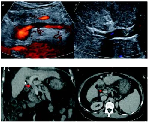
tom-ography (CT) or magnetic resonance imaging (MRI), especially when an important hepatofugal flow is present. However, CT and MRI are better for determining the extent of thrombosis (Figure 3). Contrast-enhanced US or CT imaging can help dif-ferentiate cases of benign from malignant PVT. Af-ter diagnosis, further evaluation with upper gastrointestinal endoscopy is warranted to assess the presence and degree of esophageal varices. As current imaging techniques allow the detection of asymptomatic PVT during routine US examina-tions, increasing numbers of patients with cirrhosis are being diagnosed with PVT.4
PVT is usually an incidental radiological finding in patients with cirrhosis. This is especially true for partial PVT, where it is expected that portal flow
will not be affected dramatically by the development of thromboses. In fact, in a significant proportion of patients (up to 50%) found to have PVT at the time of transplant surgery, the condition was unrecog-nized previously.8,9
In cases of acute PVT, US can show hyperechoic material in the vessel lumen with distension of the portal vein and its tributaries. Doppler US imaging shows the absence of blood flow in part or all of the lumen. CT scans without contrast can show hyper-attenuating material in the portal vein. After injec-tion of a contrast agent, there is lack of luminal enhancement, increased hepatic enhancement in the arterial phase, and decreased hepatic enhancement in the portal phase. For assessing thrombus exten-sion within the portal venous system, CT or
MRI-Table 2. Clinical presentation of PVT.17
Acute PVT Chronic PVT
Asymptomatic Asymptomatic
Abdominal pain, nausea, fever Well-tolerated UGI bleeding
Abdominal pain associated with SMVT Splenomegaly
Intestinal ischemia Hypersplenism
Hematochezia Growth retardation
Imaging Obstructive jaundice
Portal vein thrombosis on Portal cavernoma with
Doppler US collateral vessels
SMVT: superior mesenteric vein thrombosis. US: ultrasonography. UGI: upper gastrointestinal.
Figure 2. Doppler ultrasonography image of total portal vein thrombosis with absence of blood flow through the vessel is seen in A. Partial portal vein thrombosis is shown in B.
based angiography are more sensitive techniques than color Doppler ultrasonography, because the mesenteric veins are more difficult to visualize with the latter technique. Some recommend that careful screening for PVT is important for all patients with cirrhosis and for those being evaluated for liver transplantation.10
Chronic PVT is diagnosed by documenting a cav-ernoma, with abdominal US, CT, or MRI showing serpiginous structures while the main portal vein and/or its main branches are not visible. The hepat-ic arteries are usually enlarged. In the absence of cirrhosis, there might be an enlarged caudate lobe, together with an atrophic left lateral segment or right lobe of the liver. Typically, the umbilical vein is not dilated as it connects to the left portal vein branch downstream of the obstruction.1
PVT and cirrhosis
The development of PVT is a significant milestone in the natural history of cirrhosis; it is associated with worsening liver function, ascites, and the oc-currence of gastroesophageal variceal bleeding. The causal association between cirrhosis and PVT has been the subject of investigations but studies have not been able to address this association, or evalu-ate whether the development of PVT is just a conse-quence of advanced liver disease. The development of PVT was found to be more frequent in patients with advanced liver disease, but a low portal blood flow velocity was the only factor independently pre-dicting the occurrence of PVT.11 A portal blood flow velocity < 15 cm/s at initial Doppler US evaluation was associated with a higher incidence of PVT (47.8%) than a portal blood flow velocity > 15 cm/s (2%).12
Traditionally, cirrhosis has been considered a hy-pocoagulable state, and the degree of prolongation of prothrombin time (PT) and international normal-ized ratio (INR) have been taken as markers of the severity of coagulopathy. We should remember that these values were designed primarily to assess hypo-coagulability in patients being treated with vitamin K antagonists, but in patients with liver disease they probably overestimate the bleeding risk. This might explain the paradox of the poor prediction of bleeding in cirrhotic patients, even with marked pro-longation of conventional coagulation tests. It ap-pears that in the setting of hepatic synthetic impairment, both pro- and anticoagulant proteins are reduced to a similar degree and the net result in most patients with cirrhosis is a compensated
hemo-static balance with no tendency for bleeding or thrombosis. Because all of the components in the ex-trinsic coagulation pathway are produced by hepato-cytes, the degree of prolongation of the PT has been used extensively as a measure of liver synthetic function. However, even anticoagulants such as Proteins C and S as well as the levels of circulating protease inhibitors are reduced in cases of hepatic insufficiency, favoring a hypercoagulable environ-ment10 (Table 3).
A recent French multicenter prospective rand-omized trial carried out in patients with Child-Pugh types A and B cirrhosis who were subjected to Dop-pler US for screening of hepatocellular carcinoma (HCC) to identify risk factors for, and the impact of the development of, PVT. The investigators found that PVT was not a direct consequence of the pro-gression of liver disease. Furthermore, no evidence of a direct impact of the development of PVT on the progression of liver disease was found.13
PVT and HCC
PVT is a common complication of HCC with a re-ported incidence of 34% to 50%. It is one of the most negative prognostic factors and is called portal vein tumor thrombosis (PVTT). Survival in patients with PVTT is heterogeneous, depending on other clinical characteristics and hepatic function. Patient surviv-al was reported to be less than 3 months in the ab-sence of any treatment; however it has been shown to vary widely, from less than 5 months to more than 5 years depending on patient and tumor char-acteristics.
Previous staging systems of HCC did not take into account these characteristics, but more recent
Table 3. Factors promoting thrombosis in patients with
cir-rhosis.
Factors promoting thrombosis
Decreased levels of:
• Protein C. • Protein S. • Protein Z.
• Anti-thrombin a2 macroglobulin. • Heparin cofactor II.
• Plasminogen.
Increased levels of:
• Factor VIII.
Figure 4. Stratification of PVT. Grade I: < 50% portal vein thrombosis, with or without minimal extension into the superior me-senteric vein. Grade II: > 50% occlusion of the portal vein, including total occlusions, with or without minimal extension into the superior mesenteric vein. Grade III: complete thrombosis of both portal and superior mesenteric veins; the distal superior me-senteric vein is patent. Grade IV: complete thrombosis of both portal vein and proximal and distal superior meme-senteric veins.
I II III IV
staging systems, such as the Barcelona Clinic Liver Cancer (BCLC) grading system, classify all patients with vascular/portal invasion and or extrahepatic spread as having HCC stage C, and it seems that Sorafenib is the only recommended treatment. It has been suggested that vascular endothelial growth fac-tor plays a pivotal role in angiogenesis in patients with HCC and in the onset and evolution of PVTT, and that Sorafenib could exert a beneficial effect on PVTT by inhibition of the vascular endothelial growth factor receptor pathway with an antithrom-botic effect and revascularization.14 Other locore-gional therapies such as transarterial chemoembolization, radioembolization, and radio-therapy an also be considered because in theory these can suppress PVTT progression, delay intra-vascular tumor growth, and prevent the deteriora-tion of liver funcdeteriora-tion by maintaining an adequate portal blood flow.15
PVT and liver transplantation
PVT is a well-recognized complication in patients with liver cirrhosis waiting for liver transplantation (LT), and might recur after surgery. Some studies have reported a prevalence of 7.9% of PVT in this group of patients, but it can be as high as 26%.16 An accurate screening for PVT with determination of its grade of extension is mandatory, and should rou-tinely be performed in the preoperative work-up of candidates for LT.
Historically, PVT was a contraindication for LT, but in recent years the indication of LT in these
pa-tients depends on the extent of the thrombosis. Ear-ly grades (I and II) do not contraindicate transplan-tation, but a questionable contraindication can be patients with grade III and IV PVT (Figure 4). There is no ideal surgical technique for performing LT in patients with PVT.
The technical options for LT vary according to the patency of the portal vein and of other splanch-nic veins. When full patency of the portal vein has been achieved, whether with anticoagulation or with a transjugular intrahepatic portosystemic shunt (TIPS), end-to-end portal anastomosis is the first step. When full patency has not been achieved, the technical option depends upon the extent of PVT (partial or complete) and the patency of splenic and mesenteric veins.17
Alterations in liver anatomy after LT also in-creases the intrahepatic resistance to portal flow, so endothelial injury and coagulopathies could lead to a prothrombotic tendency in these patients.18 Re-currence of PVT after LT is one of the most fre-quent postoperative complications, with a rate of 3-36%; however, depending on the series studied, the incidence of de novo PVT after LT in patients without any previous evidence is generally not specified.19
Treatment
There is a growing need for optimal, evidence-based management of PVT in patients with cirrhosis because such management is currently not addressed in any consensus publication, including the recent practice guidelines on vascular disorders of the liver.4 The goal of treating acute PVT is to restore the obstructed veins, which will prevent intestinal inf-arction and portal hypertension. Correction of the causal factors should be achieved as soon as possi-ble, but is beyond the scope of this review. If fever or leukocytosis is present, antibiotics can be used. There are reports of recanalization of pylephlebitis with antibiotic therapy alone.1
Early initiation of anticoagulation therapy-prefer-ably within 30 days of symptoms-is recommended be-cause the rate of recanalization decreases from 69% to 25% when anticoagulation is instituted within the first or second week, respectively. Up to 35% of cases of acute PVT show recanalization with early anticoagulation therapy. Recanalization is less like-ly if the thrombosis is extensive because of the pres-ence of more than one prothrombotic disorder, and is associated with ascites. Early anticoagulation therapy in patients with mesenteric and portal vein thromboses minimizes serious complications such as peritonitis because of bowel necrosis, and also sig-nificantly decreases the development of complica-tions associated with esophageal varices. Recanalization occurs within 4-6 months after anti-coagulation therapy.20 Long-term anticoagulation may be recommended in patients with identified pro-thrombotic disorders, recurrent episodes of throm-bosis, or a family history of venous thrombosis. Anticoagulation can be initiated with heparin or subcutaneous low molecular weight heparin (for 2–3 weeks) because these are equally effective, although the second option makes laboratory monitoring un-necessary, with a lower risk of bleeding or immune thrombocytopenia. Later, oral vitamin K antago-nists should be given to maintain an INR of 2-3. Thrombolytic therapy with recombinant tissue plas-minogen activator, urokinase, and streptokinase in a very fresh PVT via a catheter introduced into the portal vein either transhepatically or through tran-sjugular approach might improve regional clot lysis, with 75% achieving some degree of lysis of the thrombosis. However, there is a high rate of bleed-ing (60%) so thrombolytic therapy should be re-served for patients with severe disease.21
Varices appear as early as 1 month after PVT. The treatment of acute gastrointestinal bleeding
caused by varices is similar to that used in patients with cirrhosis but without PVT. No studies have ad-dressed the role of primary prophylaxis in PVT-asso-ciated portal hypertension. There is concern at the possible extension of thromboses with beta blockers as well as vasopressors because of a decrease in splanchnic blood flow.22 Endoscopic variceal ligation is safe and highly effective in children and adults with PVT. Variceal obliteration following endoscopic sclerotherapy opens up spontaneous shunts because of a possible increase in portal pressure in 40% of patients, which in turn protects these patients from further bleeding and recurrence of varices.23
Other therapeutic strategies include TIPS.24 Re-cent information suggests that TIPS should be con-sidered a safe and feasible alternative therapy for selected patients with chronic PVT and cirrhosis, but it is not recommended for patients with a fi-brotic cord and fine collaterals instead of the origi-nal portal vein. In addition, combining traditioorigi-nal TIPS with a transhepatic or trans-splenic approach may facilitate technical success. Successful TIPS effectively reduces the portosystemic pressure gra-dient and prevents the recurrence of variceal bleed-ing. Finally, the only predictor of survival in patients with PVT and cirrhosis might be the ini-tial degree of occlusion, rather than any TIPS in-sertion.25
Some prophylactic strategies have been proposed, such as the use of enoxaparin to prevent PVT in pa-tients with advanced cirrhosis awaiting LT, because in some randomized trials a 12-month course of enoxaparin was found to be safe and effective in pre-venting PVT in patients with cirrhosis and a Child– Pugh score of 7–10.26,27 Also, enoxaparin appeared to delay the occurrence of hepatic decompensation and to improve survival.28
CONCLUSIONS
ACKNOWLEDGEMENTS
This project was support in part by a Grant from Medica Sur Clinic & Foundation.
ABBREVIATIONS
• CT: computed tomography. • FV: factor V.
• HCC: hepatocellular carcinoma. • INR: international normalized ratio. • LT: liver transplantation.
• MRI: magnetic resonance imaging.
• MTHFR: methylene tetrahydrofolate reductase. • PAI-1: plasminogen Activator Inhibitor-Type 1. • PT: prothrombin time.
• PVT: portal vein thrombosis.
• PVTT: portal vein tumor thrombosis.
• TIPS: transjugular intrahepatic portosystemic shunt.
• US: Doppler ultrasound.
REFERENCES
1. DeLeve LD, Valla DC, Garcia-Tsao G. Vascular disorders of the liver. Hepatology 2009; 49: 1729-64.
2. Ogren M, Bergqvist D, Bjorck M, Acosta S, Eriksson H, Sternby NH. Portal vein thrombosis: prevalence, patient characteristics and lifetime risk: a population study based on 23,796 consecutive autopsies. World J Gastroenterol 2006; 12: 2115-9.
3. Amitrano L, Brancaccio V, Guardascione MA, Margaglione M, Sacco M, Martino R, De Nucci C, et al. Portal vein thrombosis after variceal endoscopic sclerotherapy in cirrhotic patients: role of genetic thrombophilia. Endoscopy 2002; 34: 535-8. 4. Tsochatzis EA, Senzolo M, Germani G, Gatt A, Burroughs
AK. Systematic review: portal vein thrombosis in cirrho-sis. Aliment Pharmacol Ther 2010; 31: 366-74.
5. Rehak M, Rehak J, Müller M, Faude S, Faude F, Siegemund A, Krcova V, et al. The prevalence of activated protein C (APC) resistance and factor V Leiden is significantly high-er in patients with retinal vein occlusion without genhigh-eral risk factors. Case-control study and meta-analysis. Thromb Haemost 2008; 99: 925-9.
6. Simsek E, Yesilyurt A, Pinarli F, Eyerci N, Ulus AT. Com-bined genetic mutations have remarkable effect on deep venous thrombosis and/or pulmonary embolism occur-rence. Gene 2014; 536: 171-6.
7. Amitrano L, Guardascione MA, Brancaccio V, Margaglione M, Manguso F, Iannaccone L, Grandone E, et al. Risk fac-tors and clinical presentation of portal vein thrombosis in patients with liver cirrhosis. J Hepatol 2004; 40: 736-41. 8. Francoz C, Belghiti J, Vilgrain V, Sommacale D, Paradis V, Condat B, Denninger MH, et al. Splanchnic vein thrombosis in candidates for liver transplantation: usefulness of screening and anticoagulation. Gut 2005; 54: 691-7. 9. Dumortier J, Czyglik O, Poncet G, Blanchet MC, Boucaud
C, Henry L, Boillot O. Eversion thrombectomy for portal vein thrombosis during liver transplantation. Am J Trans-plant 2002; 2: 934-8.
10. Raja K. Portal Vein Thrombosis in Cirrhosis. J Clin Exp Hepatol 2013; doi.org/10.1016/j.jceh.2013.12.003 11. Garcia-Pagan JC, Valla DC. Portal vein thrombosis: a
pre-dictable milestone in cirrhosis? J Hepatol 2009; 51: 632-4. 12. Zocco MA, Di Stasio E, De Cristofaro R, Novi M, Ainora ME,
Ponziani F, Riccardi L, et al. Thrombotic risk factors in patients with liver cirrhosis: correlation with MELD scor-ing system and portal vein thrombosis development. J Hepatol 2009; 51: 682-9.
13. Nery F, Chevret S, Condat B, de Raucourt E, Boudaoud L, Rautou PE, Plessier A, et al. Causes and consequences of portal vein thrombosis in 1243 patients with cirrhosis: re-sults of a longitudinal study. Hepatology 2014 In press. 14. Novi M, Lauritano EC, Piscaglia AC, Barbaro B, Zocco MA,
Pompili M, Gasbarrini A. Portal vein tumor thrombosis revascularization during sorafenib treatment for hepato-cellular carcinoma. Am J Gastroenterol 2009; 104: 1852-4. 15. Yu JI, Yoon SM, Park HC, Kim JH, Kim TH, Park JW, Seong J, et al. Multicenter Validation Study of a Prognostic In-dex for Portal Vein Tumor Thrombosis in Hepatocellular Carcinoma. Cancer Res Treat 2014; 46: 348-57.
16. Gayowski TJ, Marino IR, Doyle HR, Echeverri L, Mieles L, Todo S, Wagener M, et al. A high incidence of native portal vein thrombosis in veterans undergoing liver transplanta-tion. J Surg Res 1996; 60: 333-8.
17. Francoz C, Valla D, Durand F. Portal vein thrombosis, cirrho-sis, and liver transplantation. J Hepatol 2012; 57: 203-12. 18. Ponziani FR, Zocco MA, Senzolo M, Pompili M, Gasbarrini A,
Avolio AW. Portal vein thrombosis and liver transplanta-tion: implications for waiting list period, surgical ap-proach, early and late follow-up. Transplant Rev (Orlando) 2014; 28: 92-101.
19. Duffy JP, Hong JC, Farmer DG, Ghobrial RM, Yersiz H, Hi-att JR, Busuttil RW. Vascular complications of orthotopic liver transplantation: experience in more than 4,200 pa-tients. J Am Coll Surg 2009; 208: 896-903.
20. Chawla Y, Duseja A, Dhiman RK. Review article: the mod-ern management of portal vein thrombosis. Aliment Phar-macol Ther 2009; 30: 881-94.
21. Hollingshead M, Burke CT, Mauro MA, Weeks SM, Dixon RG, Jaques PF. Transcatheter thrombolytic therapy for acute mesenteric and portal vein thrombosis. J Vasc Interv Radi-ol 2005; 16: 651-61.
22. Webster GJ, Burroughs AK, Riordan SM. Review article: portal vein thrombosis -new insights into aetiology and management. Aliment Pharmacol Ther 2005; 21: 1-9. 23. Dilawari JB, Raju GS, Chawla YK. Development of large
spleno-adreno-renal shunt after endoscopic sclerothera-py. Gastroenterology 1989; 97: 421-6.
24. Han G, Qi X, He C, Yin Z, Wang J, Xia J, Yang Z, et al. Transjugular intrahepatic portosystemic shunt for portal vein thrombosis with symptomatic portal hypertension in liver cirrhosis. J Hepatol 2011; 54: 78-88.
25. Yamashita Y, Bekki Y, Imai D, Ikegami T, Yoshizumi T, Ike-da T, Kawanaka H, et al. Efficacy of postoperative antico-agulation therapy with enoxaparin for portal vein thrombosis after hepatic resection in patients with liver cancer. Thromb Res 2014; 134: 826-31.
26. Senzolo M, Caldwell S. Portal vein thrombosis in cirrhosis: ig-nore, prevent, or treat? Gastroenterology 2013; 144: 19-20. 27. Villa E, Cammà C, Marietta M, Luongo M, Critelli R, Colopi S, Tata C, et al. Enoxaparin prevents portal vein thrombo-sis and liver decompensation in patients with advanced cirrhosis. Gastroenterology 2012; 143: 1253-60 e1-4. 28. Parikh S, Shah R, Kapoor P. Portal vein thrombosis. Am J