REVIEW ARTICLE
INTRODUCCIÓN
Los bifosfonatos (BF) son medicamentos utilizados en el manejo de la osteoporosis tanto en mujeres posmeno-páusicas como en hombres y en pacientes en tratamiento crónico con glucocorticoides; también se han utilizado en otras enfermedades metabólicas del hueso y en el tratamiento de la hipercalcemia asociada a malignidad.1 Estos fármacos han demostrado disminución en el riesgo de presentar fracturas y que mejoran la densidad mineral ósea en quienes los usan.1
OSTEONECROSIS DE MANDÍBULA ASOCIADA AL TRATAMIENTO
CON BIFOSFONATOS EN PACIENTES CON OSTEOPOROSIS: UNA REVISIÓN
OSTEONECROSIS OF THE JAW ASSOCIATED TO BISPHOSPHONATES
TREATMENT IN OSTEOPOROSIS PATIENTS: A REVIEW
JOHNAYRO GUTIÉRREZ RESTREPO1
RESUMEN. La osteonecrosis de mandíbula es una entidad que se viene describiendo desde 2003 y se deine como la presencia de un defecto en la mucosa oral que lleva a exposición del hueso de la mandíbula y del maxilar que falla en cicatrizar en un periodo de ocho semanas, en un paciente en tratamiento con bifosfonatos y sin exposición previa a radioterapia en cuello o en la cabeza. En este artículo se hace una revisión de la literatura respecto a la epidemiología, la isiopatología, los estudios diagnósticos, y los aspectos de prevención y tratamiento de esta entidad.
Palabras clave: osteonecrosis, osteoporosis, bifosfonatos.
Gutiérrez J. Osteonecrosis de mandíbula asociada al tratamiento con bifosfonatos en pacientes con osteoporosis: una revisión. Rev Fac Odontol Univ Antioq 2013; 24(2): 307-320.
ABSTRACT. Osteonecrosis of the jaw has been described since 2003 and it is deined as the presence of a defect in the oral mucosa leading
to exposure of the mandibular and maxillary bones, and failing to heal over a period of eight weeks in a patient treated with bisphosphonates and without prior exposure to radiation therapy on the head or neck. This article is a review of the literature regarding the epidemiology, pathophysiology, diagnostic studies, and several aspects related to prevention and treatment of this entity.
Key words: osteonecrosis, osteoporosis, bisphosphonates.
Gutiérrez J. Osteonecrosis of the jaw associated to bisphosphonates treatment in osteoporosis patients: A review. Rev Fac Odontol Univ Antioq 2013; 24(2): 307-320.
RECIBIDO: MAYO 6/2012-ACEPTADO: OCTUBRE 9/2012
INTRODUCTION
Bisphosphonates (BP) are drugs used in the ma-nagement of postmenopausal osteoporosis in both women and men and in patients undergoing chronic treatment with glucocorticoids; they have also been used in other metabolic bone diseases and in the treatment of hypercalcemia associated to malig-nancy.1 These drugs have proven to be effective in decreasing the risk of fractures and improving bone mineral density in those who use them.1
1 Internist and Endocrinologist, Professor at the Endocrinology Service, Universidad de Antioquia, Hospital Pablo Tobón Uribe, Medellín, Colombia.
1 Médico internista y endocrinólogo, docente del servicio de Endocrinología, Universidad de Antioquia, Hospital Pablo Tobón Uribe, Medellín, Colombia.
Their mechanism of action is based on inhibition of the enzyme farnesyl diphosphate synthase
(FPP synthase) in the mevalonic acid pathway in the osteoclast.2-6 This inhibition is performed after the BP is internalized in the cell; inhibition of FPP synthase prevents the conversion of geranyl diphosphate into farnesyl diphosphate, blocking the synthesis of signaling proteins that are essential for cell function and osteoclast survival, thus blocking the activation and maturation of immature osteoclast, and disabling them to perform its resorptive function and inducing apoptosis.2-9 These drugs are a
modiication of the molecule of pyrophosphate, and they have two radicals that allow binding to calcium crystals (R1) and deine the potency of the drug, its distribution, mechanism of action and time in the bone (R2).2-8 The presence of nitrogen radicals in the R1-aminobisphosphonates position (zoledronate, alendronate, risedronate, ibandronate, pamidronate), determines the antiresorptive potency of these drugs.2, 8, 10 Drugs without nitrogenous radicals (etidronate, clodronate) act via the ATP binding and the formation of intermediate metabolites which induce osteoclast apoptosis. Aminobisphosphonates also alter the function and assembly of the osteoclast cytoskeleton, preventing the mobility and release of substances responsible for promoting bone resorption.2-9
The characteristics of the radicals make the BP differ in their ability to bind to the bone (zoledronate> alendronate> ibandronate> risedronate> etidronate> clodronate) and inhibition of the FPP synthase ( a l e n d r o n a t e > i b a n d r o n a t e > r i s e d r o n a t e > zoledronate).9, 10
BPs have been associated with adverse effects, such as gastrointestinal symptoms (heartburn, esophagitis, nausea, abdominal pain, epigastric pain, gastrointestinal bleeding), and in the case of intravenous medications to a lu-like syndrome (fever, arthralgias, weakness, myalgia).1, 10-15 Other reported adverse effects include musculoskeletal pain, hypocalcemia, eye inlammation, transient elevations of creatinine, and atypical femoral fractures.1, 10-15 In recent years, there has been an increase in the report of cases of osteonecrosis of the jaw (ONJ) treated with these drugs, so the following paragraphs will offer a review of the most relevant aspects of this complication.
Su mecanismo de acción se basa en la inhibición de la enzima farnesil difosfato sintasa (FPPsintasa) en la vía del ácido mevalónico en el osteoclasto.2-6 Esta inhibición se lleva a cabo después de que el BF es interiorizado en la célula; la inhibición de la FPPsintasa evita la conversión del geranil difosfato a farnesil difosfato, bloqueando la síntesis de proteínas de señal esenciales para la función celular y la supervivencia del osteoclasto; de esta forma se bloquea la activación y maduración del osteoclasto inmaduro, incapacitándolo para llevar a cabo su función resortiva e induciendo su apoptosis.2-9 Estos medicamen-tos son una modificación de la molécula de pirofosfato, y tienen dos radicales que permiten la unión a los cristales de calcio (R1) y definen la potencia del medicamento, su distribución, mecanismo de acción y tiempo de permanencia en el hueso (R2).2-8 La presencia de radi-cales nitrogenados en la posición R1-aminobifosfonatos (zoledronato, alendronato, risedronato, ibandronato, pa-midronato), determina la potencia antirresortiva de estos medicamentos.2, 8, 10 Los medicamentos sin radicales nitrogenados (etidronato, clodronato) actúan mediante la unión al ATP y la formación de metabolitos intermedios que inducen la apoptosis del osteoclasto. Los aminobi-fosfonatos, también alteran la función y el ensamblaje del citoesqueleto del osteoclasto, impidiendo que se pueda movilizar y liberar las sustancias encargadas de promover la resorción del hueso.2-9
Las características de los radicales hacen que los BF di-fieran en su capacidad de fijación al hueso (zoledronato > alendronato > ibandronato > risedronato > etidronato > clodronato) y en la inhibición de la FPPsintasa (alendro-nato > ibandro(alendro-nato > risedro(alendro-nato > zoledro(alendro-nato).9, 10
Osteonecrosis of the jaw (ONJ) is deined as the presence of a defect in the oral mucosa which that leads to exposure of the mandibular or maxillary bone, which fails to heal over a period of eight weeks in a patient without prior exposure to radiation on the neck or who had been treated with BPs.16-22 ONJ is a complication of the treatment with BPs in patients with osteoporosis or other entities such as multiple myeloma, breast cancer, Paget’s disease of bone, and hypercalcemia of malignancy. This complication has been described since 2003,4 when it was reported in the U.S. and Australia.23-25 It consists of avascular necrosis of the jaw (the most common), the maxilla, or both; it may involve single or multiple lesions, which may even affect the palate.17, 19, 26, 27 In most cases (78%) it is associated with tooth extractions,16, 20, 21 and although according to its
deinition could not have been exposed to radiation, many of the patients had received chemotherapy and radiotherapy together for different malignancies (multiple myeloma, breast cancer, lung cancer, leukemia and plasmacytomas).16 ONJ has been associated with BPs and with other drugs such as denosumab,28 sunitinib29, 30 or bevacizumab31, 32 which may interfere with the function of monocytes and macrophages, alter the function of growth factor receptors and tissue revascularization phenomena, or increase the risk of infections (Actynomices). These factors facilitate the appearance of ONJ after procedures in the oral cavity.28-32
ONJ has been subclassiied into three categories: I. asymptomatic (exposed necrotic bone without evidence of infection), II. association with infection (pain, erythema, purulent discharge), III. presence of pathologic fractures, extensive fistulas or osteolysis.17, 18, 33
EPIDEMIOLOGY
Osteonecrosis of the jaw has been reported as being more common in patients receiving chemotherapy and radiation for multiple myeloma or metastatic bone disease, who in many cases should receive very high doses of BPs (12 to 15 times greater than those of patients with osteoporosis or Paget’s disease).8 In this population, the incidence of this complication is approximately 1 to 12% after 3 years of exposure to the drugs.8, 9, 17, 34
La ONM es una entidad que se define como la presencia de un defecto en la mucosa oral que lleva a exposición del hueso de la mandíbula o del maxilar, que falla en cicatrizar en un periodo de ocho semanas, en un paciente sin expo-sición previa a radioterapia en cuello o en la cabeza y que venía en manejo con BF.16-22 La ONM es una complicación del tratamiento con BF en pacientes con diagnóstico de osteoporosis u otras entidades como mieloma múltiple, cáncer de mama, enfermedad de Paget del hueso y la hipercalcemia asociada a malignidad. Esta complicación se viene describiendo desde 2003,4 cuando se reportó en Estados Unidos y Australia,23-25 y consiste en la ne-crosis avascular de la mandíbula (la más frecuente), el maxilar, o ambos; pueden ser lesiones únicas o múltiples, que incluso pueden llegar a afectar el paladar.17, 19, 26, 27 En la mayoría de los casos (78%) se asocia a extracciones de piezas dentales,16, 20, 21 y aunque, según la definición de esta entidad, no se podía haber estado expuesto a ra-diación, muchos de los pacientes habían recibido con-juntamente quimioterapia y radioterapia para diferentes neoplasias (mieloma múltiple, carcinoma de mama, pulmón, plasmocitomas y leucemia).16 La ONM se ha descrito tanto con BF como con otros medicamentos como denosumab,28 sunitinib29, 30 o bevacizumab,31, 32 medicamentos que pueden interferir de una u otra forma con la función de los monocitos y los macrófagos, alterar la función de receptores de factores de crecimiento y fenómenos de revascularización tisular o aumentando el riesgo de desarrollar infecciones (Actynomices). Lo anterior facilita la aparición de ONM tras procedimientos en la cavidad oral.28-32
La ONM se ha subclasificado en tres categorías: I. asin-tomática (hueso necrótico expuesto sin evidencia de infección); II. asociación con infección (dolor, eritema, secreción purulenta); III. presencia de fracturas pato-lógicas, trayectos fistulosos u osteolisis extensa.17, 18, 33
EPIDEMIOLOGÍA
In patients receiving BPs for the management of osteoporosis, the risk of this complication is much lower than in patients with cancer, and a lower incidence has been reported (of 1 in 1000 patients/ year of exposure), and even much lower in recent studies (1 in 100,000 patients/year treated with oral BPs), indicating how rare this complication is in daily practice, and as it will be addressed below, the risk appears to be greater in patients treated with intravenous BPs.8, 9, 17, 33, 34 The most important risk factors for the developing ONJ include advanced age; Caucasian race; the use of intravenous BPs (zoledronic acid or pamidronate, which have higher power than oral BPs) and cumulative doses (the longer the exposure the higher risk of developing ONJ); the presence of infection and, in general, trauma of the oral cavity: dentoalveolar surgery, dental extractions, dental implants, periodontal surgery with bone injury, to name just a few; anatomic variations of the oral cavity and the presence of lingual or palatal torus, and deiciency of vitamin D. Other risk factors include poorly controlled diabetes, alcohol and tobacco consumption, and cancer treatment with drugs or glucocorticoids.8, 17, 18, 22, 24, 25, 33, 34-37 Nevertheless, some patients have developed ONJ in absence of these risk factors.17, 22-24, 36
It has been reported that highly suppressed levels of bone resorption products, such as c-telopeptides, may increase the risk for ONJ due to their inability of remodeling bones by inhibition of osteoclasts; a risk classiication has even been suggested according to the values of these markers (<0.10 ng/mL: high risk; from 0.10 to 0.15 ng/mL: intermediate risk, and > 0.15 ng/mL: low risk of ONJ).38 However, recent studies have not proven this relationship as a predictor of ONJ.27, 39, 40
In a study conducted in Australia, the researchers calculated the frequency of ONJ with oral BPs (alendronate) for the treatment of osteoporosis as being 1 in 2260 to 1 in 8470 (0.01 to 0.04%), and after tooth extraction the frequency was 1 in 296 to 1 in 1130, while in patients with cancer who received venous BP the rate was much higher (1 in 10 to 1 in 15).41
En los pacientes que reciben BF para el manejo de la osteoporosis, el riesgo de esta complicación es mucho menor que en los pacientes con cáncer, y se ha reporta-do incidencia baja, de 1 caso por cada 1.000 pacientes/ año de exposición, e incluso mucho menor en estudios recientes (1 por cada 100.000 pacientes/año tratados con BF orales), lo cual indica lo poco frecuente que es esta complicación en la práctica diaria, y como se mencionará más adelante, el riesgo parece ser mayor en pacientes en tratamiento con BF intravenosos.8, 9, 17, 33, 34 Los factores de riesgo más importantes para el desarro-llo de la ONM son la edad avanzada, la raza caucásica, el uso de BF intravenosos (ácido zoledrónico o pami-dronato, que tienen mayor potencia que los BF orales) y las dosis acumuladas (a mayor tiempo de exposición mayor riesgo de desarrollar ONM), la presencia de infecciones y en general, el trauma de la cavidad oral: cirugías dentoalveolares, extracciones dentales, implan-tes orales, cirugía periodontal con lesión del hueso, entre otros; variaciones anatómicas de la cavidad oral como la presencia de torus lingual o palatino, y la deficiencia de vitamina D. Otros factores de riesgo mencionados son la diabetes mal controlada, el consumo de alcohol y de cigarrillo, y el tratamiento con medicamentos para el cáncer o con glucocorticoides.8, 17, 18, 22, 24, 25, 33, 34-37 Por otra parte, algunos pacientes han tenido ONM en ausencia de estos factores de riesgo.17, 22-24, 36
En la literatura se ha descrito que los niveles muy su-primidos de los productos de resorción ósea, como los c-telopéptidos, pueden aumentar el riesgo de presentar ONM, por la incapacidad de remodelar el hueso tras la in-hibición de los osteoclastos, e incluso se propuso una cla-sificación de riesgo de acuerdo con los valores de estos marcadores (< 0,10 ng/mL alto riesgo, 0,10-0,15 ng/mL riesgo intermedio, > 0,15 ng/mL bajo riesgo de ONM).38 Sin embargo, en estudios recientes no se comprobó esta relación como predictora de ONM.27, 39, 40
En un estudio hecho en Australia se calculó la frecuencia de presentación de ONM con BF orales (alendronato) para el tratamiento de la osteoporosis de 1 en 2.260 a 1 en 8.470 (0,01 a 0,04%), y tras la extracción dental
A descriptive study of 101 patients with ONJ found out that this condition occurred after 27 to 48 months of treatment with intravenous BPs (pamidronate and zoledronic acid, respectively) and after 67 months with alendronate,23 while other reports suggest they occurred after 9.4 to 15 months for zoledronic acid, 14.1 to 35 months for pamidronate, and 30 to 34 months for alendronate.34, 41 It appears that the risk of ONJ in patients with osteoporosis is low during the irst two to three years of therapy with oral BPs and during the irst months with venous BPs, but it may occur up to 10 years after be using these drugs.16, 17, 27, 36
In an analysis of the HORIZON group (HORIZON-PFT, GIO, Male OP and Prevention of OP) with 5903 patients without cancer who received 5 mg of zoledronic acid annually for osteoporosis treatment, compared with 5140 patients receiving placebo or other BPs (alendronate), the researchers found out that the incidence of ONJ was low with this drug, corresponding to 1 per 14,200 patients/year (there was one case of ONJ with zoledronic and another one in the placebo group).9, 42-47 As for the oral BPs most commonly used nowadays for the treatment of osteoporosis, most of the reported cases of ONJ have been described with alendronate (a drug with higher afinity for bone tissue), with an incidence of 0,5 to 2,5 cases per 100,000 patients/year, and to a lesser extent with risedronate (1.2 cases per 100,000 patients/year), and ibandronate.16, 22, 23, 33, 48-51 A recent series of ONJ in patients taking oral BPs showed a single case in 1537 patients.52 To date, there are no reports in the literature of cases of ONJ in users of osteoformers, such as teriparatide and strontium ranelate.
PATHOPHYSIOLOGY
It has been suggested that BPs may lead to a lower proliferation of epithelial cells in the oral cavity, and may even cause their apoptosis by breaking the FPP synthase, an enzyme which is necessary in cholesterol metabolism, leading to an alteration in the biosynthesis of cholesterol precursors that pre-vents the proper assembly of cell membranes.7
En un estudio descriptivo de 101 pacientes con ONM se encontró que esta entidad se presentó luego de 27 a 48 meses de tratamiento con BF intravenosos (ácido zole-drónico y pamidronato, respectivamente) y de 67 meses con alendronato,23 mientras que en otros reportes se han presentado luego de 9,4 a 15 meses para el ácido zoledrónico, 14,1 a 35 meses para pamidronato y 30 a 34 meses para alendronato.34, 41 Parece ser que el riesgo de ONM en pacientes con osteoporosis es bajo durante los primeros dos a tres años de terapia con BF orales y de los primeros meses con BF venosos, pero se puede presentar hasta 10 años después de estar usando estos fármacos.16, 17, 27, 36
En un análisis de los estudios del grupo HORIZON (HORIZON-PFT, GIO, Male OP and Prevention of OP) con 5.903 pacientes sin cáncer que recibieron 5 mg de ácido zoledrónico de forma anual para manejo de la osteoporosis, comparados con 5.140 que recibieron placebo u otros BF (alendronato), se encontró que la incidencia de ONM es baja con este medicamento, co-rrespondiendo a 1 por cada 14.200 pacientes tratados/ año (hubo un caso de ONM con zoledrónico y otro en el del grupo placebo).9, 42-47 En cuanto a los BF orales más usados en la actualidad para el tratamiento de la osteo-porosis, la mayoría de casos reportados de ONM se han descrito con alendronato (fármaco con mayor afinidad por el tejido óseo), con incidencia de 0,5-2,5 casos por 100.000 pacientes/año, y en menor proporción con risedronato (1,2 casos por 100.000 pacientes/año), e ibandronato.16, 22, 23, 33, 48-51 En una serie reciente de ONM en pacientes en tratamiento con BF orales se encontró un solo caso en 1.537 pacientes.52 Hasta el día de hoy, no hay reportes en la literatura médica de casos de ONM en usuarios de fármacos osteoformadores como el teriparatide o el ranelato de estroncio.
FISIOPATOLOGÍA
En pacientes con cáncer, metástasis óseas y función renal normal, hasta el 65% del BF aplicado por vía intravenosa (IV) se deposita dentro del hueso, y se ha reportado que si se recibe pamidronato 90 mg (IV) cada 4 semanas durante 4 años se pueden depositar entre 100 a 1.000 ng de fármaco por mg de hueso, pero se desconoce la concentración exacta de medicamento en los diferentes huesos del cuerpo (incluido el maxilar y la mandíbula) y en las zonas con mayor recambio óseo.7
Se ha sugerido que el trauma de la unidad alveolodental o la inflamación asociada a la enfermedad periodontal podrían causar liberación del BF almacenado en el hueso, el cual, al aumentar sus concentraciones, puede alcanzar niveles tóxicos para las células epiteliales que posteriormente llevarían a las lesiones típicas de esta entidad.7, 8 También se ha planteado un papel etiológico de las bacterias de la cavidad oral en esta patología, como Actinomyces sp, pues con frecuencia se ha reportado la presencia de este microorganismo en las muestras y cultivos de tejido de pacientes con ONM. Sin embargo, en muchas ocasiones es difícil diferenciar entre una infección de una colonización, y en muchos casos no se ha logrado identificar este microorganismo en los afec-tados.7, 22, 23 Se ha descrito que cuando hay exposición de hueso, este se puede colonizar con las bacterias (y en ocasiones hongos) de la región periodontal, pulpar, periapical y de la mucosa oral. 8 Este proceso infeccioso se asocia a dolor y parestesias en la región afectada, y no es infrecuente la presencia de supuración y la creación de trayectos fistulosos.8
Aún no hay claridad con respecto al efecto del BF, y se desconoce si el estado de bajo recambio óseo que genera el medicamento causa la necrosis, o si el efecto tóxico sobre la mucosa oral facilita la colonización bacteriana con la posterior infección del hueso y aparición de necrosis.8 Se piensa que con la inhibición de múltiples áreas de recambio óseo tras recibir dosis repetidas de BF hay mayor cantidad de medicamento disponible, y este puede ingresar al interior de los macrófagos y monocitos afectando su función de forma similar a como ocurre en los osteoclastos. De este modo, al generar disfunción y apoptosis de los macrófagos, se aumenta el riesgo de infecciones oportunistas por bacterias de la cavidad oral, que a través de la formación de biopelículas (una capa de glucoproteínas donde se “esconden” las bacterias de las células del sistema inmune) pueden participar direc-tamente en el proceso de daño óseo y posterior aparición de osteomielitis.8
In patients with cancer, bone metastases and normal renal function, up to 65% of the BPs intravenously applied (IV) is deposited inside bones, and it has been reported that receiving pamidronate 90 mg (IV) every 4 weeks for 4 years may leave between 100 to 1,000 ng of drug per mg of bone, but the exact concentration of drug in different bones of the body (including the upper and lower jaw) and in areas with increased bone turnover is still unknown.7
It has been suggested that trauma in the alveolodental unit or inlammation associated with periodontal disease may cause the release of BP stored in bones and therefore, by increasing its concentrations, it may reach toxic levels for the epithelial cells that would eventually lead to the lesions typical of this entity.7, 8 The etiologic role of bacteria (such as
Actinomyces sp) in the oral cavity has also been suggested, as the presence of this organism has often been reported in samples and tissue cultures of patients with ONJ. However, it is often dificult to differentiate between infection and colonization, and in many cases this organism has not been identiied in patients affected with ONJ.7, 22, 23 It has also been suggested that when the bone is exposed, it can be colonized by bacteria (and sometimes fungi) from the periodontal, pulpal, and periapical regions, as well as from the oral mucosa.8 This infectious process is associated with pain and numbness in the affected region, and in many cases with the presence of drainage and istulas.8
BPs may also affect the osteocytes by accumulating themselves in the vicinity of the vascular channels. When repetitive microfractures occur in normal conditions, osteocytes are responsible for initiating bone remodeling. When there are changes in terms of number and function of these cells, the microfractures lead to tissue damage which does not undergo repair and instead it favors the appearance of ONJ.8, 25 As previously stated, other phagocytic cells and some T cells can be affected by BP´s, leading to a state of immunosuppression that may contribute to the development of ONJ.25
Another possible mechanism of BP-induced damage seems to be related to the inhibition of angiogenesis.8 However, this last point has not been
clariied because of the impossibility of performing studies on rats or mice, since the mechanisms of bone remodeling in these animals are very different from those of humans.8 The literature has referred to inhibition of the vascular endothelial growth factor and the platelet-derived growth factor, affecting the replication of endothelial cells, which may be mediated by osteoclasts constraint, and this may favor the release of growth factors that are critical to the formation of new bone cells in response to tissue damage.8, 25
Vitamin D deficiency may be associated with increased risk of ONJ, since macrophages have enzymes that help convert 25-hydroxyvitamin D3 into its active form, 1,25 dihydroxyvitamin D3, and this, in turn, facilitates the production antibacterial products, such as cathelicidin and the expression of coreceptors such as CD14, that are required to activate antibacterial immune responses. Some studies in animals have shown that those that are deicient in vitamin D and have been exposed to BPs presented areas of exposed bone and infections consistent with ONJ.8 It should be noted that in the United States up to 67% of patients with metastatic cancer may have vitamin D levels below 30 ng/mL, which makes them more prone to ONJ.26
In a significant percentage of patients with postmenopausal osteoporosis, the levels of bone resorption markers (c-telopeptides) are usually above the normal reference range, but when they begin treatment with BPs this range greatly decreases (> 60% of baseline),38 relecting a constraint on the action of osteoclasts, and also on bone remodeling.
Los BF también pueden afectar a los osteocitos al acumularse en la vecindad de los canales vasculares. Cuando se presentan microfracturas a repetición, en condiciones normales, los osteocitos son los respon-sables de iniciar la remodelación ósea. Cuando se producen alteraciones en el número y función de estas células, las microfracturas llevan a un daño tisular que no se repara y en cambio favorece la aparición de ONM.8, 25 Como se mencionó previamente, otras células fagocíticas y algunos linfocitos T se pueden afectar por los BF, llevando a un estado de inmunosupresión que puede contribuir al desarrollo de la ONM.25
Otro posible mecanismo de daño inducido por los BF parece estar relacionado con una inhibición en la angio-génesis.8 Sin embargo, este último punto no se ha podido aclarar debido a la imposibilidad de hacer estudios en ratas o ratones, ya que los mecanismos de remodelamien-to óseo en esremodelamien-tos animales son muy diferentes a los del humano.8 Se ha mencionado en la literatura una inhibición del factor de crecimiento del endotelio vascular y del factor de crecimiento derivado de las plaquetas, que afectan la replicación de las células endoteliales, el cual puede estar mediado por el freno de los osteoclastos, los cuales al metabolizar la matriz del hueso favorecen la liberación de factores de crecimiento vitales para la formación de nuevas células óseas en respuesta al daño tisular.8, 25
La deficiencia de vitamina D se puede asociar a mayor riesgo de ONM, ya que los macrófagos tienen enzimas que ayudan a convertir la 25 hidroxivitamina D3 en su forma activa, la 1,25 dihidroxivitamina D3, y esta, a su vez, facilita la producción de productos antibacterianos, como la catelicidina y la expresión de correceptores como el CD14, que se requieren para activar respuestas inmu-nes antibacterianas. Se ha visto en estudios de animales que aquellos deficientes en vitamina D y expuestos a BF fueron los que presentaron zonas de hueso expuesto e infecciones compatibles con ONM.8 Cabe resaltar que en los Estados Unidos se ha descrito que hasta 67% de los pacientes con cáncer metastásico pueden tener niveles de vitamina D menores de 30 ng/mL, lo que los hace más propensos a presentar ONM.26
It has been suggested that highly suppressed values of bone resorption markers may be associated with a higher risk of developing ONJ.38 However, several studies have reported that suppression of bone renewal markers is not involved in ONJ, and that these values are very similar to those in premenopausal women;8 also, many cases of ONJ with BPs have been reported in patients with normal levels of c-telopeptides 9 Since most patients treated with BPs will experience suppressed levels of resorption markers, this inding seems unimportant as a predictor of risk for ONJ.9, 27, 39, 40
Finally, some studies have identiied polymorphisms in the cytochrome CYP2C8, which contains a group of enzymes that not only serve for the metabolism of drugs but also affect the angiogenesis cascade and increase the risk of ONJ by losing the revascularization mechanisms of tissues affected by trauma in the oral cavity while receiving treatment with BPs.53
RADIOLOGY
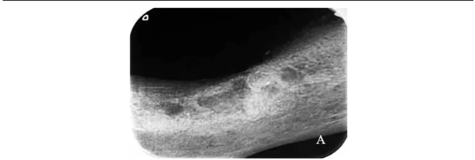
Among the most useful images to diagnose this en-tity are panoramic radiographs of the jaw, periapical radiographs of small lesions, computed tomography, and magnetic resonance imaging.7, 8 Some of the
most characteristic indings (although not pathog -nomonic of ONJ) are similar to those found in other forms of osteonecrosis, such as the presence of scle-rosis, osteolysis and a speckled trabecular pattern, periosteal reaction, and bone fragmentation.8 Other
important indings include thickening or loss of la -mina dura and extension of the periodontal ligament space.8 Nuclear medicine studies won´t show uptake of the compromised bone due to necrosis and to low blood low in the affected area. However, there may be uptake of the radiotracer in the vicinity due to increased low and to the metabolic activity of the inlammatory or infectious process.8 Figure 1 shows an X-ray of typical damage in a patient with ONJ.
TREATMENT
Before starting treatment with a bisphosphonate IV, patients should undergo a complete oral examination,
Se ha planteado que los valores muy suprimidos de los marcadores de resorción ósea se pueden asociar a un riesgo más alto de desarrollar ONM.38 Sin embargo, en varios reportes se ha informado que en la ONM no hay supresión de los marcadores de recambio óseo, y que los valores de los mismos son muy similares a los de mujeres premenopáusicas,8 y muchos de los casos de ONM con BF se han presentado en pacientes con niveles normales de c-telopéptidos.9 Ya que la mayoría de los pacientes tratados con BF van a tener los niveles de marcadores de resorción suprimidos, este hallazgo parece poco importante como predictor de riesgo de ONM.9, 27, 39, 40
Por último, se han descrito polimorfismos en el citocromo CYP2C8, que contiene un grupo de enzimas que no solo sirven para el metabolismo de medicamentos, sino que pueden afectar también la cascada de la angiogénesis y aumentar el riesgo de ONM al perderse los mecanismos de revascularización de los tejidos afectados por un trauma de la cavidad oral mientras se recibía tratamiento con BF.53
RADIOLOGÍA
Entre las imágenes más útiles para el diagnóstico de esta entidad se pueden mencionar la radiografía panorámica de la mandíbula, la radiografía periapical en lesiones peque-ñas, la tomografía computarizada y la resonancia mag-nética.7, 8 Algunos de los hallazgos más característicos aunque no patognomónicos de la ONM son similares a los encontrados en otras formas de osteonecrosis, como la presencia de esclerosis, osteolisis y un patrón moteado trabecular, reacción perióstica, fragmentación ósea y formación de secuestros.8 Otros hallazgos importantes son el engrosamiento o pérdida de la lámina dura y la ampliación del espacio del ligamento periodontal.8 En los estudios de medicina nuclear no se encontrará captación en el hueso comprometido debido a la necrosis y al bajo flujo sanguíneo de la zona afectada. Sin embargo, en las zonas aledañas puede haber captación del radiotrazador debido al aumento de flujo y de la actividad metabólica por el proceso inflamatorio o infeccioso.8 En la figura 1 se aprecia el daño típico en la radiografía de un paciente con ONM.
TRATAMIENTO
principalmente en pacientes con neoplasias que van a requerir dosis muy altas de estos medicamentos para manejo de enfermedad metastásica o de la hipercalcemia asociada a la malignidad. Cualquier diente insalvable se debe extraer y se deben hacer todos los procedimientos orales invasivos que se requieran.17 Además, se debe garantizar la óptima salud periodontal.17 En aquellos pa-cientes con osteoporosis que requieran un procedimiento oral como una extracción dental, se debe suspender el BF oral o IV por al menos tres a cuatro meses antes de la intervención, con el fin de evitar la aparición de ONM al permitir que se normalicen los marcadores de resorción ósea y la capacidad de cicatrizar adecuadamente la zona de hueso afectada.17, 27
En pacientes con enfermedades sistémicas que requieran tratamiento con BF IV, estos medicamentos también se deben suspender durante 3 meses antes y 3 meses des-pués del procedimiento odontológico, siempre y cuando la condición del paciente lo permita.17, 18 En este último escenario, se debe esperar como mínimo 14 a 21 días tras la extracción de un diente para permitir que se cubra el defecto con mucosa antes de aplicar el BF IV.17, 18
En cuanto al manejo de la ONM establecida, la mayoría de los autores sugieren la realización de procedimientos quirúrgicos menores para la remoción de tejido necrótico y de secuestros, mientras que otros hacenénfasis en que el hueso necrótico expuesto no se debe remover y se debe mantener limpio, para evitar mayor exposición por el curetaje y la deficiente cicatrización.17, 18, 23, 34, 54
especially if they present tumors that would require very high doses of these drugs for treating metastatic disease or hypercalcemia of malignancy. Any teeth that cannot be saved should be extracted and all the required invasive oral procedures must be done.17 In addition, optimal periodontal health must be guaranteed.17 In patients with osteoporosis requiring oral procedures such as tooth extraction, oral BPs or bisphosphonate IV should be discontinued for at least three to four months before the intervention, in order to prevent ONJ by allowing normalization of bone resorption markers and the ability of the affected bone area to properly heal.17, 27
In patients with systemic diseases requiring treatment with BP IV, these drugs should also be suspended for three months before and three months after the dental procedure, provided that the patient’s condition allows it.17, 18 In the latter scenario, a period of at least 14 to 21 days should be provided after the extraction of a tooth to allow covering of the defect with mucosa before applying the BP IV.17, 18
[image:9.581.54.526.56.215.2]Regarding the management of ONJ already established, most authors suggest performing minor surgical procedures for the removal of necrotic tissue and sequestrum, while others emphasize that the exposed necrotic bone should not be removed and must be kept clean, to avoid further exposure by curettage and poor healing.17, 18, 23, 34, 54
Figura 1. Hallazgos radiológicos en la ONM
Figure 1. Radiological indings in ONJ
Paciente de 85 años, con antecedente de osteoporosis en manejo con ibandronato IV durante tres años aproximadamente. La paciente desarrolló ONM tres meses después de un procedimiento de extracción dental, con osteomielitis secundaria. Las imágenes reportan el daño por osteonecrosis de mandíbula en una placa periapical.
More severe cases would require processes of resection of necrotic material and sequestrum, without leaving healthy bone exposed, and drainage of purulent collections and istulas must be performed.17, 18, 23, 34, 54 Empirical antibiotic treatment should be provided only if infection is suspected, using an oral penicillin such as amoxicillin alone or combined with metronidazole to prevent further spread of necrosis and to allow time for resolution of the necrosis already established, reserving parenteral treatment for those cases that do not respond to adequate control with oral therapy. It may also be useful, mainly in the asymptomatic forms, to use oral antiseptic mouthwash solutions such as 0.12% chlorhexidine.17, 18, 23, 34, 54 Although bacteria cultures may help identify the right antibiotic therapy, in many cases their results may be negative, and therefore some authors do not recommend this as a routine procedure.17, 18, 23, 34 In patients with neoplastic disease and ONJ requiring surgery, bone biopsy should be performed without necrosis in order to discard metastatic alterations.17, 22 Therapy with hyperbaric oxygen has also been used without encouraging results so far.17, 48
Most authors recommend suspending BPs before diagnosing ONJ, although it is unclear whether this strategy is important due the extended life of this medicine in bones (e.g., 10.6 years for alendronate).17, 18 Although information on this matter is scarce, it is believed that patients with ONJ have a high risk of recurrence if exposed to BPs again, so it is not recommended in patients with history of this entity.17, 18 However, if the medication’s beneits are greater than the risks, especially in patients with malignant disease using BP IV, treatment with this medication should be continued.17, 18, 36, 55 Concerning conservative management, rates of partial to complete response from 38 to 70% have been reported in different series, although there is much variability among the different studies in terms of deinition of responses.23, 41 There is no information regarding the role of strontium ranelate in the management of these cases by inducing new bone formation and healing the bone defect. However, the parathyroid hormone 1-34 (teriparatide) has been reported in patients who have been treated with this drug for osteonecrosis after taking BP (alendronate) for several years.48, 55-60
En casos más graves se deben practicar procesos de re-sección del material necrótico y de los secuestros, sin dejar hueso sano expuesto, además del drenaje de las colec-ciones purulentas y de los trayectos fistulosos.17, 18, 23, 34, 54 Solo si se sospecha infección se puede dar tratamiento antibiótico empírico con una penicilina oral como la amoxacilina sola o combinada con metronidazol, para evi-tar mayor extensión de la necrosis y dar tiempo a que haya resolución de la necrosis ya establecida, reservando el tra-tamiento parenteral para aquellos casos que no logren el adecuado control con la terapia oral. También puede ser de utilidad, principalmente en formas asintomáticas, el uso de enjuagues con soluciones antisépticas orales como la clorhexidina al 0,12%.17, 18, 23, 34, 54 Aunque los cultivos pueden ayudar a dirigir la terapia antibiótica, en muchos casos sus resultados van a ser negativos, por lo cual algunos autores no los recomiendan de rutina.17, 18, 23, 34 En pacientes con enfermedad neoplásica de base con ONM que requieran intervenciones quirúrgicas, se debe hacer biopsia de hueso viable sin necrosis con el fin de descartar alteración metastasica.17, 22 Se ha usado tam-bién la terapia con oxígeno hiperbárico sin que a la fecha se hayan obtenido resultados alentadores.17, 48
In these cases, the drug was used after poor response to conservative treatment (oral antiseptics, antibiotics, surgery), showing improvement in pain and local inlammatory changes and even healing mucosal lesions after 4-8 weeks of therapy, as well as complete resolution of bone alterations after 6 to 12 months of therapy with a dose of 20 ug/day.48, 55-60 One study also found signiicant increase in bone formation markers (osteocalcin), without significant changes to demonstrate resorption markers (N-telopeptides).60 To date, teriparatide is not approved for the management of ONJ, and controlled trials are needed to provide more information on its efficacy and safety in these patients. Furthermore, this treatment is contraindicated in patients with neoplastic disease, or in those who have received bone marrow radiation, due to the theoretical risk of developing osteosarcoma as observed in animal studies.
In conclusion, ONJ is a rare complication of the use of BPs in patients with osteoporosis, but it is more often reported due to the use of BPs for treating this entity and other bone-related diseases, with a higher risk in patients receiving intravenous BPs in comparison to the oral ones. Although the exact mechanisms by which BPs cause this entity remain unknown, several data in the literature link these drugs with ONJ. The best treatment strategy for ONJ is also unknown, but it is important to emphasize the need for preventive management before starting BPs in patients at high risk of this complication (malignant neoplasms), providing proper dental health and performing the necessary dental procedures before giving BPs, mainly via IV.
CORRESPONDING AUTHOR
Johnayro Gutiérrez Restrepo Universidad de Antioquia Hospital Pablo Tobón Uribe Medellín. Colombia
Telephone number: (57-4) 263 53 97 Email address: johnayro@hotmail.com
En estos casos, se usó el medicamento luego de la mala respuesta con el tratamiento conservador (antisépticos orales, antibióticos, cirugía), y se observó mejoría del dolor y de los cambios inflamatorios locales y aun cica-trización de las lesiones mucosas luego de 4-8 semanas de terapia; al igual que resolución completa de las alte-raciones óseas luego de 6 a 12 meses de terapia con una dosis de 20 ug/día.48, 55-60 En uno de los reportes se encontró también aumento significativo de los mar-cadores de formación ósea (osteocalcina), sin que se demostraran cambios significativos en marcadores de resorción (N-telopéptidos).60 Hasta la fecha, teriparatide no está aprobado para el manejo de la ONM y se requie-ren estudios clínicos controlados que permitan obtener mayor información sobre su eficacia y seguridad en este tipo de pacientes. Además, este tratamiento está contra-indicado en pacientes que tengan alteración neoplásica del hueso o que hayan recibido radiación ósea, por el riesgo teórico del desarrollo de un osteosarcoma como se ha observado en estudios en animales.
En conclusión, la ONM es una complicación muy rara del uso de BF en pacientes con osteoporosis, pero que cada vez se reporta con más frecuencia debido al uso de BF en el manejo de esta entidad y de otras enfermedades que involucran el hueso, con riesgo mayor en los pacientes que reciben BF intravenosos con respecto a los orales. Aunque aún no se conocen los mecanismos exactos por los que los BF causan esta entidad, hay muchos datos en la literatura que relacionan estos medicamentos con la ONM. Tampoco se conoce cuál sería la mejor estrategia de tratamiento para la ONM, pero se debe hacer énfasis en la necesidad del manejo preventivo antes de iniciar los BF en pacientes con alto riesgo de presentar esta complicación (neoplasias malignas), encaminado a la adecuada salud dental y a la realización de los procedi-mientos odontológicos necesarios antes de administrar BF, principalmente por vía IV.
CORRESPONDENCIA
Johnayro Gutiérrez Restrepo Universidad de Antioquia Hospital Pablo Tobón Uribe Medellín. Colombia Teléfono: (57-4) 263 53 97
REFERENCIAS / REFERENCES
1. Watts NB, John P. Bilezikian JP, Camacho PM, Greenspan SL, Harris ST, Hodgson SF et al. AACE postmenopausal osteoporosis guidelines. Endocrin Pract 2010; 16 supl 3: 1-36.
2. Fleisch H. Bisphosphonates: mechanisms of action. Endocr Rev 1998; 19(1): 80-100.
3. Miller PD. Anti-resorptives in the management of osteoporosis. Best Pract Res Clin Endocrinol Metab 2008; 22(5): 849-868.
4. Kennel KA, Drake MT. Adverse effects of bisphosphonates: implications for osteoporosis management. Mayo Clin Proc 2009; 84(7): 632-638.
5. Papapoulos S. Bisphosphonates: how do they work? Best Pract Res Clin Endocrinol Metab 2008; 22(5): 831-8474. 6. Russell RGG, Xia Z, Dunford JE, Opperman U, Kawaasi A,
Hulley PA et al. Bisphosphonates. An update on mechanisms of action and how these relate to clinical eficacy. Ann NY Acad Sci 2007; 1117: 209-257.
7. Landesberg R, Woo V, Cremers S, Cozin M, Marolt D, Vunjak-Novakovic G et al. Potential pathophysiological mechanisms in osteonecrosis of the jaw. Ann N Y Acad Sci 2011; 1218: 62-79.
8. Pazianas M. Osteonecrosis of the jaw and the role of macrophages. J Natl Cancer Inst 2011; 103(3): 232-240. 9. Grbic JT, Black DM, Lyles KW, Reid DM, Orwoll E,
McClung M et al. The incidence of osteonecrosis of the jaw in patients receiving 5 milligrams of zoledronic acid. J Am Dent Assoc2010; 141: 1365-1370.
10. Pazianas M, Cooper C, Ebetino FH, Russell GGR. Long-term treatment with bisphosphonates and their safety in postmenopausal osteoporosis. Ther Clin Risk Manag 2010; 6: 325-343.
11. Dicuonzo G, Vincenzi B, Santini D, Avvisati G, Rocci L, Battistoni F et al. Fever after zoledronic acid administration is due to increase in TNF-alpha and IL-6. J Interferon Cytokine Res 2003; 23: 649-654.
12. Kennel KA, Drake MT. Adverse effects of bisphosphonates: implications for osteoporosis management. Clin Proc 2009; 84(7): 632-638.
13. Carvalho NNC, Voss LA, Almeida MOP, Salgado CL, Bandeira F. Atypical femoral fractures during prolonged use of bisphosphonates: short-term responses to strontium ranelate and teriparatide. J Clin Endocrinol Metab 2011; 96: 2675-2680.
14. Black DM, Kelly MP, Genant HK, Palermo L, Eastell R, Bucci-Rechtweg C et al. Bisphosphonates and fractures of the subtrochanteric or diaphyseal femur. N Engl J Med 2010; 362: 1761-71.
15. Strampel W, Emkey R, Civitelli R. Safety considerations with bisphosphonates for the treatment of osteoporosis. Drug Safety 2007; 30(9): 755-763.
16. Kwon YD, Kim DY, Ohe JY, Yoo JY, Walter C. Correlation between serum C-terminal cross-linking telopeptide of type I collagen and staging of oral bisphosphonate-related osteonecrosis of the jaws. J Oral Maxillofac Surg 2009; 67: 2644-2648.
17. Ruggiero SL, Dodson TB, Assael LA, Landesberg R, Maz RE, Mehrotra B. American Association of Oral and Maxillofacial Surgeons position paper on bisphosphonate-related osteonecrosis of the jaws. J Oral Maxillofac Surg 2007; 65: 369-376.
18. Khosla S, Burr D, Cualey J, Dempster DW, Ebeling PR, Felsenberg D et al. Bisphosphonate-associated osteonecrosis of the jaw: report of a task force of the American Society for bone and mineral research. J Bone Miner Res 2007; 22: 1479-1491.
19. Wang J, Goodger NM, Pogrel MA. Osteonecrosis of the jaws associated with cancer chemotherapy. J Oral Maxillofac Surg 2003; 61: 1104.
20. Marx RE. Pamidronate (Aredia) and zoledronate (Zometa) induced avascular necrosis of the jaws: a growing epidemic. J Oral Maxillofac Surg 2003; 61: 1115.
21. Migliorati CA. Bisphosphonates and oral cavity avascular bone necrosis. J Clin Oncol 2003; 21: 4253.
22. Malden NJ, Pai AY.Oral bisphosphonate associated osteonecrosis of the jaws: three case reports. Br Dent J 2007; 203: 93-97.
23. Lazarovici TS, Yahalom R, Taicher S, Elad S, Hardan I, Yarom N. Bisphosphonate-related osteonecrosis of the jaws: a single-center study of 101 patients. J Oral Maxillofac Surg 2009; 67: 850-855.
24. Marx RE, Sawatari Y, Fortin M, Broumand V. Bisphosphonate-induced exposed bone (osteonecrosis/ osteo petrosis) of the jaw: risk factors, recognition, prevention and treatment. J Oral Maxillofac Surg 2005; 63: 1567-1575.
25. Yamashita J, McCauley L, Van Poznak C. Updates on osteonecrosis of the jaw. Curr Opin Support Palliat Care 2010; 4(3): 200-206.
26. Wang-Guillam A, Miles DA, Hutchins LF. Evaluation of vitamin D deficiency in breast cancer patients on bisphosphonates. Oncologist 2008; 13(7): 821-827. 27. Kunchur R, Need A, Hughes T, Goss A. Clinical
investigation of C-terminal cross-linking telopeptide test in prevention and management of bisphosphonate-associated osteonecrosis of the jaws. J Oral Maxillofac Surg 2009; 67: 1167-1173.
28. Akhtar NH, Afzal MZ, Ahmed AA. Osteonecrosis of jaw with the use of denosumab. J Cancer Res Ther 2011; 7(4): 499-500.
30. Koch FP, Walter C, Hansen T, Jäger E, Wagner W. Osteonecrosis of the jaw related to sunitinib. Oral Maxillofac Surg 2011; 15(1): 63-66.
31. Ngamphaiboon N, Frustino JL, Kossoff EB, Sullivan MA, O’Connor TL. Osteonecrosis of the jaw: dental outcomes in metastatic breast cancer patients treated with bisphosphonates with/without bevacizumab. Clin Breast Cancer 2011; 11(4): 252-257.
32. Francini F, Pascucci A, Francini E, Miano ST, Bargagli G, Ruggiero G et al. Osteonecrosis of the jaw in patients with cancer who received zoledronic acid and bevacizumab. J Am Dent Assoc 2011; 142(5): 506-513.
33. Assael LA. Oral bisphosphonates as a cause of bisphosphonate-related osteonecrosis of the jaws: clinical indings, assessment of risks, and preventive strategies. J Oral Maxillofac Surg 2009; 67 (supl 1): 35-43.
34. Khosla S, Burr D, Cauley J, Dempster DW, Ebeling PR, Felsenberg D et al. Bisphosphonate-associated osteonecrosis of the jaw: report of a task force of the American Society for bone and mineral research. J Bone Miner Res 2007; 22(10): 1479-1491.
35. Altundag K, Bulut N, Tezcan E, Ozen M, Purnak T. Tooth extraction: is itinciting event or sequel of osteonecrosis of the jaws associated with intravenous bisphosphonates? J Oral Maxillofac Surg 2007; 65: 154.
36. King AE, Umland EM. Osteonecrosis of the jaw in patients receiving intravenous or oral bisphosphonates. Pharmacotherapy 2008; 28(5): 667-677.
37. Dodson TB. Intravenous bisphosphonate therapy and bisphosphonate-related osteonecrosis of the jaws. J Oral Maxillofac Surg 2009; 67 (supl 1): 44-52.
38. Marx RE, Cillo JE Jr, Ulloa JJ. Oral bisphosphonate-induced osteonecrosis: risk factors, prediction of risk using serum CTX testing, prevention, and treatment. J Oral Maxillofac Surg 2007; 65(12): 2397-2410.
39. Bagan JV, Jiménez Y, Gómez D, Sirera R, Poveda R, Scully C. Collagen telopeptide (serum CTX) and its relationship with the size and number of lesions in osteonecrosis of the jaws in cancer patients on intravenous bisphosphonates. Oral Oncol 2008; 44: 1088-1092.
40. Devogelaer JP, Boutsen Y, Gruson D, Manicourt D. Is there a place for bone turnover markers in the assessment of osteoporosis and its treatment? Rheum Dis Clin N Am 2011; 37: 365-386.
41. Mavrokokki A, Cheng A, Stein B, Goss A. The nature and frequency of bisphosphonate-associated osteonecrosis of the jaws in Australia. J Oral Maxillofac Surg 2007; 65: 415-423.
42. Black DM, Delmas PD, Eastell R, Reid IR, Boonen S, Cauley JA et al. Once-yearly zoledronic acid for treatment of postmenopausal osteoporosis. N Engl J Med 2007; 356(18): 1809-1822.
43. Grbic JT, Landesberg R, Lin SQ, Mesenbrink P, Reid IR, Leung PC et al. Incidence of osteonecrosis of the jaw in women with postmenopausal osteoporosis in the health outcomes and reduced incidence with zoledronic acid once yearly pivotal fracture trial. J Am Dent Assoc 2008; 139(1): 32-40.
44. Lyles KW, Colón-Emeric CS, Magaziner JS, Adachi JD, Pieper CF, Mautalen C et al. Zoledronic acid and clinical fractures and mortality after hip fracture. N Engl J Med 2007; 357(18): 1799-1809.
45. Reid DM, Devogelaer JP, Saag K, Roux C, Lau CS, Reginster JY et al. Zoledronic acid and risedronate in the prevention and treatment of glucocorticoid-induced osteoporosis (HORIZON): a multicentre, double-blind, double-dummy, randomised controlled trial. Lancet 2009; 373(9671): 1253-1263.
46. Orwoll E, Miller PD, Adachi JD, Brown J, Adler RA, Kendler D et al. Eficacy and safety of a once yearly I. V. infusion of zoledronic acid 5 mg versus a once-weekly 70 mg oral alendronate in the treatment of male osteoporosis: a randomized, multicenter, double-blind, active-controlled study. J Bone Miner Res 2010; 25(10): 2239-2250. 47. McClung M, Miller P, Recknor C, Mesenbrink P,
Bucci-Rechtweg C, Benhamou CL. Zoledronic acid for the prevention of bone loss in postmenopausal women with low bone mass: a randomized controlled trial. Obstet Gynecol 2009; 114(5): 999-1007.
48. Kyrgidis A, Antoniades K. Could teriparatide be the treatment for osteonecrosis of the jaws? Head & Neck 2010; 33: 1382-1383.
49. Brooks JK, Gilson AJ, Sindler AJ, Ashman SG, Schwartz KG, Nikitakis NG. Osteonecrosis of the jaws associated with use of risedronate: report of 2 new cases. Oral Surg Oral Med Oral Pathol Oral Radiol Endod 2007; 103: 780-786.
50. Yamaguchi K, Oizumi T, Funayama H, Kawamura H, Sugawara S, Endo Y. Osteonecrosis of the jawbones in 2 osteoporosis patients treated with nitrogen-containing bisphosphonates: osteonecrosis reduction replacing nbp with non-nbp (etidronate) and rationale. J Oral Maxillofac Surg 2010; 68: 889-897.
51. Takagi Y, Sumi Y, Harada A. Osteonecrosis associated with short-term oral administration of bisphosphonate. J Prosthet Dent 2009; 101: 289-292.
52. Lo JC, O’Ryan FS, Gordon NP, Yang J, Hui RA, Martin D et al. Prevalence of osteonecrosis of the jaw in patients with oral bisphosphonate exposure. J Oral Maxillofac Surg 2010; 68: 243-253.
54. BarrierA, Le Scaille G, RigoletA, DescroixV, GoudotP, Ruhin B. Jaw osteonecrosis induced by oral biphosphonates 12 cases. Rev Stomatol Chir Maxillofac 2010; 111(4): 196-202.
55. Tsai KY, Huang CS, Minhuang G, Yu CT. More on the resolution of bisphosphonate-associated osteonecrosis of the jaw. J Rheumatol 2010; 37(3): 675.
56. Lee JJ, Cheng SJ, Jeng JH, Lau HP, Kok SH. Successful treatment of advanced bisphosphonate-related osteonecrosis of the mandible with adjunctive teriparatide theraphy. Head Neck 2011; 33: 1366-1371.
57. Cheung A, Seeman E. Teriparatide therapy for alendronate-associated osteonecrosis of the jaw. N Engl J Med 2010; 363(25): 2473-2474.
58. Lau AN, Adachi JD. Resolution of osteonecrosis of the jaw after teriparatide (recombinant human pth-(1-34) therapy. J Rheumatol 2009; 36(8): 1835-1837.
59. Narongroeknawin P, Danila MI, Humphreys Jr. LG, Barasch A, Curtis JR. Bisphosphonate-associated osteonecrosis of the jaw, with healing after teriparatide: a review of the literature and a case report. Spec Care Dentist 2010; 30(2): 77-82.