CASE REPORT
1 Senior Dentistry Student, School of Dentistry, Universidad de Antioquia, Medellín, Colombia.
2 Dentist, Specialist in Periodontics, Universidad de Chile, Assistant Professor, School of Dentistry, Universidad de Antioquia, Medellín, Colombia.
3 Dentist, Specialist in Oral Rehabilitation, CIEO Universidad Militar Nueva Granada, Professor, School of Dentistry, Universidad de Antioquia, Medellín, Colombia.
4 Dentist, Specialist in Comprehensive Dentistry of the Adult, Full Professor, School of Dentistry, Universidad de Antioquia, Medellín, Colombia.
TRATAMIENTO CLÍNICO DE UN PACIENTE CON PERIODONTITIS CRÓNICA
AVANZADA GENERALIZADA EN LA FACULTAD DE ODONTOLOGÍA
DE LA UNIVERSIDAD DE ANTIOQUIA, MEDELLÍN, COLOMBIA:
REPORTE DE UN CASO
CLINICAL TREATMENT OF A PATIENT WITH GENERALIZED ADVANCED
CHRONIC PERIODONTITIS AT THE SCHOOL OF DENTISTRY OF UNIVERSIDAD
DE ANTIOQUIA, MEDELLÍN, COLOMBIA. A CASE REPORT
WILLER LEANDRO RENDÓN OSORIO1, ISABEL CRISTINA GUZMÁN ZULUAGA2, INGRID XIMENA TORRES QUIROZ3, LETICIA BOTERO ZULUAGA4
RESUMEN. La enfermedad periodontal afecta la salud del paciente comprometiendo la forma, función y estética del sistema estomatognático, llevando a un deterioro notable de la calidad de vida de las personas. En este caso clínico se presenta el tratamiento integral de un paciente de sexo masculino de 43 años de edad con periodontitis crónica avanzada generalizada que incluyó: tratamiento periodontal no quirúrgico, quirúrgico y de rehabilitación oral, hecho durante dieciocho meses por un estudiante de último año del pregrado y un grupo de docentes especialistas en periodoncia y rehabilitación oral bajo la modalidad docencia-asistencial de la Facultad de Odontología de la Universidad de Antioquia (Medellín, Colombia).
Palabras clave: periodontitis crónica, desinfección de boca completa, terapia periodontal no quirúrgica, terapia periodontal quirúrgica, prótesis parcial removible.
Rendón WL, Guzmán IC, Torres IX, Botero L. Tratamiento clínico integral de un paciente con periodontitis crónica avanzada generalizada en la Facultad de Odontología de la Universidad de Antioquia, Medellín, Colombia. Reporte de un caso. Rev Fac Odontol Univ Antioq 2012; 24(1): 151-167.
ABSTRACT. Periodontal disease affects the patient’s health altering the form, function, and esthetics of the stomatognathic system and producing huge deterioration in the person’s quality of life. This clinical case presents the comprehensive treatment of a 43-year-old male patient with generalized advanced chronic periodontitis including: non-surgical and surgical periodontal treatment and oral rehabilitation during a period of eighteen months by a senior undergraduate student and a group of professors specialized in periodontics and oral rehabilitation under the modality of teaching-social assistance at the School of Dentistry of Universidad de Antioquia (Medellín, Colombia).
Key Words: chronic periodontitis, full mouth disinfection, non-surgical periodontal therapy, surgical periodontal therapy, removable partial prosthesis
Rendón WL, Guzmán IC, Torres IX, Botero L. Clinical treatment of a patient with generalized advanced chronic periodontitis at the School of Dentistry of Universidad de Antioquia, Medellín, Colombia. A case report. Rev Fac Odontol Univ Antioq 2012; 24(1): 151-167.
1 Estudiante de último año de Odontología, Facultad de Odontología, Universidad de Antioquia, Medellín, Colombia.
2 Odontóloga, especialista en Periodoncia, Universidad de Chile, profesora asistente, Facultad de Odontología, Universidad de Antioquia, Medellín, Colombia.
3 Odontóloga, especialista en Rehabilitación Oral, CIEO Universidad Militar Nueva Granada, profesora Facultad de Odontología, Universidad de Antioquia, Medellín, Colombia.
4 Odontóloga, especialista en Odontología Integral del Adulto, profesora titular, Facultad de Odontología, Universidad de Antioquia, Medellín, Colombia.
INTRODUCTION
Periodontitis is one of the principal oral diseases among the adult population; its chronic form is the most common. This disease affects the structures of dental support and, if left untreated, it may cause generalized teeth loss and deteriorate the patient’s general health as it is associated with systemic diseases.1, 2
Some risk factors may modify an individual’s vulnerability or resistance to periodontal disease. These periodontitis risk factors have been
identiied: periodontal pathogen microorganisms,
poor oral hygiene, diabetes and other systemic diseases associated to immunological dysfunction, tobacco smoking, age, sex, race, genetic predisposition, socioeconomic status, obesity, stress, immunosuppression, among others.3, 4
The main objective of periodontal therapy is to
control and eliminate inlammation by suppressing
subgingival periodontopathogenic flora; also, maintaining good oral hygiene by the patient is fundamental.5
The classical periodontal therapy includes scaling and root planing; however, more recent therapeutic approaches, such as total mouth disinfection and
the use of systemic antibiotics, have modiied
classical therapies achieving better results in cases of aggressive periodontitis or advanced chronic periodontitis.6
Several studies have demonstrated that traditional non-periodontal therapy may control periodontal disease in most cases of chronic periodontitis; however, it has some limitations, such as the
dificulty of properly instrument hard-to-reach
areas to completely remove soft and hard deposits, especially the invasive microorganisms of soft
tissues. Some scientiic evidence indicate that
besides accumulating at periodontal pockets periodontal pathogens dwell in other areas such as supragingival plaque, the tongue, the mucosa, saliva, and the tonsils; also, transmission and reinfection among these ecological niches may occur, as well as among individuals.5, 7-13
INTRODUCCIÓN
La periodontitis es una de las principales enfermedades que sufren las personas adultas, la forma crónica es la más común. Esta enfermedad compromete las es-tructuras de soporte dental y sin tratamiento oportuno puede causar la pérdida generalizada de los dientes y deteriorar la salud general debido a la asociación con enfermedades sistémicas.1, 2
Algunos factores de riesgo pueden modificar la vul-nerabilidad o resistencia de un individuo a sufrir la enfermedad periodontal. Se han identificado factores de riesgo para la periodontitis: microrganismos perio-dontopatógenos, higiene bucal inadecuada, la diabetes y otras enfermedades sistémicas asociadas a disfunción inmunológica, tabaquismo, edad, sexo, raza, predisposi-ción genética, nivel socioeconómico, obesidad, estrés, inmunosupresión, entre otros.3, 4
El objetivo primario de la terapia periodontal es el control y eliminación de la inflamación mediante la supresión de la flora periodontopatógena subgingival, además es fundamental el mantenimiento de buena higiene bucal por parte del paciente.5
La terapia clásica periodontal incluye el raspaje y alisado radicular por cuadrantes, no obstante, nuevos enfoques terapéuticos como la desinfección total de la boca y el uso de antibióticos sistémicos coadyuvantes han mo-dificado la terapia clásica logrando mejores resultados en los casos de periodontitis agresiva y periodontitis crónica avanzada.6
In cases in which deep residual periodontal pockets persist along with periodontopathogenic microbio-ta, bone defects, and muco-gingival defects after performing mechanical periodontal therapy, it is ne-cessary then to proceed to surgical therapy in order to improve access and to create a favorable osseous and gingival morphology. Defects and bifurcation damages must be carefully evaluated as there is a big probability of a regenerative processes initiating in them.14-17
Once the patient recovers, a support treatment should start consisting on periodical scheduled follow-up appointments according to evolution over time, tis-sue response, and the patient’s hygiene quality. This period is known as periodontal maintenance and is very important to periodontal health stability as it
allows obtaining a signiicant change in the quality and quantity of subgingival bacterial lora by means
of oral hygiene instruction, supra- and sub-gingival periodontal adjustment, and constant clinical and radiographic monitoring.5, 18, 19
It is important to bear in mind that due to dental loss suffered by most periodontitis patients, it is necessary to perform prosthetic treatment in order to restore patient’s functionality and esthetics. The complexity of this rehabilitating treatment depends on the effects produced by the periodontal disease.
The objective of this article is to present the compre-hensive treatment of a periodontal prosthetic patient providing dentistry students and professional den-tists with some guidelines on the interdisciplinary treatment of a challenging case.
CASE REPORT
This was a 43 year old male patient, married, with a high school diploma, and socioeconomic status No 3 (medium). He is a freelance worker living in the city of Medellín. He sought dental attention on February 2011 at the School of Dentistry of Uni-versidad de Antioquia in Medellín, Colombia. He admitted being asymptomatic but referred this as the reason for consulting: “I was told I have periodontal problems and in my gums”.
En casos en los que existan bolsas profundas residuales, persistencia de microbiota periodontopatógena, defectos óseos y defectos mucogingivales luego de hacerse la terapia periodontal mecánica, es importante entonces complementar esta terapia con la fase quirúrgica, con el objetivo de mejorar el acceso, crear una morfología ósea y gingival favorable. Los defectos y los daños de bifurcación deben ser evaluados porque existe la posibilidad de que en ellos se pueda intentar un procedimiento regenerativo.14-17
Una vez el paciente se encuentre sano, se iniciará el tra-tamiento de apoyo el cual constará de citas periódicas de revisión programadas según la evolución en el tiempo de la respuesta tisular y muy importante de la calidad de la higiene por parte del paciente, este periodo conocido como mantenimiento periodontal es muy importante y necesario para la estabilidad de la salud periodontal, en el sentido que permite conseguir un cambio significativo en la calidad y cantidad de la flora bacteriana subgingival mediante la instrucción de higiene bucal, ambientación periodontal supragingival y subgingival, además de un seguimiento clínico y radiográfico constante.5, 18, 19
Es importante tener en cuenta que debido a la pérdida dental que se presenta en la gran mayoría de pacientes con periodontitis, es necesario hacer un tratamiento pro-tésico que devuelva la funcionalidad y estética al paciente, además la complejidad de este tratamiento rehabilitador debe enfocarse dependiendo de las secuelas que dejó la enfermedad periodontal.
El objetivo de este artículo es presentar el tratamiento integral de un paciente protésico periodontal con el fin de orientar a los estudiantes de odontología y a los odontólogos, acerca del manejo interdisciplinario de un caso complejo.
REPORTE DE CASO
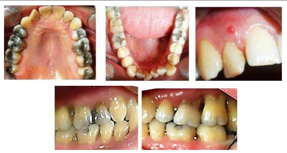
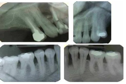
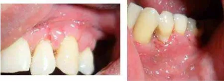
A complete clinical history was registered, and several diagnostic aids were performed, such as clinical photographs, periapical radiographic series
(igures 1, 2 and 3), and study models. The principal indings include: the patient is systemically healthy,
with family history of periodontal disease, high blood pressure, diabetes, and dental prosthesis; the functional analysis of the temporomandibular joint revealed a slight block by the end of oral opening followed by a dull noise and a sudden movement. The patient has had history of articular noise and a joint block on maximum opening. He admits brushing his teeth daily at least twice a day and not using dental
loss very often. A istulous tract was observed at the level of tooth 12 (igure 1), as well as soft and
hard dentobacterial plaque, dental mobility degree 1 and 2 at 16, 17, 26, 27, 28, 33, and 34, gingival retractions with positive bleeding clinical indexes
(30%), inlammation, suppuration, probing depth,
and loss of clinical insertion. It is important to point out the presence of lower anterior dental crowding as a local risk factor that favors bacterial plaque accumulation20, 21 (igure 1). The radiographic analysis
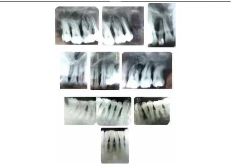
showed generalized bone loss in both vertical and
horizontal directions, and a signiicant periapical lesion on tooth 12 (igure 2).
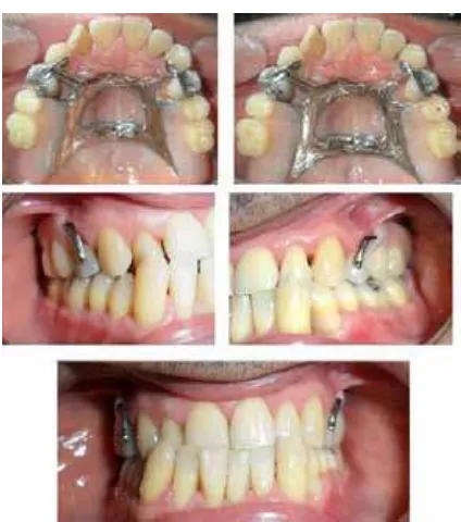
Figure 1. Initial intraoral images
[image:4.581.60.522.424.671.2]Se hizo historia clínica completa y ayudas diagnósticas como fotografías clínicas, serie radiográfica periapical (figuras 1, 2 y 3) y modelos de estudio. Entre los ha-llazgos se destacó: paciente sistémicamente sano, con antecedentes familiares de enfermedad periodontal, hipertensión arterial, diabetes y uso de prótesis dental; en el análisis funcional de la articulación temporo-mandibular se observó un leve bloqueo al final de la apertura bucal seguido de un ruido sordo y movimiento brusco, con antecedentes de ruido y bloqueo articular en máxima apertura. El paciente refirió cepillado dia-rio al menos dos veces al día, uso infrecuente de la seda dental. Se observó tracto fistuloso a nivel del 12 (figura 1), placa dentobacteriana blanda y dura, movi-lidad dental grado 1 y 2 en 16, 17, 26, 27, 28, 33, 34, retracciones gingivales con índices clínicos positivos para sangrado (30%), inflamación, supuración, pro-fundidad sondeable y pérdida de inserción clínica. Es importante resaltar la presencia del apiñamiento dental anteroinferior como factor de riesgo local que facilita la acumulación de placa bacteriana20, 21 (figura 1). En el análisis radiográfico se observó pérdida ósea generalizada de tipo horizontal y vertical y una lesión periapical significante a nivel del 12 (figura 2).
DIAGNOSIS
Systemic: healthy
Dental: occlusal active caries on 47, faulty dental restoration on 12, dental attrition on lower anterior teeth and abnormal teeth position (lower anterior crowding).
Pulpal: necrotic pulp on 12 with chronic suppurative apical periodontitis.22
Periodontal: generalized (84%) advanced chronic periodontitis with an average clinical insertion loss of 5.8 mm, bi- and trifurcation on 16, 17, 18, 26, 27, 28 (degree IIIc), 48 (degree IIb), 46, 47 (degree Ia)
(igure 3),17, 23, 24 and secondary occlusal trauma.25
Occlusal: Class III malocclusion with malposition of teeth.
TMJ: subluxation.26
Figure 2. Periapical radiographic series, initial diagnosis
DIAGNÓSTICOS
Sistémico: sano.
Dental: caries activa en oclusal del 47, restauración dental defectuosa en 12, atrición dental en dientes anteroinferio-res y anomalía en la posición de los dientes (apiñamiento anteroinferior).
Pulpar: necrosis pulpar del 12 con periodontitis apical crónica supurativa.22
Periodontal: periodontitis crónica avanzada generalizada (84%) con pérdida de inserción clínica en promedio de 5,8 mm, compromiso de bi- y trifurcación en 16, 17, 18, 26, 27, 28 (grado IIIc), 48 (grado IIb), 46, 47 (grado Ia) (figura 3).17, 23, 24 y trauma oclusal secundario.25
Oclusal: maloclusión clase III con malposiciones den-tarias.
[image:5.581.57.529.54.388.2]ATM: subluxación.26
PROGNOSIS
The general prognosis is poor due to chronic periodontal disease and abundant destruction of supporting tissue; also, the patient was a heavy smoker for almost ten years. The patient does not present systemic diseases, and quit smoking sixteen years ago.27
Individual periodontal prognosis is bad for teeth 16, 17, 18, 26, 27, and 28 because insertion loss has reached the root apex, with trifurcation degree III type c; tooth 28 does not have an opposing tooth. Individual prognosis is poor for teeth 23, 33, and 34 because bone loss has affected two thirds of dental root; it is also poor for tooth 12 due to necrotic pulp and extensive apical periodontitis, and for 46, 47, and
48 due to signiicant insertion loss and bifurcation
degree II types a and b.27
The patient sought dental attention for the irst time
on February 22nd 2011 and he was immediately told
about the importance of starting a comprehensive dental treatment. Once diagnosis and prognosis had been established, a treatment plan was designed on April 24th 2011 with approval by the patient
who signed an informed consent before initiating treatment.
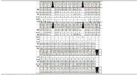
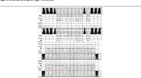
[image:6.581.52.525.54.314.2]Figure 3. Initial diagnostic periodontogram Figura 3. Periodontograma diagnóstico inicial
PRONÓSTICO
El pronóstico general es regular, debido a la cronicidad de la enfermedad periodontal y a la gran destrucción de los tejidos de soporte, además el paciente fue fumador pesado durante diez años aproximadamente; no existen enfermedades de base en el paciente y el hábito de fumar fue abandonado hace dieciseis años.27
El pronóstico periodontal individual es malo para los dien-tes 16, 17, 18, 26, 27, 28 porque la pérdida de inserción es tal que se encuentra cercana al ápice radicular, tienen daño de trifurcación grado III tipo c y el 28 no tiene diente antagonista. El pronóstico individual es regular para los dientes 23, 33, 34, porque la pérdida ósea afecta dos tercios de la raíz dental, también para el 12 debido a la necrosis pulpar y a la periodontitis apical extensa y para el 46, 47, 48 por la pérdida considerable de inserción y compromiso de bifurcación grado II tipos a y b.27
TREATMENT
Several clinical procedures were performed during the hygienic phase in order to stop periodontal infec-tion and to teach the patient how to maintain good oral hygiene; this included: patient’s motivation and education on oral health, supragingival periodontal preparation with scalers and curettes, dental pro-phylaxis with toothbrush and prophylactic paste, simple extraction for teeth with bad periodontal
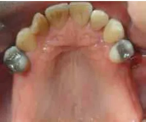
prognosis: 16, 17, 18, 26, 27, 28 (igure 4), removal
of active caries on 47, pulpal debriding and endodon-tics of tooth 12, subgingival periodontal preparation to entire mouth (total mouth disinfection)28 which
included: non-surgical scaling and root planing with curettes and sonic-scaler machine to remove subgingival irritants and disorganize adhered and
[image:7.581.97.239.332.451.2]non-adhered bacterial lora.5, 29 (igure 5).
Figura 4. Exodoncia de los dientes posterosuperiores con pronóstico periodontal malo
Figura 5. Desinfección total de la boca: raspaje y alisado radicular con curetas de gracey y scaler
Figure 4. Extraction of upper posterior teeth with bad periodontal prognosis
Figure 5. Full mouth disinfection: Scaling and root planing with Gracey curettes and scaler
It also included mouth wash with emphasis on the back of the tongue and tonsils with a 0.12% diglu-conate chlorhexidine solution (Periogard) two times a day during 15 days, and application of 0.2% chlor-hexidine gel (Dentagel) on periodontal pockets by means of subgingival irrigations two times a day.28
The process of scaling and root planing of the four quadrants was performed in less than twenty-four hours following the guidelines for total mouth disinfection suggested by Quirynen (1995).28 Due to the severity
of the patient’s periodontal disease (advanced) the prescription of systemic antibiotics was required as a complement to periodontal therapy.6, 10, 30
TRATAMIENTO
Se ejecutaron varios procedimientos clínicos dentro del periodo higiénico, cuyo fin fue detener la infección pe-riodontal y enseñar al paciente a mantener una higiene bucal adecuada, esto incluyó: educación y motivación del paciente en salud bucal, ambientación periodontal supragingival con scaler y curetas, profilaxis dental con cepillo y pasta profiláctica, exodoncia simple para dientes con pronóstico periodontal malo: 16, 17, 18, 26, 27, 28 (figura 4), eliminación de caries activa del 47, desbridamiento pulpar y endodoncia del 12, ambientación periodontal subgingival a boca completa (desinfección total de la boca)28 que incluyó: raspaje y alisado radicular no quirúrgico con curetas y aparato sónico-scaler para eliminar los irritantes subgingivales y desorganizar la flora bacteriana adherida y no adherida.5, 29 (figura 5).
Incluyó además enjuague bucal con énfasis en dorso de lengua y tonsilas con solución de digluconato de clorhexi-dina al 0,12% (Periogard) 2 veces al día durante 15 días y aplicación de gel de clorhexidina al 0,2% (Dentagel) en las bolsas periodontales a través de irrigaciones subgin-givales 2 veces al día.28
[image:7.581.300.526.336.447.2]Figure 7. Reassessment periodontogram
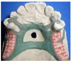
Figure 6. Installation of temporary upper esthetic plate
Amoxicillin in capsules of 500 mg was prescribed every 8 hours during 7 days because, according to
scientiic evidence,10 periodontal pathogens are sen-sible to this antibiotic. This hygienic phase inished
with the installation of an upper esthetic plate due to
the extraction of maxillary molars (igure 6).
Reassessment was performed eight weeks after periodontal disinfection—the required period for healing of periodontal tissues—.29 This step
included: evaluation of oral tissues, construction of
a new periodontogram (igure 7), and completion of
a new periapical radiographic series (January 16th 2012) (igure 8).
Se prescribió Amoxicilina en cápsulas de 500 mg cada 8 horas durante 7 días, ya que según la evidencia científica,10 los periodontopatógenos son sensibles a este antibiótico. Se culmina esta fase higiénica con la instalación de placa estética superior debido a las exodoncias de los molares maxilares (figura 6).
Ocho semanas después del tratamiento periodontal de desinfección, lapso necesario para la cicatrización de los tejidos periodontales,29 se hizo la revaluación que incluyó: evaluación de los tejidos bucales, realización de un nuevo periodontograma (figura 7) y toma de nueva serie radiográ-fica periapical (16 de enero de 2012)
(figura 8).
Figura 6. Instalación de placa estética superior temporal
[image:8.581.59.524.429.686.2]The results obtained so far were satisfactory:
con-siderable reduction of inlammation and gingival
bleeding, absence of suppuration, improvement of oral hygiene, reduction of periodontal probing, bone regeneration, and cortication of alveolar crest on affected areas.20
Based on these clinical and radiographic results, a new periodontal diagnosis was established: mild chronic periodontitis located on 23 and moderate chronic periodontitis located on 33, 34, 35 as well as on 43, 44, 45, 46, 47 y 48.23, 24 Treatment of this
new condition included extraction of 48 due to persistent periodontal pocket larger than 5 mm; bifurcation degree IIb with no opposing teeth even after rehabilitation and occlusion on upper alveolar ridge.17, 27 The surgical periodontal phase was then
planned and performed5, 14, 15, 31 for the rest of areas
affected by periodontitis, in this manner: intra-sulcus
incision, lap elevation, elimination of granulomatous
tissue, scaling and planing with curettes and scaler,
lap repositioning, atraumatic suture caliber 4/0
(Ethicon®) and periodontal dressing installation (Coe
Pack®) (igure 9). AINEs was prescribed during three
days as well as 0.12% chlorexidine (Periogard®).
Postsurgical evaluation was performed eight days later; the periodontal suture was removed and healing process was observed; no signs of infection
were found (igure 10).
Figure 8. Reassessment radiographic series. Bone regeneration can be observed on 23 mesial as well as general cortication of alveolar crest
Los resultados obtenidos hasta este momento son sa-tisfactorios: considerable disminución de la inflamación y sangrado gingival, ausencia de supuración, mejoría en la higiene bucal, disminución en el sondaje periodontal, llenado óseo y corticación de cresta ósea alveolar en sitios enfermos.20
Con estos hallazgos clínicos y radiográficos se estableció un nuevo diagnóstico periodontal: periodontitis crónica leve localizada en 23 y periodontitis crónica moderada localizada en 33, 34, 35 y en 43, 44, 45, 46, 47 y 48.23, 24 El tratamiento para esta nueva afección incluyó exodon-cia del 48 debido a la persistenexodon-cia de bolsa periodontal mayor de 5 mm, alteración de bifurcación grado IIb, con ausencia de diente antagonista aun después de la rehabi-litación y oclusión sobre el reborde alveolar superior.17, 27 Se planteó y ejecutó la fase quirúrgica periodontal 5, 14, 15, 31 para el resto de zonas afectadas por la periodontitis que incluyó acceso a sitios enfermos mediante incisión intra-sulcular, levantamiento de colgajo, eliminación de tejido granulomatoso, raspaje y alisado con curetas y scaler, lavado profuso del sitio, reposicionamiento del colgajo, sutura atraumática calibre 4/0 (Ethicon)® y colocación de apósito periodontal (Coe Pack)® (figura 9). Se prescribió AINEs durante tres días y solución de clorhexidina 0,12% (Periogard).®
[image:9.581.168.418.54.222.2]Posteriormente, a los ocho días se hizo la revisión posto-peratoria, se retiró el apósito periodontal, la sutura y se observó cicatrización en curso sin signos de infección (figura 10).
Once periodontal healing was completed (eight weeks),29 new probing was performed and no
pe-riodontal pockets were found, nor other signs or symptoms of periodontal disease.
Once periodontitis had been controlled and
maxillary edentulism classiied as Kennedy class I (topographic classiication of edentulism) (igure 4),
several prosthetic treatment options were suggested to replace missing teeth; these options ranged from using implants to adapting an acrylic metal removable partial prosthesis, but due to the patient’s biological conditions, such as little maturation time of periodontal tissue,29 poor oral hygiene history, and
scarce economic resources, the second alternative was chosen.
Type III plaster models were obtained, as well as occlusal records and tests on semi-adjustable articulator and parallelometer. Based on this analysis, a partially removable prosthesis with bilateral distal extension was planned; this required a careful design since characteristics of the residual ridge and base movement during its functioning would
[image:10.581.59.531.59.156.2]determine the occlusal eficacy of the prothesis.32
[image:10.581.159.384.204.287.2]Figura 9. Acceso quirúrgico para raspaje y alisado radicular en cuadrantes II, III y IV
Figura 10. Control postoperatorio a los ocho días, cicatrización en curso y sin signos de infección
Figure 9. Surgical access for scaling and root planing in quadrants II, III, and IV
Figure 10. Postsurgical evaluation eight days after the procedure; healing in progress, no signs of infection
Luego del periodo de cicatrización periodontal correspon-diente (ocho semanas),29 se hizo un nuevo sondaje, en el cual no se encontraron bolsas periodontales, ni otros signos y síntomas de enfermedad periodontal, culminan-do así la cicatrización del perioculminan-donto.
Con el control de la periodontitis y clasificado el edentu-lismo del maxilar como clase I de Kennedy (clasificación topográfica del edentulismo) (figura 4), se establecieron varias opciones de tratamiento protésico para remplazar los dientes ausentes, que incluían desde el remplazo dental con implantes hasta una prótesis parcial removible metal acrílico; pero por motivos biológicos del paciente, como el poco tiempo de maduración de los tejidos periodontales,29 antecedentes de higiene bucal regular, además de pocos recursos económicos, se decidió hacer la segunda alter-nativa de tratamiento.
Also, both rigidity of the major connector and maxi-mum coverage of residual ridges by the base of the prosthesis were analyzed in order to reduce the loads produced on abutment teeth.33, 34 Designing of the
upper prosthesis took into account retention, support and stability,35 so a larger connector was designed,
with a combined anterior and posterior band and RPI system (occlusal rest, proximal guide plate, and approaching I bar clasp) on 14 and 24. Then the guide planes were impressed on the mouth, as well
as the occlusal and cingular loors.
Later, the final impression was taken by using addition silicone. Next, the prosthesis design was
veriied with metal structure waxing (igure 11)
and draining was performed by means of a cobalt-chrome alloy.36 The metal structure was tried on the patient to verify it itted well, and habitual occlusion was registered (igures 12 and 13).
Teeth alignment was then performed and tested on
the patient (igure 14); acrylic was added on the
structure and the patient’s mouth was examined again. Satisfactory results were achieved in com-pliance with clinical criteria and patient’s perception
(igure 15). Finally, three follow-up appointments
for prosthesis control were scheduled.
Figure 11. Waxing of the removable partial prosthesis design Figure 12. Casting of metallic structure and test in mouth
Figure 13. Occlusal registration with aluminum wax on rings
Figure 14. Prosthetic teeth wax aligning and adaptation on mouth. Note adequate contact points
Se analizó de igual manera la rigidez del conector mayor y el máximo cubrimiento de las bases protésicas sobre los rebordes residuales para disminuir las fuerzas generadas sobre los dientes pilares.33, 34 Se estableció el diseño de la prótesis superior considerando la retención, el soporte y la estabilidad,35 por tal razón se determinó un conector mayor, tipo banda combinada anterior y posterior y el sistema de RPI (apoyo, plato proximal, gancho en I) en 14 y 24. Posteriormente se tallaron en boca los planos guía, los lechos oclusales y cingulares.
Posteriormente, se tomó la impresión definitiva utilizando silicona de adición. A continuación se verificó el diseño de la prótesis con el encerado de la estructura metálica (figura 11) y se hizo el colado en una aleación de cromo cobalto.36 Se probó la estructura metálica en el paciente verificando su asentamiento y se tomó un registro en oclusión habitual (figuras 12 y 13).
[image:11.581.106.229.424.531.2]Seguidamente se hizo el enfilado de los dientes y se probó en el paciente (figura 14); finalmente se puso acrílico a la estructura y se examinó nuevamente en boca. Se lograron resultados satisfactorios que obedecen a criterios clínicos y a la percepción del paciente (figura 15). Posteriormente se hicieron tres citas de control protésico.
[image:11.581.352.476.425.529.2]Figura 11. Encerado del diseño de la prótesis parcial removible Figura 12. Colado de la estructura metálica y prueba en boca
[image:11.581.107.229.565.671.2] [image:11.581.352.474.566.669.2]DISCUSSION
A critical part of periodontitis treatment consists on controlling and removing the irritant bacteria associated to the disease; this may be partially or totally achieved by means of non-surgical perio-dontal therapy5, 29 —a multifactorial treatment of
periodontal inlammatory lesions—.29, 37 Following
the guidelines by Quirynen (1995)28, 38 and Cobb
(2002),29 in our case the treatment team decided to
perform non-surgical periodontal treatment taking into account: severity of the disease, patient’s needs, and risk factors, trying to obtain the best possible results.
From a periodontal perspective, the clinical para-meters, either individually or in combination, may
fail to predict the disease activity in speciic areas
or to identify treatment response, except for the
loss/gain changes in insertion degrees, measured
in two different moments.21, 39
After the reassessment period of eight weeks, clinical parameters such as probing depth, clini-cal junction level, bleeding at probing, and the
inlammatory clinical signs showed improvement
[image:12.581.184.396.53.293.2]as reported by different studies.
Figura 15. Instalación definitiva de la prótesis parcial removible, hay adecuada restitución de la forma, estética y función
Figure 15. Deinite insertion of removable partial prosthesis with adequate restitution of form, esthetics and function
DISCUSIÓN
Parte importante del tratamiento de la periodontitis es el control y eliminación de los irritantes bacterianos asocia-dos a la enfermedad, tal propósito se puede alcanzar parcial o totalmente con la terapia periodontal no quirúrgica.5, 29 La terapia periodontal no quirúrgica es el tratamiento multifactorial de la lesión inflamatoria periodontal, cuyo objetivo primario es su control y eliminación.29, 37 En este caso, el grupo clínico teniendo en cuenta: la severidad de la enfermedad, las necesidades del paciente, los factores de riesgo y buscando los mejores resultados posibles, decide iniciar el tratamiento del paciente con terapia periodontal no quirúrgica, en concordancia con los conceptos de Quirynen (1995)28, 38 y Cobb (2002).29
Desde el punto de vista periodontal es posible que nin-gún parámetro clínico individual o combinado con otro, presente alta especificidad y sensibilidad para predecir actividad de la enfermedad en sitios específicos, ni para diferenciar con ellos la respuesta al tratamiento, excepto por los cambios de pérdida o ganancia en los niveles de inserción, medidos en dos momentos diferentes.21, 39
Bone loss interruption, as shown by the radiographic reassessment, as well as connective tissue reorgani-zation, as proven by the clinical reassessment, are two other positive results.21
The use of a systemic antibiotic as a complement to periodontal therapy has been widely documented; it has been demonstrated that patients treated this way present better clinical results than the ones who did not receive a systemic antibiotic.40 The
combi-nation commonly used includes Amoxicillin® plus
Metronidazole®, yielding satisfactory results;6 in this
case we prescribed Amoxicillin alone three times a day during seven days, based on a study by Botero et al (2007)10 on a Colombian population in which
the authors concluded that common periodontal pathogens, such as Porphyromonas gingivalis (P. g.) Porphyromonas subspecies (P. sp.), and Prevotella intermedia (P. i), are highly sensitive to Amoxicillin, with an effectiveness that ranges between 70 and 86.9%; they also proved that these microorganisms are resistant to Metronidazole®.
However, prescribing a systemic antibiotic may
bring some dificulties, such as treatment cost in
-crease (according to the medicines used), pathogens’ antibiotic resistance41 due to misuse, excess use or
self-medication, and side effects associated to an-tibiotics use, such as pseudomembranous colitis.42
It is important to mention the strong relationship between tobacco smoking and periodontitis, since
it is the most signiicant environmental risk factor
that may modify not only periodontitis incidence but also its evolution.27, 43 Also, it may negatively alter
treatment outcome due to a reduced immune response caused by vascular changes produced by the habit.44
Our patient was a heavy smoker for ten years, and although by the time of treatment he had already quit smoking for sixteen years, his habit may have altered aspects such as severity of periodontal destruction.
During the surgical phase, we considered necessary to apply surgical cement (Coe-pack®) after
re-positioning and suturing the lap, since it has been
reported that this material provides the patient with more comfort during the post-operatory period. Also, it offers wound protection, enables a close
lap-bone adaptation, and may prevent bleeding and
formation of excessive granulation tissue.15
La interrupción de la pérdida ósea observada en la ree-valuación radiográfica y la reorganización del tejido co-nectivo apreciable en la reevaluación clínica son también hallazgos significativos.21
El uso de antibiótico sistémico como coadyuvante de la terapia periodontal está bien documentado, se ha de-mostrado que los pacientes tratados bajo esta modalidad presentan mejores resultados clínicos que los que no recibieron antibiótico sistémico.40 Tradicionalmente se ha usado la combinación Amoxicilina más Metronidazol, la cual ha demostrado resultados satisfactorios,6 en este caso se prescribió únicamente Amoxicilina tres veces al día durante siete días, fundamentado en el estudio de Botero y colaboradores (2007)10 hecho en una población colombiana determinando que importantes patógenos periodontales como Porphyromonas gingivalis (P. g.), Porphyromonas subespecies (P. sp.) y Prevotella inter-media (P. i) presentan sensibilidad a la Amoxicilina, la cual es efectiva entre 70 y 86,9% y que la mayor resis-tencia de estos microrganismos es para el Metronidazol®.
Sin embargo, la prescripción de antibiótico sistémico puede originar inconvenientes como: elevar el costo del tratamiento según la elección del medicamento, generar resistencia antibiótica de los patógenos41 debido al mal uso, abuso y automedicación y efectos secundarios asociados a los antibióticos como la enterocolitis pseudomembranosa.42
Es de interés recordar la relación entre el tabaquismo y la periodontitis, ya que es el factor de riesgo ambiental más importante que puede modificar la incidencia y el curso de la periodontitis.27, 43 Además, puede empobrecer el resulta-do del tratamiento debiresulta-do a la respuesta inmune reducida, causada por los cambios vasculares producidos por el hábito.44 Nuestro paciente fue fumador pesado durante diez años, sin embargo, al momento del tratamiento había abandonado el hábito hacía dieciseis años, no obstante, es posible que el tabaco haya modificado aspectos como la gravedad de la destrucción periodontal.
The periapical infection of tooth 12 (suppurative chronic apical periodontitis) improved as a
conse-quence of the endodontic treatment; the istulous
tract disappeared as well as the remaining signs and symptoms of the lesion. Also, the radiographic evaluation showed that the apical lesion decreased in size, and although it still exists no clinical ob-servations indicate its progression.45, 46 Following
recommendations by the endodontics specialist, we decided to perform radiographic checkups for a year in order to evaluate whether apical surgery would be needed after that period.
Cobb (2002)29 maintains that periodontal tissues heal
during six to eight weeks after periodontal treatment, and that they achieve maturation in a period of nine to twelve months. These postulates, plus the patient’s oral hygiene history, led us to discard oral rehabili-tation by means of dental implants.
The objectives of prosthetic treatment include restitution of posterior occlusal support to avoid bite collapse, with the help of a well-designed removable partial prosthesis (RPP), adequate orientation of occlusal forces, conservation of prosthetic abutments, and maintaining good oral hygiene,47 all
of which are necessary for good treatment results and to provide patients with successful periodontal and prosthetic treatments. It is extremely important to design a personalized maintenance program for patients rehabilitated with RPP, according to their capacity to control oral hygiene, as well as to evaluate the activity of both periodontal disease and caries, alveolar ridge resorption, occlusal stability, and the conditions of RPP over time.48-51
As pointed out by Renvert & Persson (2004),19 it is
critical to include the patient in the phase of perio-dontal maintenance or support treatment, consisting on periodical follow-up appointments scheduled according to the evolution over time and tissue res-ponse, and especially according to the quality of oral hygiene by the patient. In this case, the follow-up appointments will be scheduled every three months. El cuadro infeccioso periapical del 12 (periodontitis
apical crónica supurativa) evolucionó luego del tratamiento endodóntico, desapareció el tracto fistuloso y los demás signos y síntomas clínicos de la lesión, se observó además, disminución en el tamaño de la lesión apical según el control radiográfico, no obstante dicha lesión persiste, aunque no hay hallazgos clínicos que puedan indicar progresión de la lesión;45, 46 En interconsulta con el endodoncista se determinó hacer un control radiográfico por un año y posteriormente analizar la necesidad de cirugía apical.
Cobb (2002)29 reporta que luego del tratamiento perio-dontal, los tejidos periodontales cicatrizan entre seis y ocho semanas después y que logran la maduración en un lapso de nueve a doce meses; estas considera-ciones, además de los antecedentes de higiene bucal regular nos permitieron descartar la rehabilitación oral mediante implantes dentales.
Entre los objetivos del tratamiento protésico está la restitución del soporte oclusal posterior para evitar el colapso de mordida, con base en la realización de un buen diseño de la prótesis removible (PPR), la orientación adecuada de las fuerzas oclusales, el man-tenimiento de los pilares protésicos, el control de una buena higiene bucal,47 indispensables para el buen pro-nóstico del tratamiento y para brindarle al paciente un tratamiento periodontal y protésico exitoso a lo largo del tiempo. Es de vital importancia hacer un programa de mantenimiento personalizado para los pacientes reha-bilitados con PPR, basado en la capacidad de control de la higiene oral por parte del paciente, la evaluación de la actividad de la enfermedad periodontal y caries, la reab-sorción del reborde alveolar, la estabilidad oclusal y las condiciones de la PPR con el paso del tiempo.48-51
This last treatment phase is necessary for periodontal
health stability, as it enables a signiicant change of
the quality and quantity of subgingival bacteria by means of instruction on oral hygiene, supragingival periodontal adjustment, and clinical monitoring.5
CONCLUSION
Success on the periodontal and prosthetic treatment of this patient responds to several key elements: patient´s education and active participation in his treatment, design of a comprehensive treatment
plan based on scientiic and clinical concepts that
have been veriied by previous studies, and its im
-plementation by an interdisciplinary team, under a teaching-community outreach model that allows complex dental treatments at lower costs for the community.
The comprehensive treatment of patients who have lost their teeth due to periodontitis includes reha-bilitation by means of dental prosthesis in order to restitute their teeth’s form, function and appearance, thus improving patients’ quality of life.
CORRESPONDING AUTHOR
Leticia Botero Zuluaga School of Dentistry
Universidad de Antioquia. Colombia
Email address: lbotero@odontologia.udea.edu.co
REFERENCIAS / REFERENCES
1. López R, Oyarzún M, Naranjo C, Cumsille F, Ortiz M, Baelum V. Coronary heart disease and periodontitis-a case control study in Chilean adults. J Clin Periodontol 2002; 29(5): 468-473.
2. Abnet CC, Qiao YL, Dawsey SM, Dong ZW, Taylor PR, Mark SD. Tooth loss is associated with increased risk of total death, and death from upper gastrointestinal cancer, heart disease, and stroke in a Chinese population-based cohort. Int J Epidemiol 2005; 34: 467-474.
3. Armitage GC, Offenbacher S. Consensus report on perio-dontal diseases: epidemiology and diagnosis. Ann Perio-dontology 1996; 1: 216-222.
4. Rioboo Crespo M, Bascones A. Factores de riesgo de la enfermedad periodontal: factores genéticos. Av Periodon Implantol 2005; 17(2): 69-77.
5. Botero L, Botero A, Bedoya JS, Guzmán IC. Nonsurgical periodontal therapy. Rev Fac Odontol Univ Antioq 2012; 23(2): 150-158.
6. Cionca N, Giannopoulou C, Ugolotti G, Mombelli A. Amoxicillin and metronidazole as an adjunct to full mouth scaling and root planing of chronic periodontitis. J Perio-dontol 2009; 80: 364-371.
7. Heitz-Mayield H, Trombelli L, Heitz F, Needleman I,
Moles D. A systematic review of the effect of surgical de-bridement vs. non-surgical dede-bridement for the treatment of chronic periodontitis. J Clin Periodontol 2002: 29 (supl 3): 92-102.
8. Mombelli A, Schmid B, Rutar A, Lang NP. Persistence pat-terns of Porphyromonas gingivalis, Prevotellaintermedia/ Este último periodo del tratamiento es necesario para la
estabilidad de la salud periodontal, ya que permite con-seguir un cambio significativo en la calidad y cantidad de la flora bacteriana subgingival mediante la instrucción de higiene bucal, ambientación periodontal supragingival y un seguimiento clínico.5
CONCLUSIÓN
El éxito del tratamiento periodontal y protésico de este paciente obedeció a varios elementos clave: la educa-ción del paciente y su participaeduca-ción como parte activa y fundamental en el tratamiento, la elaboración de un plan de tratamiento integral basado en conceptos científico-clínicos comprobados en la literatura y su realización a cargo de un grupo clínico interdisciplinario mediante la docencia asistencial, que además permite hacer trata-mientos odontológicos complejos a bajos costos para la comunidad.
Parte del tratamiento integral en pacientes que perdieron sus dientes debido a la periodontitis es la rehabilitación mediante prótesis dental para poder restituir la forma, función y estética, mejorando así la calidad de vida del paciente.
CORRESPONDENCIA
Leticia Botero Zuluaga Facultad de Odontología
Universidad de Antioquia. Colombia
23. Berman L, Hartwell G. Diagnóstico. En: Cohen S,
Har-graves K. Vías de la pulpa. 9.ª ed. Barcelona: Elsevier
Mosby; 2008.
24. Armitage G. American Academy of Periodontology.
Inter-national workshop for a classiication of periodontal disease
and condition. Ann Periodontol 1999; 4: 1-6.
25. Armitage G. Diagnóstico y clasiicación de las enfermeda -des periodontales. Periodontol 2000 2005; 9: 9-21.
26. Lindhe J, Lang N, Karring T, Berglundh T, Giannobile W,
Sanz M. Trauma oclusal: tejidos periodontales. En:
Periodon-tología clínica e implanPeriodon-tología odontológica. 5.ª ed. Vol. I.
Buenos Aires: Panamericana; 2009.
27. Okeson J. Signos y síntomas de los trastornos temporo-mandibulares. En: Tratamiento de oclusión y
afeccio-nes temporomandibulares. 6.ª ed. Barcelona: Elsevier
Mosby; 2008.
28. Botero L, Alvear FS, Vélez ME. Factores del pronóstico en periodoncia. Rev Fac Odontol Univ Antioq 2008; 19(2): 69-79.
29. Quirynen M. Bollen C. Full-vs. Partial-muoth disinfection in the treatment of periodontal infections: short-term clinical and microbiological observations. J Dent Res 1995; 74(8): 1459-1466.
30. Cobb CM. Clinical signiicance of non-surgical periodontal
therapy; an evidence-based perspective of scaling a root planing. J Clin Periodontol 2002; 29 (supl 2): 6-16.
31. Guzmán IC, Grisales H, Ardila CM. Adjunctive systemic
administration of moxiloxacin versus ciproloxacin plus
metronidazole in the treatment of chronic periodontitis harboring gram-negative enteric rods: microbiological and clinical effects. Rev Fac Odontol Univ Antioq 2011; 23(1): 92-110.
32. Fabrizi S, Barbieri Petrelli G, Vignoletti F, Bascones-Martínez A. Tratamiento quirúrgico vs. terapia periodontal básica: estudios longitudinales en periodoncia clínica. Av Periodon Implantol 2007; 19(3): 161-175.
33. Giraldo OL. Cómo evitar fracasos en la prótesis parcial re-movible. Rev Fac Odontol Univ Antioq 2008; 19(2): 80-88.
34. Piwowarczyk A, Köhler KC, Bender R, Büchler A, Lauer
HC, Ottl P. Prognosis for abutment teeth of removable dentures: a retrospective study. J Prosthodont 2007; 16: 377-382.
35. Nickenig HJ, Spiekermann H, Wichmann M, Andreas SK,
Eitner S. Survival and complication rates of combined
tooth-implant-supported ixed and removable partial dentures. Int
J Prosthodont 2008; 21: 131-137.
36. Kaiser F. PPR: no laboratório, en el laboratorio. 3.ª ed. São
Paulo: Quintessence; 2010.
37. Wataha JC. Biocompatibility of dental casting alloys: a review. J Prost Dent 2000; 83(2): 223-233.
nigrescens, and Actinobacillusactinomyetemcomitans after mechanical therapy of periodontal disease. J Periodontol 2000; 71(1): 14-21.
9. Slots J, Slots H. Bacterial and viral pathogens in saliva: disease relationship and infectious risk. Periodontol 2000 2011; 55(1): 48-69.
10. Botero A, Alvear FS, Vélez ME, Botero L, Velásquez H. Evaluación de los enfoques terapéuticos para las varias formas de enfermedad periodontal. Parte II: aspectos mi-crobiológicos. Rev Fac Odontol Univ Antioq 2007; 19(1): 6-20.
12. Doungudomdacha S, Rawlinson A, Walsh T, Douglas C. Effect of non-surgical periodontal treatment on clinical parameters and the numbers of Porphyromonas gingivalis, Prevotellaintermedia and Actinobacillus actinomycetemco-mitans at adult periodontitis sites. J Clin Periodontol 2001; 28: 437-445.
13. Tatsuji N, Takeyoshi K. Microbial etiology of periodontitis.
Periodontol 2000 2004; 36: 14-26.
14. Socransky SS, Haffajee AD, Ximenez LA, Feres M, Mager D. Ecological considerations in the treatment of Actinobaci-llusactinomycetemcomitans and Porphyromonas gingivalis periodontal infections. Periodontol 2000 1999; 20: 341-362.
15. Carranza FA. Técnica de colgajo para el tratamiento de
bolsas. En: Carranza FA. Periodontología clínica, 10.ª ed.
México: McGraw-Hill; 2010. p. 134-169.
16. Lindhe J, Lang N, Karring T, Berglundh T, Giannobile W,
Sanz M. Cirugía periodontal: procedimientos de acceso. En: Periodontología clínica e implantología odontológica.
5.ª ed. Vol. II. Buenos Aires: Panamericana; 2009.
17. Silvestri M, Rasperini G, Milani S. 120 Infrabony defects treated with regenerative therapy: long-term results. J Pe-riodontol 2010; 82(5): 668-675.
18. Kinaia BM, Steiger J, Neely AL, Shah M, Bhola M.
Treatment of class II molar furcation involvement: me-taanalyses of reentry results. J Periodontol 2011; 82(3): 413-428.
19. Croft LK, Nunn ME, Crawford LC, Holbrook TE, McGuire MK, Kerger MM et al. Patient preference for ultrasonic or
hand instruments in periodontal maintenance. Int J Perio-dontics Restorative Dent 2003; 23(6): 567-573.
20. Renvert S, Persson G. Supportive periodontal therapy. Periodontol 2000 2004; 36: 179-195.
21. Renvert S, Persson GR. A systematic review on the use of residual probing depth, bleeding on probing and furcation status following initial periodontal therapy to predict further attachment and tooth loss. J Clin Periodontol 2002; 29 (supl 3): 82-89.
38. Caffesse RG, Mota LF, Morrson EC. The rational for pe-riodontal therapy. Periodontol 2000 1995; 9: 7-13.
39. Quirynen M, Bollen CML. The inluence of surface rough -ness and surface-free energy on supra and subgingival plaque formation in man. A review of the literature. J Clin Periodontol 1995; 22: 1-14.
40. Lang NP, Adler R, Joss A, Nyman S. Absence of bleeding on probing an indicator of periodontal stability. J Clin Periodontol 1990; 17: 714-721.
41. Herrera D, Sanz M, Jepsen S, Needleman I, Roldán S. A systematic review on the effect of systemic antimicrobials as an adjunct to scaling and root planing in periodontitis patients. J Clin Periodontol 2002; 29 (supl 3): 136-159.
42. Kleinfelder JW, Müller RF, Lange DE. Antibiotic sus -ceptibility of putative periodontal pathogens in advanced periodontitis patients. J Clin Periodontol 1999; 26: 347-351.
43. Orellana A, Salazar E. Colitis pseudomembranosa asociada al uso de antibióticos. Acta Odontol Venez 2009; 47: 1-5.
44. Kinane D, Peterson M, Stathopoulou PG. Factores am
-bientales y otros factores que modiican las enfermedades
periodontales. Periodontol 2000 2007; 16: 107-119.
45. Pahkla E, Koppel T, Naaber P, Saag M, Loivukene K. The eficacy of non-surgical and systemic antibiotic treatment
on smoking and non-smoking periodontitis patients. Sto-matologija 2006; 8: 116-121.
46. Nair P. Fisiopatología de la periodontitis apical primaria. En:
Cohen S, Hargraves K. Vías de la pulpa. 9.ª ed. Barcelona:
Elsevier Mosby; 2008.
47. Johnson B, Witherspoon D. Cirugía perirradicular. En:
Cohen S, Hargraves K. Vías de la pulpa. 9.ª ed. Barcelona:
Elsevier Mosby; 2008.
48. Zlatariæ DK, Celebiæ A, Valentiæ-Peruzoviæ M, Jerolimov
V, Panduriæ J. A survey of treatment outcomes with remo-vable partial dentures. J Oral Rehabil 2003; 30: 847-54.
49. Vanzeveren C, D’Hoore W, Bercy P. Inluence of removable
partial denture on periodontal indices and microbiological status. J Oral Rehabil 2002; 29(3): 232-239.
50. Zlataric DK, Celebic A, Valentic Peruzovic M. The effect
of removable partial dentures on periodontal health of abutment and non-abutment teeth. J Periodontol. 2002; 73(2): 137-144.
51. Yeung AL, Lo EC, Chow TW, Clark RK. Oral health
status of patients 5-6 years after placement of cobalt-chromium removable partial dentures. J Oral Rehabil 2000; 27: 183-189.
52. Yeung AL, Lo EC, Clark RK, Chow TW. Usage and status