FACULTAD DE MEDICINA
DEPARTAMENTO DE BIOQUÍMICA Y BIOLOGÍA MOLECULAR Y FISIOLOGÍA
TESIS DOCTORAL:
Lazarillo and related Lipocalins: ligands and functions
Presentada por MARIO RUIZ GARCIA para optar al grado de
Doctor por la Universidad de Valladolid
Dirigida por:
Impreso 2T
AUTORIZACIÓN DEL DIRECTOR DE TESIS
(Art. 2.1. c de la Normativa para la presentación y defensa de la Tesis Doctoral en la UVa)
D. Diego Sánchez Romero, con D.N.I. nº29759524P, profesor del departamento de
Bioquímica y Biología Molecular y Fisiología, y
Dª. María Dolores Ganfornina Álvarez, con D.N.I. nº28873307G, profesora del
departamento de Bioquímica y Biología Molecular y Fisiología,
como Directores de la Tesis Doctoral titulada
“Lazarillo and related Lipocalins: ligands and functions”
,
presentada por D. Mario Ruiz Garcia,
alumno del programa de Investigación Biomédica impartido por el departamento de
Bioquímica y Biología Molecular y Fisiología:
Autorizan la presentación de la misma, considerando que el candidato ha superado el
nivel de formación necesario para aspirar al Título de Doctor con Mención Internacional
por la Universidad de Valladolid, mediante la realización de un proyecto de investigación
original en el que su contribución abarca desde el diseño de las preguntas científicas y el
diseño y ejecución de los experimentos, hasta la presentación en diversos formatos
(escritura de trabajos para publicación y presentaciones en congresos) de los resultados
para su difusión a la comunidad científica.
Valladolid, 28 de Febrero de 2013
La Directora de la Tesis,
El Director de la Tesis,
INDEX
1. Index ... 5
2. Abbreviations ... 9
3. Introductory Words ... 13
4. General Introduction ... 15
4.1. The Lipocalin Protein Family. ... 17
4.2. Lipocalin Family Phylogeny. ... 18
4.3. Lipocalin Structure. ... 18
4.4. Ligand Binding and Lipocalin Function. ... 23
4.5. Clade I: Arthropodan and vertebrate Lipocalin meeting point.. ... 24
4.5.1. Lazarillo ... 24
4.5.2. Drosophila Lipocalins... 25
4.5.3. Apolipoprotein D. ... 32
4.6. References ... 37
5. Objectives ... 47
6. Results ... 51
6.1. Chapter 1 ... 53
“Sex-dependent modulation of longevity by two Drosophila homologues of human Apolipoprotein D, GLaz and NLaz”. 6.1.0. Summary... 55
6.1.1. Introduction ... 57
6.1.2. Material and Methods ... 58
6.1.3. Results and Discussion ... 60
6.1.4. References ... 71
6.2. Chapter 2 ... 79
“Lipid binding properties of human ApoD and Lazarillo-related Lipocalins: Functional implications for cell differentiation”. 6.2.0. Summary... 81
6.2.1. Introduction ... 83
6.2.2. Material and Methods ... 84
6.2.3. Results and Discussion ... 85
6.2.4. References ... 96
6.2.5. Supplemental Information ... 99
6.3. Chapter 3 ... 103
“Ligand binding-dependent functions of the Lipocalin NLaz: An in vivo study in Drosophila”. 6.3.0. Summary... 105
6.3.1. Introduction ... 107
6.3.2. Material and Methods ... 108
6.3.3. Results ... 111
6.3.4. Discussion... 121
6.3.5. References ... 124
6.3.6. Supplemental Information ... 127
6.4. Chapter 4 ... 139
“Grasshopper Lazarillo, a GPI-anchored Lipocalin, increases Drosophila longevity and stress resistance, and functionally replaces its secreted homologue NLaz”. 6.4.0. Summary... 141
6.4.1. Introduction ... 143
6.4.2. Material and Methods ... 144
6.4.3. Results and Discussion ... 148
6.4.4. References ... 161
7. General Discussion ... 169
7.1. References ... 175
8. Conclusions ... 177
9. Summary in Spanish / Resumen en Castellano ... 181
9.1. References ... 186
10. Annex ... 187
9
Abbreviations
11
Abbreviations:
•
4-HNE:
4-Hydroxynonenal
•
7-T:
7(Z)-Tricosene
•
20-HE
: 20-hydroxyecdysone
•
AA:
Arachidonic Acid
•
AEA:
Anandamide
•
AGP:
α
-acid glycoprotein
•
α
-1m:
α
-1microglobulin
•
ApoD:
Apolipoprotein D
•
ApoM:
Apolipoprotein M
•
ATF3:
Activating Transcription Factor 3
•
BBP:
Bilin-Binding Protein
•
CAL
β
:
Chondrogenesis-Associated Lipocalin
β
•
CG:
Computed Gene
•
CNS:
Central Nervous System
•
CRC:
Crustacyanin
•
DHA:
Docosahexaenoic Acid
•
DNA:
Deoxyribonucleic acid
•
Ex-FABP:
Extracellular Fatty Acid Binding Protein
•
FA:
Fatty Acid
•
FABP:
Fatty Acid Binding Protein
•
FOXO:
Forkhead Box Protein O
•
GLaz:
Glial Lazarillo
•
Gp93:
Glycoprotein-93
•
GPI:
Glycosylphosphatidylinositol
•
HDL:
High Density Lipoparticles
•
Hr39:
Hormone Receptor-39
•
HSD:
High Sugar Diet
•
IIS:
Insulin/IGF1 Signaling
•
Ins:
Insecticyanin
•
InR:
Insulin Receptor
•
JNK:
Jun-N-terminal Kinase
•
KO:
Knock-Out
•
Kr:
Krüppel
•
LA:
Linoleic Acid
•
Laz:
Lazarillo
•
Lcn:
Lipocalin
•
Lip-4:
Lipase-4
•
Lola:
Longitudinals Lacking Protein
•
L-PGDS:
Prostaglandin D Synthase Lipocalin
Abbreviations
•
mRNA:
Messenger Ribonucleic Acid
•
MUP:
Major Urinary Protein
•
NAE:
N-acylethanolamines
•
NGAL:
Neutrophil Gelatinase-Associated Lipocalin
•
n-HETE:
Hydroxyeicosatetraenoic Acid
•
n-HpETE:
hydroperoxyeicosatetraenoic acid
•
NLaz:
Neural Lazarillo
•
OEA:
Oleoylethanolamine
•
OBP:
Odorant Binding Protein
•
PDB:
Protein Data Bank
•
PEA:
Palmitoylethanolamine
•
pI:
Isoelectric Point
•
PI3K:
Phosphatidyl-Inositol 3-Kinase
•
PQ:
Paraquat (1,1'-dimethyl-4,4'-bipyridinium dichloride)
•
PUFA:
Poly-Unsaturated Fatty Acid
•
RBP:
Retinol Binding Protein
•
ROS:
Reactive Oxygen Species
•
Scn:
Siderocalin
•
SCR:
Structurally Conserved Region
•
SM:
Sphingomyelin
•
SMase:
Sphingomyelinase
•
TAG:
Triacylglycerides
•
TL:
Tear Lipocalinç
13
Introductory words
Introduction
17
The Lipocalin protein family:
A brief introduction and general characteristics.
The definition of “protein family” varies according to different authors. In a simple
way, a protein family can be defined as a collection of related proteins [Pfam database:
1]. This relationship was typically based on protein function and/or primary structure.
However, with the increasing number of protein tertiary structures resolved, protein
families are also described according to its folding. Precisely, the Lipocalins belong to a
set of families mainly defined by its structural homology. The Lipocalin fold is shaped
by a
β
-barrel, forming a cup or calyx, with a central cavity that serves as a
ligand-binding site [2]. Together with fatty acid ligand-binding proteins (FABPs), avidins and
metalloproteinase inhibitors (MPI), the Lipocalins assemble the Calycin superfamily
[2].
Historically, the Lipocalin name was suggested by Pervaiz and Brew in 1987 [3]
studying the structure-function correlations among several proteins. However, the
Lipocalin protein family concept was previously proposed by Unterman et al. in 1981
[4] by comparing the amino acid sequence homology between some Lipocalin family
members.
Lipocalins are typically small (160-230 amino acid residues) extracellular proteins.
Family members are found in all five life kingdoms [5], and their numbers are growing
quickly due to the new sequenced genomes: over six hundred Lipocalins are currently
identified.
Introduction
Lipocalin Family phylogeny:
Clade division.
By using several conserved features of Lipocalins, a phylogeny of the family can be
inferred. Phylogenetic trees, based on both protein sequences and gene architecture,
have been built (fig.1A and 1B respectively) and show a similar topology [5,7-9]. The
number of tertiary structures of Lipocalins has grown lately, and it should be now
possible and desirable to update the Lipocalin tree considering protein folding as a
phylogenetic signal as well.
Focusing in the phylogenetic tree derived from protein sequence, the animal
Lipocalins are divided into twelve monophyletic clades (defined and named in fig. 1A).
Clade I is especially interesting because it encompasses the majority of arthropodan
Lipocalins and ApoD (Apolipoprotein D), the most ancestral chordate Lipocalin.
Subsequently, the family can be divided in
ancestral
vertebrate Lipocalins, those
more related to ApoD (clades III, IV, V and VI) and
modern
vertebrate Lipocalins
(clades VII-XII) (see fig 1A) [5].
Combining phylogenetic and structural information Gutierrez et al [10] conclude
that more recently evolved Lipocalins have a more flexible protein structure (based on
number of disulfide bonds), and bind smaller ligands with more efficiency than the
ancestral Lipocalins. These points are in agreement with the fact that modern Lipocalins
show a tendency to present more gene duplications and could be more specialized.
Lipocalin structure:
From genes to proteins.
Tertiary and secondary structure
Introduction
19
Figure 1. Two sets of independent characters have produced very similar phylogenetic relationships between Lipocalins.
(A) Phylogenetic tree of the Lipocalin family derived from a multiple alignment of protein sequences, rooted with a group of bacterial Lipocalins. (B) Lipocalin phylogeny based on exon-intron arrangement, rooted with a protoctist Lipocalin. Scale bars represent branch length (number of substitutions/site) [Adapted from 7].
0.5 75 97 80 78 Bger.All4 Lviv.ESP Rpip.OP 81 Ggal.CALb Hsap Lcn1c 99 72 Ggal.ExFABP Ccot.q83 84 84 85 80 Cele.Lip 82 78 76 70 79 Cint.Lip 95 100 93 98 Ddis.Lip 72 100 Dhan.Lip Plant Lipocalins Bacterial Lipocalins Bacterial Lipocalins Bacterial Lipocalins Insect Lipocalins Apolipoprotein D Insect Nitrophorins Apolipoprotein M Retinol-binding lipocalins β-lactoglobulinsDrer.PGDS
Prostaglandin D synthase-NGAL Epididymal RA-binding lipocalins
α1-microglobulin-Complement 8γ
Chemoreception Lipocalins II (VEG,OBP,VNSP)
α1-acid glycoproteins Rodent urinary proteins
Chemoreception Lipocalins I (OBP,Pbas,Aphr,VNSP)
Miscellaneous Lipocalins
Crustacean and Insect Lipocalins
Clade I Clade II Clade III Clade IV Clade V Clade VII Clade XII Clade VI Clade VIII Clade XI Clade IX Clade X 1 3 4 5 6 Nu m b e r o f i n tr on s Ddis.LipAtha.OML Msex.IcyA Same.LazDmel.GLaz Hsap.ApoDDmel.Karl Dmel.NLaz Osav.TSGP2Rnor.RBP Mmus.ApoM Rnor.PGDS Hsap.Scn
Meug.βLG
Ggal.ExFABP Hsap.AGP Mmus.MUP1 Sscr.TL Hsap.OBP2 Hsap.Glyc
Btau.βLG
Ccri.Aphr Rnor.ERBP Hsap.C8G
Mmus.α1m
0.1
A
Introduction
ligand-binding site. There is a 3
10helix at the protein N-terminus, whereas at the
C-terminus there is a canonical
α
-helix (fig.2) [2]. Most Lipocalins contain one or more
intramolecular disulphide bonds that help to stabilize the
β
-barrel and are required to get
the proper conformation and ligand-binding properties [13,14]. Several cysteine (Cys)
residues present a pattern that is conserved in the family [7].
Within this general scheme, some Lipocalins show specific divergences in their
primary and secondary structure. L-PGDS (Lipocalin-type Prostaglandin D Synthase),
α
-1m (
α
-1microglobulin) and the human version of ApoD show an unpaired Cys. The
loops L5 and L7 appear elongated in RBP (Retinol Binding Protein). An unusually long
N-terminus is present in the insect Lipocalin Karl.
Many Lipocalins present N- and/or O-linked glycosylation, and glycosylation
residues range from none to six. Glycosylation is known to confer different properties
and function, as happens for instance in Glycodelin [15], TL (Tear Lipocalin or Lcn1)
[16], AGP (
α
-acid glycoprotein) [17], and the glycosylation-dependent secretion of
α
-1m [18].
Primary structure
Lipocalins show an extreme divergence at their amino acid sequence level, and a
pairwise identity of 20-30% is common inside the family. This fact poses difficulties
when assigning newly discovered sequences to the family. However, three conserved
motifs, called structurally conserved regions (SCR), are revealed by multiple alignments
[19].
- SCR1 is defined by the residues G-x
1-W-x
2(being x
2usually Y/F/W), and it is located
in the first
β
-strand (strand A).
- SCR2 consensus sequence is T-D/N-Y-x-x-Y. Physically the SCR2 is found in the
loop L6, connecting strands F and G.
Introduction
21
three motifs are at the base of the calyx and seem to be important to achieve a proper
folding. Especially essential is the SCR1 tryptophan residue [20,21].
Following the suggestion of Flower [19], Lipocalins were conceptually divided in
two groups:
kernel
Lipocalins, where all SCR are present, and
outlier
Lipocalins that
miss at least one SCR.
Gene Structure
Some characteristic features of Lipocalins are also extended to gene structure,
exhibiting a similar arrangement of exons and introns in the coding sequence of their
genes [9].
Vertebrate Lipocalins show seven exons (1-7) and six introns (A-F), usually
including six coding exons (e1-e6). Exons 2-5 code for the entire set of
β
-strands. In
some cases, e6 appears fused with e7, as happens in human ApoM or chicken Cal
β
genes. Additionally, the intron phase pattern is well conserved (0,2,1,1,1; each number
indicates the position within the codons where the intron is inserted) [5,9].
Introd
Figur
(A) S
α-heli from
duction
re 2.Lipocal Schematic stru
ices are also 2]. (B) Side
lin structure. ucture of the L
indicated. SC view of the p
Lipocalin fold CRs are mark prototypical L
d. The β-shee ked and their
Lipocalin RB
ts are represe characteristic P (PDB code
nted by arrow c sequences d e: 1RBP). Sec
ws and labeled displayed. [A condary struc
Introduction
23
Ligand binding and Lipocalin Function:
A family overview.
Several properties of the binding pocket (depth, width, polarity and loop positions)
determine what ligands are able to bind to each Lipocalin. Biological processes often
depend on protein-ligand binding events, and this is indeed the case for Lipocalins.
Their functions cannot be completely understood without taking in account their
ligands. Some examples of the close relationship between ligand and function in
different Lipocalin clades include:
-
MrLC
is a crustacyanin (clade I) from the prawn
Macrobrachium rosenbergii
that
shows dual binding capacities. MrLC binds astaxanthin, which is responsible for the
bluish prawn coloration, but MrLC also binds 20-hydroxyecdysone (20-HE) and
regulates prawn molting [22].
Another example of coloration function involves Insecticyanin (
Ins,
also from
clade I) and its ligand biliverdin IX
γ
, which confers a green color to
Manduca sexta
larvae [23-25].
-
RBP
belongs to clade III and has been identified from fish to mammals. RBP was the
first crystallized Lipocalin [26] and is considered a prototypal Lipocalin. Actually, the
RBP ligand retinol has been demonstrated to be a common ligand
in vitro
for most
Lipocalins [27-29]. RBP transports retinol from the liver to peripheral target tissues, and
plays a key role in vision and several development processes [30,31].
Introduction
- Odorant binding proteins (
OBP
) and Major urinary proteins (
MUP
), clades X and IX
respectively, are chemosensory proteins that act as peri-receptors transporting odorant
or pheromones to the neuroepithelium, and are involved in behavior regulation [35-37].
In addition, bovine OBP has a protective role by binding 1-octen-3-ol, a potent biting
insect attractant which comes from mammal breath and sweat [38].
A set of Lipocalins shows anti-oxidant binding properties.
ApoM
binds oxidized
phospholipids selectively, thus serving as a scavenger of lipoprotein oxidation products
[39].
TL
and
OBP
bind to 4-hydroxy-2-nonenal (4-HNE), an end-product of
ω
-6 PUFA
(poly-unsaturated fatty acid) peroxidation, and prevent its cytotoxicity [40,41]. Finally,
α
-1m
binds heme and participates in its degradation [42]. Additionally,
α
-1m shows
reductase/dehydrogenase properties and may act as and oxidation repair factor for
important structural proteins as collagen [43].
Clade I: Arthropodan and vertebrate Lipocalin meeting point.
As discussed above, the Lipocalin clade I includes most arthropodan Lipocalins and
the most ancestral chordate Lipocalin: ApoD. Clade I can be subdivided into three
groups: Lazarillo-related Lipocalins, Crustacyanins (CRC) and Bilin binding-related
Lipocalins. Another group of arthropodan Lipocalins, the Nitrophorins of blood-feeding
insects, represents a large independent expansion of very divergent Lipocalin genes, and
the phylogeny reconstruction tools usually place them among more recent Lipocalin
clades (fig.1A.).
Below I will go over what is known about the ‘founding member’ of the Clade I
group: Lazarillo. I will follow by describing the Clade I Lipocalins from the model
organism
Drosophila melanogaster
, and the chordate homolog ApoD.
Lazarillo
Introduction
25
Laz shows a unique feature in the family. It is the only Lipocalin for what a
glycosylphosphatidylinositol (GPI) anchor to the plasma membrane has been
demonstrated [45]. However, a potential GPI modification was also suggested in its
plant homolog AtTIL (
Arabidopsis thaliana
temperature-inducible Lipocalin, [46]).
Nevertheless, other Lipocalin - plasma membrane associations have been reported
[47-49].
Laz is highly glycosylated. Five N-linked oligosaccharides are predicted, and
curiously all five are polarized to one side of the globular protein [50].
The natural Laz ligand or ligands are still unknown, but a set of potential candidates
have been assayed
in vitro.
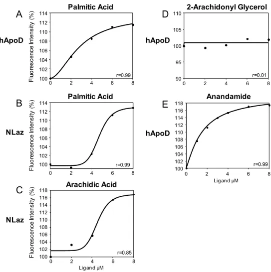
Recombinant Laz is able to bind retinoic acid and long chain
fatty acids (FA), whereas biliverdin, farnesol or juvenile hormone do not bind [51].
Looking at the spatio-temporal expression pattern, Laz shows a restricted
expression in the developing nervous system, and it is involved in axon growth and
guidance. Adding a monoclonal antibody against Laz to grasshopper embryo cultures
perturbs axon extension and causes growth cone misrouting. Moreover, outside the
nervous system Laz is associated mainly with the excretory system: Malpighian tubules
and subesophageal body [45,52].
Due to its selective expression pattern, Laz has been used as a tool for
characterizing the nervous system in economically important species as the desert locust
Schistocerca gregaria
or
Locusta migratoria
[53-56].
Drosophila Lipocalins
Eight Lipocalin genes in the Drosophila genome: Glial Lazarillo (
GLaz
), Neural
Lazarillo (
NLaz
), and
Karl
and five additional Lipocalin genes which are still identified
only by their CG (computed gene) code number.
Introd
mela
Lipo
F
Figur
(A) S Phylo
duction
anogaster
L
calins may
Following I
re 3. Drosop Schematic re ogenetic tree o
Lipocalin p
come from
I briefly rev
hila Lipocali epresentation of Lipocalin
phylogeneti
a double ev
view the dat
ns.
of the chro Clade I deriv
ic tree (fig
vent of gene
ta available
omosomal loc ved from a m
g.3B) sugg
e duplicatio
on each Dr
cation of Dr multiple alignm
gest that th
on..
osophila Lip
rosophila Lip ment of protei
he current
ipocalin.
pocalin gene in sequences
four
Introduction
27
-
CG31446
and
CG5399
(89A2 region) show 33% identity in a pairwise alignment.
At the sequence level, the C-terminus of CG5399 shows a high degree of similarity with
that of Laz, and a potential GPI modification site is predicted [big-PI Predictor: The GPI
Prediction Server. 57,58]. If confirmed experimentally, this would be the only
GPI-linked Drosophila Lipocalin.
CG5399 and CG31446 functions are unknown. However, a couple of pieces of
information are available. CG5399 expression in S2 cells appears enhanced more than
ten times when cells are treated with the insecticide methoxyfenozide (RH-2485) [59].
On the other hand, CG31446 is over-expressed in a soluble guanlyl-cyclase mutant [60],
and it is down-regulated in flies under long-term high magnetic field exposure [61].
-
CG44013
and
CG44014
(88F7 region) genes are separated by only sixteen base
pairs. Both genes are predicted to code for
kernel
Lipocalins, showing 45% identity and
64% similarity between them. CG44013 and CG44014 were considered a single ORF
called CG14872 in a previous Drosophila genome annotation.
The current functional information about these two Lipocalins comes from
expression data microarrays that considered the combined CG14872 annotation. Larvae
fed with wheat germ agglutinin (an anti-insect lectin) increase CG14872 transcription as
part of their defensive response [62].
-
CG31659
is located in the left arm of the second chromosome (22A1 region),
next to the NLaz gene. CG31659 is one of the most abundantly expressed genes in testis
and male accessory gland [63]. Indeed, the CG31659 protein has been identified as a
seminal fluid protein and is transferred from males to females during mating in
Drosophila melanogaster
and
D. yakuba
, but not in
D.simulans
[64].
-
Karl
(CG4139) is located in the X chromosome (10E3 region) and codes for an
atypical Lipocalin. Karl N-terminal is extremely long, SCR1 is not easily identifiable
and loop-2 appears expanded.
Introduction
The immune role of Karl was confirmed by Hull-Thompson [67], as Karl
over-expressing flies are more resistant to
Enterococcus faecalis
infection than the control fly
line.
Furthermore, the insertion of a p-element in the Karl gene causes an extension of
fly developing time around one hundred hours [68] and affects wing shape [69].
Finally, Karl is expressed in 25 Drosophila cell lines, including the popular
Schneider’s cell lines [70], at higher level than in whole flies at any developmental
stage [71].
GLaz:
Glial Lazarillo.
The right arm of the second chromosome (49F4 region) contains the GLaz gene.
GLaz intron-exon organization is defined by 3 introns and 4 exons, as the grasshopper
Laz gene. However, it differs from the typical insect gene arrangement: 4 introns and 5
exons, as the equivalent GLaz exons 4 and 5 are fused.
The GLaz mature protein has 192 residues with a theoretical pI of 8.62 [ProtParam
tool. 44], and only one N-glycosylation is predicted at position 16th [72]. GLaz pairwise
identities reach 26% with NLaz, 29% with Laz and 30% with ApoD. GLaz structure
presents two of unique features: the loops 2 and 3 are elongated.
At the moment, GLaz can be considered an ‘orphan’ protein. GLaz binding
properties have been reported neither
in vitro
nor
in vivo
.
In terms of expression, GLaz is found in the longitudinal glia of the developing
ventral nerve cord of the central nervous system (CNS), and in specific glial precursors
during late stages of embryogenesis. Subsequently, GLaz is absent in larvae, and again
present in pupae and the adult nervous system [50,73]. Outside the nervous system
GLaz is mainly expressed in the developing gut and salivary glands [50], and in adult
hemocytes [73].
GLaz expression level has been experimentally altered in Drosophila using genetic
tools. Male flies lacking GLaz exhibit multiple phenotypic defects. GLaz null-mutant
(KO) flies show a shortened lifespan and a reduced resistance to starvation, H
2O
2or
Introduction
29
also reflected in a lower performance in behavioral tests (phototaxis, geotropism and
flight tests), an increase of apoptotic cells and lipid peroxidation levels. Furthermore,
GLaz-KO male flies show a smaller body mass and a lower amount of triacylglycerides
(TAG) [73].
In contrast, GLaz over-expressing flies resulted in increased lifespan and enhanced
resistance to starvation and hyperoxia. Alterations in O
2concentration produce a decline
in locomotion of wild-type flies, and this deficiency is rescued by GLaz over-expression
[74]. The protective properties of GLaz were corroborated in a cellular model, as
Drosophila S2 cells over-expressing GLaz are protected against PQ and A
β
42 peptide
toxicity [75].
Flies respond to multiple stress situations (hyperoxia, high temperatures, PQ
exposure [75] and high sugar diet [76]) by increasing GLaz levels. This unspecific
regulation of expression suggests that GLaz forms part of the organism first defensive
line.
Due to GLaz ability to deal with oxidative stress, the effect of GLaz
over-expression has been studied in the context of flies that model human neurodegenerative
diseases. Friedreich’s ataxia is caused by a decreased expression of the mitochondrial
protein frataxin. This deficiency provokes a dysfunction of the mithocondrial
respiratory chain with a subsequent increase of oxidative stress, cell damage and
degeneration [77]. GLaz was able to correct the lipid peroxidation levels and mitigate
the dyslipidemia and shortened lifespan caused by frataxin reduction in a Drosophila
model of Friederich’s ataxia [78].
Finally, it has been reported that the insertion of a p-element in the GLaz promoter
does not alter ethanol sensitivity, place memory or olfaction memory [79].
NLaz:
Neural Lazarillo.
The NLaz gene is located in the 22A1region of the second chromosome. Its
intron-exon arrangement is slightly particular, because intron-exon 1 is divided in two by a small
intron.
Introduction
Phylogenetically, NLaz is the closest Drosophila gene homolog to the grasshopper
Laz and ApoD (fig. 3B). NLaz shows 39% identity with ApoD and 37% with Laz. The
NLaz C-terminal sequence is extended in comparison to most Lipocalins. This ‘tail’ is
not cleaved to GPI-anchor the protein to the plasma membrane, as it happens with
grasshopper Laz. However, this peculiarity also appears in the neural Lipocalin Gallerin
from the moth
Galleria mellonella
and in CG44014 and CG31446 fly Lipocalins.
In terms of expression, NLaz temporal pattern is similar to that of GLaz, but the
tissue profile is quite different. While GLaz is expressed by glial cells in the CNS, NLaz
is expressed by a subset of neuroblasts and neurons. Outside the nervous system NLaz
is expressed in the fat body and the developing gut [72].
A NLaz-KO fly line was generated by introducing a stop codon in the NLaz coding
sequence [82]. This fly line has been used to study the influence of NLaz in metabolism,
signaling and stress response.
Energy storage (TAG, glucose and glycogen levels) are decreased in NLaz-KO
male flies. In agreement, flies lacking NLaz show a decreased resistance to starvation,
whereas over-expression of NLaz in the fat body results in an increment of starvation
resistance. The same pattern, lower resistance in NLaz-KO and high in the
over-expressor, was achieved in longevity and PQ toxicity assays [67].
It is known that the interaction between Jun-N-terminal kinase (JNK) and
insulin/IGF1 signaling (IIS) has to be tightly regulated to ensure proper metabolic
adaptation to environmental challenges [83]. NLaz is transcriptionally regulated by JNK
signaling and is up-regulated under stress conditions. NLaz inhibits the IIS and it seems
to act at the level of the plasma membrane modulating PI3K (phosphatidyl-inositol
3-kinase) activity. As a consequence of this IIS inhibition, FOXO (Forkhead box protein
O) targets insulin receptor (InR), Lipase-4 (Lip4) and the heat shock protein 22 (hsp22)
are up-regulated and contribute to promote stress tolerance and longevity. In contrast,
when flies lacking NLaz are under stress conditions they cannot inhibit the IIS activity
and cannot attain the appropriate protective response [67].
Introduction
31
Since NLaz is expressed in neurons and neuroblast, as grasshopper Laz does, and it
is secreted by developing axons [72], a role in axon growth and pathfinding is expected
for NLaz. However, no gross defects have been observed during nervous system
development in NLaz-KO flies. Several interesting relationships have been found,
though, between NLaz and developmental processes in different tissues:
- The protein Lola is a critical transcription factor in nervous system development,
orchestrating midline crossing by CNS axons [84]. NLaz is down-regulated (fold
change: -2.40) in Lola defective embryos [85], suggesting that NLaz is a down-stream
target of Lola. In addition, Lola is also involved in construction or function of neural
circuits throughout the lifespan of the fly. This includes dendritic and axonal
development of adult olfactory projection neurons [86]. These results may indicate a
role of NLaz in axon outgrowth.
- Krüppel (Kr) is a transcription factor that plays a critical role in segmentation and
organ formation during the last stages of embryogenesis. For example, a role for Kr in
fat body formation is well documented [87]. A strong association between Kr and NLaz
has been reported by chromatin immunoprecipitation and
in vitro
binding to DNA [88].
- Gp93 is an essential protein in Drosophila since homozygous mutant animals die
at the third larval stage as a consequence of pronounced defects in midgut epithelium.
Gp93 mutant larvae exhibit a starvation-like metabolic phenotype, including
suppression of insulin signaling and extensive mobilization of amino acids and TAG.
NLaz is almost absent in fly larvae, but it is up-regulated (fold change: 6.0) in GP93
animals [89].
- A null mutant of the activating transcription factor 3 (ATF3-KO) causes a
complex larval lethal phenotype characterized by fat body lipid overload and starvation
signaling from the gut. NLaz is found up-regulated (fold change: 2.24) due to the
hyper-activation of stress cascades, including the JNK pathway [90].
- NLaz is also related with the development of sexual structures. The hormone
receptor 39 (Hr39) is required for the normal development and function of the
spermatheca in females. When Hr39 is missing, spermatheca presents obvious defects,
causing sterility. NLaz expression is highly repressed in Hr39-KO animals [91].
Additionally, NLaz has been identified as a seminal fluid protein in
D. melanogaster
Introduction
NLaz expression is also related to the circadian clock. NLaz expression cycles only
in the body when head and body gene expressions were analyzed separately [92].
Finally, as it happens with GLaz, no ligands have been described for NLaz so far.
Therefore, it would be critical to provide more details about the molecular mechanism
of action of this Lipocalin.
ApoD:
Apolipoprotein D.
ApoD orthologs are found in species from the phylum chordata. Most of the
available information comes from studies with human or mouse ApoD, and this section
will focus on them.
The human ApoD gene (hApoD) is located in the third chromosome (3q26.2), and
is composed of 3 exons and 4 exons. Chordate Lipocalins typically contain 7 exons, but
exons 4 to 7 are fused in hApoD [5]. However, 6 exons are found in the mouse ApoD
(mApoD) because of a singular 5’-UTR intron [93].
ApoD is broadly expressed and present in many fluids [94]. It is remarkable the
high levels of ApoD expression during development. Especially interesting is the
evolutionary pattern of ApoD expression in chordates. Amphioxus ApoD is expressed
in tissues derived from mesoderm and endoderm, whereas in mammals and birds there
is a shift to ectodermal derivatives [95]. Besides, cell type expression changes as well.
ApoD is expressed by subsets of neurons and glial cells in chicken [96], while adult
mouse, rat and humans express ApoD only in glial cells [97-100]. On the other hand,
ApoD is the most consistently up-regulated gene during brain aging in mammals
[101,102].
The hApoD mature protein, after secretory signal peptide removal, is composed by
169 amino acids. ApoD is a
kernel
Lipocalin with two disulfide bridges formed by Cys
8-114 and 41-165 [103]. Additionally, hApoD has a fifth Cys (residue 116) which
allows for the formation of ApoD homodimers, or heterodimers with Apo-AII or Apo-B
[104,105].
Figur
(A) H green expos colore bound repres are co
revea
broad
and
acces
gives
chain
re 4.Represe Hydrophobic
(hydrophilic) se hydrophob ed in red and d (C) and a sented (green ommon contac
aled a typic
dest site and
10 by 15 Å
ssibility to t
s a slightly
ns [106] (fig
entations of h surface repre ) over white (n ic residues. ( d positively ch hypothetical in progesteron ct points [106]
cal Lipocalin
d a length o
Å wide at its
the binding
y negative c
g.4A). This
hApoD struct esentation. Re neutral) to bro (B) Electrosta harged areas
model for A ne complex an ].
n fold. The
of around 40
s entry. Loo
pocket. Ho
charge. Lo
unusual fe
ture and its b Residues are c own (hydroph atic surface r
are in blue. ( AA (D). Lig nd brown in A
calyx has a
0 Å. The cav
ops 3 and 7
owever, Glu
oops 1, 5 a
ature in the
binding comp colored accor hobic). Loops L
representation (C, D) Top v gand contacti AA model). A
an outer dia
vity, mainly
7 are widely
u-60 is close
and 7 expo
e Lipocalin
lexes. ding their hy L1 (A/B), L5 n. Negatively view of ApoD
ng amino ac Ala44, Phe89,
ameter of ar
y hydrophob
y open and
e to the poc
ose bulky h
family is in
Introdu
ydrophobicity (E/F) and L7 y charged are D with proges cid side chai Trp127 and L
round 45 Å
bic, is 15 Å
Introduction
the speculation of ApoD interacting directly with (high dense Lipocarticles) HDL and
membranes [107].
The structures of free ApoD and its complex with progesterone are almost identical:
Only small conformational changes in side-chains are detected. Progesterone, deeply
introduced in the pocket, is tightly packed between the aromatic side chains of Phe89
and Trp127. Additionally, Tyr46 serves as an anchor forming a hydrogen bond with the
ketone group of the progesterone [106].
Arachidonic acid (AA) is another well characterized ApoD ligand [108,109]. The
ApoD crystal structure is compatible with AA binding, and a model of its binding
shows ligand-protein contact points in common with the progesterone-ApoD crystal
[106] (fig.4C and 4D).
The wide calyx of ApoD allows the binding of hydrophobic molecules of different
nature. Besides progesterone [108-112] and AA [108,109], other molecules that ApoD
can bind are retinol and retinoic acid [28]. The ability of ApoD to bind other molecules
such as cholesterol [108,113] or bilirubin [109,114] is less clear.
hApoD has two glycosylation sites: Asn45 and Asn78. The hApoD crystal was
obtained from recombinant protein produced in
Escherichia coli
, and thus glycosylation
is absent in the resolved structure. In any case, two different glycosylation patterns have
been reported: in plasma and in apocrine secretion. Interestingly, both, Asn45 and
Asn78 oligosaccharide trees are terminated in sialic acid in plasma, whereas in apocrine
secrection, only the Asn45 tree terminates in sialic acid [115,116].
Many studies in the mouse as a model organism have pointed out the protective
properties of ApoD. Endogenously, mApoD is up-regulated under oxidative stress
caused by PQ exposure [117], and this up-regulation is mediated by the JNK pathway
[118], sharing activation networks with NLaz [67]. The ectopic expression of hApoD in
flies and mice confers higher resistance to PQ [75,117] and OC43 coronavirus [119]. In
contrast, mice lacking ApoD show a decreased resistance to PQ, higher levels of lipid
peroxidation in the brain, and an altered transcriptional response to oxidative stress
[117,118,120]. Moreover, ApoD presence is required to get a proper response after
sciatic nerve injury [121].
Introduction
35
have provided a really interesting mechanistic model. n-HETE (hydroxyeicosatetraenoic
acid) and n-HpETE (hydroperoxyeicosatetraenoic acid) are products of AA oxidation.
ApoD will bind HpETEs and, with the involvement of Met93, will catalyze their
reduction to the corresponding HETEs [122,123]. Met93 is conserved in mammalian
ApoD and chicken ApoD but absent in more ancestral ApoD orthologs, as for example
in salmon and amphioxus. Surprisingly, ApoD protein from amphioxus also shows
antioxidant activity
in vitro
[124].
Interestingly, ApoD-KO mice show behavioral differences in locomotor and
memory tests [117,118]. A putative explanation could be found in the fact that
ApoD-KO mice suffer alterations in brain receptor and neurotransmiters composition
[118,125-127].
In addition to the control of lipid peroxidation, many connections between ApoD
and lipids are often established. Indeed, ApoD received its name because it was the
fourth apolipoprotein discovered [128]. ApoD was identified in human plasma as a
component of HDL, and less abundant in other lipoparticles [114,129]. Inside HDL
particles, ApoD is predominant in HDL3 [130,131], which are small, dense and protein
enriched lipoparticles. Typically, minor bioactive lipid components are preferentially
associated with dense HDL3 particles [132].
Looking at fatty acids composition, ApoD-KO mice show an increase in a set fatty
acid in brain extracts. Highlights linoleic acid (LA, 18:2), eicosadienoic acid (20:2),
both are
ω
-6 FA, and docosahexaenoic acid (DHA, 22:6) [133]. An influence of ApoD
over FA management was also observed in a cellular model. Cells tranfected with ApoD
are able to incorporate higher amounts of AA. This fact suggested that ApoD could help
to stabilize the plasma membrane and avoid AA oxidation [134].
In humans, ApoD is up-regulated in many pathological situations, including
schizophrenia [135], Alzheimer’s [136-138], Parkinson’s disease [139] and multiple
sclerosis [140]. Additionally, ApoD levels are altered in several types of cancers
[94,141-143].
Introduction
affecting mRNA processing. Three distinct missense mutations have been identified in
an African-black population associated with alterations in lipid metabolism [146]. None
of these mutations (F36V, Y108C, T158K) are located within the ligand-binding cavity,
but the ApoD tertiary structure could still be compromised. For instance, Y108C is
located really close to C114 and C116, and could likely affect the formation of disulfide
bridges and result in a loss of function [106].
Introduction
37
References
1. Punta M, Coggill PC, Eberhardt RY, Mistry J, Tate J, et al. (2012) The Pfam protein families database. Nucleic Acids Res 40: D290-301.
2. Flower DR, North AC, Sansom CE (2000) The lipocalin protein family: structural and sequence overview. Biochim Biophys Acta 1482: 9-24.
3. Pervaiz S, Brew K (1987) Homology and structure-function correlations between alpha 1-acid glycoprotein and serum retinol-binding protein and its relatives. FASEB J 1: 209-214.
4. Unterman RD, Lynch KR, Nakhasi HL, Dolan KP, Hamilton JW, et al. (1981) Cloning and sequence of several alpha 2u-globulin cDNAs. Proc Natl Acad Sci U S A 78: 3478-3482.
5. Sanchez D, Ganfornina M, Gutierrez G, Gauthier-Jauneau A, Rislier J, et al. (2006) Lipocalin Genes and their Evolutionary History. In: Akerstrom B, Borregaard N, Flower D, Salier J, editors. Lipocalins. 1st Edition ed. Georgetown, Texas: Landes Bioscience. pp. 5-16. 6. Akerström B, Borregaard N, Flover D, Salier J (2006) Lipocalins. Georgetown, Texas. 204 p. 7. Ganfornina M, Sanchez D, Greene L, Flover D (2006) The Lipocalin Protein Family: Protein
Sequence, Structure and Relationship to the Calycin Superfamily. In: Akerstrom B, Borregaard N, Flower D, Salier J, editors. Lipocalins. 1st Edition ed. Georgetown, Texas: Landes Bioscience. pp. 17-27.
8. Ganfornina MD, Gutierrez G, Bastiani M, Sanchez D (2000) A phylogenetic analysis of the lipocalin protein family. Mol Biol Evol 17: 114-126.
9. Sanchez D, Ganfornina MD, Gutierrez G, Marin A (2003) Exon-intron structure and evolution of the Lipocalin gene family. Mol Biol Evol 20: 775-783.
10. Gutierrez G, Ganfornina MD, Sanchez D (2000) Evolution of the lipocalin family as inferred from a protein sequence phylogeny. Biochim Biophys Acta 1482: 35-45.
11. Gasymov OK, Abduragimov AR, Glasgow BJ (2010) pH-Dependent conformational changes in tear lipocalin by site-directed tryptophan fluorescence. Biochemistry 49: 582-590.
12. Gasymov OK, Abduragimov AR, Yusifov TN, Glasgow BJ (2004) Interstrand loops CD and EF act as pH-dependent gates to regulate fatty acid ligand binding in tear lipocalin. Biochemistry 43: 12894-12904.
13. Gasymov OK, Abduragimov AR, Glasgow BJ (2011) The conserved disulfide bond of human tear lipocalin modulates conformation and lipid binding in a ligand selective manner. Biochim Biophys Acta 1814: 671-683.
14. Liu J, Guo C, Yao Y, Lin D (2008) Effects of removing a conserved disulfide bond on the biological characteristics of rat lipocalin-type prostaglandin D synthase. Biochimie 90: 1637-1646.
15. Seppala M, Koistinen H, Koistinen R, Chiu PC, Yeung WS (2007) Glycosylation related actions of glycodelin: gamete, cumulus cell, immune cell and clinical associations. Hum Reprod Update 13: 275-287.
16. Le Danvic C, Guiraudie-Capraz G, Abderrahmani D, Zanetta JP, Nagnan-Le Meillour P (2009) Natural ligands of porcine olfactory binding proteins. J Chem Ecol 35: 741-751. 17. Hochepied T, Berger FG, Baumann H, Libert C (2003) Alpha(1)-acid glycoprotein: an acute
Introduction
18. Wester L, Fast J, Labuda T, Cedervall T, Wingardh K, et al. (2000) Carbohydrate groups of alpha1-microglobulin are important for secretion and tissue localization but not for immunological properties. Glycobiology 10: 891-900.
19. Flower DR, North AC, Attwood TK (1993) Structure and sequence relationships in the lipocalins and related proteins. Protein Sci 2: 753-761.
20. Katakura Y, Totsuka M, Ametani A, Kaminogawa S (1994) Tryptophan-19 of beta-lactoglobulin, the only residue completely conserved in the lipocalin superfamily, is not essential for binding retinol, but relevant to stabilizing bound retinol and maintaining its structure. Biochim Biophys Acta 1207: 58-67.
21. Greene LH, Chrysina ED, Irons LI, Papageorgiou AC, Acharya KR, et al. (2001) Role of conserved residues in structure and stability: tryptophans of human serum retinol-binding protein, a model for the lipocalin superfamily. Protein Sci 10: 2301-2316. 22. Yang F, Wang MR, Ma YG, Ma WM, Yang WJ (2011) Prawn lipocalin: characterization of
a color shift induced by gene knockdown and ligand binding assay. J Exp Zool A Ecol Genet Physiol 315: 562-571.
23. Holden HM, Rypniewski WR, Law JH, Rayment I (1987) The molecular structure of insecticyanin from the tobacco hornworm Manduca sexta L. at 2.6 A resolution. EMBO J 6: 1565-1570.
24. Riddiford LM, Palli SR, Hiruma K, Li W, Green J, et al. (1990) Developmental expression, synthesis, and secretion of insecticyanin by the epidermis of the tobacco hornworm, Manduca sexta. Arch Insect Biochem Physiol 14: 171-190.
25. Kayser H (2005) Lipocalins and structurally related ligand-binding proteins; Gilbert LI, Iatrou K, Gil lS, editors. Oxford: Elsevier.
26. Newcomer ME, Jones TA, Aqvist J, Sundelin J, Eriksson U, et al. (1984) The three-dimensional structure of retinol-binding protein. EMBO J 3: 1451-1454.
27. Ahnstrom J, Faber K, Axler O, Dahlback B (2007) Hydrophobic ligand binding properties of the human lipocalin apolipoprotein M. J Lipid Res 48: 1754-1762.
28. Breustedt DA, Schonfeld DL, Skerra A (2006) Comparative ligand-binding analysis of ten human lipocalins. Biochim Biophys Acta 1764: 161-173.
29. Peng Y, Liu J, Liu Q, Yao Y, Guo C, et al. (2010) Conformational and biochemical characterization of a rat epididymis-specific lipocalin 12 expressed in Escherichia coli. Biochim Biophys Acta 1804: 2102-2110.
30. Clagett-Dame M, DeLuca HF (2002) The role of vitamin A in mammalian reproduction and embryonic development. Annu Rev Nutr 22: 347-381.
31. Goodman DS (1980) Plasma retinol-binding protein. Ann N Y Acad Sci 348: 378-390. 32. Correnti C, Strong RK (2012) Mammalian siderophores, siderophore-binding lipocalins, and
the labile iron pool. J Biol Chem 287: 13524-13531.
33. Coudevylle N, Geist L, Hotzinger M, Hartl M, Kontaxis G, et al. (2010) The v-myc-induced Q83 lipocalin is a siderocalin. J Biol Chem 285: 41646-41652.
34. Correnti C, Clifton MC, Abergel RJ, Allred B, Hoette TM, et al. (2011) Galline Ex-FABP is an antibacterial siderocalin and a lysophosphatidic acid sensor functioning through dual ligand specificities. Structure 19: 1796-1806.
35. Matarazzo V, Zsurger N, Guillemot JC, Clot-Faybesse O, Botto JM, et al. (2002) Porcine odorant-binding protein selectively binds to a human olfactory receptor. Chem Senses 27: 691-701.
Introduction
39
37. Machnes Z, Avtalion R, Shirak A, Trombka D, Wides R, et al. (2008) Male-specific protein (MSP): a new gene linked to sexual behavior and aggressiveness of tilapia males. Horm Behav 54: 442-449.
38. Ramoni R, Vincent F, Grolli S, Conti V, Malosse C, et al. (2001) The insect attractant 1-octen-3-ol is the natural ligand of bovine odorant-binding protein. J Biol Chem 276: 7150-7155.
39. Elsoe S, Ahnstrom J, Christoffersen C, Hoofnagle AN, Plomgaard P, et al. (2012) Apolipoprotein M binds oxidized phospholipids and increases the antioxidant effect of HDL. Atherosclerosis 221: 91-97.
40. Grolli S, Merli E, Conti V, Scaltriti E, Ramoni R (2006) Odorant binding protein has the biochemical properties of a scavenger for 4-hydroxy-2-nonenal in mammalian nasal mucosa. FEBS J 273: 5131-5142.
41. Lechner M, Wojnar P, Redl B (2001) Human tear lipocalin acts as an oxidative-stress-induced scavenger of potentially harmful lipid peroxidation products in a cell culture system. Biochem J 356: 129-135.
42. Allhorn M, Berggard T, Nordberg J, Olsson ML, Akerstrom B (2002) Processing of the lipocalin alpha(1)-microglobulin by hemoglobin induces binding and heme-degradation properties. Blood 99: 1894-1901.
43. Olsson MG, Allhorn M, Larsson J, Cederlund M, Lundqvist K, et al. (2011) Up-regulation of A1M/alpha1-microglobulin in skin by heme and reactive oxygen species gives protection from oxidative damage. PLoS One 6: e27505.
44. Gasteiger E, Hoogland C, Gattiker A, Duvaud S, Wilkins MR, et al. (2005) Protein Identification and Analysis Tools on the ExPASy Server;. In: Walker JM, editor. The Proteomics Protocol Handbook: Humana Press. pp. 571-607.
45. Ganfornina MD, Sanchez D, Bastiani MJ (1995) Lazarillo, a new GPI-linked surface lipocalin, is restricted to a subset of neurons in the grasshopper embryo. Development 121: 123-134.
46. Frenette Charron JB, Breton G, Badawi M, Sarhan F (2002) Molecular and structural analyses of a novel temperature stress-induced lipocalin from wheat and Arabidopsis. FEBS Lett 517: 129-132.
47. Martins PA, Gomes F, Vaz WL, Moreno MJ (2008) Binding of phospholipids to beta-Lactoglobulin and their transfer to lipid bilayers. Biochim Biophys Acta 1778: 1308-1315.
48. Nishi K, Sakai N, Komine Y, Maruyama T, Halsall HB, et al. (2002) Structural and drug-binding properties of alpha(1)-acid glycoprotein in reverse micelles. Biochim Biophys Acta 1601: 185-191.
49. Saaren-Seppala H, Jauhiainen M, Tervo TM, Redl B, Kinnunen PK, et al. (2005) Interaction of purified tear lipocalin with lipid membranes. Invest Ophthalmol Vis Sci 46: 3649-3656.
50. Sanchez D, Ganfornina MD, Bastiani MJ (2000) Lazarillo, a neuronal lipocalin in grasshoppers with a role in axon guidance. Biochim Biophys Acta 1482: 102-109. 51. Sanchez D, Ortega-Cubero S, Åkerström B, Herrera M, Bastiani MJ, et al. (2008) Molecular
interactions of the neuronal GPI-anchored lipocalin Lazarillo. Journal of Molecular Recognition 21: 313-323.
Introduction
53. Boyan G, Posser S, Ludwig P, Guntner M, Williams L (2004) Ontogeny of identified cells from the median domain in the embryonic brain of the grasshopper Schistocerca gregaria. Arthropod Struct Dev 33: 125-137.
54. Boyan GS, Williams JL (2004) Embryonic development of the sensory innervation of the antenna of the grasshopper Schistocerca gregaria. Arthropod Struct Dev 33: 381-397. 55. Boyan GS, Williams JL, Posser S, Braunig P (2002) Morphological and molecular data
argue for the labrum being non-apical, articulated, and the appendage of the intercalary segment in the locust. Arthropod Struct Dev 31: 65-76.
56. Graf S, Ludwig P, Boyan G (2000) Lazarillo expression reveals a subset of neurons contributing to the primary axon scaffold of the embryonic brain of the grasshopper Schistocerca gregaria. J Comp Neurol 419: 394-405.
57. Eisenhaber B, Bork P, Eisenhaber F (1998) Sequence properties of GPI-anchored proteins near the omega-site: constraints for the polypeptide binding site of the putative transamidase. Protein Eng 11: 1155-1161.
58. Eisenhaber B, Bork P, Eisenhaber F (1999) Prediction of potential GPI-modification sites in proprotein sequences. J Mol Biol 292: 741-758.
59. Mosallanejad H, Badisco L, Swevers L, Soin T, Knapen D, et al. (2010) Ecdysone signaling and transcript signature in Drosophila cells resistant against methoxyfenozide. J Insect Physiol 56: 1973-1985.
60. Riedl CA, Neal SJ, Robichon A, Westwood JT, Sokolowski MB (2005) Drosophila soluble guanylyl cyclase mutants exhibit increased foraging locomotion: behavioral and genomic investigations. Behav Genet 35: 231-244.
61. Herranz R, Larkin OJ, Dijkstra CE, Hill RJ, Anthony P, et al. (2012) Microgravity simulation by diamagnetic levitation: effects of a strong gradient magnetic field on the transcriptional profile of Drosophila melanogaster. BMC Genomics 13: 52.
62. Li HM, Sun L, Mittapalli O, Muir WM, Xie J, et al. (2009) Transcriptional signatures in response to wheat germ agglutinin and starvation in Drosophila melanogaster larval midgut. Insect Mol Biol 18: 21-31.
63. Fischer BE, Wasbrough E, Meadows LA, Randlet O, Dorus S, et al. (2012) Conserved properties of Drosophila and human spermatozoal mRNA repertoires. Proc Biol Sci 279: 2636-2644.
64. Findlay GD, Yi X, Maccoss MJ, Swanson WJ (2008) Proteomics reveals novel Drosophila seminal fluid proteins transferred at mating. PLoS Biol 6: e178.
65. Wertheim B, Kraaijeveld AR, Hopkins MG, Walther Boer M, Godfray HC (2011) Functional genomics of the evolution of increased resistance to parasitism in Drosophila. Mol Ecol 20: 932-949.
66. Irving P, Ubeda JM, Doucet D, Troxler L, Lagueux M, et al. (2005) New insights into Drosophila larval haemocyte functions through genome-wide analysis. Cell Microbiol 7: 335-350.
67. Hull-Thompson J, Muffat J, Sanchez D, Walker DW, Benzer S, et al. (2009) Control of metabolic homeostasis by stress signaling is mediated by the lipocalin NLaz. PLoS Genet 5: e1000460.
68. Mensch J, Lavagnino N, Carreira VP, Massaldi A, Hasson E, et al. (2008) Identifying candidate genes affecting developmental time in Drosophila melanogaster: pervasive pleiotropy and gene-by-environment interaction. BMC Dev Biol 8: 78.
Introduction
41
70. Schneider I (1972) Cell lines derived from late embryonic stages of Drosophila melanogaster. J Embryol Exp Morphol 27: 353-365.
71. Cherbas L, Willingham A, Zhang D, Yang L, Zou Y, et al. (2011) The transcriptional diversity of 25 Drosophila cell lines. Genome Res 21: 301-314.
72. Sanchez D, Ganfornina MD, Torres-Schumann S, Speese SD, Lora JM, et al. (2000) Characterization of two novel lipocalins expressed in the Drosophila embryonic nervous system. Int J Dev Biol 44: 349-359.
73. Sanchez D, Lopez-Arias B, Torroja L, Canal I, Wang X, et al. (2006) Loss of glial lazarillo, a homolog of apolipoprotein D, reduces lifespan and stress resistance in Drosophila. Curr Biol 16: 680-686.
74. Walker DW, Muffat J, Rundel C, Benzer S (2006) Overexpression of a Drosophila Homolog of Apolipoprotein D Leads to Increased Stress Resistance and Extended Lifespan. Current Biology 16: 674-679.
75. Muffat J, Walker DW, Benzer S (2008) Human ApoD, an apolipoprotein up-regulated in neurodegenerative diseases, extends lifespan and increases stress resistance in Drosophila. Proc Natl Acad Sci U S A 105: 7088-7093.
76. Pasco MY, Leopold P (2012) High sugar-induced insulin resistance in Drosophila relies on the lipocalin Neural Lazarillo. PLoS One 7: e36583.
77. Marmolino D (2011) Friedreich's ataxia: past, present and future. Brain Res Rev 67: 311-330.
78. Navarro JA, Ohmann E, Sanchez D, Botella JA, Liebisch G, et al. (2010) Altered lipid metabolism in a Drosophila model of Friedreich's ataxia. Hum Mol Genet 19: 2828-2840.
79. LaFerriere H, Guarnieri DJ, Sitaraman D, Diegelmann S, Heberlein U, et al. (2008) Genetic dissociation of ethanol sensitivity and memory formation in Drosophila melanogaster. Genetics 178: 1895-1902.
80. Gupta R, Jung E, Brunak S (2004) Prediction of N-glycosylation sites in human proteins. Denmark: Center for Biological Sequence Analysis, Denmark.
81. Baycin-Hizal D, Tian Y, Akan I, Jacobson E, Clark D, et al. (2011) GlycoFly: a database of Drosophila N-linked glycoproteins identified using SPEG--MS techniques. J Proteome Res 10: 2777-2784.
82. Rong YS, Titen SW, Xie HB, Golic MM, Bastiani M, et al. (2002) Targeted mutagenesis by homologous recombination in D. melanogaster. Genes & Development 16: 1568-1581. 83. Karpac J, Jasper H (2009) Insulin and JNK: optimizing metabolic homeostasis and lifespan.
Trends Endocrinol Metab 20: 100-106.
84. Crowner D, Madden K, Goeke S, Giniger E (2002) Lola regulates midline crossing of CNS axons in Drosophila. Development 129: 1317-1325.
85. Gates MA, Kannan R, Giniger E (2011) A genome-wide analysis reveals that the Drosophila transcription factor Lola promotes axon growth in part by suppressing expression of the actin nucleation factor Spire. Neural Dev 6: 37.
86. Spletter ML, Liu J, Su H, Giniger E, Komiyama T, et al. (2007) Lola regulates Drosophila olfactory projection neuron identity and targeting specificity. Neural Dev 2: 14.
87. Hoshizaki DK (1994) Kruppel expression during postembryonic development of Drosophila. Dev Biol 163: 133-140.
Introduction
89. Maynard JC, Pham T, Zheng T, Jockheck-Clark A, Rankin HB, et al. (2010) Gp93, the Drosophila GRP94 ortholog, is required for gut epithelial homeostasis and nutrient assimilation-coupled growth control. Dev Biol 339: 295-306.
90. Rynes J, Donohoe CD, Frommolt P, Brodesser S, Jindra M, et al. (2012) Activating transcription factor 3 regulates immune and metabolic homeostasis. Mol Cell Biol 32: 3949-3962.
91. Allen AK, Spradling AC (2008) The Sf1-related nuclear hormone receptor Hr39 regulates Drosophila female reproductive tract development and function. Development 135: 311-321.
92. Ceriani MF, Hogenesch JB, Yanovsky M, Panda S, Straume M, et al. (2002) Genome-wide expression analysis in Drosophila reveals genes controlling circadian behavior. J Neurosci 22: 9305-9319.
93. Yoshida K, Cleaveland ES, Nagle JW, French S, Yaswen L, et al. (1996) Molecular cloning of the mouse apolipoprotein D gene and its upregulated expression in Niemann-Pick disease type C mouse model. DNA Cell Biol 15: 873-882.
94. Rassart E, Bedirian A, Do Carmo S, Guinard O, Sirois J, et al. (2000) Apolipoprotein D. Biochim Biophys Acta 1482: 185-198.
95. Wang L, Zhang S, Liu Z, Li H, Wang Y, et al. (2007) Characterization and expression of amphioxus ApoD gene encoding an archetype of vertebrate ApoD proteins. Cell Biol Int 31: 74-81.
96. Ganfornina MD, Sanchez D, Pagano A, Tonachini L, Descalzi-Cancedda F, et al. (2005) Molecular characterization and developmental expression pattern of the chicken apolipoprotein D gene: implications for the evolution of vertebrate lipocalins. Dev Dyn 232: 191-199.
97. del Valle E, Navarro A, Astudillo A, Tolivia J (2003) Apolipoprotein D expression in human brain reactive astrocytes. J Histochem Cytochem 51: 1285-1290.
98. Sanchez D, Ganfornina MD, Martinez S (2002) Expression pattern of the lipocalin apolipoprotein D during mouse embryogenesis. Mech Dev 110: 225-229.
99. Ong WY, Lau CP, Leong SK, Kumar U, Suresh S, et al. (1999) Apolipoprotein D gene expression in the rat brain and light and electron microscopic immunocytochemistry of apolipoprotein D expression in the cerebellum of neonatal, immature and adult rats. Neuroscience 90: 913-922.
100. Hu CY, Ong WY, Sundaram RK, Chan C, Patel SC (2001) Immunocytochemical localization of apolipoprotein D in oligodendrocyte precursor-like cells, perivascular cells, and pericytes in the human cerebral cortex. J Neurocytol 30: 209-218.
101. Loerch PM, Lu T, Dakin KA, Vann JM, Isaacs A, et al. (2008) Evolution of the aging brain transcriptome and synaptic regulation. PLoS One 3: e3329.
102. de Magalhaes JP, Curado J, Church GM (2009) Meta-analysis of age-related gene expression profiles identifies common signatures of aging. Bioinformatics 25: 875-881. 103. Yang CY, Gu ZW, Blanco-Vaca F, Gaskell SJ, Yang M, et al. (1994) Structure of human
apolipoprotein D: locations of the intermolecular and intramolecular disulfide links. Biochemistry 33: 12451-12455.
104. Blanco-Vaca F, Pownall HJ (1993) Disulfide linked dimers of apolipoprotein D in urine. Electrophoresis 14: 1086-1087.
Introduction
43
106. Eichinger A, Nasreen A, Kim HJ, Skerra A (2007) Structural insight into the dual ligand specificity and mode of high density lipoprotein association of apolipoprotein D. J Biol Chem 282: 31068-31075.
107. Bishop RE (2000) The bacterial lipocalins. Biochim Biophys Acta 1482: 73-83.
108. Morais Cabral JH, Atkins GL, Sanchez LM, Lopez-Boado YS, Lopez-Otin C, et al. (1995) Arachidonic acid binds to apolipoprotein D: implications for the protein's function. FEBS Lett 366: 53-56.
109. Vogt M, Skerra A (2001) Bacterially produced apolipoprotein D binds progesterone and arachidonic acid, but not bilirubin or E-3M2H. J Mol Recognit 14: 79-86.
110. Dilley WG, Haagensen DE, Cox CE, Wells SA, Jr. (1990) Immunologic and steroid binding properties of the GCDFP-24 protein isolated from human breast gross cystic disease fluid. Breast Cancer Res Treat 16: 253-260.
111. Lea OA (1988) Binding properties of progesterone-binding Cyst protein, PBCP. Steroids 52: 337-338.
112. Pearlman WH, Gueriguian JL, Sawyer ME (1973) A specific progesterone-binding component of human breast cyst fluid. J Biol Chem 248: 5736-5741.
113. Patel RC, Lange D, McConathy WJ, Patel YC, Patel SC (1997) Probing the structure of the ligand binding cavity of lipocalins by fluorescence spectroscopy. Protein Eng 10: 621-625.
114. Goessling W, Zucker SD (2000) Role of apolipoprotein D in the transport of bilirubin in plasma. Am J Physiol Gastrointest Liver Physiol 279: G356-365.
115. Schindler PA, Settineri CA, Collet X, Fielding CJ, Burlingame AL (1995) Site-specific detection and structural characterization of the glycosylation of human plasma proteins lecithin:cholesterol acyltransferase and apolipoprotein D using HPLC/electrospray mass spectrometry and sequential glycosidase digestion. Protein Sci 4: 791-803.
116. Zeng C, Spielman AI, Vowels BR, Leyden JJ, Biemann K, et al. (1996) A human axillary odorant is carried by apolipoprotein D. Proc Natl Acad Sci U S A 93: 6626-6630. 117. Ganfornina MD, Do Carmo S, Lora JM, Torres-Schumann S, Vogel M, et al. (2008)
Apolipoprotein D is involved in the mechanisms regulating protection from oxidative stress. Aging Cell 7: 506-515.
118. Bajo-Graneras R, Ganfornina MD, Martin-Tejedor E, Sanchez D (2011) Apolipoprotein D mediates autocrine protection of astrocytes and controls their reactivity level, contributing to the functional maintenance of paraquat-challenged dopaminergic systems. Glia 59: 1551-1566.
119. Do Carmo S, Jacomy H, Talbot PJ, Rassart E (2008) Neuroprotective effect of apolipoprotein D against human coronavirus OC43-induced encephalitis in mice. J Neurosci 28: 10330-10338.
120. Bajo-Graneras R, Sanchez D, Gutierrez G, Gonzalez C, Do Carmo S, et al. (2011) Apolipoprotein D alters the early transcriptional response to oxidative stress in the adult cerebellum. J Neurochem 117: 949-960.
121. Ganfornina MD, Do Carmo S, Martínez E, Tolivia J, Navarro A, et al. (2010) ApoD, a glia-derived apolipoprotein, is required for peripheral nerve functional integrity and a timely response to injury. Glia 58: 1320-1334.