42
www.medigraphic.com
Key words: Urinary endothelin-1, diabetesmellitus, renal damage.
Palabras clave: Endotelina-1 urinaria, diabetes mellitus, daño renal.
Recibido: 10/12/2007 Aceptado: 21/12/2007
Resumen
Antecedentes/Objetivo: La excreción de endotelina-1 uri-naria (UET-1) es mayor en pacientes con enfermedad renal de cualquier etiología, comparados con sujetos normales, por lo que ésta podría servir potencialmente como un marcador de daño renal. Metodología: Comparamos la excreción de UET-1 en 67 pacientes con diabetes tipo 2 (DM2) con y sin hipertensión (HTA), y en presencia o ausencia de microalbuminuria (MA). Estudiamos 14 sujetos sanos como grupo control. La microalbúmina y la endotelina fueron medi-das en orina de 24 horas. La diferencia entre los grupos fue determinada mediante prueba ANOVA. Utilizamos una prue-ba de correlación múltiple para buscar asociación entre la UET-1 y el tiempo de evolución, control metabólico y daño renal. Resultados: La excreción de UET-1 fue mayor en el grupo de diabetes sin MA con respecto al grupo de diabetes con MA (p < 0.04). Se encontró diferencia significativa entre la excreción de UET-1 del grupo control y el grupo de diabe-tes sin HTA. Conclusiones: En nuestro estudio, la excreción de UET-1 tiende a ser mayor en pacientes diabéticos sin HTA al compararlos con pacientes diabéticos con hipertensión. Consideramos que es necesario continuar estudiando la par-ticipación de la UET-1 en la patología renal con modelos que además de evaluar la función glomerular incluyan otros sitios como el túbulo proximal.
Abstract
Background/Aims: As urinary endothelin-1 excretion (UET-1) is greater in patients with any cause of renal disease when compared to normal subjects, it eventually may be considered as a renal disease marker. Methods: We compared the UET-1 excretion in 67 type 2 diabetes mellitus patients (DM2) with and without hypertension (HT), and with and without microalbuminuria (MA). And in 14 healthy subjects as a con-trol. MA and UET-1 were assessed in 24 h urine, and the difference between groups was tested by ANOVA. The asso-ciation between UET-1 and variables like evolution time, meta-bolic control, and renal disease was tested by a multiple corre-lation test. Results: UET-1 was greater in the group of diabet-ics without MA when compared to the group of hypertensive diabetics with MA (p < 0.04). UET-1 was different between the control group and the group of diabetics without HT. Con-clusion: In our study, the ET-1 excretion tends to be greater in diabetic patients without hypertension as compared with diabetic hypertensive patients. We think the role played by the urinary ET-1 excretion on renal pathophysiology should be clarified in glomerular and proximal tubule pathology.
Urinary levels of endothelin-1
in type 2 diabetes mellitus
patients: a non-defined marker
of early renal damage
Maciste H Macías-Cervantes, Carlos Kornhauser, Elva L Pérez-Luque, M Eugenia Garay-Sevilla, Antonio E Rivera-Cisneros**
* Instituto de Investigaciones Médicas, Universidad de Guanajuato. ** Facultad de Psicología, Universidad de Guanajuato.
Corresponding author: Carlos Kornhauser M.D.
Instituto de Investigaciones Médicas
20 de Enero 929, Col. Obregón 37320, León, Gto., México Tel: 52 477 714 3812 Fax: 52 477 716 7623
43
www.medigraphic.com
Introduction
iabetic nephropathy is the main complica-tion in type 2 diabetes mellitus patients (DM2).1,2 Some hemodynamic factors like
arteri-al hypertension (HT), glomerular hypertension and glomerular hyperfiltration may increase the urinary excretion of albumin and enhance the de-velopment of the diabetic nephropathy.3,4
Endot-helin-1 (ET-1) shows a wide spectrum of biolog-ical activities such as vasoconstriction, mithogenesis, inhibition of the renal Na/Kasa, va-sopressin stimulation and regulation of the atrial natriuretic factor liberation.5
The vasoconstrictor effect of ET-1 is greater than that of angiotensin II, renal vessels being ten times more sensible than the rest of the systemic vessels.6 When administered in vivo ET-1 induces
an increment in systemic blood pressure which is associated to an increase in the afferent and effer-ent renal arteriolar resistance, mesangial contrac-tion and proliferacontrac-tion, and with a reduccontrac-tion of the glomerular plasma flow and of the ultrafiltration coefficient,7,8 that determine a 30 to 50% fall in
the single nephron glomerular filtration rate.9,10
There are some experimental and clinical data involving ET-1 in chronic renal disease. Experi-mental data support the role of ET-1 in main-taining high blood pressure in spontaneously hy-pertensive rats as well as in rats treated with deoxicorticosterone and salt,11-15 and show that
renal dysfunction is reduced by inhibiting the en-dothelin receptors in experimentally produced diabetic animals. Clinical data have shown that plasma ET-1 is increased in diabetic patients,16-17
although a recent study failed to demonstrate any relationship between ET-1 plasma concen-tration and blood pressure levels in type 2 dia-betes patients.18
The urinary excretion of ET-1 (UET-1) is
great-er in patients with renal disease of any etiology com-pared to normal subjects;16,19 however, UET-1 has
not been studied in type 2 diabetes patients with
early renal disease and normal or high blood pres-sure. For this reason, we think that if UET-1 is
shown to be related with the presence of microal-buminuria in type 2 hypertensive diabetes patients, it might be used as a marker of renal disease.
In this work we compared the UET-1 in type
2 diabetes mellitus patients with and without high blood pressure and with and without microalbu-minuria.
Material and methods
We did a cross sectional, comparative study in a total of 67 type 2 diabetes mellitus patients from March 2003 to February 2005 at the Instituto de Investigaciones Médicas, University of Guanajuato.
Diabetic patients were grouped according to the presence or absence of both arterial hyper-tension (HT) and microalbuminuria (MA). Group 1 was formed with eleven hypertensive diabetic patients with MA, group 2 consisted in 22 hyper-tensive diabetic patients without MA, group 3 in-cluded 11 diabetic patients with MA but no HT, and group 4 had 23 diabetic patients without HT or MA. A control group was formed with 14 healthy subjects in order to obtain the reference values for the UET-1.
We included subjects either sex, 36 to 67 years old, with no smoking habits, no history of anti-conceptive use in the last two years, no renal, or cardiological diseases, no secondary hypertension, hematuria, systemic or local infectious diseases. Blood pressure readings had to be no greater than
179 over 109 mmHg.20 High blood pressure was
defined by a reading of 140/90 or greater. A complete clinical history was done in every subject. Blood pressure was determined as the media of three readings obtained in the supine and prone position, with a mercury sphygmomanom-eter and an appropriate cuff at the dominant arm. Height (shoeless) and weight (with clothes but shoeless) were obtained at first visit. Body mass index was calculated by the Quetelet formula.
44
www.medigraphic.com
Patients who accepted inclusion and were on blood pressure medication were advised to stop treatment during 15 days and their blood pres-sure was strictly followed for this time. After this period 24 h urine was collected, and after 12 h fasting, 15 mL of morning venous blood were drawn from every subject.
Glycated hemoglobin (HbA1c) was assessed by chromatography or cationic interchange (Bio-systems, Barcelona, Spain). Creatinine assay was done by the Jaffé reaction (Spinreact, Spain), Trig-lycerides, total cholesterol and its fractions HDL, LDL, and VLDL were measured by an enzymatic colorimetric method (Spinreact Spain). Serum insulin was tested by a commercial method based on radioimmunoassay (EURO/DPC Ltd. KHAD1, England) with an intraessay variation coefficient of 10.5%. Creatinine and albumin were deter-mined on the collected 24 h urine. Samples of this urine were freezed for posterior determina-tion of albuminuria (EURO/DPC Ltd. KHAD1), and ET-1 by radioimmunoanalysis using a highly sensible commercial method (Amersham Bio-sciences code RPA 545). Urinary ET-1 was mea-sured with an intraessay variation coefficient of 7.45%. The assay is based on the competition between unlabelled ET-l and a fixed quantity of [125I]-labelled ET-l (synthetic) for a limited num-ber of binding sites on an ET-l specific antibody. With fixed amounts of antibody and radioactive ligand, the amount of radioactive ligand bound by the antibody will be inversely proportional to the concentration of added nonradioactive ligand. The antibody bound ET-l is then reacted with the Amerlex-M second antibody reagent which con-tains second antibody that is bound to magnetiz-able polymer particles. Separation of the antibody bound fraction is effected by either magnetic sep-aration or centrifugation of the Amerlex-M sus-pension and decantation of the supernatant.
Measurement of the radioactivity in the pellet enables the amount of labelled ET-l in the bound fraction to be calculated. The concentration of
unlabelled ET-l in the sample is then determined by interpolation from a standard curve.
Glomerular filtration rate (GFR) was calculat-ed according to the formula of the MDRD study: GFR= 186.3 x (Crs)-1.154 x age-0.203 x (0.742 in
women).21 MA was defined as the presence of 20
to 200 μg/min22 and insulin resistance was
calcu-lated by the homeostatic model.23
Statistics
Mean and standard deviation were obtained for normal variables. Median with the 25 - 75 quar-tiles for variables with non normal distribution. The 4 groups were compared by ANOVA. An additional comparison of the urinary ET-1 was made between the patient groups and the con-trol group. Kruskal Wallis test was used to ana-lyze MA. Difference between groups was anaana-lyzed by the LSD post hoc test. A multiple regression test was done taking as dependent variable uri-nary ET-1 and as regressor candidates the levels of HbA1c, glycemia, serum insulin and time of duration of the disease. We tested the effect of the urinary ET-1 increase on the risk of develop-ing MA by means of a logistic regression model. Statistical difference was defined by p < 0.05.
Ethical considerations
This investigation was approved by the Institu-tional Ethics Committee. All subjects signed an informed consent.
Results
45
www.medigraphic.com
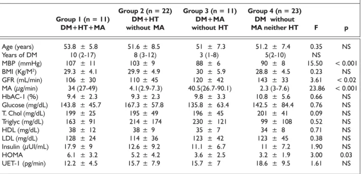
greater in the group of the diabetic hyperten-sive patients with MA, as compared with the group of diabetic patients with microalbumin-uria, and without HT, and with the group of diabetic patients without MA and without HT. The levels of the insulin resistance index were significantly lower in the group of diabetic pa-tients without MA and without HT as compared with the groups of diabetic patients with hy-pertension.
Calculated GFR was greater in the group of diabetics without MA or HT as compared with the two groups of hypertensive diabetic patients (p < 0.02). The two groups of non hypertensive diabetic patients had similar values of calculated GFR.
Figure 1 shows that urinary ET-1 was greater in the group of normotensive diabetic patients
without MA as compared with the group of dia-betic hypertensive patients with MA (18.6 ± 9.5
pg/min vs 12.2 ± 4.5 pg/min, p = 0.02), and
with the control group (18.6 ± 9.5 pg/min vs
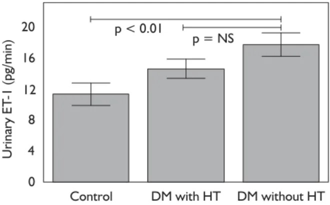
11.2 ± 5.3pg/min). Figure 2 shows that there was a significant difference in the urinary ET-1 excre-tion of the control group (mean 11.2 ± 5.4) as compared with the non hypertensive diabetic group (mean 17.6 ± 8.7) but there was no dif-ference with the diabetic hypertensive group (mean 14.5 ± 7.1).
Multiple regression analysis was unable to show any correlation among the urinary ET-1 excre-tion and the levels of HbA1c, glycaemia, serum insulin and time of diagnosis of the disease.
The logistic regression analysis shows that the urinary ET-1 excretion does not represent a risk factor to develop MA.
Table I. Clinical and Biochemical Characteristics of the Diabetic Groups.
Group 2 (n = 22) Group 3 (n = 11) Group 4 (n = 23)
Group 1 (n = 11) DM+HT DM+MA DM without
DM+HT+MA without MA without HT MA neither HT F p
Age (years) 53.8 ± 5.8 51.6 ± 8.5 51 ± 7.3 51.2 ± 7.4 0.35 NS
Years of DM 10 (2-17) 8 (3-12) 3 (1-8) 5(2-10) NS
MBP (mmHg) 107 ± 11 103 ± 9 88 ± 6 90 ± 8 15.50 < 0.001
BMI (Kg/M2) 29.3 ± 4.1 29.9 ± 4.9 30 ± 5.9 28.8 ± 4.5 0.23 NS
GFR (mL/min) 106 ± 30 110 ± 45 120 ± 42 143 ± 33 3.61 < 0.02
MA (µg/min) 34 (27-49) 4.1(2.9-7.3) 40.5(26.7-90.1) 2.3 (3-7.6) 23.86 < 0.001
HbAC-1 (%) 9.4 ± 2.3 9.3 ± 2.3 9.8 ± 3.3 10.8 ± 5.6 0.66 NS
Glucose (mg/dL) 143.8 ± 45.7 167.3 ± 57.8 135.8 ± 63.4 142.5 ± 84.4 0.76 NS
T. Chol (mg/dL) 199 ± 25 195 ± 49 196 ± 45 201 ± 41 0.09 NS
Triglyc (mg/dL) 163 ± 91 214 ± 174 230 ± 121 99 ± 108 0.52 NS
HDL (mg/dL) 38 ± 12 38 ± 9 35 ± 7 34 ± 8 0.71 NS
LDL (mg/dL) 128 ± 24 114 ± 36 123 ± 42 123 ± 45 0.38 NS
Insulin (µUI/mL) 17.9 ± 9 12.6 ± 9.2 11.1 ± 6.7 11 ± 7.2 1.90 NS
HOMA 6.1 ± 3.2 5.2 ± 4.2 3.6 ± 2.5 3.2 ± 1.9 3.00 0.03
UET-1 (pg/min) 12.2 ± 4.5 15.7 ± 7.9 15.7 ± 7 18.6 ± 9.5 1.61 NS
46
www.medigraphic.com
ESTE DOCUMENTO ES ELABORADO POR MEDIGRAPHIC
Discussion
Consistent with the data reported by Shin et al,24
we found in our study that the diabetic patients with no hypertension or microalbuminuria had greater urinary ET-1 excretion than healthy sub-jects. The reduced urinary ET-1 excretion sug-gests that ET-1 synthesis is reduced or its catab-olism is enhanced.25-26 Also, our group of diabetic
patients with no hypertension or microalbumin-uria had the greater GFR. This may be explained by the diuretic effect of the ET-1 on the tubules, as described by Lariviere27 and Ohta,16 who
as-sociate the urinary ET-1 excretion levels with the excretion of the N-Acetyl-²-D-glucosaminidase (NAG), which is a marker of proximal tubular damage.27 This association of urinary ET-1 and
NAG may support the possibility that the elevat-ed urinary ET-1 excretion seen in some nephrop-athies is the result of proximal tubular damage, and not of glomerular damage.
The lack of association between the urinary ET-1 excretion and the presence of microalbuminur-ia found in our study, is consistent with the find-ing of De Mattia.28
On the other hand in the study done by Lee et al, the urinary ET-1 excretion levels were greater in diabetic patients with albuminuria as compared with a control group.29
However, this finding might be explained by the inclusion of patients with albuminuria over 200 ¼g/min, while in our study we included only pa-tients with microalbuminuria, in order to study exclusively incipient renal damage.
The lack of association between the urinary ET-1 excretion levels and glycaemia in our study may be explained because our patients were in a bet-ter metabolic control, as measured by the HbA1c than the patients studied by Shin et al, in whom they found a positive association between hyper-glycemia and urinary ET-1 excretion.24
There was no association between the evolu-tion time of the diabetes or the high blood pres-sure. This finding may be due to the fact that the urinary excretion of ET-1 depends more on the metabolic control than in the time of evolution of the disease.30
In conclusion, our study shows that the uri-nary excretion of ET-1 tends to be greater in the diabetic patients without hypertension as com-pared with the diabetic hypertensive patients. On the other hand the diabetes evolution time and the hypertension evolution time did not corre-late with the urinary ET-1 excretion.
There is no correlation of urinary ET-1 excre-tion and the mean blood pressure, the metabolic control as measured by the HB1Ac levels, the insulin levels and the GFR.
Urinary ET
-1 Excretion (pg/min)
DM+HT +MA
DM+HT no MA
DM+ MA no HT
DM no HT or MA
5 10 15 20 25
30 p < 0.005 p < 0.02
Control
35
0
Figure 1. Urinary ET-1 excretion on all the groups of study.
Urinary ET
-1 (pg/min)
20
16
12
4 8
0
p < 0.01
p = NS
Control DM with HT DM without HT
47
www.medigraphic.com
As clinical studies on the urinary ET-1 excre-tion are still few, we think it is worth to continue trying to clarify the role played by the urinary ET-excretion on the renal pathophysiology, in-cluding models evaluating the proximal tubular and glomerular pathology.
Bibliography
1. Bloomgarden Z. The epidemiology of complications. Diabetes Care 2002; 25: 924-932.
2. Sabag-Ruiz E, Alvarez-Feliz A, Celiz-Zepeda S, Gomez-Alcala AV. Chronic complications of diabetes mellitus. What is the prevalence of diabetes in a family medical unit? Rev Med Inst Mex Seguro Soc 2006; 44: 415-421.
3. Remuzzi G, Bertanio T. Pathophysiology of progressive nephr-opathies. N Engl J Med 1998; 339: 1448-1456.
4. Gnudi L, Raij L. The link between Glut-1 and hypertension in diabetes nephropathy. Curr Hypertens 2006; Rep 8: 79-83. 5. Yanagisawa M, Kurihara H, Kimura S, Tomobe Y, Kobayashi M
et al. A novel potent vasoconstrictor peptide produced by endothelial cells. Nature 1998; 332: 411-415.
6. López-Farre A, Gómez-Garre D, Bernabeu F, Montanes I, Mill-as I, López-Novoa JM. Renal effects and mesangial cell contrac-tion induced by endothelin are mediated by PAF. Kidney Int
1991; 39: 624-30.
7. Swislocki AL, Hoffman BB, Reaven GM. Insulin resistance, glu-cose intolerance and hyperinsulinemia in patients with hyper-tension. Am J Hypertens 1989; 2: 419-423.
8. Denton KM, Shweta A, Finklestein L, Floerr RL, Evans RG. Effect of endothelin-1 on regional kidney blood flow and renal arteriole caliber in rabbits. Clinic Exp Pharmacol Physiol 2004; 31: 494-501.
9. King AJ, Brenner BM. Endothelium-derived vasoactive factors and the renal vasculature. Am J Physiol 1991; 260 (4 Pt 2); R653-662.
10.Vercellotti GM, Tolins JP. Endothelial activation and the kidney: vasomediator modulation and antioxidant strategies. Am J Kid-ney Dis 1993; 21: 331-343.
11.Haynes WG, Webb DJ. The endothelin family of peptides: local hormones with diverse roles in health and disease? Clin Sci 1993; 84: 485-500.
12.Sugimoto K, Tsuruoka S, Fujimura A. Renal protective effect of YM598, a selective endothelin ET(A) receptor antagonist, against diabetic nephropathy in OLETF rats. Eur J Pharmacol 2002; 450: 183-189.
13.Rothermund L, Traupe T, Dieterich M, Kossmehl P, Yagil Ch et al. Nephroprotective effects of the ETa receptor antagonist durasten in salt sensitive genetic hypertension. Eur J Pharmacol
2003; 468: 209-216.
14.Benigni A, Colosio V, Brena C, Bruzzi I, Bertani T, Remuzzi G. Unselective inhibition of endothelin receptors reduce renal dys-function in experimental diabetes. Diabetes 1998; 47: 450-456. 15.Hynynen MM, Khalil RA. The vascular endothelin system in hypertension –recent patens and discoveries. Recent Pat Cardio-vas Drug Discov 2006; 1: 95-108.
16.Ohta K, Hirata Y, Shichiri M, Kanno K, Emori T et al. Excretion of endothelin-1 in normal subjects and patients with renal dis-ease. Kidney Int 1991; 39: 307-311.
17.Dhaun N, Goddard J, Webb DJ. The endothelin system and its antagonism in chronic kidney disease. J Am Soc Nephrol 2006; 17: 943-955.
18.Mather KJ, Mirzamohammadi B, Lteif A, Steinberg HO, Baron AD. Endothelin contributes to basal vascular tone and endothe-lial dysfunction in human obesity and type 2 diabetes. Diabetes
2002; 51: 3517-3523.
19.Bruno CM, Meli S, Marcinno M, Ierna D, Sciacca C, Neri S. Plasma endothelin-1 levels and albumin excretion rate in nor-motensive, microalbuminuric type 2 diabetic patients. J Biol Regul Homeost Agents 2002; 16: 114-117.
20.Verna LR: National Heart, Lung, and Blood Institute Releases New Guidelines for the treatment of hypertension. Am Fam Physician 1998; 57: 362-364.
21.Coresh J, Astor BC, McQuillan G, Kusek J, Greene T et al. Calibration and random variation of the serum creatinine assay as critical elements of using equations to estimate glomerular filtration rate. Am J Kidney Dis 2002; 39: 920-929.
22.American Diabetes Association: Position Statement Diabetic Nephropathy. Diabetes Care 2003; 26: s94-s98.
23.Katsuki A, Sumida Y, Gabazza E, Murashima S, Furuta M et al. Homeostasis model assessment is a reliable indicator of insulin resistance during follow-up of patients with type 2 diabetes.
Diabetes Care 2001; 24: 362-365.
24.Shin SJ, Hsiao PJ, Hsieh MC, Lee YJ, Tsai JH. Increased urinary endothelin-1 excretion in newly diagnosed type 2 diabetic pa-tients. Kaohsiung J Med Sic 1999; 15: 589-596.
25.Hoffman A, Grossman E, Goldstein DS, Gill JRJ, Keiser H. Urinary excretion rate of endothelin-1 in patients with essen-tial hypertension and salt-sensitivity. Kidney Int 1994; 45: 556-560.
26.Zoccali C, Leonardis D, Parlongo S, Mallamaci F, Postorino M. Urinary and plasma endothelin-1 in essential hypertension and in hypertension secondary to renoparenchymal disease. Nephrol Dial Transplant 1995; 10: 1320-1323.
27.Lariviere R, Lebel M. Endothelin-1 in chronic renal failure and hypertension. Can J Physiol Pharmacol 2003; 81: 607-621. 28.De Mattia G, Cassone FM, Bellin C, Bravi M, Laurenti O et al.
Role of plasma and urinary endothelin-1 in early diabetic and hypertensive nephropathy. Am J Hypertension 1998; 11: 983-988.
29.Lee YJ, Shin SJ, Tsai JH. Increased urinary endothelin-1-like im-munoreactivity excretion in NIDDM patients with albuminuria.
Diabetes Care 1994; 17: 263-266.