ABSTRACT. The Visual Colorimetric Immunoassay (Cholera SMARTTM) [ NEW HORIZONS DIA G-NOSTICS] is among the quick diagnostic techniques developed during recent years for the direct detec-tion of Vibrio cholerae O:1 in fecal material. In this work, this immunoassay was used together with the culture technique (Laboratorios Nacionales de Salud Pública e Instituto Nacional de Diagnóstico y Refer-encia Epidemiológicos de la SSA. de México) to detect V. cholerae O:1 in 50 oyster samples. The samples were collected in Mexico City, from markets and roadside stands, during the period from October 1994 to March 1995. Of the 50 samples analyzed, only one was found positive by both techniques. These results indicate that the Cholera SMARTTM [NHD] kit represents a quick and simple technique for the detection of V. cholerae O:1 in oysters. Apart from looking for the V. cholerae O:1, various members of the Vi-brionaceae family were isolated and identified from the same oyster samples. The findings were as fol-lows: V. cholerae NO O:1 (26 %), V. parahaemolyticus (8%), V. alginolyticus (46 %), other species of the Vibrio spp. genus (6 %) and species from the Aeromonas spp. genus (12 %).
RESUMEN. Dentro de las técnicas rápidas de diagnóstico desarrolladas en los últimos años para la deter-minación directa de Vibrio cholerae O:1 en materia fecal está el Inmunoensayo Colorimétrico Visual (Cholera SMARTTM) [ NEW HORIZONS DIAGNOSTICS]. En el presente trabajo se aplicó este Inmu-noensayo junto con la técnica de cultivo (Laboratorios Nacionales de Salud Pública e Instituto Nacional de Diagnóstico y Referencia Epidemiológicos de la SSA de México) para la determinación de Vibrio chole-rae O:1 en 50 muestras de ostiones. Las muestras fueron obtenidas en mercados y puestos callejeros de la Ciudad de México, en el período comprendido de octubre de 1994 a marzo de 1995. De las 50 muestras analizadas solamente una fue positiva por ambas técnicas. En función de los resultados obtenidos, se con-sidera que el Cholera SMARTTM [NHD] funciona como una técnica rápida y sencilla para la determina-ción de Vibrio cholerae O:1 en ostiones. Además de la búsqueda de V. cholerae O:1, también se aislaron e identificaron en las muestras estudiadas varios miembros de la familia Vibrionaceae, encontrándose V. cholerae NO O:1 (26 %), V. parahaemolyticus ( 8%), V. alginolyticus (46 %), otras especies del género Vi-brio spp. (6 %) y especies del género Aeromonas spp. (12 %).
Palabras clave: Vibrio cholerae, Inmunoensayo colorimétrico.
Detection of
Vibrio cholerae
O:1 in Oysters by the Visual
Colorimetric Immunoassay and the Culture Technique
Sergio Villalpando-Guzmán, M. Guadalupe Eusebio-Hernández*, and David Avilés-Ruíz
Departamento de Microbiología, Escuela Nacional de Ciencias Biológicas, IPN. Prolongación de
Carpio y Plan de Ayala, S/N, Col. Casco de Santo Tomás, 11340 México, D.F., Mexico.
Tel. (5) 729 60 00 ext. 62374, Fax (5) 7 29 62 07
.E-mail: svillalpando71@hotmail.com
*Corresponding author and COFAA fellowship holder
Received 8 September 1999/Accepted 13 April 2000
INTRODUCTION
Vibrio cholerae O:1 is the causal agent of cholera, which is a disease that continues to be prevalent in many developing countries.5,6,11,19 The strains of V. cholerae be-longing to the O:1 serogroup have been classified into three serotypes: Ogawa, Inaba and Hikojima. These sero-types contain the classical and ElTor1 biosero-types15. In Peru, in 1991, 107,064 cases of this disease were registered, and in Mexico, during the same year, the first case within the same pandemia, was reported.11,19 Transmission of cholera
occurs by the inges tion of water or food contaminated with
commonly implicated in the appearance of cholera cases are: fish and shell fish, fruit and vegetables and other foods such as rice, potatoes, lentils, beans, egg, chicken, etc.9,13,14,20
Fish and other sea food may be contaminated if they have been collected from water contaminated with fecal material or from those aquatic environments in which V. cholerae O:1 is naturally present.3,21 Oysters (bivalve mo l-lusks from the genus Crassotrea spp.), are among the sea-food that are implicated in the occurrence of cholera cases, in particular since they are frequently eaten raw.7,22
host and the microorganism. Symptoms include, the abrupt appearance of nausea, vomiting, diarrhea and abdominal spasms, there are also metabolic alterations such as deh y-dration, acidosis, hypocalcimia and electrolyte imba l-ance.12 Laboratory diagnosis is carried out by the isolation and identification of the causal agent or may be carried out through techniques such as, phase contrast and dark field microscopy, immunoflourescesnce, latex agglutination, coagulation, enzyme linked immunoabsorbent assay (ELISA) and the polymerase chain reaction (PCR).2,4,9
In 1994, Hasan and cols. reported the development and evaluation of a test based on the use of colloidal gold part i-cles, known as the Visual Colorimetric Immunoassay [Cholera SMARTTM (Sensitive Membrane Antigen Rapid Test)], which allows direct detection of V. cholerae O:1 from samples of fecal material. The basis of the test is as follows: a sample suspected of containing V. cholerae O:1 is reacted with monoclonal antibodies against bacterial lipopolysaccharide factor A, which are marked with colloi-dal gold particle. If V. cholerae O:1 is present in the sam-ple, V. cholerae O:1-monoclonal antibody complexes are formed, which are subsequently captured and concentrated by polyclonal antibodies linked to a solid phase matrix. Following the depositing of colloidal gold, and after a minimum incubation time of 5-10 minutes, the reaction appears at first sight as a developing pink stain. In the ab-sence of V. cholerae O:1, the complex does not form, in which case the pink test coloring does not appear.10
Today there is the tendency within medical and food microbiology to develop and apply quick diagnostic tech-niques, that allow results to be obtained with a high degree of reliability, such that the spread of infectious diseases is prevented by cutting the diagnostic time. This is the case for the Cholera SMARTTM technique, which is analyti-cally sensitive and specific, and therefore reliable. In other words, this technique can detect 6 x 10⁶ CFU/ml of V. cholerae O:1 and is a test which is 100 % specific for this microorganism. The work presented here shows the first steps towards determining the application of this kit in the area of sanitary microbiology.
The objetive of this work is compare the Visual Colori-metric Immunoassay technique (Cholera SMARTTM) [NHD] with the culture technique (Laboratorios Nacion-ales de Salud Pública e Instituto Nacional de Diagnóstico y Referencia Epidemiológicos de la Secretaría de Salud), in order to detect V. cholerae O:1 and thus to validate the use of this immunoassay with oyster samples.
MATERIAL AND METHODS
50 oyster samples were collected from local markets and street stands in different parts of Mexico City. The quantity of each sample collected corresponded to one or-der of oysters (approximately 10 large pieces or 20 small pieces).8 The oysters did not contain sauce, onion or any
other ingredient usually used in its preparation in the form of a cocktail. The conditions of conservation in which the oysters were kept at the site of sale are as follows: a) oys-ters removed from their shells, kept on aluminum trays on beds of ice (29 samples collected); b) oysters removed from their shells, kept on aluminum trays or in plastic buckets, at ambient temperature (4 samples collected); and c) oysters in their shells kept at ambient temperature (17 samples collected). For this final group, the mollusks were taken out of their shell at the place of sale. Each sample was placed in disposal cups (plastic or unicel) covered with plastic lids, and each cup was placed in a polythene bag. Once the samples were obtained they were transported in an icebox for a maximum time of one hour, before the analysis was carried out.
Preliminary assays. Before applying the Cholera SMARTTM [NHD] test and the culture technique, two preliminary assays were carried out. The first of these was to determine the minimum enrichment time, at 37ºC, of a series of dilutions of pure V. cholerae O:1 strain, diluted in alkaline peptone water. The objective of this procedure was to determine the end point at which a positive reaction was observed when the Cholera SMARTTM [NHD] test was applied. In the second preliminary assay, a sample of oysters was inoculated with a strain of V. cholerae O:1 and the minimum time necessary for enrichment, in order to obtain a positive reaction with the Cholera SMARTTM [NHD] kit, was determined. In this way it was possible to determine the influence of both the components of the oys-ter and the time necessary for enrichment under which this "kit" functions.
Detection of V. cholerae O:1 in oysters by the cul-ture technique. This technique is described in the National Public Heath Laboratories of the Mexican Health Minis-try.8 It is based on the methodologies provided by the Food and Drug Administration of the United States of America. In this investigation we only used tryptone agar with 1 % NaCl, following which we applied different tests in the order recommended by the INDRE (Instituto Nacional de Diagnóstico y Referencia Epidemiológicos).9
were incubated at 37 ºC for 24 h. The 1:10 dilution, incu-bated at 37 ºC, was kept in these conditions for 24 h. Subsequently, three suspected colonies of V. cholerae
were selected from each plate. Each selected colony was transferred to an agar plate with 1 % tryptone and 1 % NaCl (T1N1 agar) and isolated by cross streaking, with incubation at 37 ºC for 24 h.
Following colony isolation in T1N1 agar, Gram stain-ing was carried out to observe the morphology microscopi-cally. Identification was then carried out by inoculation in the following media: Kligler’s iron agar, lysine iron agar, indol ornithin mobility media, Simmon’s citrate agar, mythl- red Voges Proskauer broth, tryptone broth with 0, 1, 3, 6, 8 and 10 % NaCl, and arginine broth. All the media were incubated at 37 ºC for 24 h. If the biochemical tests at 24 h confirmed the presence of V. cholerae, the oxidase test was carried out from the lysine-iron agar and the thread test, from Kligler’s iron agar. If both the oxidase test and the thread test confirmed the presence of V. chol-erae, the serological confirmation was made with polyva-lent antiserum, to identify the O:1 serogroup and then the serotype was identified using Ogawa and Inaba specific antisera. When the biochemical tests for V. cholerae coin-cided, but there was no agglutination with the polyvalent antiserum, the presence of V. cholerae NO O:1 was re-ported.
Detection of V. cholerae O:1 in oysters by visual col-orimetric immunoassay (Cholera SMARTTM) [NEW HORIZONS DIAGNOSTICS]. The test was carried out using the enrichment broth incubated at 37 ºC for 8 and 24 h . An aliquot of enrichment broth was taken and trans-ferred to a filtering device to half its capacity, and was ho-mogenized for one minute by gentle shaking (it was not necessary to use the extraction regulating solution included in the commercial kit, since the sample was in a enrich-ment broth). Then, three drops of the liquid contained in the filtering device were extracted and added to the reac-tion vial in which three drops of reconstitureac-tion solureac-tion had previously been added and this was shaken gently. Later, the contents of the vial were absorbed with the sterile da-cron bud which was included in the kit. The bud was place in the upper compartment of the SMART device, which was closed in the indicated area. This was incubated for 20 min at ambient temperature, and finally the test areas and negative control included in the lower compartment of the SMART device, were read. The positive control included within the same kit was run simultaneously.
Identification of other species from the Vibrio spp. Genus and other members of the Vibrionaceae family obtained from oyster samples. Identification of other mi-croorganisms present in the samples studies was carried out using those colonies whose morphologies differed from those belonging to V. cholerae isolated on thiosulphate citrate bile salt saccharose (TCBS) agar. In addition, tests were carried out on those colonies with morphologies simi-lar to V. cholerae, but which, when biochemical tests were
carried out were found to be different.
The procedure for identifying the above microorgan-isms was similar to that used for V. cholerae, including gelatin agar, urea broth and phenol red broth with the addi-tion of manitol. 2 % NaCl was added to Kligler’s iron agar, lysine-iron agar, indol ornithin motility media, methyl red Voges Proskauer broth and gelatin agar. To identify Vibrio parahaemolyticus it was necessary to use the L-leucine assimilation test. In addition, the following media were used: bile esculin agar, blood agar and phenol red broth with the addition of arabinose to id entify species from the
Aeromonas sp. genus.1,8,9,15
RESULTS
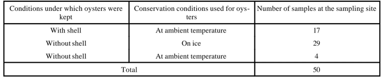
In table 1 the conservation conditions under which the oysters were kept at the sampling site are shown. The pre s-ence or abss-ence of shells at the site of sale is also shown. The results of the preliminary assay show that the min i-mum enrichment time of a pure V. cholerae O:1 strain, at 37 ºC, is 8 h. Such that, at the end of this incubation time a positive reaction was registered when the Cholera SMARTTM [NHD] kit was applied using an inoculation of 30 CFU/ml. When a starting concentration of 10 and 3 CFU/ml of V. cholerae O:1 was used, the test was positive at 9 h. With respect to the other preliminary assay, in which a sample of oysters was used, a positive test was registered using a starting inoculation of 10 CFU/ml, with 9 h incubation at 37 ºC. Table 2 shows results obtained using the culture technique and the Cholera SMARTTM [NHD] test to detect V. cholerae O:1 in oysters. Here 49 samples are shown which were negative both with the Cholera SMARTTM [NHD] test, applied after 8 and 24 h incubation, and with the culture technique. Only one pos i-tive sample was obtained by applying the immunoassay after 8 and 24 h incubation, which was confirmed by the culture technique. The strain detected was identified as V. cholerae O:1 Ogawa serotype, ElTor biotype. In table 3 microorganisms belonging to the Vibrionaceae family are shown, identified from the oyster samples processed by the culture technique.
DISCUSSION
cholerae O:1 are necessary to obtain an unequivocally positive result.10 The presence of the microorganism in oyster samples was determined, using samples kept in very different micro -environmental conditions compared to samples of clinical origin (fecal material) for which this immunoassay was designed. The origin of the sample used is directly reflected in the number of CFU/ml of V. chol-erae O:1 detected in the 2 sample types. When the Cholera SMARTTM [NHD] kit was used in the first preliminary assay, a positive result was obtained after incubating the V. cholerae suspension, diluted to 30 CFU/ml in APA, for 8 h. Hence, starting from this concentration of bacterial cells,
the immunoassay developed a positive result after 8 h incu-bation, and the tests applied before this incubation time were negative. In the same way, positive results were ob-tained with the "kit" after 9 h incubation using a suspen-sion of 10 and 3 CFU/ml of V. cholerae O:1. Furthermore, from the preliminary assay, in which an oyster sample was used, it was possible to determine if the components of the oyster itself interfered with the sensitivity of the test. It was found that after 9 h incubation, the oyster sample, diluted in APA and inoculated with an initial concentration of 10 CFU/ml, tested positive the same way as the assay in which only microorganisms in APA were used. This indi-cates that oyster components did not interfere with the test. Based on these results, the Cholera SMARTTM [NHD] test was used fo llowing incubation of the enrichment broth for 8 h, at 37 ºC, in the same way as the culture technique. The result was then confirmed after 24 h incubation. It is important to point out that the sample that resulted positive for V. cholerae O:1 was obtained form a road side stand, in which the oysters, without their shells, were kept on alumi-num trays on a bed of ice. Furthermore, they were col-lected during the last week of March, which is important to point out given that there is seasonality involved with out-breaks of cholera cases.12 The detection of at least one sample as positive for V. cholerae O:1 by both techniques, has important public health implications, and direct reper-cussions on the prevalence of the disease in urban areas such Mexico City. Hence, we are carrying out an evalu a-tion of potential risks associated with the consumpa-tion of oysters which represent a transmitter of cholera within Mexico City and its environs. In addition we are investi-gating the various sources of contamination that allow the entrance of V. cholerae O:1 into oysters, for example, the contamination of origin and contamination via oyster han-dlers from the point of harvest to the site of sale to the con-Table 1. Conservation conditions used for oyster samples at the sampling site.
Conditions under which oysters were kept
Conservation conditions used for oys-ters
Number of samples at the sampling site
With shell At ambient temperature 17
Without shell On ice 29
Without shell At ambient temperature 4
50 Total
Table 2. Detection of Vibrio cholerae O:1 in oysters by the culture technique and the visual colorimetric immunoassay.
Number of negative samples for both techniques.
Number of positive samples detected by culture only.
Number of positive samples detected by the Cholera
SMARTTM test only.
Number of positive samples detected by both techniques
49 0 0 1
Table 3. Microorganisms isolated from oysters by the cul-ture technique.
Microorganisms Percentage of micro-organism isolation
Vibrio alginolyticus 46
Vibrio cholerae NO O:1 26
Vibrio parahaemolyticus 8
Aeromonas caviae 6
Vibrio harvegi 4
Aeromonas sobria 4
Aeromonas hydrophila 2
Vibrio cholerae O:1 serotipo Oga-wa
2
sumer.
With respect to the culture technique and dilutions, all the oyster flesh was homogenized along with the valve liq-uid. This allowed us to obtain adequate samples for analy-sis. Furthermore, this preparation did not have a negative effect on the viability of V. cholerae O:1, as may occur with the ext rinsic and intrinsic parameters that are present in the food before analysis.3,14
Observing the data shown in table 1, the imp ortance of obtaining samples kept on ice at the place of sale, can be appreciated. Since, it is within this group that the sample, in which V. cholerae O:1 was demonstrated, was found. Furthermore, it is important to mention that at the moment of acquiring this positive sample, the oysters were removed from their shells. In table 1, the conservation temperature can be seen (ambient or on ice) as well as the influence of the mollusc shell on the viability of V. cholerae O:1. With respect to the immunoassay, no results were ob-tained that were only positive using this test. That is, false positive results were not registered, which indicates that the technique did not develop cross reactions when con-fronted with the variety of microorganisms that may be found in this type of sample.16 Furthermore, positive sam-ples were not detected by the culture technique alone, such that, false negatives were not registered. This indicates to us that the Cholera SMARTTM technique functioned well, since there was no reaction in those samples which were found to be negative for microorganisms by culture. Hence, all the reagents and the lyophilized conjugate showed specific activity.10,18
The low incidence of V. cholerae O:1 in the oyster samples analyzed in this work may be related to the time of year in which the study was carried out. The study took place during autumn and winter, and during only two weeks of spring, and although cholera cases occur throug h-out the whole year, they increase in summer.3,12 Further-more, the general handling of the oysters should also been considered, from when they were taken from their natural habitat until the places where they were consumed, in which a very important role is played by microbial comp e-tition.
The results in table 3, give an idea of the variety of mi-croorganisms that may be present in this type of mollusk, which is strongly related to their natural characteristics (as biological concentrator organisms). Most importantly it can be seen that within these microorganisms are included potential pathogens for human beings, such as V. cholerae
NO O:1, V. parahaemolyticus, V. alginolyticus and Aero-monas hydrophila.16
One of the most interesting aspects of the development of this work, was to determine the function of this immu-noassay when confronted with a type of sample which is different from that for which the kit was originally de-signed. The experience obtained in this study showed that this technique can be applied to oysters, since handling of the samples was not complicated. As has already been
shown, we decided to enrich the oyster samples in order to increase the probability o f obtaining a positive result, since there is a direct relationship between the analytical sensi-tivity of the immunoassay and the concentration of the mi-croorganisms in the oyster sample. That is, the immunoas-say does not detect concentrations of the microorganism lower than 6X10⁶ CFU/ml, and the concentration of V. cholerae O:1 in oysters does not necessarily reach this con-centration, however there are reports of infective doses of 1X10⁶ CFU/g.10,12 In spite of requiring an enrichment time
of 8 h to apply the immunoassay to the oyster sample, there is still an important reduction in the time required to obtain results, compared to the minimum of 78-80 h re-quired to obtain results from culture. This type of product, therefore represents an important quick technique for the detection of V. cholerae O:1. Based on the advantages that this immunoassay offers, we propose that only those sam-ples that result positive by this technique should be pro c-essed by culture, above all in those regions of the country in which epidemic outbreaks occur. This would allow the rapid application of preventative action in order to avoid the dissemination of the disease through foods such as mo llusks.
As a consequence of the results obtained, it was not possible to apply any statistical model to describe the rela-tionship between both techniques. However, it is very in-teresting that the total of oyster samples analyzed that re-sulted negative for V. cholerae O:1 by the Cholera SMARTTM test, were also found to be negative by the culture technique. In addition, the sample that resulted positive by the immunoassay was also positive by the cul-ture technique. All this data has allowed us to learn more about this technique. However, it is necessary to keep a line of investigation that provides more information on its application to samples taken from their original enviro n-ment (water, aquatic sedin-ments, Phytoplankton and biofilms, etc.) as well as from food samples. Among, the other applications of this type of immunoassay, is the pos-sibility of detecting viable forms of V. cholerae O:1 that are not possible to cultivate, taking as reference the isola-tion data of the microorganism in water, fish and seafood.3
In summary, it is important to point out the range of applications that may be obtained from this technique within the field of sanitary and environmental microbiol-ogy in Mexico, which will eventually provide fast and reli-able detection of V. cholerae O:1. In this way, quality con-trol will become more efficient and as a consequence pre-ventive or corrective measures will be applied more rap-idly to combat the problem of cholera epidemics.
REFERENCES
1. Baumann, P. and R. H. W. Schubert. 1984. Family II
Vibrionaceae. Veron 1965, 5245AL, p. 516-538. In: N. R. Krieg and J.G. Holt (Ed.). Bergey´s Manual of Sy s-tematic Bacteriology, Vol. 1 .The Williams and Wil-kins, Baltimore.
2. Bradford, A. K., C. A. Bopp and J. G. Wells. 1994. Iso-lation and Identification of Vibrio cholerae O:1 from fecal specimens, p. 3-12. In: I.K. Wachsmuth, P.A. Blake And O. Olsvik (Ed.). Vibrio cholerae and Chol-era. Molecular to global perspectives. ASM-Press, Washington.
3. Colwell, R. R. and W. M. Spira. 1992. The Ecology of
Vibrio cholerae, p. 107-127. In: D. Barua and W. B. Greennough III (Ed). Cholera. Plenum Medical Book, New York.
4. Colwell, R. R., J. A. K. Hasan, A. Huq, L. Loomis, R. J. Siebeling, M. Torres, S. Galvez, S. Islam, M. T. Tamplin And D. Bernstein. 1992. Development and Evaluation of a Rapid, Simple, Sensitive, Monoclonal Antibody-based Co-agglutination test for Direct Detec-tion of Vibrio cholerae O:1. FEMS Microbiol. Lett. 97:215-220.
5. Dirección General de Epidemiología de la Secretaría de Salud, México. 1995. Tablas del Informe Semanal de Epidemiología (cólera). Boletín Semanal de Vigilancia Epidemiológica 1:13.
6. Dirección General de Epidemiología de la Secretaría de Salud, México. 1995. Situación del Cólera en México (hasta la semana 10 de 1995). Boletín Semanal de Vigi-lancia Epidemiológica del Cólera en México 1:1-2. 7. Eyles, M. J. And G. R. Davey. 1988. Vibrio cholerae
and Enteric Bacteria in Oysters Producing Areas of Two Urban Estuaries in Australia. JFM 6:207-218. 8. Gallardo, R. R., M. C. Rodríguez y P. A. Vargas-Tapia
(Ed.). 1992. Manual de Técnicas y Procedimientos para la Investigación de Vibrio cholerae en Agua y Alimen-tos. Secretaría de Salud. Subsecretaría de Regulación y Fomento Sanitario. Laboratorio Nacional de Salud Pú-blica, México.
9. Giono, C. S., L. Gutiérrez C. Y A. M. Hinojosa A. 1991. Manual de Procedimientos para el Aislamiento y Caracterización de Vibrio cholerae O:1. Publicación Técnica del INDRE # 10. Subsecretaría de Organiza-ción y Desarrollo. Instituto Nacional de Diagnóstico y Referencia Epidemiológicos, México.
10. Hasan, J. A. K., A. Huq, M. L. Tamplin, R. J. Siebeling And R. R. Colwell. 1994. A Novel Kit for Rapid
Detec-tion of Vibrio cholerae O:1. J. Clin Microbiol. 32:249-252.
11. Instituto Nacional de Diagnóstico y Referencia Epid e-miológicos de la Secretaría de Salud. 1991. Situación del Cólera en México hasta el 30 de junio de 1991. Bol. Quincenal cólera/Diarreas Infecciosas 1:469-486. 12. Kaper, J. B., J. G. Morris And M. H. Levine. 1995.
Cholera. Clin. Microbiol. Rev. 8:48-86.
13. Kaysner, C. A., M. L. Tamplin and R. M. Twedt. 1992. Vibrio, p. 451-473. In: C. Vanderzant And D. Splittstaesser (Ed.). Compendium of Methods for the Microbiological Examination of foods, 3rd ed. Ameri-can Public Health Association, Washington, D.C. 14. Kaysner, C. A. and W. E. Hill. 1994. Toxigenic Vibrio
cholerae O:1 in Food and Water, p. 27-39. In: I.K. Wachsmuth, P.A. Blake And O. Olsvik ( eds.). Vibrio cholerae and Cholera. Molecular to global perspec-tives. ASM-Press, Washington.
15. Kelly, M. T., F. W. Hickaman-Brenner and J. J. Farmer III. 1991. Vibrio, p. 384-395. In: W. J. Hausler, K. L. Herrman, H. D. Isenberg And H. J. Shadomy ( eds. ). Manual of Clinical Microbiology, 5th ed. ASM, Wash-ington.
16. Matte, G. R., M. H. Matte, I. G. Rivera And M. T. Mar-tins. 1994. Distribution of Potentially Pathogenic Vi-brios in Oysters from a Tropical Region. J. Food Prot. 57:870-873.
17. Mintz, E. D., T. Popovic And P. A. Blake. 1994. Trans-mission of Vibrio cholerae O:1, p. 345-356. In: I.K. Wachsmuth, P.A. Blake And O. Olsvik (eds.). Vibrio cholerae and Cholera. Molecular to global perspec-tives. ASM-Press, Washington.
18. New Horizons Diagnostics Corporation. 1993. Cholera SMARTTM Test: A Colorimetric Immunoassay for the Direct Detection of Vibrio cholerae O:1. New Horizons Diagnostics Co., Columbia, Maryland, USA.
19. OPS. 1991. Epidemia de Cólera en el Perú y Pautas para su Control. Bol. Of. Sanit. Panam. 110:277-297. 20. OPS. 1993. El cólera y la Comercialización de los
Ali-mentos. Bol. Of. Sanit. Panam. 115:458-463.
21. Popovic, T., O. Olsvik, P. A. Blake and K. Waschmuth. 1993. Cholera in the Americas: Foodborne Aspects. J. Food Prot. 56:811-821.