Otras secciones de este sitio:
☞ ☞ ☞ ☞
☞ Índice de este número
☞ ☞ ☞ ☞
☞ Más revistas
☞ ☞ ☞ ☞
☞ Búsqueda
Others sections in this web site:
☞ ☞ ☞ ☞
☞ Contents of this number ☞
☞ ☞ ☞
☞ More journals ☞
☞ ☞ ☞ ☞ Search Artículo:
Some biological features of Mollicutes
Derechos reservados, Copyright © 2002: Asociación Mexicana de Microbiología, AC Revista Latinoamericana de Microbiología
Número Number 2
Abril-Junio April-June2002 Volumen
Volume44
* Laboratorio de Micoplasmas-Centro de Investigaciones Microbiológicas del Insti-tuto de Ciencias de la Benemérita Universidad Autónoma de Puebla.
Revista
Latinoamericana
Vol. 44, No. 2 April - June. 2002 pp. 53 - 57
Some biological features of Mollicutes
José Antonio Rivera-Tapia,* María Lilia Cedillo-Ramírez* & Constantino Gil Juárez*
INTRODUCTION
Mollicutes is a class of microorganisms which includes
176 species. These microorganisms show special features that
make them different to bacteria and even between them
36(Ta-ble 1). Mollicutes are the smallest cell free living bacteria that
are able to replicate by themselves. Their genome ranges from
577 to 2220 Kbp.
3They lack of cell wall so they are
pleomor-phic, their colonies have the typical “fried egg” form, showing
a dense central zone. Mollicutes have different habitats
in-cluding insects, plants, animals and humans.
33Mollicutes have been studied in different aspects like
their interaction with the immune system,
6macrophage
ac-tivation,
25cytokine induction
23and the evasion of the
im-mune response by surface components,
37these studies
have lead us to a better understanding of their
pathogenici-ty. Some human Mycoplasmas are able to penetrate host
cells and are considered as cofactors of AIDS.
5Spiroplas-mas, Acholeplasmas and Anaeroplasmas play important
roles as pathogens of different hosts.
11The main purpose of this review is to let you know some
recent studies of Mollicutes using animals models, their
RESUMEN. Los Micoplasmas son un grupo de bacterias que
perte-necen a la clase Mollicutes, la cual comprende a los Micoplasmas,
Spiroplasmas y Acholeplasmas. Se han descrito 176 especies y se
caracterizan por ser los procariontes más pequeños de vida libre que existen, carecen de pared celular, el tamaño de su genoma oscila en-tre 577 y 2220 kpb, son exigentes desde el punto de vista nutricional y por lo tanto difíciles de cultivar, por lo que el desarrollo de técni-cas de biología molecular ha permitido su detección en un mayor número de hospederos. Los Micoplasmas se han asociado a enfer-medades agudas y crónicas en animales y el hombre principalmente, mientras que los spiroplasmas se han encontrado en los artrópodos, plantas y flores, causando o no daño.
Algunos estudios recientes han mostrado el papel que juegan algu-nos componentes estructurales de los Micoplasmas en la patogenici-dad, tales como proteínas relacionadas con el citoesqueleto y las ad-hesinas, así como la influencia que tienen ciertas características ge-néticas en el desarrollo de una enfermedad infecciosa.
Palabras clave: Micoplasmas, enfermedades crónicas,
citoesque-leto.
ABSTRACT. Mycoplasmas are a bacterial group that is classified
in the Mollicute class which includes Mycoplasmas, Spiroplasmas and Acholeplasmas. One hundred and seventy six species have been described in this group. Mycoplasmas are the smallest self living prokaryotes, they do not have a bacterial wall, their genomic size ranges from 577 to 2220 bpk, they are nutritional exigent so it is hard to culture them, but the development of molecular biology techniques has let us detect more mycoplasmas in different hosts.
Mycoplasmas have been associated to acute and chronic diseases
mainly in animals and humans while spiroplasmas have been found in arthropods, plants and flowers producing or not damage. Some recent studies have shown the role of some structural compo-nents of Mycoplasmas in pathogenesis, such as cytoskeleton pro-teins and adhesins, and the influence of some genetic characteristics on the development of an infectious disease.
Key words: Mycoplasmas, chronic diseases, cytoskeleton.
isolation from clinical cases and from their hosts, showing
their importance as pathogenic agents.
MORPHOLOGY AND STRUCTURE
Mollicutes are surrounded by a plasmatic membrane,
because of the lack of a cell wall they show different
forms including pear cell form, a bowl with a terminal-tip
or long helicoidal filaments, these forms suggest the
pres-ence of a cell skeleton.
31Some Mycoplasmas are able to
move on solid surface, that is the case of Mycoplasma
pneumoniae that posses motility and chemiotactic activity
but the genes involved in these activities have not been
studied at all.
22Spiroplasmas have helicoidal morphology and motility.
These functions have relationship with the presence of fibers
in their cytoskeleton, these fibers are proteins of 59 Kda, the
gene that codifies this protein has been cloned.
38,45Rivera-Tapia et al. Some biological features of Mollicutes
Rev Latinoam Microbiol 2002; 44 (2): 53-57
54
Cytoskeletal proteins show different migration patterns
when electrophoresis is performed, probably because of a
proline rich part in the molecular structure.
29Mollicutes do not differ from prokaryotes in the way
they divide, they do binary fission. In the typical binary
fis-sion, the cytoplasm division take place at the same time
that the genomic replication, but in Mycoplasmas,
cyto-plasmic division may be delayed once the genomic
replica-tion have occurred, including the formareplica-tion of
multinucle-us filaments.
31The mechanisms that rules cellular division
in Mollicutes have not been completely studied, but the
ge-nomic information that is now available has helped to
ana-lyze and evaluate their structure.
One non motile mutant of Spiroplasma citri was
gener-ated using the transposon Tn 4001, this transposon was
in-serted in the Scml gene, the gene was obtained from a
mo-tile wild strain inserted in Spiroplasma citri. Transfection
of the non motile mutant with the recombinant plasmid
re-stores motility suggesting that the product of gene smc1 is
involved in the motility of Spiroplasma citri.
18GENOME AND COMPOSITION
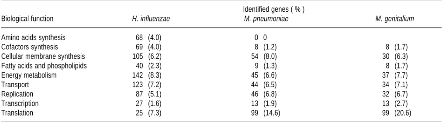
Although Mycoplasmas posses a small number of
genes compared with prokaryotes they are free living
mi-croorganisms. It is probably that mycoplasmas evolved
from gram positive bacteria and during evolution they
lost the ability to synthesize cell wall,
35but they kept the
ability to synthesize other cell structures. When we
com-pare Haemophilus influenzae, M. pneumoniae and M.
genitalium we observed that Mycoplasmas have a less
percentage of genes involved in the synthesis of essential
amino acids, they also show deficiency in the genes that
codify the energetic metabolism but genes involved in the
replication, transcription and translation are in greater
percentage than in H. influenzae (Table 2). There is
in-traspecific variation in the genomic size in Spiroplasma
that is promoted by the insertion of viral sequences that is
the case of Spiroplasma citri in which the viral sequence
is 150 Kb.
2Mollicutes have a small and non uniform G + C content.
M. pneumoniae genes that codify for P1 and OERG adhesins
show a great G + C content (56 % mol) while in other
repli-cations extremes there is a small G + C content 26 %
com-pared with the average of 40 % of G + C (3, 16). This
varia-tion is important from the phylogenetical point of view
be-cause it means that genes are conserved for the synthesis of
RNAr and RNAt and the possibility of an exogen origin of
the genes that codify the synthesis of adhesins.
17Spiroplasmas and acholeplasmas are frequently infected
by virus (phages) while Mycoplasmas are infrequently
in-fected.
48One of these lisogenic virus that infect
mycoplas-mas is MAV1, it infect M. arthritidis enhancing its ability
to induce arthritis.
43The genome of these viruses ranges
from 4 to 40 Kb.
7The presence of plasmids has only been reported in
Spiroplasma citri and M. mycoides but the biological role
have not been determined.
20,21The decrease in the genetic information of
Mycoplas-mas is probably the result of their parasitic way of life,
al-though they have developed special components that attack
host cells and let them survive.
ECOLOGY AND HABITATS
There is an increasing list of host for Mollicutes because
they are widely distributed in nature, they infect humans,
mammals, birds, fishes, insects and plants.
New molecular techniques have led to an easier
identifica-tion and classificaidentifica-tion of new species and strains (Table 1).
Table 1. Representative properties of the Mollicutes.
Family Genome G + C Content Requirement of
(Species number) size (kpb) (mol %) cholesterol Host
Mycoplasmataceae 577-1350 23-40 + Human,
(108) animals
Spiroplasmataceae 780-2220 24-31 + Plants,
(50) insects
Acholeplasmataceae 1500-1650 26-36 – Animals,
(13) plants,
insects
Anaeroplasmataceae 1500-1600 29-34 + Ovine and
(5) bovine rumen
edigraphic.com
Some studies support the presence of structures similar
to Mycoplasmas in animal tissues including humans.
Wirostko´s describe the presence of fastidious
microorgan-isms that produce uveitis and probably other human
diseas-es, they have been identified so Mollicutes.
19The first Mycoplasmas isolated from Bartholin glands of
humans was Mycoplasma hominis, other species have been
isolated from humans being Mycoplasma penetrans the
lat-est to be described
26, encouraging microbiologist to search.
Mycoplasmas show tissue and host specificity as a result
of their nutritional needs and parasitic way of live. Although
this host specificity some human Mycoplasmas may induce
experimental infections in animals, that is the case of M.
pneumoniae that is able to induce experimental tract
infec-tions in hamsters and a natural pneumonia in humans.
9The natural habitats of Mycoplasmas are the respiratory
and urogenital tracts, eyes, mammary glands and joints.
Some Mycoplasmas enter the body using the respiratory or
urogenital tract and then they reach other tissues that is the
case of M. pneumoniae and M. genitalium that have been
isolated from urogenital and respiratory tract.
10,16The increasing number of patients with
immunodefi-ciencies associated with hipogammaglobulinemia like
AIDS and the treatment of patients with
immunosuppres-sive drugs have favored the isolation of Mycoplasmas.
Some Mycoplasmas and Ureaplasmas are considered
normal flora of the urogenital tract but recently they have
been isolated from blood of patients with AIDS or
immun-osupressed patients who are more susceptible to suffer
uro-genital infections by Mycoplasma hominis and
Ureaplas-ma urealyticum. The bacteria Ureaplas-may invade the respiratory
tract and joints producing damage.
12,28The development of new techniques of molecular
biolo-gy have improved the diagnostic of Mycoplasma infections
because these microorganisms are fastidious and their
identification can only be performed by PCR amplifying
genes of the 16S subunit of RNAr.
24,36Spiroplasmas are motile and show helicoidal morphology,
they are frequently isolated from intestine, salivary glands,
homocele of insects and the surface of plants and flowers.
46,13Spiroplasma melliferum and Spiroplasma apis are pathogenic
for bees, they cross the intestine barrier reaching hemolinfa
where they reproduce and induce death of their host.
4Spiro-plasma have also been reported in Aedes aegypti mosquito
re-ducing the fertility of the insects, opening the possibility of
their use as biological control of mosquito.
Several neotropical species of fruit flies (Drosophila)
and cockroaches are susceptible to the infection by
Spiro-palsma poulsonii and Spiroplasma floricola respectively.
Spiroplasma may establish a mutualism relationships.
27,47Spiroplasma mirum was isolated from rabbit ticks and is
considered pathogenic to chicken embryos, new born
ro-dents and adult rabbits. SMCA strain induces a high
inci-dence of cataracts in new born rodents, while GT-48 strain
induces fatal encephalitis.
39,40Acholeplasmas have been isolated from feces of healthy
horses and feces of pathogen free rabbits.
1, 15Acholeplas-mas have also been reported in hemolinfa of fire flies,
be-ing the first report of a Spiroplasma in the homocele of
in-sect.
41Anaeroplasmas have only been isolated from bovine
and ovine rumen.
32CONCLUSSION
Mycoplasmas are bacteria with special genetic features
that make them different of other prokaryotes, these
char-acteristics let them interact closely with their host
produc-ing plant, insect, animal and human infections.
REFERENCIAS
1. Angulo, A.F.; M. Doeksen; A. Hill & A.A. Polak-Vogelzang. 1987. Isolation of Acholeplasmatales from rabbit feces. Lab. Anim. 21: 201-204.
Table 2. Comparison of the genetic participation in the biological processes in prokaryotes.
Identified genes ( % )
Biological function H. influenzae M. pneumoniae M. genitalium
Amino acids synthesis 68 (4.0) 0 0
Cofactors synthesis 69 (4.0) 8 (1.2) 8 (1.7)
Cellular membrane synthesis 105 (6.2) 54 (8.0) 30 (6.3)
Fatty acids and phospholipids 40 (2.3) 9 (1.3) 8 (1.7)
Energy metabolism 142 (8.3) 45 (6.6) 37 (7.7)
Transport 123 (7.2) 44 (6.5) 34 (7.1)
Replication 87 (5.1) 46 (6.8) 32 (6.7)
Transcription 27 (1.6) 13 (1.9) 13 (2.7)
Translation 25 (7.3) 99 (14.6) 99 (20.6)
Rivera-Tapia et al. Some biological features of Mollicutes
Rev Latinoam Microbiol 2002; 44 (2): 53-57
56
2. Bebear, C.M.; P. Aullo; J.M. Bove & J. Renaudin. 1996.
Spiroplas-ma citri virus SpV1: characterization of viral sequence present in
the spiroplasmal host chromosome. Curr. Microbiol. 32: 134-140. 3. Bove, J.M. 1993. Molecular features of Mollicutes. Clin. Infect.
Dis. 17(Suppl. 1):S10-S31.
4. Bove, J.M. 1997. Spiroplasmas: Infectious agents of plants, arthro-pods and vertebrates. Wien. Klin. Wochenschr. 109: 604-612. 5. Brenner, C.; O. Neyrolles & A. Blanchard. 1996. Mycoplasmas and
HIV infection: from epidemiology to their interaction with immune cells. Frontiers Biosc. 54: 42-54.
6. Cole, B.C. 1996. Mycoplasma interactions with the immune sys-tem: implications for disease pathology. ASM News 62: 471-475. 7. Dybvig, K. & L.L. Voelker. 1996. Molecular biology of
mycoplas-mas. Annu. Rev. Microbiol. 50: 25-57.
8. Fleiscmann, R.D.; M.D. Adams; O. White; R.A. Clayton; E.F. Kirk-ness & A.R. Kerlavage. 1995. Whole-genome random sequencing and assembly of Haemophilus influenzae. Rd. Science 269: 496-512.
9. Franzoso, G.; P.C. Hu; G. Meloni & M.F. Barile. 1994. Immunob-lot analysis of chimpanzee sera after infection and after immuniza-tion and challenge with Mycoplasma pneumoniae. Infect. Immun. 62: 1008-1014.
10. Fraser, C.M.; J.D. Gocayne; O. White; M.D. Adams; R.A. Clayton; R.D. Fleischmann; C.J. Bult; A.R. Kerlavage; G. Sutton; J.M. Kelley; J.L. Fritchman; J.F. Weidman; K.V. Small; M. Sandusky; J. Fuhrmann; D. Nguyen; T.R. Utterback; D.M. Saudek; C.A. Phil-lips; J.M Merrick; J.F. Tomb; B.A. Dougherty; K.F. Bott; P.C. Hu; T.S. Lucier; S.N. Petterson; H.O. Smith; C.A. Hutchinson III & J.C. Venter. 1995. The minimal gene complement of Mycoplasma
geni-talium. Science 270: 397-403.
11. Garnier, M.; X. Foissac; P. Gaurivaud; F. Laigret; J. Renaudin; C. Saillard & J.M. Bove. 2001. Mycoplasmas, plants, insects vectors: a matrimonial triangle. Acad. Sci. 324:923-928.
12. Gass, R.; J. Fisher; D. Badesch; M. Zamora; A. Weinberg; H. Mel-sness; F. Grover; J.G. Tully & F.C. Fang. 1996. Donor-to-host transmission of Mycoplasma hominis in lung allograft recipients. Clin. Infect. Dis. 22:567-568.
13. Gaurivaud, P.; J.L. Danet; F. Laigret; M. Garnier & J.M. Bove. 2000. Fructose utilization and phytopathogenicity of Spiroplasma
citri. Mol. Plant. Microbe. Interact. 13:1145-1155.
14. Goulet, M.; R. Dular; J.G. Tully; G. Billowers & S. Kasatiya. 1995. Isolation of Mycoplasma pneumoniae from the human urogenital tract. J. Clin. Microbiol. 33:2823-2825.
15. Heitmann, J.; H. Kirchhoff; C. Chercheletzi; E Jonas & E. Deegen. 1982. Isolation of Acholeplasmas from horse feces. Vet. Microbiol. 7:273-276.
16. Himmelreich, R.; H. Hilbert; H. Plagens; E. Pirkl; B.C. Li & R. Herrman. 1996. Complete sequence analysis of the genome of the bacteria Mycoplasma pneumoniae. Nucleic. Acids Res. 24:4420-4449.
17. Himmelreich, R.; H. Plagens; H. Hilbert; B. Reiner & R. Herrmann. 1997. Comparative analysis of the genomes of the bacteria
Myco-plasma pneumoniae and MycoMyco-plasma genitalium. Nucleic Acids
Res. 25:701-712.
18. Jacob, C.; F. Nouzieres; S. Duret; J.M. Bove & J. Renaudin. 1997. Isolation, characterization, and complementation of a motility mu-tant of Spiroplasma citri. J. Bacteriol. 179:4802-4810.
19. Johnson, L.; E. Wirostko; W. Wirostko & B. Wirostko. 1996.
My-coplasma-like organisms in Hodgkin disease. Lancet 347:901-902.
20. King, K.W. & K. Dibvig. 1994. Mycoplasmal cloning vectors de-rived from plasmid pKMK1. Plasmid 31:49-59.
21. King, K.W. & K. Dibvig. 1994. Transformation of Mycoplasma
capricolum and examination of DNA restriction modification in M. capricolum and M. mycoides subsp. mycoides. Plasmid 31:308-311.
22. Kirchhoff, H. 1992. Motility, p. 289-306. In: J. Maniloff; R.N.
McElhaney; L.R. Finch & J.B. Baseman (ed.). Mycoplasma: molec-ular biology and pathogenesis. American Society for Microbiology, Washington, D.C.
23. Kita, M.; Y. Ohmoto; N. Yamaguchi & J. Imanishi. 1992. Induction of cytotoxic activity in human peripheral blood mononuclear cells by Mycoplasmas. Microbiol. Immunol. 36:507-516.
24. La Scola, B.; G. Michel & D. Raoult. 1997. Use of amplification and sequencing of the 16S rRNA gene to diagnose Mycoplasma
pneumoniae osteomyelitis in a patients with
hipogammaglobuline-mia. Clin. Infect. Dis. 24:1161-1163.
25. Li, Y.H.; Z.Q. Yan; J.S. Jensen; K. Tullus & A. Brauner. 2000. Ac-tivation of nuclear kB and induction of inductible nitric oxide syn-thase by Ureaplasma urealyticum in macrophages. Infect. Immun. 68:7087-7093.
26. Lo, S.C.; M. Hayes; R.Y.H. Wang; P.F. Pierce; H. Kotani & W.K. Shih. 1991. Newly discovered Mycoplasmas isolated from patients infected with HIV. Lancet 338:1415-1418.
27.McGarrity, G.J.; D.L. Williamson. 1989. Spiroplasma pathogenicity
in vivo and in vitro, p. 365-392. In: R.F. Whitcomb & J.G. Tully
(Ed.). The Mycoplasmas, vol 5. Academic Press, New York. 28. Meyer, R.D. & W. Clough. 1993. Extragenital Mycoplasma
homin-is infections in adults: emphashomin-is on immunosuppression. Clin.
In-fect. Dis. 17(Suppl. 1):S243-S249.
29. Proft, T.; H. Hilbert; G. Layh-Schmitt & R. Hermann. 1995. The proline-rich P65 protein of Mycoplasma pneumoniae is a compo-nent of the Triton X-100-insoluble fraction and exhibits size poly-morphism in the strain M129 and FH. J. Bacteriol. 177: 3370-3378. 30. Proft, T.; H. Hilbert; H. Plagens & R. Hermann. 1996. The P200 protein of Mycoplasma pneumoniae shows common features with the cytadherence-associated protein HMW1 and HMW·. Gene 171:79-82.
31. Razin, S. 1978. The Mycoplasmas. Microbiol. Rev. 42:414-470. 32. Razin, S. 1991. The genera Mycoplasma, Ureaplasma,
Acholeplas-ma, AnaeroplasAcholeplas-ma, and AsteroplasAcholeplas-ma, p. 1937-1959. In: A. Balow;
H.G. Truper; M. Dworkin; W. Harder & K.-H. Schleifer (ed.). The prokaryotes, vol. 2, 2nd ed. Springer-Verlag, New York, N.Y.
33. Razin, S. 1992. Mycoplasma taxonomy and ecology, p. 3-22. In: J. Maniloff; R.N. McElhaney; L.R. Finch & J.B. Baseman (ed.).
My-coplasmas: molecular biology and pathogenesis. American Society
for Microbiology, Washington, D.C.
34. Razin, S. 1994. DNA probes and PCR in diagnostic of
Mycoplas-mas infections. Mol. Cell. Probes 8:497-511.
35. Razin, S. 1997. Comparative genomics of Mycoplasmas. Wien. Klin. Wochenschr. 109:551-556.
36. Razin, S.; D. Yogev & Y. Naot. 1998. Molecular biology and pathogenicity of Mycoplasmas. Microbiol. Mol. Biol. Rev. 62: 1094-1156.
37. Theiss, P.; A. Karpas & K. Wise. 1996. Antigenic topology of the P29 surface lipoprotein of Mycoplasma fermentans: differential dis-play of epitopes results in high-frequency phase variation. Infect. Immun. 64:1800-1809.
38. Trachtenberg, S. & R. Gilad. 2001. A bacterial linear motor: cellu-lar and molecucellu-lar organization of the contractile cytoskeleton of the helical bacterium Spiroplasma melliferum BC3. Mol. Microbi-ol. 41:827-848.
39. Tully, J.G.; R.F. Whitcomb; H.F. Clark & D.L. Williamson. 1977. Pathogenic spiroplasmas: cultivation and vertebrate pathogenicity of a new spiroplasma. Science 195:892-894.
40. Tully, J.G.; R.F. Whitcomb; D.L. Rose; J.M. BOVE. 1982.
Spiro-plasma mirum, a new species from the rabbit tick (Haemaphysalis leporispalustris). Int. J. Syst. Bacteriol. 32:92-100.
edigraphic.com
42. Vazeille-Falcoz, M.; A.M. Perchec-Merien & F. Rodhain. 1994. Ex-perimental infection of Aedes aegypti mosquitoes, suckling mice, and rats with four mosquito Spiroplasma. J. Invertebr. Pathol. 63:37-42. 43. Voelker, L.L.; K.E. Weaver; L.J. Ehle & L.R. Washburn. 1995.
As-sociation of lysogenic bacteriophage MAV1 with virulence of
My-coplasma arthritidis. Infect. Immun. 63:4016-4023.
44. Weisburg, W.G.; J.G. Tully; D.L. Rose; J.P. Petzel; H. Oyaizu; D. Yang; L. Mandelco; J. Sechrest; T.G. Lawrence & J. Van Teten. 1989. A phylogenetic analysis of the Mycoplasmas: basis for their classification. J. Bacteriol. 171:6455-6467.
45. Williamson, D.L.; J. Renaudin & J.M. Bove. 1991. Nucleotide se-quence of the Spiroplasma citri fibril protein gene. J. Bacteriol. 173:4353-4362.
46. Wiiliamson, D.L.; R.F. Whitcomb; J.G. Tully; G.E. Gasparich; D.L. Rose; P. Carle; J.M. Bove; K.J. Hackett; J.R. Adams; R.B. Henegar; M. Konai; C. Chastel & F.E French. 1998. Revised group classifica-tion of the genus Spiroplasma. Int. J. Syst. Bacteriol. 48:1-12. 47. Williamson, D.L.; B. Sakaguchi; K.J. Hackett; R.F. Whitcomb; J.G.
Tully; P. Carle; J.M. Bove; J.R. Adams; M. Konai & R.B. Henegar.
1999. Spiroplasma poulsonii sp. Nov., a new species associated with male-lethality in Drosophila willstoni, a neotropical species of fruit fly. Int. J. Syst. Bacteriol. 49:2611-2618.
48. Zou, N.; K. Park & K. Dybvig. 1995. Mycoplasma virus P1 has a linear, double-stranded DNA genome with inverted terminal re-peats. Plasmid 33:41-49.
Correspondence to:
José Antonio Rivera-Tapia,
Universidad Autónoma de Puebla. Edificio 76, Complejo de Ciencias, Ciudad Universitaria. C.P. 72570, Puebla, Pue., México.
Tel. 2 33 20 10 ext. 21, Fax. 2 33 20 10 ext. 25.