In vitro
aging promotes endoplasmic reticulum (ER)-mitochondria Ca
2 +
cross talk and loss of store-operated Ca
2 +
entry (SOCE) in rat
hippocampal neurons
María Calvo-Rodríguez
a,1, Mónica García-Durillo
a, Carlos Villalobos
a,⁎
, Lucía Núñez
a,b aInstituto de Biología y Genética Molecular (IBGM), Consejo Superior de Investigaciones Científicas (CSIC), Valladolid, Spainb
Departamento de Bioquímica y Biología Molecular y Fisiología, Universidad de Valladolid, Valladolid, Spain
a b s t r a c t
a r t i c l e i n f o
Article history:
Received 23 February 2016 Received in revised form 27 July 2016 Accepted 4 August 2016
Available online 05 August 2016
Aging is associated to cognitive decline and susceptibility to neuron death, two processes related recently to sub-cellular Ca2+homeostasis. Memory storage relies on mushroom spines stability that depends on store-operated Ca2+entry (SOCE). In addition, Ca2+transfer from endoplasmic reticulum (ER) to mitochondria sustains energy production but mitochondrial Ca2+overload promotes apoptosis. We have addressed whether SOCE and ER-mitochondria Ca2+transfer are influenced by culture time in long-term cultures of rat hippocampal neurons, a model of neuronal aging. We found that short-term cultured neurons show large SOCE, low Ca2+store content and no functional coupling between ER and mitochondria. In contrast, in long-term cultures reflecting aging neu-rons, SOCE is essentially lost, Stim1 and Orai1 are downregulated, Ca2+stores become overloaded, Ca2+release is enhanced, expression of the mitochondrial Ca2+uniporter (MCU) increases and most Ca2+released from the ER is transferred to mitochondria. These results suggest that neuronal aging is associated to increased ER-mitochondrial cross talking and loss of SOCE. This subcellular Ca2+remodeling might contribute to cognitive de-cline and susceptibility to neuron cell death in the elderly.
© 2016 Published by Elsevier B.V. Keywords:
Aging
Hippocampal neurons Store-operated calcium entry ER-mitochondria cross talking Stim1
Orai1
1. Introduction
Aging is associated to cognitive decline and increased risk of neuron cell death related to excitotoxicity and/or neurodegenerative diseases. These processes have been linked to the remodeling of intracellular Ca2+homeostasis including changes in voltage-gated Ca2+channels and glutamate receptors sensitive toN-Methyl-D-Aspartate (NMDA). Interestingly, some of these changes are mimickedin vitroafter long-term culture of rat hippocampal neurons[1,2]. In fact, after several weeks in culture, rat hippocampal neurons display important hallmarks of neuronal agingin vivoincluding accumulation of reactive oxygen spe-cies (ROS), lipofuscin granules, heterochromatic foci, activation of the Jun N-terminal protein kinase (pJNK) and p53/p21 pathways, gradual loss of cholesterol, and, as stated above, changes in Ca2+channel densi-ty and NMDA receptor expression[1–5]. Accordingly, long-term cul-tures of hippocampal neurons may provide a suitable model for investigating Ca2+remodeling in aging hippocampal neurons.
In the last few years, it has been reported that memory storage in the hippocampus depends on store-operated Ca2+entry (SOCE) which is
required for mushroom spines stability[6]. SOCE is triggered by the re-lease of Ca2+from intracellular stores induced by phospholipase C acti-vation after receptor stimulation[7]. At the molecular level, SOCE is induced by interaction of stromal interaction molecule 1 (Stim1)[8], a Ca2+sensor at the endoplasmic reticulum (ER), and Orai1, a pore forming protein of store-operated channels (SOCs) at the plasma mem-brane[9,10]. SOCE has been studied in detail in non-excitable cells. However, it is also present in excitable cells including neurons[7,11]. It has been proposed that the Ca2+sensor at ER that triggers SOCE in hippocampal neurons is Stim2 rather than Stim1[12]. However, this view is controversial and recent results indicate that both Stim proteins are involved in Ca2+homeostasis in neurons. STIM1 mainly activates SOCE, whereas Stim2 regulates resting Ca2+levels in the ER and Ca2+ leakage with the additional involvement of Stim1[13]. Interestingly, re-cent data suggest that Stim2 is downregulated in aging animals, animal models of AD and AD patients[6]suggesting an important role of Stim2 and SOCE in aging and AD.
Subcellular Ca2 +homeostasis is critical for neuron cell damage induced by different insults including excitotoxicity and neurode-generation. For example, activation of NMDA receptors promotes mitochondrial Ca2 +overload and cell death in long-term cultured hippocampal neurons[4,5]. Likewise, oligomers of the amyloidβ peptide (Aβ), the most likely neurotoxin in AD, promote Ca2+ entry, mitochondrial Ca2+ overload and neuron cell death in ⁎ Corresponding author.
E-mail address:carlosv@ibgm.uva.es(C. Villalobos).
1
Present address: Alzheimer Research Unit, Department of Neurology, Massachusetts General Hospital and Harvard Medical School, 114, 16th St. Charlestown, MA 02129, USA.
http://dx.doi.org/10.1016/j.bbamcr.2016.08.001
0167-4889/© 2016 Published by Elsevier B.V.
Contents lists available atScienceDirect
Biochimica et Biophysica Acta
cerebellar granule cells[14]. Both Aβand NMDA promote Ca2+influx but they also mobilize Ca2+from stores. Specifically, Aβmobilizes ER Ca2+viaIP
3dependent and independent mechanisms[15]. Aβand NMDA receptor activation cause mitochondrial dysfunction involving ER Ca2+release[16]. Thesefindings suggest a relevant role for Ca2+ transfer from ER to mitochondria in neuron cell death. The role of Ca2+transfer from ER to mitochondria in aging has not been addressed. In fact, mitochondria Ca2+handling has remained elusive due to the constrains of monitoring accurately mitochondrial Ca2+concentration ([Ca2+]
mit) in individual neurons. However, bioluminescence imaging of targeted probes like aequorin has revealed the presence of subpopu-lations of mitochondria able to sense high Ca2+domains (Ca2+ micro-domains) near Ca2+channels at the plasma membrane[17,18]and the ER[19,20]. More recent data indicates that Ca2 +transfer from ER to mitochondria depends on the formation of close contacts sites between ER and mitochondria[21]and specialized structures named mitochondria-associated membranes (MAMs)[22,23]. Ca2+ transfer from the ER to mitochondria is involved in cell survival as well. Absence of this transfer results in enhanced phosphorylation of pyruvate dehydrogenase and AMP kinase (AMPK) activation, pro-moting prosurvival macroautophagy. Thus, constitutive InsP3R de-pendent Ca2+ release to mitochondria is an essential cellular process required for efficient mitochondrial respiration and mainte-nance of bioenergetics[24]. Whether this transfer is affected by aging has not been addressed. Inasmuch as mitochondrial potential (ΔѰ), the main driving force for mitochondrial Ca2+uptake, de-creases with age[4,25], it is possible that aging may influence Ca2+ transfer from ER to mitochondria. Here we have used long-term cul-tures of rat hippocampal neurons to address whether SOCE and ER– mitochondria Ca2+cross talk are influenced byin vitroaging.
2. Methods
2.1. Animals and reagents
Wistar rat pups were obtained from the Valladolid University an-imal facility. All anan-imals were handled according to ethical standards approved by the animal housing facility of the Valladolid University Medical School (Valladolid, Spain) in agreement with the European Convention 123/Council of Europe and Directive 86/609/EEC. Fura2/AM, TMRM, coelenterazine and lipofectamine® 2000 are from Invitrogen (Barcelona, Spain). Fetal bovine serum (FBS) is from Lonza (Barcelona, Spain). Horse serum, neurobasal medium, HBSS medium, B27,L-glutamine and gentamicin are from Gibco (Barcelona, Spain). Papain solution is from Worthington (Lakewood, NJ). The poly-D-lysine and Annexin V are from BD (Madrid, Spain).
DNase I and antibody against mitochondrial calcium uniporter (MCU) are from Sigma (Madrid, Spain). Antibodies against Stim1 and Orai1 are from Alomone (Jerusalem, Israel). Antibody against βIII tubulin is from Covance (Princeton, NJ, USA). Other reagents and chemicals were obtained either from Sigma or Merck. Plasmids for mitochondria-targeted aequorin fused to GFP are a kind gift from Prof. P. Brulet (CNRS, France).
2.2. Primary hippocampal neuron culture
Hippocampal neurons were prepared from Wistar rat pups under sterile conditions as reported by Brewer et al.[26]with further modifi -cations by Perez-Otaño et al.[27]. Briefly, newborn rat pups were decapitated and, after brain removal, meninges were discarded and hip-pocampi were separated from cortex. Hippocampal tissue was cut in small pieces, transferred to papain solution (20 u/ml) and incubated at 37 °C for 30 min. After 15 min, DNase I (50μg/ml) was added. Tissue pieces were washed with Neurobasal Medium and cell suspension was obtained using afire-polished pipette in the same medium supplement-ed with 10% FBS. Cells were centrifugsupplement-ed at 160gfor 5 min and pellet was
suspended in Neurobasal medium. Hippocampal cells were plated onto poly-D-lysine-coated, 12 mm diameter glass coverslips at 30 × 103cells/ dish (plating density, 169 cells/mm2), and grown in Neurobasal medi-um supplemented withL-glutamine (2 mM), gentamicin (1μg/ml), 2% B27 and 10% FBS, and maintained in a humidified incubator at 37 °C with 5% CO2without further media exchange. Cells were cultured for 4–8 daysin vitro(DIV) for resembling“young”neurons or 15–21 DIV for mimicking“aged”neurons before experiments. Other details have been reported in detail elsewhere[4,28].
2.3. Fluorescence imaging of cytosolic free Ca2+concentration
Hippocampal cells were cultured for 4–8 DIV or 15–21 DIV and washed in standard external medium (SEM) containing (in mM) NaCl 145, KCl 5, CaCl21, MgCl21, glucose 10 and Hepes/NaOH 10 (pH 7.4). Cells were incubated in the SEM containing fura2/AM (4μM) for 60 min at room temperature in the dark. Then coverslips were placed on the perfusion chamber of a Zeiss Axiovert 100 TV inverted microscope, perfused continuously with pre-warmed (37 °C) SEM and epi-illuminated alternately at 340 and 380 nm light using afilter wheel. Light emitted at 520 nm wasfiltered with the dichroic mirror and recorded every 5 s with a Hamamatsu ER camera (Hamamatsu Photonics France). Pixel by pixel ratios of con-secutive frames were captured and [Ca2+]
cytvalues from regions of interest (ROIs) corresponding to individual neurons were averaged and expressed as the ratio offluorescence emission following excita-tion at 340 and 380 nm as reported in detail previously[4,14]. For calculations of cytosolic [Ca2+] the Grynkiewicz equation was used: [Ca2+] = Kd
∗(R−Rmin) / (Rmax−R)∗F380max/F380min[29] where Kd is the dissociation constant of fura2 (224 nM), R is the ob-servedfluorescence ratio at both wavelengths (F340/F380); Rmin is the minimum ratio value (in absence of Ca2+); Rmax is the maxi-mum ratio value (when Fura-2 is saturated by Ca2+) and F380max/ F380min is a scaling factor (fluorescence intensity at 380 nm excita-tion in the absence of Ca2+and at Ca2+saturation). We used the fol-lowing values for our imaging setup: Rmax = 1.4; Rmin = 0.1, respectively. F380max/F380min = 2.7. For best comparison both ra-tios and calculated cytosolic [Ca2 +] are shown.
For measurements of SOCE, fura2-loaded cells were treated with the sarcoplasmic and endoplasmic reticulum Ca2+ATPase (SERCA) pump blocker thapsigargin (Tg, 1μM) for 10 min in SEM devoid of extracellu-lar Ca2+before imaging. Then cells were subjected tofluorescence im-aging and stimulated with 5 mM Ca2 + to monitor the SOCE-dependent rise in [Ca2+]
cyt. SOCE recordings were made in the presence of tetrodotoxin (TTX) to prevent activation of voltage-gated Ca2+ chan-nels by connected neurons. For estimation of Ca2+store content, we measured the rise in [Ca2+]
cytinduced by low concentrations of the Ca2+ionophore ionomycin (400 nM) added in the absence of extracel-lular Ca2+. Ionomycin releases Ca2+ stored by making holes in endomembranes. Accordingly, the rise in [Ca2+]
cytdepends solely on the amount of stored Ca2+and not on expression of G-protein coupled receptors and or IP3receptors. This procedure has been used extensively to estimate Ca2+store content[11].
For quantification of rises in [Ca2+]
cyt, the maximum rise in ratio was computed for responsive cells. In addition, we calculated the fraction of responsive cells. Finally, we calculated the product of the maximum rise in ratio by the fraction of responsive cells in order to have an estimation of the increase in [Ca2+] in response to a given agonist in the whole cell population. This procedure has been described in the same cells in detail previously[4].
2.4. Bioluminescence imaging of mitochondrial free Ca2+concentration
and a GFP sequence to select transfected neurons for bioluminescence imaging. Transfection efficiency was 10–20%. After 24 h, cells were washed with SEM and incubated for 2 h with 4μM coelenterazine at room temperature in the dark. Then, cells were washed again and placed into a perfusion chamber coupled to Zeiss Axiovert S100 TV inverted microscope. Cells were perfused at 5–10 ml/min with pre-warmed (37 °C) test solutions made in SEM. At the end of each experi-ment, cells were permeabilised with 0.1 mM digitonin solved in SEM containing 10 mM CaCl2in order to release all the residual photonic emissions, a value that is required for calibration. For permeabilisation, cells were treated with a digitonin 50μM solved in intracellular stan-dard medium (ISM) (130 mM KCl, 10 mM NaCl, 1 mM MgCl2, 1 mM K3PO4, 0.2 mM EGTA, 1 mM ATP, 20μM ADP, 2 mM succinate, and 20 mM HEPES, pH 6.8). Then, cells were incubated with ISM containing EGTA-buffered, low [Ca2+] (200 nM) for 5 min before perfusing with ISM containing 10μM [Ca2 +] for 1 min. This medium is intended to test mitochondrial Ca2 +uptake in the presence of a given rise in [Ca2+]
cytlarge enough to promote mitochondrial Ca2+uptake. Biolumi-nescence images were taken with a Hamamatsu VIM photon counting camera handled with an Argus-20 image processor. For analysis, regions of interest (ROIs) corresponding to individual neurons selected for their morphology and photonic emissions were integrated for 10 s periods and converted into values of mitochondria free Ca2+concentration ([Ca2+]
mit) as reported previously[17,18,28,30].
2.5. Mitochondrial potential
Hippocampal neurons were washed in SEM and loaded with the mi-tochondrial potential probe tetramethylrhodamine, methyl ester (TMRM, 10 nM) for 30 min at room temperature in the dark. Then cov-erslips containing cells were placed on a Zeiss Axiovert 100 TV inverted microscope and subjected tofluorescence imaging. Fluorescence images were captured with the rhodaminefilter set with a Hamamatsu ER-Orca
fluorescence camera as reported previously[4,14,31].
2.6. Immunofluorescence
Hippocampal cells in culture werefixed withp-formaldehyde 4% and incubated with antibodies against MCU (1:200), Stim1 (1:50), Orai1 (1:50) andβIII tubulin (1:300) at 4 °C overnight. Immuno-positive cells were revealed using Alexafluor 488-tagged antibodies (1:300). Size of neurons in short-term and long-term cultured was different. Accordingly,fluorescence emission per area unit (optical density) was measured in selected ROIs corresponding to individual neurons using ImageJ software. Further details have been reported elsewhere[4].
2.7. Statistics
Changes influorescence ratio are expressed as A.U.C. (area under curve) and maximum increase in ratio (Δratio). Calculations were performed using Origin Lab 7.0. Data are presented as mean ± SEM. When only two means were compared, Student'st-test was used. For more than two groups, statistical significance of the data was assessed by one-way ANOVA and compared using Bonferroni's multiple comparison tests. Differences were considered significant at pb0.05.
3. Results
3.1. In vitro aging downregulates store-operated Ca2+entry in rat hippo-campal neurons
Store-operated Ca2+entry (SOCE), the Ca2+entry pathway activat-ed by the emptying of intracellular Ca2+stores, was tested in 4–8 DIV and 15–21 DIV hippocampal neurons usingfluorescence imaging. For
this end, fura2-loaded hippocampal cells were treated with the SERCA pump blocker thapsigargin (Tg) in Ca2+free medium to deplete intra-cellular Ca2+stores. Then cells were subjected to
fluorescence imag-ing and the effects of extracellular Ca2+addition on [Ca2+]
cytwere monitored. Imaging experiments were carried out in the presence of tetrodotoxin (TTX) to prevent activation of the neural network formedin vitroand/or to Ca2+entry secondary to activation of ligand and/or voltage-activated Ca2+channels. Neurons were selected ac-cording to morphologic characteristics.Fig. 1shows that addition of external Ca2+to cells not treated with Tg induces an small rise in [Ca2+]
cytthat is similar in 4–8 DIV and 15–21 DIV neurons and not sensitive to La3+. However, after treatment with Tg, addition of external Ca2+induces a very large rise in [Ca2 +]
cytin 4–8 DIV neu-rons that is reversed largely by the classic SOCE antagonist La3 +, thus reflecting SOCE activation. In contrast, Ca2+addition to 15– 21 DIV neurons treated with Tg induced a much smaller rise than in 4–8 DIV neurons that was also partially sensitive to La3+but only slightly larger than the rise recorded in neurons not treated with Tg (Fig. 1A, B).Fig. 1C shows the quantification of SOCE in 4– 8 DIV and 15–21 DIV neurons after removal of the rise in [Ca2+]
cyt in cells not treated with Tg. In other wordsFig. 1C shows the rise in [Ca2+]cytelicited by empting of intracellular Ca2 +stores and sensi-tive to La3+. The results indicate that SOCE is much larger in 4– 8 DIV neurons than in 15–21 DIV neurons suggesting thatin vitro aging is associated to downregulation of SOCE in rat hippocampal neurons.
3.2. In vitro aging is associated to downregulation of Stim1 and Orai1
Orai1 and Stim1 are the most important molecular players involved in SOCE. We have investigated whether these molecular players are expressed in short-term and long-term cultures of rat hippocampal neurons.Fig. 2A shows immunofluorescence images of rat hippocampal neurons cultured for several DIV corresponding to Orai1 and Stim1 ex-pression. Data suggest that expression of both Orai1 and Stim1 de-creases with culture time. Quantitative analysis of optical density of immunofluorescence images indicates that expression of both molecu-lar players decreases significantly in 15–21 DIV cultures relative to 4– 8 DIV cultures (Fig. 2A, B). However, immunostaining ofβIII tubulin, a neuronal marker whose expression does not change with aging, is sim-ilar in young and aged neurons (Fig. 2C).
3.3. In vitro aging increases Ca2+store content and caffeine-induced Ca2+
release in rat hippocampal neurons
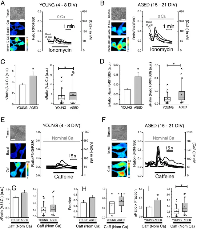
Ca2 +store content was tested in rat hippocampal neurons by monitoring the rise in [Ca2 +]
cytinduced by the Ca2 +ionophore ionomycin added in medium devoid of external Ca2 +. We found that ionomycin-induced rises in [Ca2 +]
cytin hippocampal neurons were small in 4–8 DIV cultures (Fig. 3A) and increased significantly in 15–21 DIV cultures (Fig. 3B). In addition, resting levels of [Ca2 +]
cytrecorded before removal of extracellular Ca2 +were also lower in 4–8 DIV neurons than in 15–21 DIV neurons (Fig. 3A, B). For comparisons, we have calculated the area under the curves
(Fig. 3C) as well as the maximum rise in [Ca2 +]
cytfrom the basal line (Fig. 3D). Results show that both parameters reflecting Ca2 + stores content are significantly larger in 15–21 DIV neurons than in 4–8 DIV neurons, thus suggesting thatin vitroaging in rat hippocam-pal neurons is associated to enhanced resting [Ca2 +]
cytas previously reported[4]and increased Ca2 +stores content.
Ca2+release activated by ryanodine receptors was tested next in short and long-term cultured hippocampal neurons. For this end, the ef-fects of caffeine on [Ca2 +]
permissive for Ca2+-induced Ca2+release. We found that caffeine in-creased [Ca2+]
cytin 4–8 DIV and 15–21 DIV neurons (Fig. 3E, F). In ad-dition, Ca2+responses to caffeine were seemingly larger in 15–21 DIV neurons than in 4–8 DIV cultures. Ca2+responses to agonists in primary cultures are always heterogeneous, often with large differences from cell to cell. As reported previously[4], to quantify Ca2 +responses of the whole cell population in cells populations containing a mix of re-sponsive and unrere-sponsive cells we computed the average change in
fluorescence ratio (ΔRatio F340/F380) in the responsive cell population
(Fig. 3G) and the fraction of responsive cells (Fig. 3H). Then we calculate
the product of both parameters, a value that reflects properly the re-sponse of the whole cell population (Fig. 3I). Results show that Ca2+ re-lease induced by caffeine in the whole cell population is significantly larger in 15–21 DIV neurons than in 4–8 DIV neurons. Thus, suggesting thatin vitroaging is associated to enhance caffeine-induced release of Ca2+in rat hippocampal neurons.
3.4. In vitro aging increases Ca2+release induced by ACh rat hippocampal
neurons
ACh is an important agonist for hippocampal neurons. We also in-vestigated effects ofin vitroaging on Ca2+responses induced by acetyl-choline (ACh).Fig. 4shows representative pseudocolor images (Fig. 4A, B) and representative [Ca2+]cytrecordings obtained in 4–8 DIV and 15– 21 DIV neurons stimulated with ACh. Cytosolic Ca2 +responses were larger in 15–21 DIV neurons than in 4–8 DIV cultures (Fig. 4A, B). How-ever, differences were seemingly due to both changes in the size of [Ca2 +]
cytrises in responsive cells (Fig. 4C) and the relative number (fraction) of responsive cells (Fig. 4D). As a whole, Ca2+responses to ACh are larger in 15–21 DIV neurons relative to 4–8 DIV cultures
(Fig. 4E).
Ca2+responses to ACh may be due to both Ca2+release and Ca2+ entry. To investigate the influence ofin vitroaging on Ca2 +release
1 1
0
Transm
Tg+ (5 Ca)
A
B
C
0.00 0.04 0.08 0.12
*
YOUNG AGED
0.15 0.20 0.25 0.30 0.35 0.40
30 s
5 Ca
0 Ca
TTX
La
3+Rat
io F
340/
F
380
Tg (-)Tg (+)
“YOUNG” (4 - 8 DIV)
“AGED” (15 - 21 DIV)
5 Ca
0 Ca
TTX
La
3+Δ
R
a
ti
o
F340/F380
(a
.u
.)
Transm
Tg+ (5 Ca)
0.0 0.1 0.2 0.3
Δ
Ratio F340/F380
(a.u.)
YOUNG AGED
0
30
*
60 80 100 135 180
[Ca2+] in nM
Fig. 1.In vitroaging decreases store-operated Ca2+entry (SOCE) in rat hippocampal neurons. Store-operated Ca2+entry was tested in hippocampal neurons from short-term (4–8 DIV)
and long-term (15–21 DIV) cultures. Cells were loaded with fura2, treated with thapsigargin (Tg, 1μM) in external Ca2+free medium for 15 min and subjected tofluorescence Ca2+
imaging. Cells were perfused with external medium containing extracellular Ca2+
5 mM in the presence of TTX (500 nM). A. Pictures show transmission (Transm) images as well as calcium images (Ratio F340/F380) of the same neurons coded in pseudocolor after addition of Ca2+
5 mM young and aged cultures. Pseudocolor scales for Ratio F340/F380 are shown at right. Bar corresponds to 10μm. B. Representative Ratio F340/F380 recordings (mean ± SEM) from 4 to 5 cells in control cells not treated with Tg (Tg−, black recordings) o treated with Tg (Tg+, red recordings). Cytosolic [Ca2+
] scale is shown at right. Addition of La3+
inhibits SOCE. C. Left bars are average (mean ± SEM) values ofΔRatio F340/F380 reflecting SOCE in young and aged neurons. The rises in [Ca2+
]cytin cells not treated with thapsigargin (black recordings) were subtracted for rises in Tg treated cells to estimate the size of
induced by ACh we tested the effects of this agonist on [Ca2+] cytin the absence of extracellular Ca2+. ACh increased [Ca2+]cytin the absence of extracellular Ca2+indicating release of Ca2+from intracellular stores. This effect was abolished in the presence of atropine, an antagonist of muscarinic receptors, thus indicating that Ach induces Ca2 +release upon activation of muscarinic receptor (data not shown). We found that ACh-induced Ca2+release is larger in 15–21 DIV cultures than in 4–8 DIV cultures as shown inFig. 4F, G. Interestingly, despite that Ca2 +store content is larger in the in vitroaged neurons, rises in [Ca2 +]
cytinduced by Ca2 +release evoked by ACh in responsive cells are similar in 4–8 DIV and 15–21 DIV cultures (Fig. 4H) suggesting in-creased buffering in 15–21 DIV neurons.
The relative number (fraction) of ACh responsive cells was larger in 15–21 DIV neurons than 4–8 DIV neurons (Fig. 4I). Specifically, whereas only 35% of all neurons in short-term cultures are responsive to ACh in
Ca2 +-free medium, almost 80% of all long-term cultured neurons re-leased Ca2+in response to ACh. Due to this difference, the product of theΔCa2+in responsive neurons by the fraction of responsive cells is larger in 15–21 DIV neurons (Fig. 4J). Taken together, these data suggest differences in the way that young and aged neurons handle Ca2 + re-lease induced by physiological agonists. We tested next whether ER mi-tochondria coupling could be involved in the differential handling.
3.5. In vitro aging increases Ca2+transfer from ER to mitochondria
We tested next the effects of preventing mitochondrial Ca2 + up-take using the mitochondrial uncoupler FCCP on the rises in [Ca2 +]
cyt induced by Ca2 +release evoked by ACh in 4–8 DIV and 15–21 DIV cultures (Fig. 5). We found that ACh-induced [Ca2 +]
cytincreases in Ca2 +free medium are dramatically enhanced in 15–21 DIV neurons
A
B
YOUNG
AGED
Transm
STIM 1
YOUNG AGED
*
YOUNG
AGED
YOUNG AGED
0 20 40 60 80 100 120 140
0 5 10 15 20 25 30 35 40
*
Optical Density (a.u.)
Transm
ORAI 1
YOUNG AGED
0 10 20 30 40 50 60
0 5 10 15 20 25 30
*
YOUNG AGED
Optical Density (a.u.)
*
C
YOUNG
AGED
Transm
-Tubulin III
YOUNG AGED
20 30 40 50 60 70 80
YOUNG AGED
Optical Density (a.u.)
0 20 40 60
80 90
β
treated previously with FCCP. However, prevention of mitochondrial Ca2 +uptake with FCCP has only a minor effect on the response to ACh in 4–8 DIV neurons (Fig. 5A–D). In other words, the effects of FCCP on the rise in [Ca2+]
cytinduced by ACh were much larger in 15– 21 DIV neurons than in 4–8 DIV neurons (Fig. 5E). In addition, FCCP has no effect on the relative number (fraction) of cells responsive to A-Ch. These results suggest that Ca2 +released from the ER after IP
3
receptor activation is barely seen by mitochondria in 4–8 DIV neurons. However, in 15–21 DIV neurons, a large fraction of Ca2 +released from the ER is efficiently transferred to surrounding mitochondria.
To support further the above view we have monitored directly the rise in mitochondrial Ca2+concentration ([Ca2+]mit) elicited by Ca2+release induced by ACh in medium devoid of external Ca2+(Fig. 6). For this end, hippocampal neurons were transfected with a mitochondria-targeted
0.0 0.2 0.4 0.6 0.8 R at io F340/ F380
1 min
0 CaYOUNG (4 - 8 DIV)
0.0 0.2 0.4 0.6 0.8 R at io F340/ F380
1 min
0 CaAGED (15 - 21 DIV)
T
ransm
Basal (1 Ca)
Ionom (0 Ca)
A
B
C
D
YOUNG AGED 0.00 0.05 0.10 0.15 0.20 Δ Ratio ( R a tio F 3 4 0 /F 3 8 0 ) (a.u.)*
0 0.8 0 0.8 YOUNG AGED 0 2 4 6*
Basal (1 Ca) Basal (1 Ca) Δ Ratio ( A.U.C.) (a.u.)Ionomycin
Ionomycin
T ransmBasal (1 Ca)
Ionom (0 Ca)
0.0 0.2 0.4 0.6 YOUNG AGED
*
Δ Ratio ( R a tio F 3 4 0 /F 380) (a.u.) 0.0 0.5 1.0 1.5 0.0 0.5 1.0 1.5*
0.0 0.2 0.4 0.6 0.8 1.015 s
R at io F340/ F 38 0Caffeine
Caffeine
0.8 0 Tr a n s m Ba s a l Ca ffYOUNG (4 - 8 DIV)
AGED (15 - 21 DIV)
0.8 0 0.0 0.4 0.8 0.2 0.6 1.0
Nominal Ca
Nominal Ca
Δ Ratio ( A.U.C.) (a.u.) Fraction Δ
Ratio x Fraction
Caff (Nom Ca)
YOUNG AGED YOUNG AGED YOUNG AGED
Caff (Nom Ca) Caff (Nom Ca)
E
F
G
H
15 s
R at io F340/ F 38 0 0.0 0.4 0.8 0.2 0.6 1.0 Tr a n s m Ba s a l Ca ff 0.0 1.0 2.0 3.0 4.0Caff (Nom Ca)
YOUNG AGED 0.0
0.4 0.8 1.2
YOUNG AGED
Caff (Nom Ca)
0.5 1.5 2.5
YOUNG AGED
Caff (Nom Ca)
*
0 60 180 340 680[Ca2+] in nM
0 60 180 340 680
[Ca2+] in nM
[Ca2+] in nM
0 180 680 60 340 1350
[Ca2+] in nM
0 180 680 60 340 1350 0 5 10 15 20
*
Δ Ratio ( A.U.C.) (a.u.) YOUNG AGED 1.0 2.0 0.0I
Fig. 3.In vitroaging increases Ca2+store content and caffeine-induced Ca2+release in rat hippocampal neurons. Hippocampal neurons were loaded with fura2 and subjected to fluorescence imaging for monitoring Ca2+store content and Ca2+release. Cells were perfused with ionomycin 400 nM (Ionom) in external medium devoid of Ca2+(0 Ca). Pictures
show transmission (Transm) and pseudocolor images offluorescence ratios in basal conditions - Basal (1 Ca) - and after ionomycin - Ionom (0 Ca) - in short-term (A, 4–8 DIV) and long-term (B, 15–21 DIV) cultured neurons. Pseudocolor scales shown at right. Bar represents 10μm. Traces are representative recordings offluorescence ratios in individual neurons identified by their morphology. Cytosolic [Ca2+
] scale is shown at right. Bars are average (mean ± SEM) areas under traces (C) or maximum rises in ratios (D) and corresponding box plot data in 4–8 DIV and 15–21 DIV cultures (n= 46, 53 individual cells from 4 independent cultures (*pb0.05). Box plots of the same data are shown Ca2+
aequorin and subjected to bioluminescence imaging for monitoring of [Ca2+]
mitin transfected neurons. Transfection efficiency was similar (15 ± 5%) for both short-term and long-term cultured neurons. Transfected neurons in the microscopicfield were selected by their GFP
fluorescence and subjected to bioluminescence imaging.Fig. 6A shows
representativefluorescence and bioluminescence images of transfected neurons and representative traces of the rises in [Ca2+]
mitinduced by ACh in Ca2+free medium. Results show that both the rises in [Ca2+]mit in-duced by ACh and the relative number of responsive cells (fraction) are significantly larger in 15–21 DIV neurons than in 4–8 DIV neurons
1 0 Tr a n s m Ba s a l AC h (1 C a )
A
0.0 0.4 0.8ACh
1 Ca
ACh
1 Ca
*
ACh (1 Ca)
YOUNG AGED 0 2 4 6 8 10 0.0 0.2 0.4 0.6 0.8 1.0
ACh (1 Ca)
0 2 4 6 8 10
ACh (1 Ca)
YOUNG (4 - 8 DIV)
AGED (15 - 21 DIV)
Rat
io F
340/
F
380
YOUNG AGED YOUNG AGED 0.2
0.6 1.0
Fraction
Δ
Ratio x Fraction
Δ Ratio ( A.U.C.) (a.u.) 1 0
B
C
D
E
15 s
15 s
0.0 0.4 0.8 Rat io F 340/ F 380 0.2 0.6 1.0 Tr a n s m B a sa l AC h (1 C a ) 0 10 20 30ACh (1 Ca)
YOUNG AGED 0.2
0.4 0.6 0.8 1.0 1.2
ACh (1 Ca)
YOUNG AGED 0
10 20 30
ACh (1 Ca)
YOUNG AGED
*
15 s
0 Ca
0 Ca
YOUNG (4 - 8 DIV)
AGED (15 - 21 DIV)
R a ti o F3 4 0 /F3 8 0 0.0 0.4 0.8 0.2 0.6 1.0 Tr a n s m Ba s a l AC h (0 C a ) 1 0 1 0
G
F
15 s
Tr a n s m Ba s a l AC h (0 C a ) Rat io F 340/ F 380 0.0 0.4 0.8 0.2 0.6 1.0 0 1 2 3 0 1 2 3*
0.0 0.2 0.4 0.6 0.8 1.0*
Δ Ratio ( A.U.C.) (a.u.)ACh (0 Ca)
YOUNG AGED
ACh (0 Ca) ACh (0 Ca)
YOUNG AGED YOUNG AGED
Fraction
Δ
Ratio x Fraction
J
I
H
0 2 4 6 8 10ACh (0 Ca)
YOUNG AGED
0.0 0.4 0.8 1.2
ACh (0 Ca)
YOUNG AGED 0
1 2 3 4 5 6 7
ACh (0 Ca)
YOUNG AGED
ACh
ACh
*
*
0 180 675[Ca2+] in nM
60 340 1350
[Ca2+] in nM
[Ca2+] in nM [Ca2+] in nM
0 180 675 60 340 1350 0 180 675 60 340 1350 0 180 675 60 340 1350
Fig. 4.In vitroaging increases Ca2+
responses to acetylcholine in rat hippocampal neurons. Hippocampal neurons were loaded with fura2 and subjected tofluorescence imaging for monitoring the effects of ACh in short-term (A, 4–8 DIV) and long-term (B, 15–21 DIV) cultured neurons. Cells were stimulated with acetylcholine 100μM in Ca2+
-containing medium. Pictures show transmission images (Transm) and pseudocolor images offluorescence ratios taken before (Basal) and after acetylcholine - ACh (1 Ca). Pseudocolor scale shown at right. Bar corresponds to 10μm. Traces are representative recordings offluorescence ratios of individual neurons identified by their morphology in 4–8 DIV and 15–21 DIV cultures. Bars correspond to the average (mean ± SEM) areas under traces in ACh-responsive cells (C), the fraction of responsive cells (D) or the product of both parameters (E) and corresponding box plot data.n= 25 and 22 cells from 3 independent experiments (*pb0.05). Ca2+
release induced by ACh in Ca2+
-free medium was monitored in short-term (F, 4–
(Fig. 6B). Consistently with the FCCP results shown above, biolumines-cence imaging of [Ca2+]
mitindicate that mitochondria from 4 to 8 DIV neurons are essentially blind to Ca2+release induced by ACh. In contrast,
in 15–21 DIV neurons, a fraction of mitochondria become functionally coupled to Ca2+stores consistently with efficient Ca2+transfer from the ER to mitochondria Ca2+.
A
C
E
B
D
Cntrl FCCP 12
0 3 6 9
Cntrl FCCP
0.0 0.2 0.4 0.6 0.8
1.0 CntrlFCCP
0 2 4 6 8
*
, #
#
*
, #
#
*
Δ
Ratio (
A.U.C.)
(a.u.)
Fraction
Δ
Ratio x Fraction
ACh (0 Ca)
YOUNG AGED
ACh (0 Ca)
YOUNG AGED
ACh (0 Ca)
YOUNG AGED
0.0 0.5 1.0 1.5
R
at
io F340/
F380
ACh
0 Ca + FCCP
0.0 0.5 1.0 1.5
R
at
io F340/
F380
ACh
0 Ca
0.0 0.5 1.0 1.5
R
at
io F340/
F380
ACh
0 Ca
15 s
0.0 0.5 1.0 1.5
R
at
io F340/
F
380
ACh
0 Ca + FCCP
15 s
15 s
15 s
T
ransm
Basal
Ach (0 Ca)
Control
0 1.5
YOUNG (4 - 8 DIV)
AGED (15 - 21 DIV)
0 1.5
T
ransm
Basal
Ach (0 Ca)
Control
FCCP
0 1.5
T
ransm
Basal
Ach (0 Ca)
0 1.5
T
ransm
Basal
Ach (0 Ca)
FCCP
0 10 20 30
ACh (0 Ca)
YOUNG AGED
Δ
Ratio (
A.U.C.)
(a.u.)
0.0 0.4 0.8 1.2 1.6
Fraction
ACh (0 Ca)
YOUNG AGED
#
#
Cntrl FCCP
0 5 10 15 20
Δ
Ratio x Fraction
ACh (0 Ca)
YOUNG AGED
#
#
*
Cntrl
FCCP CntrlFCCP
*
*
0 270 1350
[Ca2+] in nM
80 600
[Ca2+] in nM
[Ca2+] in nM
[Ca2+] in nM
0 270 1350
80 600
0 270 1350
80 600
0 270 1350
80 600 4000
Fig. 5.Most Ca2+released from the ER is taken up by mitochondria in 15–21 DIV neurons but not in 4–8 DIV neurons. Hippocampal neurons were loaded with fura2 and subjected to fluorescence imaging for monitoring the effects of the mitochondrial uncoupler FCCP on the rise in [Ca2+]
cytevoked by ACh-induced Ca2+release. Pictures show transmission images
(Transm) and pseudocolor images offluorescence ratios taken before (Basal) and after acetylcholine - ACh (0 Ca) - in 4–8 DIV (A, 4–8 DIV) and 15–21 DIV (B, 15–21 DIV) cultured neurons. Traces are representative recordings offluorescence ratios of individual neurons identified by their morphology in 4–8 DIV and 15–21 DIV cultures in the absence (A,B) or the presence of FCCP (C,D). E. Bars correspond to the average (mean ± SEM) areas under traces in responsive cells (left panel), the fraction of responsive cells (middle panel) or the product of both parameters (right panel) and corresponding box plot data. n = 101, 84, 109 and 34 cells from 4 to 8 independent experiments (*pb0.05vs.control; #pb0.05vs.4–
3.6. The mitochondrial Ca2+uniporter is upregulated in 15–21 DIV rat
hip-pocampal neurons
One of the most important players involved in mitochondrial Ca2+ uptake is the recently cloned mitochondrial Ca2 +uniporter (MCU)
[32,33]. We have tested changes in relative expression of MCU in
hippo-campal neurons along with time in culture using specific immunofl uo-rescence and optical density quantification in 4–8 DIV and 15–21 DIV cultures.Fig. 7A shows immunofluorescence images of rat hippocampal neurons cultured for several DIV corresponding to MCU expression. Data suggest that MCU expression increases with culture time. Quanti-tative analysis of optical density of immunofluorescence images
indicates that expression of MCU in individual neurons is increased sig-nificantly in 15–21 DIV cultures relative to 4–8 DIV cultures (Fig. 7B). Notice that optical density of immunofluorescence images ofβIII tubu-lin, a neural marker whose expression does not change, is similar in 4–8 DIV cultures than in 15–21 DIV cultures (Fig. 2C).
3.7. In vitro aging is associated to mitochondrial depolarization and de-creased mitochondrial Ca2+uptake in permeabilised neurons
Finally, we have also investigated mitochondrial Ca2 +uptake in permeabilised neurons andΔΨ, the driving force for mitochondrial Ca2+uptake in 4–8 DIV and 15–21 DIV hippocampal neurons. For this
A
B
0 2 4 6
0 Ca
ACh
0 Ca
ACh
[Ca
2+
]
mit
(µM)
30 sec 30 sec
YOUNG AGED
0 1 2 3 4 5
ACh 100 µM
0.0 0.2 0.4 0.6 0.8 1.0
*
Fr
ac
tion
YOUNG AGED
ACh 100 µM
1 2 3
*
Δ
Ca
2+
x
Fr
ac
tion
YOUNG AGED
ACh 100 µM
0
GFP Fluorescence
Aequorin Bioluminescence
GFP Fluorescence
Aequorin Bioluminescence
ACh 100 µM ACh 100 µM
0 2 4 6
[Ca
2+
]
mit
(µM)
YOUNG (4 - 8 DIV)
AGED (15 - 21 DIV)
0 2 4 6 8 10
Δ
[Ca
2+
]
mit(µM)
Δ
[Ca
2+
]
mit(µM)
YOUNG AGED
ACh 100 µM
0.0 0.4 0.8 1.2
Fr
ac
tion
YOUNG AGED
ACh 100 µM
*
0 1 2 3 4 5
Δ
Ca
2+
x
Fr
ac
tion
YOUNG AGED
ACh 100 µM
*
Fig. 6.Mitochondrial Ca2+uptake induced by Ca2+release in short-term and long-term cultured hippocampal neurons. A. Hippocampal neurons were transfected with
mitochondria-targeted, GFP aequorin and subjected to bioluminescence imaging for monitoring mitochondrial Ca2+uptake. Pictures showfluorescence (GFPfluorescence) and bioluminescence
(Aequorin Bioluminescence) images of short-term (4–8 DIV) and long-term (15–21 DIV) cultured hippocampal neurons. Photonic emissions representing mitochondrial Ca2+
uptake were obtained during perfusion with ACh 100μM in Ca2+
free medium (0 Ca). Traces are representative recordings of the rises in mitochondrial Ca2+
concentration ([Ca2+
]mit)
induced by Ca2+
release evoked by ACh 100μM in Ca2+
-free medium. B. Bars correspond to the average (mean ± SEM) rises in [Ca2+
]mitin responsive cells (left panel), the fraction
end, transfected neurons expressing a low-affinity, mitochondria-targeted GFP aequorin, were permeabilised in with digitonin solved in internal medium containing very low Ca2+concentration (200 nM) to mimic [Ca2+]
cyt. Then neurons were stimulated with internal medium containing 10μM Ca2 +, a concentration required for mitochondrial Ca2 +uptake[17,18].Fig. 8A, B shows that both 4–8 DIV and 15– 21 DIV neurons undergo rises in mitochondrial Ca2+uptake when ex-posed to 10μM Ca2+. Interestingly, the rise in [Ca2+]mitwas signifi cant-ly larger in 4–8 DIV neurons than in 15–21 DIV neurons. These results are unexpected as 15–21 DIV display enhanced expression of the MCU. However, as stated above, we and others have shown that in vitroaging is associated to mitochondria depolarization[4]. We
con-firmed this view by monitoring TMRM staining in 4–8 DIV and 15– 21 DIV neurons. TMRM accumulates inside mitochondria according to
ΔΨ. Fig. 7C shows representative images of TMRM fluorescence
confirming that TMRM accumulates largely in mitochondria of 4– 8 DIV but only poorly in mitochondria from 15 to 21 DIV cultures. These results indicate that long-term cultures of rat hippocampal neu-rons show depolarized mitochondria and decreased global mitochon-drial Ca2+uptake when exposed to similar concentrations of Ca2+.
4. Discussion
Here we show that long-term culture of rat hippocampal neurons, a model ofin vitroaging, is associated to downregulation of SOCE and in-creased transfer of Ca2+from ER to mitochondria (ER-mitochondrial Ca2+cross talk). These results have been obtained in anin vitromodel of aging and, therefore, may not reflect entirely the physiological aging of the brain. However, in support of thefindings, it has been pre-viously shown that long-term cultured rat hippocampal neurons ac-quire important hallmarks of neuronal agingin vivoincluding ROS accumulation, lipofuscin granules, heterochromatic foci, activation of pJNK and p53/p21 pathways and loss of cholesterol among others[1,3, 4]. Therefore, the remodeling of subcellular Ca2+in aging neurons re-ported here may contribute to cognitive decline and the susceptibility to neuron cell death that is characteristic of the elderly.
SOCE is a widespread calcium entry pathway involved in different physiological functions in multiple types of cells[7]. In hippocampal neu-rons, the presence and functions of SOCE have remained elusive. Recent evidence indicates, however, that SOCE is required for the sustained acti-vation of calmodulin-kinase II (CamKII) and the stabilization of mature
A
B
2 DIV 8 DIV 15 DIV 21 DIV 0
3 6 9 12 15 18
*
, #
*
*
, #
Optical Density (a.u.)
2 DI
V
15 DI
V
8
DI
V
2
1
DIV
Transm
MCU
Dapi
Merge
0 10 20 30 40
Optical Density (a.u.)
2 DIV 8 DIV 15 DIV 21 DIV
#
*
Fig. 7.Aging increases expression of the mitochondrial Ca2+
mushroom spines[34]. Mushroom spines between excitatory neurons are considered stable memory spines that make functionally stronger synapsis responsible of memory storage[35]. Accordingly, downregula-tion of SOCE in aging neurons could compromise mushroom spines stabil-ity and cognitive performance. It is tempting to speculate that SOCE downregulation with aging may contribute to impaired stability of mush-room spines leading to reduced ability for learning and memory storage. Interestingly, a similar mechanism has been recently put forward to ex-plain exacerbated cognitive decline associated to AD[6].
Molecular players involved in SOCE in hippocampal neurons are not completely understood. It has been proposed that, at variance with most cell types, SOCE in hippocampal neurons is driven by Stim2, rather than Stim1[12]. However, recent results suggest that Stim1 mainly ac-tivates SOCE, whereas Stim2 regulates resting Ca2+levels in the ER and Ca2+leakage with the additional involvement of Stim1[13]. We show
here that Stim1 and Orai1 are downregulated in aged neuronsin vitro while expression ofβIII tubulin, a neural marker that does not change with aging, remains similar in young and aged neurons (Fig. 2C). There-fore, loss of SOCE in long-term cultured rat hippocampal neurons could be mediated at least in part by downregulation of Stim1 and Orai1. Fur-ther research is required to ascertain wheFur-ther these molecular players are downregulated with agingin vivo.
Additional factors may contribute to SOCE downregulation in aging neurons. For example, SOCE depends on mitochondrial Ca2+uptake re-quired for preventing Ca2+-dependent inactivation of store-operated channels[31,36,37]. In fact, it has been shown that mitochondrial depo-larization prevents mitochondrial Ca2+uptake and inhibits SOCE in dif-ferent cell types[38]. Accordingly, it is possible that the mitochondrial depolarization characteristic of aged neurons may lead also to Ca2 + -dependent inactivation of SOC channels and SOCE downregulation in
0 50 100 150 200 250 300
1 min
0 50 100 150 200 250 300
1 min
0 50 100 150 200 250 300
*
A
GFP
Fluorescence
Aequorin
Bioluminescence
GFP
Fluorescence
Aequorin
Bioluminescence
YOUNG (4 - 8 DIV)
AGED (15 - 21 DIV)
Ca2+ 10 µM Ca2+ 10 µM
[Ca
2+
]
mit(µM)
[Ca
2+
]
mit(µM)
Ca
2+Ca
2+Δ
[Ca
2+
]
mit(µM)
YOUNG AGED
Ca
2+10 µM
B
C
2 D
IV
8 D
IV
15 D
IV
Tra n sm
TM R M
0 100 200 300 400
Δ
[Ca
2+
]
mit(µM)
YOUNG AGED
Ca
2+10 µM
*
Fig. 8.In vitroaging is associated to mitochondria depolarization and decreased mitochondrial Ca2+uptake in permeabilised neurons. A. Cells were transfected with mutated,
mitochondria-targeted, GFP aequorin and subjected to bioluminescence imaging for monitoring mitochondrial Ca2+uptake induced by 10μM Ca2+in permeabilised cells. Pictures
showfluorescence (GFP Fluorescence) and bioluminescence (Aequorin Bioluminescence) images in“young”(4–8 DIV) and“aged”(15–21 DIV) cultured hippocampal neurons. Traces are representative recordings of rises in mitochondrial Ca2+
concentration ([Ca2+
]mit) induced by 10μM Ca2+in 4–8 DIV and 15–21 DIV cultures. B. Bars correspond to averaged
(mean ± SEM) rises in [Ca2+
]mitin 4–8 DIV and 15–21 DIV cultures and corresponding box plot data. Data are from 10 and 11 cells from 5 independent experiments (*pb0.05). C.
aging. Further research is required to ascertain more precisely the mechanisms involved in SOCE downregulation in aging neurons.
The other mayor change in subcellular Ca2 +homeostasis in 15
– 21 DIV neurons is the enhanced Ca2+transfer from the ER to mitochon-dria. Evidences in support of this view are several fold. First, direct mea-surements of [Ca2 +]
mitin transfected neurons using mitochondria-targeted aequorin and bioluminescence imaging shows that only mito-chondria from 15 to 21 DIV neurons undergo rises in [Ca2+]
mitevoked by ACh-induced Ca2 +release. In contrast, mitochondria from 4 to 8 DIV neurons are blind to ACh-induced Ca2+release. Second, inhibition of mitochondrial Ca2+uptake with FCCP enhances dramatically the rise in [Ca2 +]cytinduced by Ca2 +release in 15–21 DIV neurons whereas having a much lower effect in 4–8 DIV neurons. These results indicate that a large part of Ca2+released from the ER is actually taken up by mi-tochondria in long-term cultured neuronsin vitrobut not in 4–8 DIV neurons.
Several mechanisms may contribute to the ER-mitochondria cross talking including changes in ER, mitochondria and ER-mitochondria in-terface. Regarding the ER, thefirst mechanism is the size of Ca2+stores in 15–21 DIV neurons that is enhanced relative to 4–8 DIV neurons. Therefore, ACh-induced Ca2 +release may form Ca2+microdomains large and sustained enough to promote mitochondrial Ca2+uptake in surrounding mitochondria. Enhanced Ca2+store content could be mediated by the larger resting [Ca2+]
cytobserved in 15–21 DIV neurons reported previously[4]and confirmed here which favours SERCA activ-ity. Enhanced resting [Ca2+]
cytin aging neurons could be mediated by age-related loss of endogenous Ca2+buffers such as calbindinD28k as recently reported[39,40]. In addition to Ca2+store content, Ca2+ re-lease is also enhanced in 15–21 DIV neurons compared to 4–8 DIV cul-tures as shown by the rises in [Ca2+]
cytinduced by caffeine and ACh in Ca2+free medium. These effects could be mediated by age-related changes in expression and/or activity of Ca2+release channels[41]. Re-garding mitochondria, increased Ca2+transfer from ER to mitochondria could also be facilitated by enhanced expression of the MCU, the Ca2+ channel responsible for mitochondrial Ca2+uptake. Our present results using immunofluorescence against MCU in morphologically identified hippocampal neurons support this possibility. As a caution note, as stat-ed above, results on MCU expression in aging should be confirmed in aged rats as well.
In spite of increased expression of MCU in 15–21 DIV, permeabilised neurons show decreased mitochondrial Ca2+uptake when challenged with 10μM Ca2+which is consistent with the loss of mitochondrial potential observed in“aged”neurons. To reconcile these seemingly con-tradictory (paradoxical) results we must take into account that, as pre-viously reported, functional coupling between ER and mitochondria is mediated by the so-called mitochondria associated ER membranes (MAMs), a specialized domain of close contact sites between the ER and mitochondria involved in maintaining a dynamic cross-talk between the two organelles[22,23]. This domain enables formation of high Ca2+ microdomains restricted in space and time that, together with enhanced MCU expression, would explain increased transfer of Ca2+from ER to mi-tochondria in aging in the face of mimi-tochondrial depolarization. In fact, it could be hypothesized that enhanced ER-mitochondria cross-talking in aging is meant to compensate the loss of mitochondrial potential in order to maintain the signal. Further research is required to ascertain the molecular basis for enhanced ER-mitochondrial coupling in aging neurons.
ER-mitochondrial Ca2+cross talk may have important physiological consequences for the aging neuron, particularly related to energy decay and susceptibility to neuron cell death in the elderly. For example, as mentioned above, a Ca2+leak from ER to mitochondria is required to sustain the Krebs cycle, oxidative phosphorylation and ATP synthesis [24]. In the absence of such aflow, Krebs cycle slows down, electron transfer and ATP/AMP ratio decrease, thus leading to activation of AMP kinase (AMPK) and macro-autophagia. This pathway is intended to provide energy to metabolically compromised neurons[24] by
providing a constantflow of Ca2+from ER to mitochondria that is re-quired by the Krebs cycle to keep rolling. However, if excessive Ca2+is transferred, then it follows mitochondrial Ca2+overload leading to ap-optosis. Thus, the possible age-associated, ER-mitochondria Ca2+cross talk reported here may favour energy production in aging neurons at the expense of increased risk of neuron cell death associated to mito-chondrial Ca2+overload during, for example, excitotoxicity and AD. In-terestingly, changes in ER-mitochondria Ca2+cross talk have been recently linked to AD. For example, PS2 overexpression, FAD-linked PS2 mutants and Aβfavour the interaction between ER and mitochon-dria, thus facilitating mitochondrial Ca2+uptake and risk of neuron damage[41,42].
In summary, we report here thatin vitroaging of rat cultured hippo-campal neurons is associated to loss of SOCE and enhanced ER-mitochondria cross talking. These changes are mediated at least in part by downregulation of Stim1 and Orai1, the molecular players in-volved in SOCE, and changes in Ca2+store content, Ca2+release, upreg-ulation of MCU and possible changes in ER-mitochondria domains in in vitroaged neurons. Further research is required to address whether the above remodeling of subcellular Ca2+may contribute to the cogni-tive decline (related to SOCE loss) and increased susceptibility to neu-ron cell death (related to mitochondrial Ca2+overload) in the elderly. The molecular basis of the above remodeling might provide novel op-portunities for neuroprotection against age-related brain disorders.
Conflict of interest
None.
Transparency document
TheTransparency documentassociated with this article can be
found, in online version.
Acknowledgements
This work was supported by grants BFU2012-37146 and BFU2015-70131R from Ministry of Economy and Competitiveness (Spain) and grants VA145U13 and BIO/VA33/13 from Regional Government of Castilla y León (Spain). MCR was supported by a pre-doctoral fellowship from Re-gional Government of Castilla y León (Spain) and the European Social Fund.
Appendix A. Supplementary data
Supplementary data to this article can be found online athttp://dx.
doi.org/10.1016/j.bbamcr.2016.08.001.
References
[1] N.M. Porter, O. Thibault, V. Thibault, K.C. Chen, P.W. Landfield, Calcium channel den-sity and hippocampal cell death with age in long-term culture, J. Neurosci. 17 (1997) 5629–5639.
[2] L.D. Brewer, O. Thibault, J. Staton, V. Thibault, J.T. Rogers, G. García-Ramos, S. Kraner, P.W. Landfield, N.M. Porter, Increased vulnerability of hippocampal neurons with age in culture: temporal association with increases in NMDA receptor current, NR2A subunit expression and recruitment of L-type calcium channels, Brain Res. 1151 (2007) 20–31.
[3] A.O. Sodero, C. Weissmann, M.D. Ledesma, C.G. Dotti, Cellular stress from excitatory neurotransmission contributes to cholesterol loss in hippocampal neurons aging in vitro, Neurobiol. Aging 32 (2011) 1043–1053.
[4] M. Calvo, S. Sanz-Blasco, E. Caballero, C. Villalobos, L. Núñez, Susceptibility to excitotoxicity in 15-21 DIV hippocampal cultures and neuroprotection by non-steroidal anti-inflammatory drugs: role of mitochondrial calcium, J. Neurochem. 132 (2015) 403–417.
[5] M. Calvo-Rodríguez, L. Núñez, C. Villalobos, Non-steroidal anti-inflammatory drugs (NSAIDs) and neuroprotection in the elderly: a view from the mitochondria, Neural Regen. Res. 10 (2015) 1371–1372.
protein knock-in mouse model of Alzheimer's disease, J. Neurosci. 35 (2015) 13275–13286.
[7] A.B. Parekh, J.W. Putney, Store-operated calcium channels, Physiol. Rev. 85 (2005) 757–810.
[8] J. Liou, M.L. Kim, W.D. Heo, J.T. Jones, J.W. Myers, J.E. Ferrell, T. Meyer, STIM is a Ca2+
sensor essential for Ca2+
-store-depletion-triggered Ca2+
influx, Curr. Biol. 15 (2005) 1235–1241.
[9] S. Feske, G. Gwack, M. Prakriya, S. Srikanth, S.H. Puppel, B. Tanasa, P.G. Hogan, R.S. Lewis, M. Daly, A. Rao, A mutation in Orai1 causes immune deficiency by abrogating CRAC channel function, Nature 441 (2006) 179–185.
[10] P.G. Hogan, R.S. Lewis, A. Rao, Molecular basis of calcium signaling in lymphocytes: STIM and ORAI, Annu. Rev. Immunol. 28 (2010) 491–533.
[11] C. Villalobos, J. García-Sancho, Capacitative Ca2+
entry contributes to the calcium
in-flux induced by thyrotropin-releasing hormone (TRH) in GH3pituitary cells,
Pflugers Arch. 430 (1995) 923–935.
[12] A. Berna-Erro, A. Braun, R. Kraft, C. Kleinschnitz, M.K. Schuhmann, D. Stegner, T. Wultsch, J. Eilers, S.G. Meuth, G. Stoll, B. Nieswandt, STIM2 regulates capacitive Ca2+entry in neurons and plays a key role in hypoxic neuronal cell death, Sci.
Sig-nal. 2 (93) (2009) ra67.
[13] J. Gruszczynska-Biegala, P. Pomorski, M.B. Wisniewska, J. Kuznicki, Differential roles for STIM1 and STIM2 in store-operated calcium entry in rat neurons, PLoS One 6 (4) (2011), e19285.
[14] S. Sanz-Blasco, R.A. Valero, I. Rodríguez-Crespo, C. Villalobos, L. Núñez, Mitochondri-al Ca2+
overload underlies Aβoligomers neurotoxicity providing an unexpected mechanism of neuroprotection by NSAIDs, PLoS One 3 (7) (2008), e2718.
[15] L.E. Jensen, G. Bultynck, T. Luyten, H. Amijee, M.D. Bootman, H.L. Roderick, Alzheimer's disease-associated peptide Aβ42 mobilizes ER Ca2+via InsP3
R-dependent and -inR-dependent mechanisms, Front. Mol. Neurosci. 6 (2013) 36.
[16] I.L. Ferreira, E. Ferreiro, J. Schmidt, J.M. Cardoso, C.M. Pereira, A.L. Carvalho, C.R. Oliveira, A.C. Rego, Aβand NMDAR activation cause mitochondrial dysfunction in-volving ER calcium release, Neurobiol. Aging 36 (2015) 680–692.
[17] C. Villalobos, L. Núñez, P. Chamero, M.T. Alonso, J. García-Sancho, Mitochondrial [Ca2+
] oscillations driven by local high-[Ca2+
] domains generated by spontaneous electric activity, J. Biol. Chem. 276 (2001) 40293–40297.
[18] C. Villalobos, L. Núñez, M. Montero, A.G. García, M.T. Alonso, P. Chamero, J. Alvarez, J. García-Sancho, Redistribution of Ca2+
among cytosol and organella during stimula-tion of bovine chromaffin cells, FASEB J. 16 (2002) 343–353.
[19] L. Núñez, L. Senovilla, S.I. Sanz-Blasco, P. Chamero, M.T. Alonso, C. Villalobos, J. García-Sancho, Bioluminescence imaging of mitochondrial calcium in sympathetic neurons from adult rat dorsal root ganglion, J. Physiol. Lond. 580 (2007) 385–395.
[20] L. Núñez, C. Villalobos, J. García-Sancho, Coupling or not coupling of mitochondria to Ca2+sources in neurones. Soma and neurites differ, Physiol. News 70 (2008) 23–24.
[21]R. Rizzuto, P. Pinton, W. Carrington, F.S. Fay, K.E. Fogarty, L.M. Lifshitz, R.A. Tuft, T. Pozzan, Close contacts with the endoplasmic reticulum as determinants of mito-chondrial Ca2+responses, Science 280 (1998) 1763–1766.
[22] C. Giorgi, D. De Stefani, A. Bononi, R. Rizzuto, P. Pinton, Structural and functional link between the mitochondrial network and the endoplasmic reticulum, Int. J. Biochem. Cell Biol. 41 (2009) 1817–1827.
[23] C. Giorgi, S. Missiroli, S. Patergnani, J. Duszynski, M.R. Wieckowski, P. Pinton, Mitochondria-associated membranes: composition, molecular mechanisms, and physiopathological implications, Antioxid. Redox Signal. 22 (2015) 995–1019.
[24] C. Cárdenas, R.A. Miller, I. Smith, T. Bui, J. Molgó, M. Müller, H. Vais, K.H. Cheung, J. Yang, I. Parker, C.B. Thompson, M.J. Birnbaum, K.R. Hallows, J.K. Foskett, Essential regulation of cell bioenergetics by constitutive InsP3receptor Ca2+transfer to
mito-chondria, Cell 142 (2010) 270–283.
[25] W. Dong, S. Cheng, F. Huang, W. Fan, Y. Chen, H. Shi, H. He, Mitochondrial dysfunc-tion in long-term neuronal cultures mimics changes with aging, Med. Sci. Monit. 17 (2011) BR91–BR96.
[26]G.J. Brewer, J.R. Torricelli, E.K. Evege, P.J. Price, Optimized survival of hippocampal neurons in B27-supplemented neurobasal, a new serum-free medium combination, J. Neurosci. Res. 35 (1993) 567–576.
[27]I. Pérez-Otaño, R. Luján, S.J. Tavalin, M. Plomann, J. Modregger, X.B. Liu, E.G. Jones, S.F. Heinemann, D.C. Lo, M.D. Ehlers, Endocytosis and synaptic removal of NR3A-containing NMDA receptors by PACSIN1/syndapin1, Nat. Neurosci. 9 (2006) (2006) 611–621.
[28] M. Calvo-Rodríguez, C. Villalobos, L. Núñez, Fluorescence and bioluminescence im-aging of subcellular Ca2+
in aged hippocampal neurons, J. Vis. Exp. 106 (2015), e53330.
[29]G. Grynkiewicz, M. Poenie, R.Y. Tsien, A new generation of Ca2+
indicators with greatly improvedfluorescence properties, J. Biol. Chem. 260 (1985) 3440–3450.
[30]M. Montero, M.T. Alonso, E. Carnicero, I. Cuchillo-Ibañez, A. Albillos, A.G. García, J. García-Sancho, J. Alvarez, Chromaffin-cell stimulation triggers fast millimolar mito-chondrial Ca2+
transients that modulate secretion, Nat. Cell Biol. 2 (2000) 57–61.
[31] R.A. Valero, L. Senovilla, L. Núñez, C. Villalobos, The role of mitochondrial potential in control of calcium signals involved in cell proliferation, Cell Calcium 44 (2008) 259–269.
[32] J.M. Baughman, F. Perocchi, H.S. Girgis, M. Plovanich, C.A. Belcher-Timme, Y. Sancak, X.R. Bao, L. Strittmatter, O. Goldberger, R.L. Bogorad, V. Koteliansky, V.K. Mootha, In-tegrative genomics identifies MCU as an essential component of the mitochondrial calcium uniporter, Nature 476 (2011) 341–345.
[33] D. De Stefani, A. Raffaello, E. Teardo, I. Szabò, R. Rizzuto, A forty-kilodalton protein of the inner membrane is the mitochondrial calcium uniporter, Nature 476 (2011) 336–340.
[34] S. Sun, H. Zhang, J. Liu, E. Popugaeva, N.J. Xu, S. Feske, C.L. White 3rd., I. Bezprozvanny, Reduced synaptic STIM2 expression and impaired store-operated calcium entry cause destabilization of mature spines in mutant presenilin mice, Neuron 82 (2014) 79–93.
[35] J. Bourne, K.M. Harris, Do thin spines learn to be mushroom spines that remember? Curr. Opin. Neurobiol. 17 (2007) (2007) 381–386.
[36] M. Hoth, C.M. Fanger, R.S. Lewis, Mitochondrial regulation of store-operated calcium signaling in T lymphocytes, J. Cell Biol. 137 (1997) 633–648.
[37] J.A. Gilabert, A.B. Parekh, Respiring mitochondria determine the pattern of activation and inactivation of the store-operated Ca2+
current ICRAC, EMBO J. 19 (2000)
6401–6407.
[38] L. Núñez, R.A. Valero, L. Senovilla, S. Sanz-Blasco, J. García-Sancho, C. Villalobos, Cell proliferation depends on mitochondrial Ca2+
uptake: inhibition by salicylate, J. Physiol. Lond. 571 (2006) 57–73.
[39] D. Riascos, D. de Leon, A. Baker-Nigh, A. Nicholas, R. Yukhananov, J. Bu, C.K. Wu, C. Geula, Age-related loss of calcium buffering and selective neuronal vulnerability in Alzheimer's disease, Acta Neuropathol. 122 (2011) 565–576.
[40] S.S. Ahmadian, A. Rezvanian, M. Peterson, S. Weintraub, E.H. Bigio, M.M. Mesulam, C. Geula, Loss of calbindin-D 28K is associated with the full range of tangle pathology within basal forebrain cholinergic neurons in Alzheimer's disease, Neurobiol. Aging 36 (2015) 3163–3170.
[41] E. Zampese, C. Fasolato, M.J. Kipanyula, M. Bortolozzi, T. Pozzan, P. Pizzo, Presenilin 2 modulates endoplasmic reticulum (ER)-mitochondria interactions and Ca2+
cross-talk, Proc. Natl. Acad. Sci. U. S. A. 108 (2011) 2777–2782.