Apoptosis, Cytotoxicity and Cell Proliferation
4
thedition
Apoptosis, Cytoto
xicity and Cell P
rolif
er
ation
4
th
edition
Published by
Roche Diagnostics GmbH Roche Applied Science 68298 Mannheim Germany
© 2008 Roche Diagnostics GmbH All rights reserved.
Intended use
Our preparations are exclusively intended to be used in life science research applications. They must not be used in or on human beings since they were neither tested nor intended for such utilization.
Preparations with hazardous substances
Our preparations may represent hazardous substances to work with. The dangers which, to our knowledge, are involved in the handling of these preparations (e.g., harmful, irri-tant, toxic, etc.), are separately mentioned on the labels of the packages or on the pack inserts; if for certain preparations such danger references are missing, this should not lead to the conclusion that the corresponding preparation is harmless. All preparations should only be handled by trained personnel.
Preparations of human origin
The material has been prepared exclusively from blood that was tested for Hbs antigen and for the presence of antibodies to the HIV-1, HIV-2, HCV and found to be negative.
Nevertheless, since no testing method can offer complete assurance regarding the absence of infectious agents, products of human origin should be handled in a manner as recom-mended for any potentially infectious human serum or blood specimen.
Liability
The user is responsible for the correct handling of the products and must follow the in-structions of the pack insert and warnings on the label.
Roche Diagnostics shall not assume any liability for damages resulting from wrong han-dling of such products.
Impressum
© 2008 by Roche Diagnostics GmbH
Apoptosis, Cytotoxicity and Cell
Proliferation
Contents
Chapter 1
Introduction
How cells die: Apoptosis and other cell death pathways ... 2
Introduction ... 2
Terminology of Cell Death ... 3
Molecular Basics of Apoptosis ... 6
Assays for Apoptosis ... 10
Apoptosis Product Selection Guide ... 16
Chapter 2
Apoptosis –
Caspase Activity
Assays that Measure Apoptosis-induced Proteases (Caspases) ... 20M30 CytoDEATH/ M30 CytoDEATH, Fluorescein ... 21
Caspase 3 Activity Assay ... 26
Homogeneous Caspases Assay, fluorimetric ... 30
Anti-Poly (ADP-Ribose) Polymerase (Anti-PARP) ... 34
Chapter 3
Apoptosis –
Membrane Alterations
Assays that Measure Membrane Alterations ... 38Annexin-V-FLUOS/Annexin-V-FLUOS Staining Kit/ Annexin-V-Alexa 568 ... 39
Annexin-V-Biotin ... 45
Assays that Use DNA Stains (DAPI, Propidium iodide) ... 49
Chapter 4
Apoptosis –
DNA Fragmentation in
Cell Populations
Assays that Measure DNA Fragmentation in Cell Populations ... 54Apoptotic DNA Ladder Kit ... 57
Chapter 5
Apoptosis –
DNA Fragmentation in
Individual Cells
Assays that Measure DNA Fragmentation in Individual Cells
(TUNEL labeling assays) ... 70
In Situ Cell Death Detection Kit, Fluorescein / In Situ Cell Death Detection Kit, TMR ... 74
In Situ Cell Death Detection Kit, AP / In Situ Cell Death Detection Kit, POD ... 79
Chapter 6
Cytotoxicity -
Introduction
Relationship between Cytotoxicity, Apoptosis and Necrosis .... 88Cytotoxicity Product Selection Guide ... 90
Chapter 7
Cytotoxicity in
Cell Populations
Assays that Measure Plasma Membrane Leakage ... 92Cytotoxicity Detection Kit (LDH) ... 94
Cytotoxicity Detection KitPLUS (LDH) ... 98
Assays that Measure DNA Synthesis ... 101
Cellular DNA Fragmentation ELISA ... 103
Chapter 8
Cell Proliferation -
Introduction
Terminology of Cell Proliferation and Viability ... 110Cell Cycle ... 111
Cell Proliferation / Viability Assay Methods ... 113
Chapter 9
Cell Proliferation –
Metabolic Activity
Methods for Studying Cell Proliferation and Viability in
Cell Populations ... 116
Assays that Measure Metabolic Activity ... 116
Cell Proliferation Reagent WST-1 ... 120
Cell Proliferation Kit I (MTT) ... 124
Cell Proliferation Kit II (XTT) ... 127
ATP Bioluminescence Assay Kit H5II / ATP Bioluminescence Assay Kit CLS II ... 130
Chapter 10
Cell Proliferation –
DNA Synthesis in
Cell Populations
Assays that Measure DNA Synthesis in Cell Populations ... 1365´-Bromo-2´-deoxy-uridine Labeling and Detection Kit III (POD) ... 139
Cell Proliferation ELISA, BrdU (colorimetric) ... 143
Cell Proliferation ELISA, BrdU (chemiluminescent) ... 143
Chapter 11
Cell Proliferation –
DNA Synthesis in
Individual Cells
Assays that Measure DNA Synthesis in Individual Cells ... 1505´-Bromo-2´-deoxy-uridine Labeling and Detection Kit I / 5´-Bromo-2´-deoxy-uridine Labeling and Detection Kit II ... 151
In Situ Cell Proliferation Kit, FLUOS ... 156
Anti-Bromodeoxyuridine Antibodies ... 160
Chapter 12
Appendix
Apoptosis - related Parameters - Abbreviations and References ... 168General Abbreviations ... 173
Ordering Information ... 175
Introduction
1
How cells die: Apoptosis and other cell death pathways 2
Introduction 2
Terminology of Cell Death 3
Molecular Basics of Apoptosis 6
Assays for Apoptosis 10
How cells die: Apoptosis and other cell death pathways
1
How cells die: Apoptosis and other
cell death pathways
by Klaus Schulze-Osthoff
Introduction
Cell death is an essential part of normal development and continues into adulthood. The human body, for instance, is composed of approximately 1014 cells. Every day billions of cells die an altruistic death in order to secure the functionality of the whole organism. Thus, we remain the same size only because cell division exactly balances cell death.
During development, cell death helps sculpt organs or separate fingers and toes. It also eliminates structures that once served a function but are no longer needed, such as the tail of a tadpole during amphibian metamorphosis. Most of the neurons die during development before having any chance to function in the nervous system. Cell death also eliminates most newly formed lymphocytes, especially those that are useless or danger-ous, by targeting self-antigens. Neutrophils, for instance, are produced continuously in the bone marrow, but the vast majority die within a few days. This apparently futile cycle of cell proliferation and cell death serves to maintain a supply of cells that can be readily mobilized when needed [1].
As cell death is intimately linked to tissue homeostasis, its disruption has, not surpris-ingly, been implicated in numerous pathological conditions. A reasonable estimate is that either too little or too much cell death contributes to approximately half of all medical illnesses, for many of which no adequate therapy exists. Abnormalities in cell death regulation can be a significant component of diseases such as cancer, autoimmune syndromes, AIDS, ischemia, liver diseases and neurodegenerative disorders including Parkinson’s and Alzheimer’s disease [2]. Consequently, considerable interest has emerged in devising therapeutic strategies aimed at modulating cellular life-and-death decisions.
Since it was first described more than 40 years ago, tremendous progress has been made in our understanding of programmed cell death [3]. The importance of these discoveries was highlighted by the award of the 2002 Nobel Prize for Physiology or Medicine to S. Brenner, R. Horvitz and J. Sulston for their discoveries of the genetic regulation of organ development and programmed cell death in the nematode worm Caenorhabditis elegans [4]. Together with many other investigations, these have underscored the high degree of conservation of cell death pathways from nematodes to humans. Much of the attention in the field has been focused on a major cell death mechanism, apoptosis, which is often considered to be synonymous with cell death
However, the elucidation of additional programmed cell death pathways is gradually changing this notion. Indeed, it is now evident that cells have many options for dying, and that apoptosis is just one particular, albeit certainly the most prominent, form of cell death.
How cells die: Apoptosis and other cell death pathways
1
Terminology of Cell Death
As our understanding of programmed cell death has evolved, it has become evident that cells can die by various mechanisms. A recent classification delineated eight different types of cell death, while some researchers describe as many as eleven pathways of cell death in mammals [5,6]. However, whether these various cell fates are indeed based on distinct molecular mechanisms remains unclear. Historically, three types of cell death have been distinguished in mammalian cells by morphological criteria.
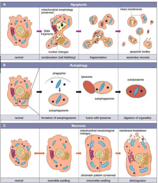
Apoptosis, also termed type I cell death, is defined by characteristic changes in the nuclear morphology, including chromatin condensation and fragmentation, overall cell shrinkage, blebbing of the plasma membrane and formation of apoptotic bodies that contain nuclear or cytoplasmic material.
Autophagic cell death, also known as type II cell death, is characterized by a massive accumulation of double-membrane containing vacuoles known as autophagosomes, which subsequently fuse with lysosome vacuoles.
Type III cell death, better known as necrosis, is often defined in a negative manner as death lacking the characteristics of the type I and type II processes.
To simplify matters I will explain these three major cell death mechanisms (Figure 1). Distinction of different cell death forms is not only relevant for semantic reasons, but can also have important clinical implications when considering the potential therapeutic targeting of cell death processes. Nevertheless, in some conditions these distinct cell modes may only be the extremes, and there are numerous examples in which cell death demonstrates a continuum of intermediate features, for instance of both apoptosis and necrosis. In the literature we occasionally find newly coined expressions such as
necrapoptosis or aponecrosis. As we shall discuss later, the inhibition of a particular type of cell death does not prevent cell death in many cases, but simply shifts the phenotype to alternative modes of cell death [7].
How cells die: Apoptosis and other cell death pathways
1
Apoptosis
How cells die: Apoptosis and other cell death pathways
1
provoking an inflammatory response. Moreover, the engulfment of apoptotic cells bymacrophages triggers the production of anti-inflammatory cytokines.
Because apoptotic cells are eaten and digested so quickly, there are usually few dead cells to be seen in tissue sections, even when large numbers of cells have died. This probably explains why apoptosis was neglected by pathologists for a long time. Looking inside the cell, one of the most noticeable features of apoptosis is the condensation of the nucleus and its fragmentation into smaller pieces, a highly distinctive event that is not seen in other forms of cell death. Another defining feature is the extensive hydrolysis of nuclear DNA into internucleosomal fragments (Figure 1A).
Apoptosis is the major cell death pathway for removing unwanted and harmful cells in a clean or silent manner during embryonic development, tissue homeostasis and immune regulation. In addition, most anti-cancer therapies rely on the activation of apoptotic pathways. As the alterations of apoptosis are stereotypical and similar in all cell types irrespective of the death stimulus, the biochemical mechanisms underlying these changes also follow a similar built-in program. In nematodes, insects and human cells, most, if not all, morphological alterations of apoptosis are mediated by the activation of an evolutionarily conserved and unique class of intracellular proteases known as caspases [9].
Autophagy
Like apoptosis, autophagy is a highly conserved and genetically controlled process involving a cascade of molecular events [10]. Autophagy (meaning self-eating) is classically activated in response to nutrient starvation, but is also observed during development, differentiation and various forms of environmental stress. In addition, defective autophagy underlies a number of pathological conditions, including myopa-thies, neurodegenerative diseases, liver diseases, and some forms of cancer [11].
Autophagy is a major catabolic mechanism by which long-lived proteins and organelle components are directed to lysosomes and recycled in order to maintain energy and protein synthesis. It is characterized by the appearance of numerous cytosolic vacuole-like structures known as autophagosomes, which are formed by the assembly of double-layered, membrane-bound structures of still largely undefined origin. The autophago-somes encapsulate cytosolic materials and subsequently fuse with lysoautophago-somes, which causes the autophagosomal contents to degrade (Figure 1B).
Although the role of the autophagic process in protein and organelle degradation, and in protection during nutrient starvation is readily accepted, its function in programmed cell death is controversial [12]. This is partly because the term ‘autophagic cell death’ has been applied to two distinct observations: cell death associated with autophagy and cell death through autophagy.
Under normal physiological conditions autophagy occurs at low basal levels, contribut-ing to the turnover of cytoplasmic components and promotcontribut-ing cell adaptation and survival during stress, e.g. starvation. Excess autophagy, on the other hand, leads to autophagic cell death.
How cells die: Apoptosis and other cell death pathways
1
Necrosis
Necrotic cell death is often defined negatively as a form of cell death that lacks signs of apoptosis or autophagy. Necrotic cells typically show cytoplasmic swelling and vacuola-tion, rupture of the plasma membrane, dilation of organelles (mitochondria, endoplas-mic reticulum and Golgi apparatus), as well as moderate chromatin condensation. When cells swell and burst they spill their contents over their neighbors and elicit a damaging inflammatory response [14] (Figure 1C).
Necrosis is usually considered to be an uncontrolled and accidental cell death which, unlike apoptosis, is not energy-dependent. Biochemically, most prominent features include massive energy depletion, the formation of reactive oxygen species and the activation of non-apoptotic proteases. All these events result in a loss of function of homeostatic ion pumps and damage to membrane lipids with cell membrane swelling and rupture.
Furthermore, during necrosis a substantial rise in intracellular calcium is observed. The elevated calcium levels in the cytosol trigger mitochondrial calcium overload, leading to depolarization of the inner mitochondrial membrane and a shut-down of ATP produc-tion [15]. While depleproduc-tion of ATP impedes the funcproduc-tion of membrane channels, in-creased calcium activates calcium-dependent proteases, e.g. calpains. Calcium fluxes, ATP depletion and oxidative stress involve complex and interactive feedback loops that self-amplify and potentiate each other, leading to exaggerated cell death. Such processes are most relevant under conditions of excessive trauma and ischemia-reperfusion. Necrosis, however, can also be observed in response to death receptor activation or chemotherapy, conditions that were originally believed to mediate cell death exclusively via apoptosis.
In addition, the inhibition of specific proteins involved in regulating apoptosis or autophagy can switch the type of cell death to necrosis. Finally, secondary or post-apop-totic necrosis occurs when massive apoptosis overwhelms the scavenging activity of phagocytes, thereby resulting in leakage of the cell contents with induction of inflamma-tory responses.
There is increasing evidence that necrosis is more tightly regulated than previously thought and underlies a genetic control that might be relevant in multiple physiological and pathological scenarios [14]. Necrosis might therefore serve as a backup cell death mechanism when apoptosis or autophagic cell death fails.
Molecular Basics of Apoptosis
Although any form of regulated cell death is potentially relevant, we will focus below on apoptosis, which is the best-defined and most prevalent form of cell death. In view of the recent explosion of publications, it is often difficult for newcomers in the field to grasp the essentials of apoptosis regulation. As we will see later, the basic molecular machinery of apoptosis is relatively simply, even though its complexity has increased through evolution.
The demolition machinery
prote-How cells die: Apoptosis and other cell death pathways
1
residues within specific tetra- or pentapeptide recognition sequences [16]. The fourteendifferent caspases identified in mammals are expressed in most cell types. To preclude unwarranted cell death, however, each caspase is maintained as an inactive zymogen consisting of a prodomain followed by two subunits with the catalytic domain. Caspases operate in hierarchical cascades that serve to amplify the apoptotic signal.
Based on their structure and sequence in cell death pathways, caspases can be divided into upstream initiators and downstream effectors of apoptosis. Effector caspases such as caspase-3, -6, and -7 contain only a short prodomain and cleave diverse cellular sub-strates, whereas initiator caspase-8, -9, and -10 have a long prodomain and exert regulatory roles by activating downstream effector caspases. While it was previously assumed that all caspases are activated by proteolytic cleavage, recent results suggest that the mode of activation differs considerably between initiator and effector caspases [17].
Effector caspases are constitutive homodimers that are always activated by initiator caspases through proteolytic cleavage.
By contrast, the zymogens of initiator caspases are predominantly present as mono-mers. Their activation is achieved by the binding of their long prodomains to high-mo-lecular weight adaptor complexes that stimulate the homodimerization of the initiator caspases. The dimerization in turn causes a conformational change in the initiator caspases, resulting in their activation. Thus, the most important event for initiator caspase activation is their recruitment into caspase activation platforms.
Once activated, effector caspases cleave an estimated 400 substrates, including proteins involved in scaffolding of the cytoplasm, signal transduction and transcription-regulato-ry proteins, cell cycle-controlling components and proteins involved in DNA replication and repair [18]. For many of the identified substrates the functional consequences of their cleavage are unknown and have only been inferred from their normal functions. In other cases, proteolysis of certain components can be linked to discrete morphological changes. A classical example is the DNase inhibitor ICAD (inhibitor of caspase-activated DNase). Cleavage of ICAD by caspase-3 liberates the active CAD nuclease which, in turn, mediates apoptotic DNA fragmentation. The cleavage of several other substrates, including gelsolin as well as the kinases ROCK-1 and PAK2, has been implicated in membrane blebbing of apoptotic cells.
Moreover, caspases destroy several proteins required for maintenance of the cytoskeletal architecture, such as the intermediate filament cytokeratin-18, which is involved in filament organization. A large percentage of caspase substrates are further involved in cell adhesion or mediate cell–cell communication in adherens and gap junctions.
How cells die: Apoptosis and other cell death pathways
1
Activation pathways
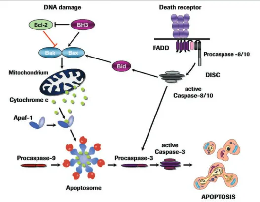
Caspase activation is initiated by two major signaling routes, namely the extrinsic death receptor pathway and the intrinsic mitochondrial pathway (Figure 2). Death receptor-mediated apoptosis plays a fundamental role in the maintenance of tissue homeostasis, especially in the immune system, whereas the mitochondrial pathway is used extensively in response to extracellular cues and internal insults such as DNA damage [21,22].
Death receptors form a subgroup of the tumor necrosis factor (TNF) receptor superfam-ily that includes TNF-R1, CD95 (Fas/APO-1) and receptors binding to the TNF-related apoptosis-inducing ligand (TRAIL). All death receptors are characterized by an intracel-lular motif, termed the death domain, which is important for transmission of the apoptotic signal. Upon ligand binding, the death receptors interact via their death domain with a corresponding protein motif in adapter proteins such as FADD. These adapters also contain a second homotypic protein interaction motif, the death effector domain, which facilitates their binding to the prodomain of the initiator caspase-8. Taken together, these components form the death-inducing signaling complex (DISC) which, when assembled, activates caspase-8 through a proximity-induced dimerization mechanism [23,24]. At this point a high local concentration of dimerized caspase-8 leads to its autocatalytic activation and the subsequent activation of downstream effector caspases.
How cells die: Apoptosis and other cell death pathways
1
The death receptor pathway plays an important role in the immune system. For instance,when we become infected by a virus, cytotoxic T lymphocytes are activated and then express CD95 ligand. By binding to its receptor, CD95 ligand induces apoptosis in the infected cells to prevent the virus from replicating and spreading to other cells. Cytotoxic T cells, in addition, utilize a granule-exocytosis pathway for the elimination of virus-infected cells. Cytotoxic granules contain a pore-forming protein, perforin, and serine proteases known as granzymes. Granzyme B can directly cleave proteins after aspartate residues and can therefore also activate caspase-3. In this way the upstream events of death receptor signaling are bypassed, and apoptosis is induced directly (Figure 2).
Most cell death, however, proceeds via the mitochondrial pathway, which integrates signals generated by a variety of stressors including DNA damage, loss of adhesion, growth factor withdrawal and others [25]. As a key event these apoptotic stimuli evoke the release of cytochrome c from the mitochondrial intermembrane space into the cytosol. Cytochrome c normally functions in electron transport processes of the respira-tory chain to generate ATP. In the cytosol of apoptotic cells, however, it serves as a cofactor for the adapter protein Apaf-1. Upon binding of cytochrome c, Apaf-1 oli-gomerizes and recruits the initiator caspase-9 to trigger the formation of the apopto-some [26]. In this case the binding of caspase-9 to Apaf-1 is mediated by a shared homotypic interaction motif known as the caspase recruitment domain (CARD). Thus, like the DISC, the apoptosome is a high-molecular weight complex that serves as a caspase activation platform. Once assembled in the apoptosome, caspase-9 becomes activated and subsequently triggers the caspase cascade (Figure 2).
Although, at first glance, both pathways of caspase activation appear to trigger apoptosis in a straightforward manner, the circuitry of the system is often more complicated. In some cell types, for example, activation of caspase-8 at the DISC is insufficient to stimulate the direct activation of effector caspases, and this reaction requires an amplifi-cation step with cytochrome c-dependent apoptosome formation. In this scenario caspase-8 cleaves and activates the pro-apoptotic Bcl-2 protein Bid, which catalyzes cytochrome c release. Bid cleavage thereby links the death receptor to the mitochondrial pathway. Furthermore, in addition to these two pathways, a number of other pathways of caspase activation have been reported, including an initiator role of caspase-2 or of caspase-12 in apoptosis triggered by endoplasmic reticulum stress. However, unlike the well-established role of the death receptor and mitochondrial pathway, the physiological relevance of these death routes remains controversial and is currently not supported by the phenotype of knockout models.
Intracellular controls
The decision to die must be tightly controlled, and so it is not surprising that the death program is regulated not only by proapoptotic mediators, but also by various apoptosis inhibitors. In the death receptor pathway this is accomplished by the caspase-8 inhibitor c-FLIP, which has a structure very similar to caspase-8 but without the catalytic protease domains. Upregulation of c-FLIP expression can therefore compete efficiently with caspase-8 for DISC binding [24].
How cells die: Apoptosis and other cell death pathways
1
Meanwhile, more than 20 members of the Bcl-2 family have been identified, which are all defined by the presence of one to four Bcl-2 homology (BH) domains [29]. Bcl-2 proteins come in two flavors including, firstly, anti-apoptotic proteins that prevent the release of cytochrome c and, secondly, proapoptotic proteins that trigger MOMP.
The proapoptotic Bcl-2 proteins can be further subdivided into two subfamilies based on the sharing of BH domains. BH multidomain proteins, such as Bax and Bak, are the triggers of MOMP, most likely as a result of their ability to form pores in the outer mitochondrial membrane. The other subfamily, the BH3-only proteins, which contain only the BH3 domain, act as upstream regulators by controlling the allosteric activation of the gatekeepers Bax and Bak. It is therefore not surprising that the activity of BH3-only proteins is under tight transcriptional and posttranslational control. While, in healthy cells, BH3-only proteins are either not expressed or are conformationally restrained, they rapidly become activated following exposure to cellular stresses. Interest-ingly, different types of stresses can evidently activate distinct sets of BH3-only proteins, suggesting that BH3-only proteins act as essential sensors of different death stimuli [29].
In addition to cytochrome c, activation of the proapoptotic Bcl-2 proteins results in the release of toxic mitochondrial mediators, including the protein Smac and the serine protease Omi/HtrA2 [30]. Both proteins can promote apoptosis by counteracting the inhibitor-of-apoptosis proteins (IAPs), which comprise a family of endogenous caspase inhibitors. Interestingly, Omi/HtrA2 is also thought to promote caspase-independent cell death, apparently due to its serine protease activity.
Another group of proteins released from mitochondria comprises apoptosis-inducing factor (AIF) and endonuclease G. Both can promote cell death in a caspase-independent manner by inducing chromatin condensation and DNA degradation. Thus, if for some reason cells do not activate caspases after MOMP, these mediators might still ensure that cell death proceeds.
Assays for Apoptosis
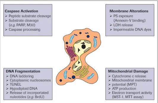
Since apoptosis occurs via a tightly regulated cascade, there are many possibilities to measure the activity of these regulators or the functional consequences of their action (Figure 3). A large number of apoptosis assays for detecting and counting apoptotic cells have been devised. All of these assays have advantages and disadvantages. For instance, certain features of apoptosis might only appear transiently, while others might partially overlap with necrosis. It is therefore crucial to employ two or more distinct assays to confirm that cell death is occurring via apoptosis. In addition, certain assays might be suitable for cultured cells, but inappropriate for investigating apoptosis in tissue sections. Therefore, when choosing methods of apoptosis detection in cells, tissues or organs, we should understand the pros and cons of each assay
Caspase activation
How cells die: Apoptosis and other cell death pathways
1
cytokeratin-18 (see M30 CytoDEATH, page 21), are particularly convenient for detectingearly apoptosis in cellular assays and even in archived tissue biopsies by means of immunohistochemistry.
An alternative that is often used to obtain evidence for the involvement of a particular caspase is to monitor its proteolytic cleavage as an indicator of caspase activation. While the proteolytic processing of effector caspase-3, -6 and -7 faithfully reflects their activa-tion, this does not hold true for initiator caspases [17]. As mentioned above, in contrast to a widespread misinterpretation seen in many publications, the mere cleavage of an initiator caspase does not activate it, and therefore this cannot be taken as sufficient evidence for its activation. Furthermore, certain caspases are also cleaved by non-caspase proteases at a non-relevant site, which might incorrectly indicate that caspase-mediated proteolytic activation has occurred.
Caspase activity is most frequently detected in synthetic substrates assays. The substrates are generated by fusing a preferred tetrapeptide cleavage sequence at the aspartate residue to a fluorogenic or colorimetric reporter, so that a signal is produced upon substrate cleavage (see Homoegeneous Caspases Assay, fluorimetric, page 30). The knowledge of the inherent substrate specificity of individual caspases has further been employed to construct peptide-based substrate reporters that are often claimed to measure the activity of specific caspases. However, although these substrates are useful for characterizing individual purified caspases, they are unable to distinguish a role of a certain caspase in cell lysates. This is due to a significant overlap between individual consensus sequences, because the caspases are promiscuous on these sequences. This specificity problem, however, can be solved by the use of ELISA-based capture assays for individual caspases that are coupled with a substrate reaction (see Caspase 3 Activity Assay, page 26). Thus, by employing adequate tools and appropriate assays, detection of caspase activity or its cleavage products might still constitute a specific approach for measuring apoptosis.
How cells die: Apoptosis and other cell death pathways
1
Membrane alterations
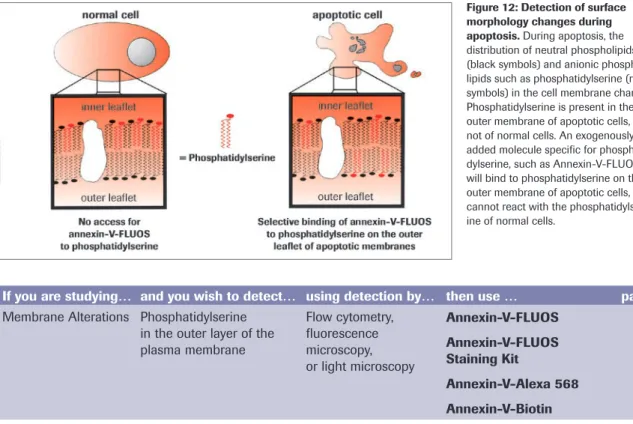
Another characteristic feature of apoptosis is the exposure of the phospholipid phos-phatidylserine (PS) to the outer cell membrane. PS is normally confined to the inner plasma membrane in healthy cells, but is translocated to the outer membrane leaflet in response to proapoptotic stimuli. This flip-flop mechanism is caspase-dependent, although how caspases promote PS externalization remains a mystery. PS exposure is important for the elimination of apoptotic cells, because it represents an ‘eat-me’ signal for the engulfment by professional phagocytes following binding to a putative PS receptor. This process therefore ensures the early uptake of apoptotic cells with no release of cellular contents and without provoking an inflammatory response [31].
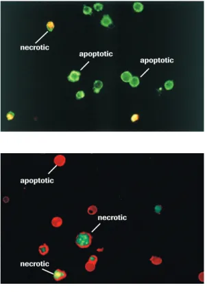
PS exposure is experimentally detected most commonly using annexin-V, a PS-binding protein. Various annexin-V derivatives coupled to different fluorochromes are available, providing versatile possibilities for apoptosis measurement, for instance by multicolor flow cytometry or fluorescence microscopy (see Annexin-V products on page 39-45). The advantages are sensitivity and rapidity; the disadvantage is that necrotic cells are labeled upon rupture of their plasma membrane. Therefore, it important to control the membrane integrity of the PS-positive cells by double-staining with membrane-imper-meable DNA dyes such as propidium iodide. In these assays, healthy cells are doubly negative to annexin-V and propidium iodide, whereas cells in the early phases of
apoptosis are annexin-V-positive but propidium iodide-negative, and secondary necrotic cells are doubly positive to annexin-V and propidium iodide.
DNA fragmentation
The internucleosomal cleavage of DNA is a classical feature of apoptosis and fulfills the physiological function of eliminating DNA that is highly immunogenic. The failure to degrade DNA can give rise to autoantibodies that can cause lupus erythematosus-like autoimmune disease. DNA fragmentation during apoptosis is induced by caspase-medi-ated cleavage of ICAD (inhibitor of caspase-activcaspase-medi-ated DNase), with the ensuing activa-tion of CAD (caspase-activated DNase). This event then leads to characteristic internu-cleosomal DNA double-strand breaks with fragments of multiples of 180 base pairs in size. In contrast, necrotic cell death is accompanied by late and random DNA tion through the release of lysosomal DNases. Techniques that detect DNA fragmenta-tion are thus not necessarily specific to apoptosis, but may detect DNA damage in a variety of cell death paradigms.
Detection of DNA fragmentation is currently one of the most frequently used tech-niques for highlighting apoptotic cells in tissues. A traditional method for demonstrating internucleosomal DNA degradation is gel electrophoresis of genomic DNA (see Apop-totic DNA Ladder Kit, page 57). In these experiments, apopApop-totic cells show a characteris-tic DNA ladder, while necrocharacteris-tic cells reveal a smear of randomly degraded DNA. Another assay is the detection of hypodiploid nuclei from apoptotic cells, which is usually performed in a flow cytometer using DNA-binding fluorochromes such as propidium iodide. The technique determines the DNA content, and therefore the number of apoptotic cells, but also allows the simultaneous analysis of cell cycle parameters of surviving cells. It should be stressed, however, that necrotic cells sometimes also display a certain degree of DNA degradation that may result in hypodiploid nuclei. Therefore, the presence of a hypodiploid DNA peak does not provide unequivocal proof of apoptotic cell death but should be adequately controlled.
tech-How cells die: Apoptosis and other cell death pathways
1
In tissue sections, the TUNEL (terminal dUTP nick end-labeling) method is widely usedto measure DNA fragmentation (see In Situ Cell Death Detection Kits, page 74-79). The principle of the assay is that endonuclease-generated DNA breaks are enzymatically labeled by terminal transferase with UTP derivatives coupled to fluorochromes or biotin that can be detected in an immunoperoxidase reaction. This assay is very sensitive, and allows DNA fragmentation to be assessed quantitatively by light and fluorescence microscopy or by flow cytometry. However, since this method can also be subject to pitfalls, such as false positives from necrotic cells, it should be paired with additional assays.
Mitochondrial changes
Apoptosis-specific alterations of mitochondria are more difficult to detect. The mito-chondrial pathway begins with the permeabilization of the mitomito-chondrial outer mem-brane by proapoptotic members of the Bcl-2 family, resulting in a release of cytochrome c and other toxic proteins from the intermembrane space into the cytosol. The release of these proteins is generally determined by immunocytochemistry or by Western blotting of cytosolic, mitochondrial and nuclear fractions.
In addition to MOMP, another event observed during cell death is the loss of the inner mitochondrial transmembrane potential, the electrochemical proton gradient generated by the respiratory chain. This process is called mitochondrial permeability transition (MPT) and involves the opening of a largely undefined, non-selective pore in the inner mitochondrial membrane [28]. MPT also causes mitochondrial swelling, which can eventually result in rupture of the outer mitochondrial membrane and release of cytochrome c and other intermembrane proteins. It has therefore previously been claimed that MPT and the loss of the mitochondrial transmembrane potential can actively contribute to cytochrome c release.
The mitochondrial transmembrane potential can be measured easily using a variety of potentiometric dyes, together with flow cytometry or fluorescence microscopy. In many publications MPT is considered to be an early marker of mitochondrial alterations, which is thought to precede cytochrome c release and apoptosome formation. However, with rare exceptions, this hypothesis seems to be largely incorrect. Only a few cytotoxic (in fact necrotic rather than apoptotic) stimuli, such as calcium overload or oxidative stress, seem to cause cytochrome c release downstream of permeability transition. In most cases, MOMP caused by proapoptotic Bcl-2 proteins, but not by the transmem-brane potential, is the primary mechanism of cytochrome c release. This assumption is supported by the fact that loss of MPT does not precede cytochrome c release and is generally prevented by caspase inhibition. Therefore, the loss of the transmembrane potential should be regarded rather as a good marker of mitochondrial damage that may occur in late apoptosis, but also during necrosis.
Caveats and cautionary notes
How cells die: Apoptosis and other cell death pathways
1
Another important issue concerns the timing of the experiments. Apoptosis proceeds very rapidly, often within a few hours, whereas necrosis is typically much slower. Thus, if an injured cell fails to undergo apoptosis and remains viable for a few hours, it might well die through alternative pathways a few hours later. In many instances, the suppres-sion of signs of apoptosis has been misinterpreted as inhibition of cell death, which is often an incorrect statement. Therefore, I strongly recommend the use of additional clonogenicity and viability assays. Traditional assays measuring cell proliferation (see chapter 9), ATP content (see ATP Bioluminescence Assay Kits, page 130) or the release of marker enzymes such as lactate dehydrogenase (see Cytotoxicity Detection KitPLUS (LDH), page 98) are well-suited for this purpose. Implementation of those methods will provide us not only with better insights into cell death mechanisms, but can also demonstrate whether short-term inhibition of cell death is connected to long-term survival.
Klaus Schulze-Osthoff
Professor of Molecular Medicine Institute of Molecular Medicine University of Düsseldorf D-40225 Düsseldorf, Germany
References
Raff MC. Social controls on cell survival and cell death. Nature. 1992; 356:397-400. 1.
Fischer U, Schulze-Osthoff K. New approaches and therapeutics targeting apoptosis 2.
in disease. Pharmacol Rev. 2005; 57:187-215.
Lockshin RA, Williams CM. Programmed cell death. I. Cytology of degeneration in 3.
the intersegmental muscles of the Pernyi Silkmoth. J Insect Physiol 1965; 11:123–133. Horvitz HR. Nobel lecture. Worms, life and death. Biosci Rep. 2003; 23:239-303. 4.
Kroemer G, El Deiry WS, Golstein P, Peter ME, Vaux D, Vandenabeele P, Zhivotovsky 5.
B, Blagosklonny MV, Malorni W, Knight RA, Piacentini M, Nagata S, Melino G. Classification of cell death: Recommendations of the nomenclature committee on cell death. Cell Death Differ 2005; 12:1463 7.
Melino G, Knight RA, Nicotera P. How many ways to die? How many different 6.
models of cell death? Cell Death Differ 2005; 12:1457 62.
Chipuk JE, Green DR. Do inducers of apoptosis trigger caspase-independent cell 7.
death? Nat Rev Mol Cell Biol. 2005; 6:268-75.
Kerr JFR, Wyllie AH, Currie AR. Apoptosis: A basic biological phenomenon with 8.
wide-ranging implications in tissue kinetics. Br J Cancer. 1972; 26:239–57.
Taylor RC, Cullen SP, Martin SJ. Apoptosis: controlled demolition at the cellular level. 9.
Nat Rev Mol Cell Biol. 2008; 9:231-41.
Klionsky DJ. Autophagy: from phenomenology to molecular understanding in less 10.
than a decade. Nat Rev Mol Cell Biol. 2007; 8:931-7.
Levine B, Kroemer G. Autophagy in the pathogenesis of disease. Cell. 2008; 11.
132:27-42.
Levine B, Yuan J. Autophagy in cell death: an innocent convict? J Clin Invest. 2005; 12.
115:2679 –2688.
Maiuri MC, Zalckvar E, Kimchi A, Kroemer G. Self-eating and self-killing: crosstalk 13.
between autophagy and apoptosis. Nat Rev Mol Cell Biol. 2007; 8:741-52.
Festjens N, Vanden Berghe T, Vandenabeele P. Necrosis, a well-orchestrated form of 14.
How cells die: Apoptosis and other cell death pathways
1
Los M, Wesselborg S, Schulze-Osthoff K. The role of caspases in development,16.
immunity, and apoptotic signal transduction: lessons from knockout mice. Immu-nity. 1999; 10:629-39.
Fuentes-Prior P, Salvesen GS. The protein structures that shape caspase activity, 17.
specificity, activation and inhibition. Biochem J. 2004; 384:201-32.
Fischer U, Janicke RU, Schulze-Osthoff K. Many cuts to ruin: a comprehensive 18.
update of caspase substrates. Cell Death Differ. 2003; 10:76-100.
Schwerk C, Schulze-Osthoff K. Non-apoptotic functions of caspases in cellular 19.
proliferation and differentiation. Biochem Pharmacol. 2003; 66:1453-8.
Kuranaga E, Miura M. Nonapoptotic functions of caspases: caspases as regulatory 20.
molecules for immunity and cell-fate determination. Trends Cell Biol. 2007; 17:135-44.
Danial NN, Korsmeyer SJ. Cell death: critical control points. Cell. 2004; 116:205-219 21.
Meier P, Vousden KH. Lucifer’s labyrinth--ten years of path finding in cell death. Mol 22.
Cell. 2007; 28:746-54.
Debatin KM, Krammer PH. Death receptors in chemotherapy and cancer. Oncogene. 23.
2004; 23:2950–2966.
Peter ME, Budd RC, Desbarats J, Hedrick SM, Hueber AO, Newell MK, Owen LB, 24.
Pope RM, Tschopp J, Wajant H, Wallach D, Wiltrout RH, Zörnig M, Lynch DH. The CD95 receptor: apoptosis revisited. Cell. 2007; 129:447-50.
Green DR, Kroemer G. The pathophysiology of mitochondrial cell death. Science. 25.
2004 ;305: 626-9.
Jiang X, Wang X. Cytochrome C-mediated apoptosis. Annu Rev Biochem. 2004; 26.
73:87-106.
Chipuk JE, Green DR. How do BCL-2 proteins induce mitochondrial outer mem-27.
brane permeabilization? Trends Cell Biol. 2008; 18:157-64.
Kroemer G, Galluzzi L, Brenner C. Mitochondrial membrane permeabilization in cell 28.
death. Physiol Rev. 2007; 87:99-163.
Youle RJ, Strasser A. The BCL-2 protein family: opposing activities that mediate cell 29.
death. Nat Rev Mol Cell Biol. 2008; 9:47-59.
Ekert PG, Vaux DL. The mitochondrial death squad: hardened killers or innocent 30.
bystanders? Curr Opin Cell Biol. 2005; 17: 626-30.
Lauber K, Blumenthal SG, Waibel M, Wesselborg S. Clearance of apoptotic cells: 31.
getting rid of the corpses. Mol Cell. 2004; 14:277-87.
Apoptosis Product Selection Guide
1
DNA fragmentation, quantitative
K Cell Death Detection ELISAPLUS 11 774 425 001
11 920 685 001
DNA fragmentation
K In Situ Cell Death Detection Kit,
TMR red 12 156 792 910
K In Situ Cell Death Detection Kit,
Fluorescein 11 684 795 910
Membrane modification
K Annexin-V-Alexa 568 03 703 126 001
K Annexin-V-FLUOS 11 828 681 001
K Annexin-V-FLUOS Staining Kit 11 858 777 001 11 988 549 001
Caspase activity, indirect
K M30 CytoDEATH, Fluorescein 12 156 857 001
K M30 CytoDEATH 12 140 322 001 12 140 349 001 If you are studying
cell
populations single cells Start
Caspase activity, quantitative
K Caspase 3 Activity Assay 12 012 952 001
K Homogeneous Caspases
Assay, fluorimetric 12 236 869 00103 005 372 001
using detection by
light microscopy FACS or
fluorescence microscopy
Caspase activity, indirect
K Anti-Poly (ADP-Ribose)
Polymerase (PARP) 11 835 238 001
DNA fragmentation, qualitative
K Apoptotic DNA Ladder Kit 11 835 246 001
using detection by
using detection by
colorimetric
ELISA fluorimetricELISA western blot or gel electrophoresis
ELISA
using detection by
western blot gel
electro-phoresis
BrdU incorporation in DNA fragments, quantitative
K Cellular DNA Fragmentation
ELISA 11 585 045 001
Apoptosis Product Selection Guide
1
DNA fragmentation
K In Situ Cell Death Detection Kit, POD 11 684 817 910
K In Situ Cell Death Detection Kit, AP 11 684 809 910
Caspase activity, indirect
K M30 CytoDEATH 12 140 322 001 12 140 349 001
Caspase activity, indirect
K M30 CytoDEATH 12 140 322 001 12 140 349 001
Membrane modification
K Annexin-V-Biotin 11 828 690 001
DNA fragmentation
K In Situ Cell Death Detection Kit,
POD 11 684 817 910
K In Situ Cell Death Detection Kit, AP 11 684 809 910
cell cultures, blood cells, etc.
tissue sections
DNA fragmentation
K In Situ Cell Death Detection Kit,
TMR red 12 156 792 910
K In Situ Cell Death Detection Kit,
Fluorescein 11 684 795 910
Caspase activity, indirect
K M30 CytoDEATH, Fluorescein 12 156 857 001
K M30 CytoDEATH 12 140 322 001 12 140 349 001
FACS or fluorescence microscopy
using detection by
light microscopy
in in
Follow the selection guide below to determine the appropriate Roche Applied Science product for the study of apoptosis to meet your needs.
See page 90 for products to study cytotoxicity and page 114 for products to study cell proliferation. If you need additional help, please visit
How cells die: Apoptosis and other cell death pathways
1
Apoptosis Autophagy Necrosis
Morphological features
Membrane blebbing, no
loss of integrity Cell membrane stays intact, weak membrane blebbing
Loss of membrane integrity
Aggregation of chromatin
at the nuclear membrane Marginal chromatin con-densation No chromatin condensa-tion
Begins with shrinking of cytoplasm and condensa-tion of nucleus
Begins with sequestration of cytoplasmic material in autophagosomes and autolysosomes
Begins with swelling of cytoplasm and mitochon-dria
Mitochondria preserve normal ultrastructure, outer membrane permeabilization
Damaged mitochondria degraded within autopha-gosomes
Disintegration (swelling) of organelles
Ends with fragmentation of cell into small vesicles (apoptotic bodies)
No cell lysis, ends with
self-digestion Ends with total cell lysis
Biochemical features
Tightly regulated process involving mediators and enzymes
Tightly regulated process involving various regula-tory steps
Loss of ion homeostasis
Energy (ATP)-dependent
(active process)
Generates ATP in the absence of exogenous energy supply
No energy requirement (passive process)
Non-random internu-cleosomal DNA fragmenta-tion (ladder pattern after agarose gel electropho-resis)
No DNA fragmentation Random digestion of DNA (smear of DNA after aga-rose gel electrophoresis)
Release of several apop-togenic mitochondrial proteins (cytochrome c, AIF etc.)
Involvement of Atg (autophagy related gene) products; ubiquitin-like activation cascades
Oxidative stress, calcium overload, ATP depletion
Activation of caspase
cascade Activation of lysosomal proteases and hydrolases Activation of calcium-dependent proteases
Loss of plasma membrane asymmetry (i.e., transloca-tion of phosphatidylserine from the inner to the outer side of the membrane)
Physiological significance
Serves to eliminate dam-aged, transformed or infected cells; functions in organ development and regulation of immune responses
Serves primarily to main-tain cellular energy and to recycle damaged organ-elles
Physiological role unclear, potential backup mecha-nism during apoptosis failure.
Induced by physiological stimuli (death ligands), growth factor deple-tion and different cellular stresses
Triggered by nutrient deprivation and other cel-lular stresses.
Apoptosis – Caspase Activity
2
Assays that Measure Apoptosis-induced Proteases (Caspases) 20 M30 CytoDEATH/ M30 CytoDEATH, Fluorescein 21
Caspase 3 Activity Assay 26
Assays that Measure Apoptosis-Induced Proteases (Caspases)
2
Assays that Measure Apoptosis-
Induced Proteases (Caspases)
Several caspases are thought to mediate very early stages of apoptosis10. For instance, one of these, caspase 3 (CPP32) is required for the induction of apoptosis by certain effectors [especially tumor necrosis factor and the cytotoxic T cell ligand effector, CD95 (also called Fas)] Enari et al. (1996), Nature 380, 723–726.
These proteases cleave numerous substrates at the carboxy site of an aspartate residue. All are synthesized as pro-enzymes; activation involves cleavage at aspartate residues that could themselves be sites for the caspase family. As caspases are probably the most important effector molecules for triggering the biochemical events which lead to apoptotic cell death, assays for determination of caspase activation can detect apoptosis earlier than many other commonly used methods.
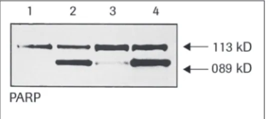
The most elucidatory assay for these caspases involves western blot detection of prote-olytic cleavage products found in apoptotic cells. An antibody, Anti-PARP, sold by Roche Applied Science, can be used in such an assay. The antibody can detect intact and cleaved forms of Poly-ADP-Ribose Polymerase, a target for some caspases.
For specific and quantitative measurement of caspase activity Western blotting is not suitable. To quantify caspase activation enzyme activity assays based on detection of cleaved caspase substrates have been developed recently. However most of the caspase substrates are not exclusively cleaved by a specific caspase but only preferentially, while other members of the caspases family act on these substrates to a lower extent. Roche Applied Science offers a caspase 3 activity assay with highest specificity by the use of an immunosorbent enzyme assay principle.
If you are studying… and you wish to detect… using detection by… then use … page Caspase Activity Caspase cleavage of
cytokeratin 18
Western blot, flow cytometry, fluorescence microscopy, or light microscopy
M30 CytoDEATH M30 CytoDEATH, Fluorescein
21 21
Caspase 3 activity Fluorescence ELISA Caspase 3 Activity Assay 26
Caspases Fluorescence ELISA Homogeneous Caspases
Assay, fluorimetric
30
Caspase cleavage of PARP Western blot, immunoprecipitation, immunohistology
Anti-Poly (ADP-Ribose) Polymerase (PARP)
M30 CytoDEATH / M30 CytoDEATH, Fluorescein
2
M30 CytoDEATH*
Cat. No. 12 140 322 001 50 tests Cat. No. 12 140 349 001 250 tests
M30 CytoDEATH, Fluorescein*
Cat. No. 12 156 857 001 250 testsType Monoclonal antibody, clone M30, IgG2b, mouse
Useful for Detection of apoptosis in epithelial cells and tissues (formalin grade)
Sample material Adherent cells, tissue samples (routinely fixed and paraffin-embedded tissue sections, cryostat sections)
Method Detect apoptosis by applying the M30-antibody to fixed samples, then using secondary detection systems. Suitable for immunohistochemistry, immunocytochemistry, and flow cytometry
Significance of
reagent Use the M30 CytoDEATH antibody for the determination of early apoptotic events in cells and tissue sections by detection of a specific epitope of cytokeratin 18 that is presented after cleavage by caspases.
Specificity The M30 CytoDEATH antibody binds to a caspase-cleaved, formalin-resistant epitope of the cytokeratin 18 (CK 18) cytoskeletal protein. The immunoreactivity of the
M30 CytoDEATH antibody confined to the cytoplasma of apoptotic cells.
Time 2 h for immunofluorescence on cells, 3.5 h for staining of tissues (excluding dewaxing)
Antibody supplied as
Mouse monoclonal antibody (clone M30), lyophilized, stabilized. Formalin grade.
Background information
During Apoptosis, vital intracellular proteins are cleaved. The proteases that mediate this process are called caspases (Cysteinyl-aspartic acid proteases). Caspases are expressed as zymogenes, which are activated by different apoptosis inducers. Once activated, a single caspase activates a cascade of caspases.
It has been shown that the M30 antibody recognizes a specific caspase cleavage site within cytokeratin 18 that is not detectable in native CK18 of normal cells. Consequent-ly, the M30 CytoDEATH antibody is a unique tool for the easy and reliable determina-tion of very early apoptotic events in single cells and tissue secdetermina-tions.
Benefits Detect early apoptosis in epithelial cells and tissue sections with high sensitivity.
Determine caspase activity − even in formalin-fixed, paraffin-embedded tissue.
Choose from two antibody formats to meet your specific application needs.
Easily perform fluorescence and FACS analysis with M30 CytoDEATH, Fluores-cein.
Use in dual-labeling applications, for example, in combination with the In Situ Cell Death Detection Kit, TMR red (TUNEL technique, see page 71-73).
M30 CytoDEATH / M30 CytoDEATH, Fluorescein
2
How to use the reagent
I.a Assay procedure overview
for immunofluorescence and flow cytometry on cells for M30 CytoDEATH and M30 Cy-toDEATH, Fluorescein:
1 Fix cells. Add Anti-Mouse-Ig-Fluorescein
(not necessary for M30 CytoDEATH, Fluorescein).
Add M30 CytoDEATH antibody or M30 CytoDEATH, Fluorescein
Analyze under a fluorescence microscope or in FACS.
Staining procedure for fluorescence microscopy and flow cytometry (FACS)
M30 CytoDEATH M30 CytoDEATH, Fluorescein
Wash cells with PBS
Fix cells in ice-cold pure methanol (–15 to –25°C) for 30 min
Wash cells twice with PBS/BSA
Incubate with M30 CytoDEATH working solution (for 30 min, at 15–25°C)
Incubate with M30 CytoDEATH, Fluores-cein working solution (for 30 min, at
15–25°C)
Wash cells twice with PBS
Incubate with Anti-Mouse-Ig-Fluorescein (for 30 min, at 15–25°C)
Wash cells twice with PBS
M30 CytoDEATH / M30 CytoDEATH, Fluorescein
2
I.b Assay procedure overview
for formalin-embedded tissue for M30 CytoDEATH
1 Dewax formalin-fixed, paraffin- embedded tissue sections.
Add Streptavidin-POD.
Retrieve antigen by heating in citric acid buffer.
Add substrate solution (DAB or AEC).
Add M30 antibody. Counterstain with Harries hematoxilin.
Add Anti-Mouse-Biotin. Analyze under a light microscope.
Staining procedure for immunohistochemistry
Incubate paraffin-embedded sections (over night) at 37°C
Dewax formalin-fixed, paraffin-embedded tissue sections: 2x xylol, 2x ethanol (96%), 1x ethanol (70%), 1x methanol/H2O2 (3%)
(10 min, RT). Rinse for 10 min in demineralized water
Antigen retrieval:
Prepare 500 ml of 10 mM citric acid buffer. Incubate in a microwave oven at 750 W until boiling. Place slides into the heated citric acid solution. Incubate once more at 750 W. When solution is boiling, turn setting of microwave oven to “keep warm” (about
100 W). Incubate for 15 min. Cool the slides down (for 5 min, at 15–25°C)
Rinse three times in PBS; incubate 2 min in a separate jar of PBS
Block with PBS + 1% BSA (for 10 min, at 15–25°C)
Remove blocking solution. Add M30 CytoDEATH working solution (1 h, 15–25°C)
Wash slides three times in PBS
Cover with Anti-Mouse-Biotin (for 30 min, at 15–25°C)
Wash slides three times in PBS
Cover with Streptavidin-POD (for 30 min, at 15–25°C)
Wash slides three times in PBS
Incubate slides in a freshly prepared substrate solution (DAB or AEC at 15–25°C) until a clearly visible color develops
M30 CytoDEATH / M30 CytoDEATH, Fluorescein
2
Typical results with the reagent
Figure 4: Detection of apoptosis in HeLa cells, treated with TNF and Actinomycin D, using M30 CytoDEATH. Secondary detection with Anti-Mouse-Fluorescein and propidium iodide.
Figure 6: FACS analysis of apoptosis in HeLa cells, using M30 CytoDEATH, Fluorescein.
White: untreated control cells. Red: Cells treated with TNF and Actinomycin D.
Figure 5: Detection of apoptosis in human colon using M30 CytoDEATH (blue filter). Secondary detection with Anti-Mouse-Biotin, Streptavidin-POD and AEC as substrate, counterstained with hematoxi-lin.
References
M30 CytoDEATH
Epidermal growth factor abrogates hypoxia-induced apoptosis in cultured human 1.
trophoblasts through phosphorylation of BAD serine 112
Rachel G. Humphrey, Christina Sonnenberg-Hirche, Steven D. Smith, Chaobin Hu, Aaron Barton, Yoel Sadovsky, and D. Michael Nelson
Endocrinology, Feb 2008; 10.1210/en.2007-1253.
Binary PAH mixtures cause additive or antagonistic effects on gene expression but 2.
synergistic effects on DNA adduct formation
Yvonne C.M. Staal, Dennie G.A.J. Hebels, Marcel H.M. van Herwijnen, Ralph W.H. Gottschalk, Frederik J. van Schooten, and Joost H.M. van Delft
Carcinogenesis, Dec 2007; 28: 2632 - 2640.
Desmoglein-2: A Novel Regulator of Apoptosis in the Intestinal Epithelium 3.
Gerner-M30 CytoDEATH / Gerner-M30 CytoDEATH, Fluorescein
2
NAC-1 Controls Cell Growth and Survival by Repressing Transcription ofGadd45G-4.
IP1, a Candidate Tumor Suppressor
Kentaro Nakayama, Naomi Nakayama, Tian-Li Wang, and Ie-Ming Shih Cancer Res., Sep 2007; 67: 8058 - 8064.
Endothelin-1 Attenuates Apoptosis in Cultured Trophoblasts From Term Human 5.
Placentas
M. Cervar-Zivkovic, C. Hu, A. Barton, Y. Sadovsky, G. Desoye, U. Lang, and D.M. Nelson
Reproductive Sciences, Jul 2007; 14: 430 - 439.
The ABC transporter BCRP/ABCG2 is a placental survival factor, and its expression 6.
is reduced in idiopathic human fetal growth restriction
Denis A. Evseenko, Padma Murthi, James W. Paxton, Glen Reid, B. Starling Emerald, K. M. Mohankumar, Peter E. Lobie, Shaun P. Brennecke, Bill Kalionis, and J. A. Keelan
FASEB J, Nov 2007; 21: 3592 - 3605.
Prognostic Value of Apoptosis in Rectal Cancer Patients of the Dutch Total Mesorec-7.
tal Excision Trial: Radiotherapy Is Redundant in Intrinsically High-Apoptotic Tumors
Elza C. de Bruin, Cornelis J.H. van de Velde, Simone van de Pas, Iris D. Nagtegaal, J. Han J.M. van Krieken, Marleen J.E.M. Gosens, Lucy T.C. Peltenburg, Jan Paul Medema, and Corrie A.M. Marijnen
Clin. Cancer Res., Nov 2006; 12: 6432 - 6436.
Effect of Simultaneous Inhibition of Epidermal Growth Factor Receptor and 8.
Cyclooxygenase-2 in HER-2/Neu-Positive Breast Cancer
Susan Lanza-Jacoby, Randy Burd, Francis E. Rosato, Jr., Kandace McGuire, James Little, Noel Nougbilly, and Sheldon Miller
Clin. Cancer Res., Oct 2006; 12: 6161 - 6169.
The Histidine Triad Protein Hint1 Triggers Apoptosis Independent of Its Enzymatic 9.
Activity
Jörg Weiske and Otmar Huber
J. Biol. Chem., Sep 2006; 281: 27356 - 27366.
Adenoviral vector saturates Akt pro-survival signaling and blocks insulin-mediated 10.
rescue of tumor-necrosis-factor-induced apoptosis
Kathryn Miller-Jensen, Kevin A. Janes, Yun-Ling Wong, Linda G. Griffith, and Douglas A. Lauffenburger
J. Cell Sci., Sep 2006; 119: 3788 - 3798.
M30 CytoDEATH, Fluorescein
Epithelial Cells Remove Apoptotic Epithelial Cells During Post-Lactation Involution 1.
of the Mouse Mammary Gland
Jenifer Monks, Christine Smith-Steinhart, Ellen R. Kruk, Valerie A. Fadok, and Peter M. Henson
Biol Reprod, Dec 2007; 10.1095/biolreprod.107.065045.
Quercetin enhances TRAIL-mediated apoptosis in colon cancer cells by inducing the 2.
accumulation of death receptors in lipid rafts
Faiy H. Psahoulia, Konstantinos G. Drosopoulos, Lenka Doubravska, Ladislav Andera, and Alexander Pintzas
Mol. Cancer Ther., Sep 2007; 6: 2591 - 2599
The Induction and Suppression of the Apoptotic Response of HSV-1 in Human 3.
Caspase 3 Activity Assay
2
Caspase 3 Activity Assay
Cat. No. 12 012 952 001 96 testsType Immunosorbent enzyme assay, fluorometric
Useful for Specific, quantitative in vitro determination of caspase 3 activity
Sample material Cell lysates, recombinant caspase 3
Method Cell lysis, followed by capturing of caspase 3 by a specific antibody and fluorometric determination of proteolytic cleavage of the substrate
Test principle
Significance of kit This kit allows specific, quantitative detection of caspase 3 activity in cellular lysates after induction of apoptosis. Caspase 3 activation play a key role in initiation of cellular events during the early apoptotic process. The immunosorbent enzyme assay principle of this kit guarantees high specificity without cross-reactions with other known caspases. The fluorochrome generated by proteolytic cleavage of the caspase substrate is proportional to the concentration of activated caspase 3 in the lysates.
Sensitivity In a model system, caspase 3 activity was clearly detectable in lysates of 106 cells with 5 % apoptotic cells (Figure 7). However, the lower limit for determination of caspase 3 activity in cellular lysates of dying cells in a particular sample varies with the kinetics of the apoptotic process, the apoptotic agent used, and the number of affected cells within the total cell population.
Specificity This fluorometric immunosorbent enzyme assay is highly specific for caspase 3 by the use of an anti-caspase 3-specific monoclonal capture antibody in combination with a specific caspase substrate. Enzyme activity of natural and recombinant human caspase 3 is detected by this assay. Cross-reactions with other caspases are not known.
Time Approx. 5 h (after induction of apoptosis)
Benefits Specifically detect caspase 3 activity. Detect only natural and recombinant human caspase 3 activity in research samples.
Detect low levels of caspase 3 activity – even in populations where as little as 5% of cells are apoptotic.
Detect and obtain semiquantitative data from samples undergoing the early stages of apoptosis.
Perform kinetic assays. Lysates are stable for up to six months, allowing studies at multiple time points.
Caspase 3 Activity Assay
2
How to use the kit
I.
Assay procedure overview
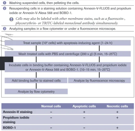
The assay uses a fluorometric immunosorbent enzyme assay (FIENA) principle. The procedure involves:
1 Inducing apoptosis in cells by desired method (for instance 2 x 10⁶ cells). After the
induction, the cells are washed and pelleted by centrifugation.
Preparing samples by resuspending and incubating cells in lysis buffer. After lysis and following centrifugation, samples can be removed for direct analysis or storage.
Coating microplate with anti-caspase 3 solution and blocking of unspecific binding.
Transferring a sample to the anti-caspase 3-coated well of a microplate and capturing of caspase 3.
Washing the immobilized antibody-caspase 3 complexes three times to remove cell components that are not immunoreactive.
Incubating sample with caspase substrate (Ac-DEVD-AFC) that is proteolytically cleaved into free fluorescent AFC.
Measuring generated AFC fluorometrically.
Sample preparation
Treat sample (2 x 10⁶ cells) with apoptosis-inducing agent. Include a negative control
without induction (1–24 h)
Wash treated and control cells with ice-cold PBS and centrifuge (300 x g) (5 min, 15–25°C)
Incubate cell pellet in lysis buffer (1 min, on ice)
Centrifuge at maximum speed in a tabletop centrifuge (1 min, 15–25°C)
Remove 100 µl sample for direct analysis or storage for 1 week at –15 to –25°C
Coating microplate
Incubate microplate with anti-caspase 3 coating solution (either at 37°C for 1h or at 2-8°C over night)
Block unspecific binding by incubation with blocking buffer (30 min, 15–25°C)
Remove blocking solution and wash 3 times with incubation buffer (3 x 1 min, 15–25°C)
Assaying protease activity
Add sample (100 µl lysate, positive control) into microplate well (1 h, 37°C)
Remove sample and wash 3 times with incubation buffer (3 x 1 min, 15–25°C)
Caspase 3 Activity Assay
2
II. Kit content
1. Coating buffer, 10 x 2. Anti-caspase-3, 20 x
3. Blocking buffer, ready-to-use 4. Incubation buffer, 5x 5. DTT, 100 x
6. Substrate solution Ac-DEVD-AFC, 20 x 7. AFC
8. Positive control, apoptotic U937 cell lysate 9. Microplate modules (12 x 8-wells) 10. Adhesive plate cover
Typical results with the kit
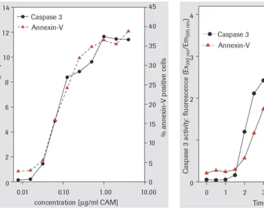
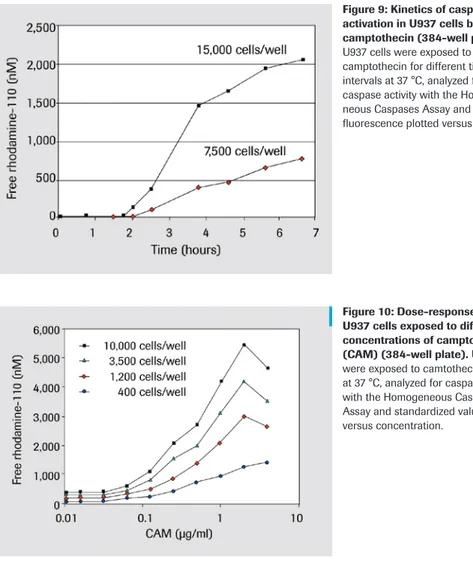
The caspase 3 activity assay has been used to detect caspase 3 activation in U937 cells exposed to different concentrations of the apoptosis inducing agent camptothecin (CAM) (Figure 7, dose response curve). In this model system, the induction of apoptotis in only 5% of U937 cells is sufficient for detection of caspase 3 activation. Caspase 3 activity/fluorochrome development is proportional to the percentage of apoptotic cells.
Figures 7 and 8, demonstrate that Caspase 3 activity and Annexin-V binding correlate very closely in both dose-response and kinetic studies
Figure 7: Dose-response experiment analyzed by the caspase 3 Activity Assay. U937 cells were exposed to different concentrations of camptothecin (CAM) for 4 h at 37°C. Lysates were analyzed for caspase 3 activity and standardized values are plotted versus concentration. Additionally, an aliquot of the same cells was analyzed for Annexin-V binding.
Caspase 3 Activity Assay
2
References
Proliferative and Protective effects of Growth Hormone Secretagogues on Adult Rat 1.
Hippocampal Progenitor cells
Inger Johansson, Silvia Destefanis, N. David Åberg, Maria A.I. Åberg, Klas Blomgren, Changlian Zhu, Corrado Ghè, Riccarda Granata, Ezio Ghigo, Giampiero Muccioli, Peter S. Eriksson, and Jörgen Isgaard
Endocrinology, Jan 2008; 10.1210/en.2007-0733.
ALL1 fusion proteins induce deregulation of EphA7 and ERK phosphorylation in 2.
human acute leukemias
Hiroshi Nakanishi, Tatsuya Nakamura, Eli Canaani, and Carlo M. Croce PNAS, Sep 2007; 104: 14442 - 14447.
Ginkgolide B induces apoptosis and developmental injury in mouse embryonic stem 3.
cells and blastocysts Wen-Hsiung Chan
Hum. Reprod., Nov 2006; 21: 2985 - 2995.
The mechanism of methylselenocysteine and docetaxel synergistic activity in prostate 4.
cancer cells
Rami G. Azrak, Cheryl L. Frank, Xiang Ling, Harry K. Slocum, Fengzhi Li, Barbara A. Foster, and Youcef M. Rustum
Mol. Cancer Ther., Oct 2006; 5: 2540 - 2548.
Plasminogen inhibits TNF-induced apoptosis in monocytes 5.
Jennifer W. Mitchell, Nagyung Baik, Francis J. Castellino, and Lindsey A. Miles Blood, Jun 2006; 107: 4383 - 4390.
Tumour necrosis factor signalling through activation of Kupffer cells plays an 6.
essential role in liver fibrosis of non-alcoholic steatohepatitis in mice
K Tomita, G Tamiya, S Ando, K Ohsumi, T Chiyo, A Mizutani, N Kitamura, K Toda, T Kaneko, Y Horie, J-Y Han, S Kato, M Shimoda, Y Oike, M Tomizawa, S Makino, T Ohkura, H Saito, N Kumagai, H Nagata, H Ishii, and T Hibi
Gut, Mar 2006; 55: 415 - 424.
Inhibition of lung cancer cell growth by quercetin glucuronides via G2/M arrest and 7.
induction of apoptosis
Jen-Hung Yang, Te-Chun Hsia, Hsiu-Maan Kuo, Pei-Dawn Lee Chao, Chi-Chung Chou, Yau-Huei Wei, and Jing-Gung Chung
Drug Metab. Dispos., Feb 2006; 34: 296 - 304.
BRCA1 Phosphorylation Regulates Caspase-3 Activation in UV-Induced Apoptosis 8.
Sarah A. Martin and Toru Ouchi
Cancer Res., Dec 2005; 65: 10657 - 10662.
Hyperglycemia Reduces Survival and Impairs Function of Circulating Blood-Derived 9.
Progenitor Cells
Nicolle Kränkel, Volker Adams, Axel Linke, Stephan Gielen, Sandra Erbs, Karsten Lenk, Gerhard Schuler, and Rainer Hambrecht
Arterioscler. Thromb. Vasc. Biol., Apr 2005; 25: 698 - 703.
Endogenous IGF-I protects human intestinal smooth muscle cells from apoptosis by 10.
regulation of GSK-3 activity John F. Kuemmerle