Predictive role BLVRA mRNA expression
in hepatocellular cancer
Kristýna Kubícková,* Iva Subhanová,† Renáta Konícková,†
Linda Matousová,† Petr Urbánek,* Hana Parobková,‡ Martin Kupec,§ Jirí Pudil,|| Libor Vítek†,¶
* Department of Internal Medicine, 1st Faculty of Medicine, Charles University in Prague, and Military University Hospital, Prague, Czech Republic. † Institute of Medical Biochemistry and Laboratory Diagnostics, General University Hospital, and 1st Faculty of Medicine, Charles University in Prague, Prague, Czech Republic. ‡ Department of Radiology, Military University Hospital, Prague, Czech Republic. § Department of Oncology, 1st Faculty of Medicine, Charles University in Prague, and Thomayer Hospital, Prague, Czech Republic, Prague, Czech Republic. || Department of Surgery, 2nd Faculty of Medicine, Charles University in Prague, and Military University Hospital, Prague, Czech Republic. ¶ 4th Department of Internal Medicine, General University Hospital, 1st Faculty of Medicine, Charles University in Prague, Prague, Czech Republic.
A B S T R A C T A B S T R A C T A B S T R A C T A B S T R A C T A B S T R A C T
Introduction and aim. Introduction and aim.Introduction and aim. Introduction and aim.
Introduction and aim. Hepatocellular carcinoma (HCC) is the most common primary malignant liver tumor. It is primarily caused by hepatic cirrhosis or chronic viral hepatitis. Hepatic carcinogenesis is associated with increased oxidative stress. Thus, the aim of our study was to assess expression of the genes involved in the homeostasis of oxidative stress in patients with HCC. MaterialMaterialMaterialMaterialMaterial and methods.
and methods.and methods. and methods.
and methods. The study was performed on 32 patients with primary HCC (verified by liver histology in 29 patients) and 27 control subjects (in 11 subjects, liver histology was available either with no or minimal changes in the liver tissue). Gene expressions of heme oxygenase 1 (HMOX1), biliverdin reductase A/B (BLVRA/B), NADPH oxidase 2 (NOX2) and p22phox were analyzed in the
liv-er and pliv-eriphliv-eral blood leukocytes (PBL) in the subjects. Results.Results.Results.Results.Results. Compared to controls, almost a 3 times higher mRNA level of BLVRA was detected in livers of HCC patients (p = 0.002); while those of BLVRB as well as HMOX1 were unchanged (p > 0.05). In accord with these results in the liver tissue, BLVRA mRNA levels in PBL were also significantly increased in HCC pa-tients (p = 0.012). mRNA levels of NOX2 and p22phox in the liver tissue, although higher in HCC patients, did not differ significantly
compared to control subjects (p > 0.05). Nevertheless, NOX2 mRNA level in PBL was significantly higher in HCC patients (p = 0.003). Conclusions.Conclusions.Conclusions.Conclusions. BLVRA mRNA levels in the liver as well as in PBL are significantly higher in HCC patients most likelyConclusions. as a feedback mechanism to control increased oxidative stress associated with HCC progression.
Key words. Key words.Key words. Key words.
Key words. Biliverdin reductase. Heme catabolic pathway. Heme oxygenase. Liver cirrhosis. Oxidative stress.
INTRODUCTION
Hepatocellular carcinoma (HCC) is the most common primary malignant liver tumor. The global mortality rate is 694,000 cases per year.1 Worldwide, HCC is the fifth most common cancer in men and the seventh in women; repre-senting the third most frequent cause of cancer-related death.2 The incidence of HCC has different geographical distributions; sub-Saharan Africa, China, Hong Kong and Taiwan being among those regions with the highest inci-dence rates of HCC (i.e., more than 15 cases per 100,000 population per year.3 Conversely, North and South
Amer-ica and most of Europe are among those countries with a lower incidence. However, in recent years, the incidence rates have increased even in these regions, and this trend is expected to continue.4
This malignant disease arises in patients with chronic liver disease, mostly at the stage of liver cirrhosis. Almost 90 percent of cases are due to underlying cirrhosis or chronic hepatitis B and C virus infections.5 Well-defined etiological agents for the development of HCC are aflatoxin and exces-sive alcohol intake. Also, non-alcoholic steatohepatitis due to obesity, metabolic syndrome, and diabetes contribute significantly to the incidence of HCC.6
The Official Journal of the Mexican Association of Hepatology, the Latin-American Association for Study of the Liver and
the Canadian Association for the Study of the Liver
Manuscript received: Manuscript received:Manuscript received:
Manuscript received:Manuscript received: March 22, 2016. Manuscript accepted:Manuscript accepted:Manuscript accepted:Manuscript accepted:Manuscript accepted: April 13, 2016. DOI:10.5604/16652681.1222104.
>
>
>
As far as the risk factors are known, screening programs for the risk groups can be established with an aim to detect tumors in the early stages. However, according to the available data, only 30% of patients with HCC are diag-nosed in the early stages, when curative treatment is still possible.7 The recommended method of surveillance of HCC is a liver ultrasound at 6-month intervals;8 having sensitivity of about 65-80%, and a specificity of almost 90%.9
A combination of liver ultrasound and serum α 1-feto-protein (AFP) had been recommended in the previous guidelines. However, even combinations of these proce-dures is not sufficiently sensitive or specific to be used as a surveillance assay. AFP is typically increased in advanced tumors,10 but can be elevated in cholangiocarcinoma, liver metastases of colorectal cancer, gastric, testicular, or ovari-an covari-ancer; ovari-and it is also raised in cirrhosis. At the time of diagnosis, over 30% of HCC patients have normal serum levels of AFP.11 According to current AASLD guidelines, AFP serology is still considered an inadequate screening test for HCC.
Thus, new biomarkers are needed for early diagnosis of HCC. In fact, several of them are now under investigation including oxidative stress markers, angiogenic growth fac-tors, or other markers such as glypican-3,12 lectin-bound AFP or des-γ carboxyprotrombin.13 However, so far, none of these, has been adequately investigated to be recom-mended as a screening test.
Hepatic carcinogenesis is a complex, multi-step proc-ess involving all pro-oncogenic and protective mecha-nisms. Increased production of reactive oxygen (ROS) and nitrogen species (RONS) is considered to be a trigger point in hepatic carcinogenesis.
NADPH oxidase (NOX) is a multiprotein enzyme complex importantly involved in ROS production,14 a phenomenon believed to contribute significantly to the apoptosis of liver cells.15NOX2, NADPH oxidase proto-typic isoform is activated by the p22phox protein, which stabilizes and binds it to other subunits.16
The important enzyme in the antioxidant defense is heme oxygenase (HMOX), having two isoforms
-HMOX1, highly inducible by oxidative stress, and
HMOX2, the constitutive isoenzyme.17HMOX catalyzes
the degradation of heme to biliverdin, carbon monoxide, and iron. Although controversies exist on the role of
HMOX1 in carcinogenesis,18 both biliverdin and carbon
monoxide exert important protective effects against oxida-tive stress.17
Another key enzyme in the heme catabolic pathway is biliverdin reductase (BLVR), reducing biliverdin to bilirubin, believed to be the most potent endogenous antioxidant substance.19 BLVR exists in two isoforms
-BLVRA, the major enzyme in adults, and BLVRB, the
predominant isoform in in the fetus.20BLVRA has multiple additional functions also acting as a transcription factor,21 a unique serine/threonine/tyrosine kinase,22 as well as cell membrane receptor involved in the immune response.23 Its role in carcinogenesis still remains to be elucidated.24
The aim of our study was to assess the expressions of those genes involved in the homeostasis of oxidative stress in patients with HCC.
MATERIAL AND METHODS
Subjects
The study was performed on 32 patients with primary HCC (verified by liver histology in 29 patients) and 38 control subjects. The HCC patients were diagnosed, fol-lowed, and treated in the Military University Hospital in Prague between 2011 - 2014. Diagnosis of HCC was made by clinical, laboratory, and imaging (CT, MRI) examina-tion. Liver histology was available from 29 patients (23 from CT-guided biopsies, in the remaining 6 patients the material was obtained from surgically-resected tissue).
Blood samples were analyzed in 32 patients with HCC – those with a verified diagnosis by histological examina-tion; plus those with a likely diagnosis of HCC without histological verification, but diagnosed radiologically (typ-ical imaging features were present in a contrast-enhanced study via dynamic CT-scan or MRI). A liver biopsy was not performed in these patients due to disapproval of the patient, advanced stage of the disease or contraindication of a liver biopsy.
As controls, 27 healthy volunteers (blood donors or employees of General Faculty Hospital and 1st Faculty of Medicine, Charles University in Prague) were used for gene expression studies in PBL. Eleven subjects who un-derwent a liver biopsy which resulted in no or minimal changes in the liver tissue (5 with non-alcoholic fatty liver disease, 3 with minimal changes, and 3 with normal liver histology) were used as controls for gene expression stud-ies in liver tissue.
The study was registered under ID: NCT00842205 (www.clinicaltrial.gov). The study protocol conformed to the ethical guidelines of the 1975 Declaration of Helsinki. All subjects involved in the study had provided prior writ-ten informed consent.
Material sampling and storage
collected into PAXgene Blood RNA Tubes (PreAnalytix, Hombrechtikon, Switzerland) and stored at -80°C until total RNA isolation.
Total RNA isolation and reverse transcription
Homogenization of liver tissue and isolation of total RNA was performed using RNeasy Mini (Qiagen, Dallas, TX, USA), isolation total RNA from PBL using a PAX-gene kit (Qiagen, Dallas, TX, USA), according to the man-ufacturer’s instructions. DNase treatment with RNase-free DNase (Qiagen, Dallas, TX, USA); prior to cDNA synthesis was carried out according to the manu-facturer’s instructions. First-strand cDNA was synthe-sized from 0.2 μg of total RNA in a final volume of 20 μg using a High-Capacity cDNA kit (Applied Biosystems, Foster City, CA, USA) according to the manufacturer’s in-structions.
Gene expression quantification
The BLVRA, BLVRB, HMOX1 and hypoxanthine
phos-phoribosyl transferase (HPRT) primer sequences were used as described previously.25 Primers for NOX2 and p22phox were designed using Primer 3 software (http:// frodo.wi.mit.edu/primer3/, accessed 2013 Feb 01) and syn-thesized by Generi Biotech (Hradec Králové, Czech Re-public) (Table 1).
To determine the relative gene expression level of all data analysis, HPRT mRNA expressions were measured as internal controls. The fold change was calculated as 2-ΔΔct. The qPCR was performed in a 20 μL reaction volume, con-taining 4 μL of five-fold diluted cDNA template from a completed RT reaction, 1x SYBR Green Master Mix (Ap-plied Biosystems, Foster City, CA, USA), and 200 nM (400 nM for BLVRB, 1000 nM for p22phox) of forward and re-verse primers. All RT-PCR were set up in 96-well optical plates, and run on an ABI PRISM 7500 Sequence Detector System (Applied Biosystems, Foster City, CA, USA).
The cycling conditions included polymerase activation at 95°C for 10 min, followed with 40 cycles of 95°C for 15 s, and 60°C for 60 s. PCR products were subjected to a melting curve analysis. All samples were analyzed in trip-licates. PCR efficiencies for target and housekeeping cDNA were 96 -105%.
Serum biochemistry
Serum markers of liver injury (ALT, AST, GGT, ALP) and bilirubin were analyzed by routine assays on an auto-mated analyzer (Cobas R8000 Modular analyzer, Roche Diagnostics GmbH, Mannheim, Germany).
Hematologic parameters were also analyzed on automat-ed analyzers - INR on ACL500 (Instrumentation Laborato-ry, Bedford, LaboratoLaborato-ry, Bedford, Massachusetts, USA); hemoglobin and platelets on a Sysmex XE-5000 a XT-2000i (Sysmex Corporation, Kobe, Japan), respectively.
Statistical analysis
Due to the non-normal distribution, data are described as median and IQ range. Differences between the studied groups were evaluated using the Mann-Whitney rank sum test. All analyses were performed with alpha set to 0.05.
RESULTS AND DISCUSSION
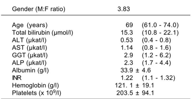
The basic clinical and laboratory characteristics of our HCC patients are shown in table 2. The median age of our HCC patients was 69 years, HCC was almost 4 times more frequent in men than in women. The most prevalent underlying cause of HCC was non-alcoholic steatohepati-tis (NASH), followed with alcoholic liver disease (ALD) (Table 3).
Hepatic carcinogenesis is a complex process, the un-derstanding of which is still far from complete. Neverthe-less, the role of increased oxidative stress and a dysfunctional antioxidant defense system seems to con-tribute significantly to the manifestation and progression
Table 1. Primer sequences for target and internal control genes.
Genes Forward primer Reverse primer Fragment (bp)
HMOX1 GGGTGATAGAAGAGGCCAAGA TTTGAGGAGTTGCAGGAGCT 67
BLVRA TCCCTCTTTGGGGAGCTTTC GGACCCAGACTTGAAATGGAAG 180
BLVRB CCACGTGGTAGTGGGAGATG TCGTGGGACTGAGGTCATTG 110
p22phox CTTCACCCAGTGGTACTTTGG GGCGGTCATGTACTTCTGTCC 130
NOX2 GATTCTCTTGCCAGTCTGTCG ATTCCTGTCCAGTTGTCTTCG 94
HPRT CACTGGCAAAACAATGCAGAC GGGTCCTTTTCACCAGCAAG 96
of HCC (reviewed in reference 26). For instance, mice deficient in CuZn superoxide dismutase, which converts superoxide to H2O2, exhibit increased an incidence of HCC.27 Increased oxidative stress induced by hepatitis C virus infection, resulting in increased hepatic tumorigen-esis, was also reported.28 The role of NOX, the major pro-ducer of superoxide in the mitochondria in mediating transforming growth factor (TGF)-β-induced hepatic fi-brosis and carcinogenesis is also well recognized.29 In this context, it is interesting to note that bilirubin, one of the most important endogenous antioxidant substances,30 is a potent inhibitor of NOX.31,32
For decades the heme catabolic pathway has only been recognized only as a pathway required for the disposal of heme degradation products, but is now believed to play an important role in protection from increased oxidative stress.19 This pathway includes two important enzymes, HMOX and BLVR, reducing biliverdin to bilirubin, the major endogenous antioxidant. HMOX1, an inducible iso-form, is a matter of controversy in terms of its role in car-cinogenesis.18 While in some cancers HMOX1 gene expression may be viewed as a negative prognostic factor,33 clinical studies show that subjects with a more active
HMOX1 gene variant are less likely to develop a variety of
tumors (for review see reference 34). The protective role
of HMOX1 was also reported in an animal model of
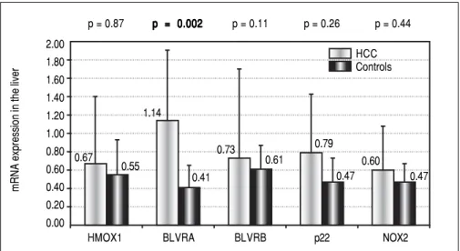
he-patic carcinogenesis, demonstrating increased malignancy when HMOX1 was downregulated.35 However, in our study, we were not able to identify HMOX1 mRNA ex-pression to be differentially modulated in HCC patients, either in the tumor tissue (0.67 ± 0.73 vs. 0.55 ± 0.38, p > 0.05) (Figure 1) or in PBL (1.91 ± 2.1 vs. 1.51 ± 0.62, p > 0.05). Nevertheless, BLVRA mRNA level was significantly upregulated in our HCC patients, both in tumor tissue and PBL. In fact, an almost 3 times higher mRNA levels of
BLVRA were detected in livers of HCC patients
com-pared to controls (1.14 ± 0.76 vs. 0.41 ± 0.24, p = 0.002). In accord with results in the liver tissue, BLVRA mRNA lev-el in PBL was also significantly increased in our HCC pa-tients (1.17 ± 0.46 vs. 0.90 ± 0.29, p = 0.012).
These results are in accord with recent data by De Giorgi, et al. on patients with HCV-induced HCC36 as well as our own results demonstrating increased BLVRA
mRNA expression in HCV infected patients.25 Our data are also corroborated by the immunohistological study by Arena, et al., who showed increased protein expression of BLVR in tumor tissues of patients with melanoma.37 Overexpression of a BLVRA protein was also reported in clinical renal cancers,38 as well as vaginal carcinomas.39
BLVRB, the other BLVR isoenzyme being predominantly
important during fetal life, was reported to be upregulated on a protein level in HCC patients by Melle, et al.;40 and its possible pro-carcinogenic role in HCC was also de-scribed in a recent experimental study by Huan, et al.41 However, we were not able to confirm this data, since only a mild and non-significant elevation of BLVRB
mRNA levels was found in our HCC patients compared to controls (0.73 ± 0.97 vs. 0.61 ± 0.26, p > 0.05) (Figure 1). The functional significance of increased BLVRA
mRNA expression remains to be answered. One expla-nation might be feedback stimulation of the antioxidant defense, which is what we believe is true in HCV-infected patients; those who responded to antiviral therapy had much higher BLVRA mRNA expression compared to non-responders.25 The beneficial role of
BLVRA in preventing oxidative stress-induced
senes-cence was also reported,42 supporting this hypothesis. On the other hand, BLVRA silencing in renal cells had a pro-apoptotic effect,43 and BLVRA, surprisingly serving as a transcription factor, is a known activator of multiple pro-proliferative intracellular signaling pathways.24
BLVRA is also a sensor of intracellular hypoxia; indeed,
its expression has been shown to be significantly in-creased in response to hypoxia.44 Thus, it seems that several mechanisms are behind the up-regulated
BLVRA observed in biological studies.
Table 3. Etiology of the HCC.
Etiology Patients (n)
NASH 10
ALD 7
HCV 5
NASH+ALD 3
HBV 2
Fibrosis (unknown cause) 1
Hemochromatosis 1
NASH: non-alcoholic steatohepatitis. ALD: alcoholic liver disease. HCV: viral hepatitis C. HBV: viral hepatitis B.
Table 2. Clinical and laboratory characteristics of HCC patients.
Gender (M:F ratio) 3.83
Age (years) 69 (61.0 - 74.0) Total bilirubin (μmol/l) 15.3 (10.8 - 22.1) ALT (μkat/l) 0.53 (0.4 - 0.8) AST (μkat/l) 1.14 (0.8 - 1.6) GGT (μkat/l) 2.9 (1.2 - 6.2) ALP (μkat/l) 2.3 (1.7 - 4.4) Albumin (g/l) 33.9 ± 4.6
INR 1.22 (1.1 - 1.32)
Hemoglobin (g/l) 121. 1 ± 19.1 Platelets (x 109/l) 203.5 ± 94.1
HCC Controls
Our explanation of increased BLVRA mRNA expres-sion due to increased oxidative stress might be plausible, as evidenced by increased NOX2 mRNA levels in the PBL of our HCC patients. Although mRNA levels of NOX2
and p22phox in the liver tissue only showed a
non-signifi-cantly higher trend in our HCC patients (0.60 ± vs. 0.47, and 0.79 ± 0.64 vs. 0.47 ± 0.26, respectively, p > 0.05 for both comparisons) (Figure 1), mRNA level of NOX2 in PBL was significantly higher in these HCC patients (1.91 ± 1.21 vs. 1.22 ± 0.52, p = 0.003). Thus, BLVRA may act as a feedback mechanism to scavenge superoxide overpro-duced by increased NOX2.26
It is also important to emphasize the importance of the PBL as a biological material to be used for screening ex-pression studies. The PBL are easily available from blood sampling, and their gene expression profiles are more reli-able compared to liver cancers often containing necrotic tissues.44
CONCLUSION
In conclusion, we observed increased BLVRA mRNA level in the liver as well as in PBL in HCC patients, which seems to be a feedback mechanism to control in-creased oxidative stress associated with HCC progression, as evidenced by increased NOX2 mRNA levels in PBL of these patients. We were not able to assess either the BLVRA protein levels or BLVRA enzyme activities in our biological samples; thus further studies aimed to deeper analyze BLVRA as a possible therapeutic target are certain-ly needed.
ABBREVIATIONS
• AFP:α1-fetoprotein. • ALD: alcoholic liver disease.
• ALT: alanine aminotransferase. • AST: aspartate aminotransferase. • ALP: alkaline phosphatase. • BLVR: biliverdin reductase. • BLVRA: biliverdin reductase A. • BLVRB: biliverdin reductase B. • GGT: gamma glutamyl transpeptidase. • HBV: viral hepatitis B.
• HCC: hepatocellular carcinoma. • HCV: viral hepatitis C.
• HMOX1: heme oxygenase 1.
• HPRT: hypoxanthine phosphoribosyl transferase. • INR: international normalized ratio of prothrombin
time.
• NASH: non-alcoholic steatohepatitis.
• NOX2: NADPH oxidase 2.
• PBL: peripheral blood leukocytes. • RONS: reactive nitrogen species. • ROS: reactive oxygen species. • TGF: transforming growth factor.
ACKNOWLEDGEMENTS
This work was supported by grant IGA MZ NT 13092-4/2012 from the Czech Ministry of Health.
REFERENCES
1. Ferlay J, Shin R, Bray F, al. e. GLOBOCAN 2008 v1.2, Can-cer Incidence and Mortality Worldwide: IARC CanCan-cerBase No.10. Available at http://www.iarc.fr/en/media-centre/iarc-news/2010/globocan2008.php. Access: Feb 15, 2016. 2. Torre LA, Bray F, Siegel RL, Ferlay J, Lortet-Tieulent J, Jemal
A. Global cancer statistics, 2012. CA Cancer J Clin 2015; 65: 87-108.
Figure 1. Figure 1.Figure 1.
Figure 1.Figure 1. mRNA levels of selected genes in the liver of HCC patients. HMOX1: heme oxyge-nase 1. BLVRA: biliverdin reductase A. BLVRB: biliverdin reductase B. p22: gene encoding for p22 phox protein, NOX2: NADPH oxidase 2.
mRNA expression in the liver
2.00 1.80 1.60 1.40 1.20 1.00 0.80 0.60 0.40 0.20 0.00
HMOX1 BLVRA BLVRB p22 NOX2
0.67
0.55 1.14
0.41 0.73
0.61
0.47 0.47
0.79
3. Kew MC. Epidemiology of hepatocellular carcinoma in sub-Saharan Africa. Ann Hepatol 2013; 12: 173-82.
4. Llovet JM. Updated treatment approach to hepatocellular car-cinoma. J Gastroenterol 2005; 40: 225-35.
5. Perz JF, Armstrong GL, Farrington LA, Hutin YJ, Bell BP. The contributions of hepatitis B virus and hepatitis C virus infec-tions to cirrhosis and primary liver cancer worldwide. J Hepatol 2006; 45: 529-38.
6. Caldwell SH, Crespo DM, Kang HS, Al-Osaimi AM. Obesity and hepatocellular carcinoma. Gastroenterology 2004; 127: S97-S103.
7. Llovet JM, Di Bisceglie AM, Bruix J, Kramer BS, Lencioni R, Zhu AX, Sherman M, et al. Design and endpoints of clinical trials in hepatocellular carcinoma. J Natl Cancer Inst 2008; 100: 698-711.
8. Bruix J, Sherman M. Management of hepatocellular carcino-ma: an update. Hepatology 2011; 53: 1020-2.
9. Singal A, Volk ML, Waljee A, Salgia R, Higgins P, Rogers MA, Marrero JA. Meta-analysis: surveillance with ultrasound for early-stage hepatocellular carcinoma in patients with cirrho-sis. Aliment Pharmacol Ther 2009; 30: 37-47.
10. Sherman M. Serological surveillance for hepatocellular carci-noma: time to quit. J Hepatol 2010; 52: 614-5.
11. Colombo M. Screening for cancer in viral hepatitis. Clin Liver Dis 2001; 5: 109-22.
12. Capurro M, Wanless IR, Sherman M, Deboer G, Shi W, Miy-oshi E, Filmus J. Glypican-3: a novel serum and histochemi-cal marker for hepatocellular carcinoma. Gastroenterology
2003; 125: 89-97.
13. Marrero JA, Su GL, Wei W, Emick D, Conjeevaram HS, Fon-tana RJ, Lok AS. Des-gamma carboxyprothrombin can dif-ferentiate hepatocellular carcinoma from nonmalignant chronic liver disease in American patients. Hepatology 2003; 37: 1114-21.
14. Quinn MT, Gauss KA. Structure and regulation of the neu-trophil respiratory burst oxidase: comparison with non-phagocyte oxidases. J Leukoc Biol 2004: 76: 760-81. 15. Lee YS, Kang YS, Lee JS, Nicolova S, Kim JA. Involvement
of NADPH oxidase-mediated generation of reactive oxygen species in the apototic cell death by capsaicin in HepG2 hu-man hepatoma cells. Free Radic Res 2004; 38: 405-12.
16. Bedard K, Krause KH. The NOX family of ROS-generating NADPH oxidases: physiology and pathophysiology. Physiol Rev 2007; 87: 245-313.
17. Ryter SW, Alam J, Choi AM. Heme oxygenase-1/carbon mon-oxide: from basic science to therapeutic applications. Physi-ol Rev 2006; 86: 583-650.
18. Was H, Dulak J, Jozkowicz A. Heme oxygenase-1 in tumor biology and therapy. Curr Drug Targets 2010; 11: 1551-70. 19. Vitek L, Schwertner HA. The heme catabolic pathway and
its protective effects on oxidative stress-mediated diseases.
Adv Clin Chem 2007; 43: 1-57.
20. Cunningham O, Gore MG, Mantle TJ. Initial-rate kinetics of the flavin reductase reaction catalysed by human biliverdin-IX-beta reductase (BVR-B). Biochem J 2000; 345, Pt. 2: 393-9. 21. O’Brien L, Hosick PA, John K, Stec DE, Hinds TD, Jr.
Biliver-din reductase isozymes in metabolism. Trends Endocrinol Metab 2015; 26: 212-20.
22. Lerner-Marmarosh N, Shen J, Torno MD, Kravets A, Hu Z, Maines MD. Human biliverdin reductase: a member of the in-sulin receptor substrate family with serine/threonine/tyro-sine kinase activity. Proc Natl Acad Sci USA 2005; 102: 7109-14.
23. Wegiel B, Baty CJ, Gallo D, Csizmadia E, Scott JR, Akhavan A, Chin BY, et al. Cell surface biliverdin reductase mediates biliverdin-induced anti-inflammatory effects via
phosphati-dylinositol 3-kinase and Akt. J Biol Chem 2009; 284:
21369-78.
24. Gibbs PE, Miralem T, Maines MD. Biliverdin reductase: a tar-get for cancer therapy? Front Pharmacol 2015; 6: 119. 25. Subhanova I, Muchova L, Lenicek M, Vreman HJ, Luksan O,
Kubickova K, Kreidlova M, et al. Expression of biliverdin re-ductase A in peripheral blood leukocytes is associated with treatment response in HCV-infected patients. PloS One
2013; 8: e57555.
26. Choi J, Corder NL, Koduru B, Wang Y. Oxidative stress and hepatic Nox proteins in chronic hepatitis C and hepatocellu-lar carcinoma. Free Radic Biol Med 2014; 72: 267-84. 27. Elchuri S, Oberley TD, Qi W, Eisenstein RS, Jackson
Rob-erts L, Van Remmen H, Epstein CJ, et al. CuZnSOD defi-ciency leads to persistent and widespread oxidative damage and hepatocarcinogenesis later in life. Oncogene
2005; 24: 367-80.
28. Tsukiyama-Kohara K. Role of oxidative stress in hepatocar-cinogenesis induced by hepatitis C virus. Int J Mol Sci 2012; 13: 15271-8.
29. Crosas-Molist E, Bertran E, Fabregat I. Cross-Talk Between TGF-beta and NADPH Oxidases During Liver Fibrosis and Hepatocarcinogenesis. Curr Pharm Des 2015; 21: 5964-76. 30. Pal S, Polyak SJ, Bano N, Qiu WC, Carithers RL, Shuhart M,
Gretch DR, et al. Hepatitis C virus induces oxidative stress, DNA damage and modulates the DNA repair enzyme NEIL1. J Gastroenterol Hepatol 2010; 25: 627-34.
31. Lanone S, Bloc S, Foresti R, Almolki A, Taille C, Callebert J, Conti M, et al. Bilirubin decreases nos2 expression via inhibi-tion of NAD(P)H oxidase: implicainhibi-tions for protecinhibi-tion against endotoxic shock in rats. FASEB J 2005; 19: 1890-2.
32. Fujii M, Inoguchi T, Sasaki S, Maeda Y, Zheng J, Kobayashi K, Takayanagi R. Bilirubin and biliverdin protect rodents against diabetic nephropathy by downregulating NAD(P)H oxidase. Kidney Int 2010; 78: 905-19.
33. Berberat PO, Dambrauskas Z, Gulbinas A, Giese T, Giese N, Kunzli B, Autschbach F, et al. Inhibition of heme oxygen-ase-1 increases responsiveness of pancreatic cancer cells to anticancer treatment. Clin Cancer Res 2005; 11: 3790-8.
34. Exner M, Minar E, Wagner O, Schillinger M. The role of heme oxygenase-1 promoter polymorphisms in human disease.
Free Rad Biol Med 2004; 37: 1097-104.
35. Caballero F, Meiss R, Gimenez A, Batlle A, Vazquez E. Immu-nohistochemical analysis of heme oxygenase-1 in preneo-plastic and neopreneo-plastic lesions during chemical hepatocarcinogenesis. Int J Exp Pathol 2004; 85: 213-21. 36. De Giorgi V, Buonaguro L, Worschech A, Tornesello ML,
Izzo F, Marincola FM, Wang E, et al. Molecular signatures as-sociated with HCV-induced hepatocellular carcinoma and liv-er metastasis. PLoS One 2013;8: e56153.
37. Arena V, Pennacchia I, Guerriero G, Mancuso C. The heme oxygenase/biliverdin reductase system in skin cancers. J Biol Regul Homeost Agents 2015; 29: 259-64.
38. Maines MD, Mayer RD, Erturk E, Huang TJ, Disantagnese A. The oxidoreductase, biliverdin reductase, is induced in hu-man renal carcinoma-pH and cofactor-specific increase in activity. J Urol 1999; 162: 1467-72.
39. Hellman K, Alaiya AA, Becker S, Lomnytska M, Schedvins K, Steinberg W, Hellstrom AC, et al. Differential tissue-specific protein markers of vaginal carcinoma. Br J Cancer 2009;
100: 1303-14.
41. Huan L, Bao C, Chen D, Li Y, Lian J, Ding J, Huang S, et al. MiR-127-5p targets the biliverdin reductase B/NF-kappaB pathway to suppress cell growth in hepatocellular carcino-ma cells. Cancer Sci 2016; 10.1111/cas.12869.
42. Kim SY, Kang HT, Choi HR, Park SC. Biliverdin reductase A in the prevention of cellular senescence against oxidative stress. Exp Mol Med 2011; 43: 15-23.
43. Miralem T, Hu ZB, Torno MD, Lelli KM, Maines MD. Small inter-ference RNA-mediated gene silencing of human biliverdin re-ductase, but not that of heme oxygenase-1, attenuates arsenite-mediated induction of the oxygenase and increases apoptosis in 293A kidney cells. J Biol Chem 2005; 280: 17084-92.
44. Kim SS, Seong S, Lim SH, Kim SY. Biliverdin reductase plays a crucial role in hypoxia-induced chemoresistance in human glioblastoma. Biochem Biophys Res Commun 2013; 440: 658-63.
Correspondence and reprint request:
Libor Vítek, MD, PhD
Institute of Medical Biochemistry and Laboratory Diagnostics, 1st Faculty of Medicine, Charles University in Prague, Na Bojisti
3, Praha 2, 12000, Czech Republic Tel.: +420 2 2496 4203, Fax: +420 2 2496 4203
E-mail: [email protected]