Otras secciones de este sitio:
☞ ☞ ☞ ☞
☞ Índice de este número
☞ ☞ ☞ ☞
☞ Más revistas
☞ ☞ ☞ ☞
☞ Búsqueda
Others sections in this web site:
☞ ☞ ☞ ☞
☞ Contents of this number ☞
☞ ☞ ☞
☞ More journals ☞
☞ ☞ ☞ ☞ Search Artículo:
Alcoholic intake predisposes to more interface hepatitis in chronic hepatitis C
Copyright © 2005: Mexican Association of Hepatology
ANNALS OF HEPATOLOGY Number 3 July-September 2 0 0 5 Volume 4
Annals of Hepatology 4(3) 2005: 176-183
MG
176
edigraphic.com
Annals of Hepatology 2005; 4(3): July-September: 176-183
Annals of Hepatology
1 Gastroenterology Clinic, Heliopolis Hospital. 2 LIM-14, School of Medicine, University of São Paulo.
3 Department of Hematology, Pro-sangue-Hemocentro of São Paulo.
Address for correspondence: Maria de Fátima GS Ribeiro.
Rua Peixoto Gomide, 581/122 – ZC: 01409-001 São Paulo - SP. Brasil.
Tel: 11-3284-9415 Fax: 3287-9357 E-mail: [email protected]
Abbreviation used: HCV: hepatitis C virus; CHC: chronic hepatitis C; ALT: alanine aminotransferase; AST: aspartate aminotransferase; GGT: gamma glutamyl transferase; BMI: body mass index; HBsAg: Hepatitis B surface antigen; IgG: immunoglobulin G; IgM: immunoglobulin M.
Manuscript received and accepted: 1 May and 12 June, 2005. Abstract
Progression of liver disease in chronic hepatitis C de-pends on several factors related to the host, virus and the environment which deserves further investiga-tions. 120 candidates for blood donation with hepatitis C virus were divided into three groups according to alcohol intake: abstainers41, light drinkers36 and heavy drinkers43. Liver histopathology alterations, namely architectural staging, periportal and lobular inflam-mation as well as portal inflammatory infiltrate were graded from 0 to 4 and afterwards divided into light (0 to 2) and severe (3 to 4). There were more drinkers among men (83.5%) than among women (41.5%). Re-garding the three groups, mild periportal inflamma-tion was significantly related with abstainers and light drinkers groups whereas severe periportal inflamma-tion was more predominant in heavy drinkers (p = 0.033). When we compared mild with severe histo-pathological alterations older age was significantly (p = 0.004) associated with severe fibrosis, periportal in-flammation and portal inflammatory infiltrate. In re-lation to enzyme levels a significant difference in fibro-sis and lobular activity was found for ALT, AST and GGT. Only AST was a marker of greater portal in-flammatory infiltrate. Additionally, platelets were sig-nificantly lower in severe fibrosis and in periportal
Original Article
Alcoholic intake predisposes to more interface
hepatitis in chronic hepatitis C
María de Fátima Gomes de Sá Ribeiro;1 Luiz Carlos da Costa Gayotto;2
Dalton de Alencar Fischer Chamone;3 Edna Strauss2
inflammation. Logistic regression analysis identified AST and platelets as independent predictors for severe fibrosis. In conclusion, a correlation was found be-tween alcohol consumption and periportal inflamma-tion. Fibrosis correlated with age, high enzymes levels and low platelets. AST and platelets were the best pre-dictors for severe fibrosis.
Key words: Hepatitis C virus, fibrosis, platelets, etha-nol ingestion, blood donors.
Introduction
Hepatitis C virus (HCV) is recognized as the major
cause of chronic hepatitis worldwide.1 Once HCV
infec-tion develops, it progresses silently to chronic hepatitis in about 80% of the cases. Cirrhosis and hepatocellular
car-cinoma will develop in respectively 20% and 4%.2 The
natural history of HCV infection remains speculative, mainly due to the absence of symptoms that would re-quire medical attention. Predictive factors for the progres-sion of the disease continue to be a major challenge, mainly in asymptomatic patients, as candidates for blood donation. Many studies have been developed in order to try to identify which are the potential prognostic markers
that could lead to the development of cirrhosis.3 Various
factors of the host, the virus and the environment have been investigated, including older age, gender, age at contamination, viral load, genotype, alcohol intake and
disease transmission patterns.4-6
The interaction between HCV infection and alcohol in-take contributes to the severity of liver disease. Accord-ing to many studies, excessive use of alcohol influences
the progression of HCV infection.7-10 However, alcohol
consumption alone may cause damage, but only 20%
de-velop cirrhosis,11 suggesting that other factors may
con-tribute to hepatic disorders. Some investigators, examin-ing HCV carriers with different levels of alcohol con-sumption, found fibrosis progression especially in obese
and diabetes patients.12
Controversies exist concerning histological findings in
chronic hepatitis C13 and in general, hepatic enzyme
val-ues are considered to correlate poorly with
MF Gomes de Sá Ribeiro et al. Alcoholic intake predisposes to more interface hepatitis in chronic hepatitis C 177
edigraphic.com
namely degree of fibrosis, are clinically relevant for stag-ing chronic hepatitis C, the degree of the inflammatory process should also be considered in the natural history or the progression of the disease.
The aim of our study was to evaluate the relationship between different alcohol consumption and grading of histopathological variables.
Patients and methods
During a period of two years, 120 consecutive outpatients at the Hemocentro of São Paulo (Clinic Hospital of FM-USP) were studied. Inclusion of these candidates for blood donation was made according to the following criteria: a) Anti-HCV positive, confirmed by supplementary recombi-nant immunoblot assay (RIBA or LIA and HCV RNA by PCR); b) alanine aminotransferase (ALT) levels above nor-mal; c) age ranging from 18 to 65 years for both genders; d) a written informed consent of the patients, as approved by our hospital ethics committee; e) acceptance of liver biopsy performance. In all patients the following clinical and demo-graphic information were obtained: gender, age, Body Mass Index (BMI) (calculated as weight in kilograms divided by height in meters squared), source of HCV infection (transfu-sion, intravenous drug use, surgery, tattoos, acupuncture, sexual contacts), estimated duration of HCV infection (de-fined as the number of years since the time of the earliest ex-posure to an identified risk factor), laboratory data at the time of liver biopsy: serum alanine aminotransferase (ALT), se-rum aspartate aminotransferase (AST), sese-rum gamma
glutamyl transferase (GGT), platelet number (mm3), serum
Immunoglobulin M (IgM), Immunoglobulin G (IgG). Exclu-sion criteria were: presence of nuclear antibodies, anti-smooth muscle antibodies, anti-liver-kidney microsomes an-tibodies, anti-mitochondrial antibodies in titles > 1/80; Anti-HIV, HBsAg and hepatocellular carcinoma.
Alcohol quantification. All patients and family mem-bers were interviewed to obtain informations about the type of alcoholic drink, the amount of alcohol consumed per day, duration and complete stopping of alcoholic in-take. The quantification of the daily alcohol intake was submitted to the following formula: volume in ml multi-plied by 0.8 (constant) and by the alcoholic degree of the beverage, divided by 100 and express in g/day. In this study the patients were divided into three groups: Group A: abstainers; Group B: light alcohol intake as < 40 g/day for men and < 20 g/day for women and Group C: heavy alcohol intake, defined as > 40 g/day for men and > 20 g/ day for women. Complete stopping was considered only after six months of abstinence. At the time of liver biop-sy, complete stopping was observed in 24.3% of patients in group B and in 29.5% of patients in group C.
Liver Histology. Liver biopsy samples were obtained by percutaneous punction ultrasound oriented. Mean length of biopsy samples was 1.8 ± 0.5 cm and they were formalin-fixed, paraffin-embedded and stained with
Hematoxylin-Eosin, Perls, Masson’s trichrome and reticulin impregna-tion by silver salts. The mean number of portal tracts was 10 ± 4. The histological inflammatory activity (grade) and degree of architectural derangement or fibrosis (stage) of the viral hepatitis were assessed according to the Brazilian
classification of the chronic hepatitis17 as follows: the stage
of fibrosis (F) or architectural alterations was assessed on a four-point scale: F0 = normal lobular architecture; F1 = fi-brous portal expansion; F2 = fifi-brous portal expansion with portal-portal septa; F3 = partial preservation of the lobular architecture with portal-portal and portal-central septa, with occasional sketch of nodules; F4 = cirrhosis. Peripor-tal activity (PA) was graded according to the intensity of necroinflammatory lesions: PA0 = absence of inflamma-tion; PA1 = spill-over of inflammatory cells without inter-face hepatitis; PA2 = mild interinter-face hepatitis; PA3 = moder-ate interface hepatitis; PA4 = severe interface hepatitis. Lob-ular activity (LA) was graded as following: LA0 =normal hepatocytes; LA1 = mild alterations in hepatocytes, includ-ing ballooninclud-ing degeneration or acidophil body, associated with lymphocytes/histiocytes infiltrate and rare foci of necro-sis; LA2 = foci necrosis of hepatocytes, numerous areas of mononuclear aggregates; LA3 = foci necrosis of hepatocytes surrounded by mononuclear aggregates in several areas, lim-ited areas of confluent necrosis; LA4 = foci necrosis of hepa-tocytes surrounded by mononuclear aggregates in numerous areas, with extensive/multiple confluent necrosis. Portal In-filtrate (PI) was graded: PI0 = rare portal lymphocytes; PI1 = mild increase of the number of portal lymphocytes; PI2 = moderate increase of the number of portal lymphocytes; PI3 = marked increase of the number of portal lymphocytes; PI4 = very marked increase of the number of portal lymphocytes. Steatosis was scored as present or absent.
For statistical comparisons between groups or vari-ables, two levels of histopathological alterations were considered: grades 0, 1 and 2 were grouped as light (ab-sent or mild) and grades 3 and 4 as severe.
Statistical Analysis. Chi-square test was used for analy-sis of qualitative variables, whereas quantitative variables as age, BMI, duration of infection and platelet were as-sessed using analysis of variance (ANOVA). When signif-icant, the Tukey or the Student’s t-test was used to discrim-inate the differences. Kruskal-Wallis test, Dunn test or Wil-coxon rank-sum test were also used to analyze variables as ethanol (g/d), duration of alcohol intake, ALT, AST, GGT, IgG, IgM, and abstinence. Multiple logistic regression analysis with stepwise of selected variables was performed to correlate the degrees and stages of the histopathological alterations with other clinical and biochemical data. Odds ratios and 95% confidence intervals were calculated. Val-ues of p < 0.05 were considered to be significant.
Results
ac-MG
178
edigraphic.com
:rop odarobale FDP
VC ed AS, cidemihparG
arap
acidémoiB arutaretiL :cihpargideM
sustraídode-m.e.d.i.g.r.a.p.h.i.c sustraídode-m.e.d.i.g.r.a.p.h.i.c
cihpargidemedodabor
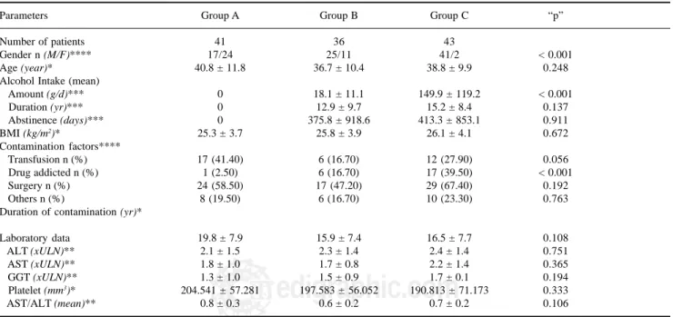
cording to alcohol intake as follow: the group A, abstain-ers: n = 41; group B, light drinkabstain-ers: n = 36 and group C, heavy drinkers: n = 43. Male patients were common in groups B and C (69.4% and 95.3% respectively), whereas female patients more common in group A (58.5%) (p < 0.001). The epidemiological, clinical and demographic data of the 120 patients at the time of the liver biopsy are shown in table I. The three groups were not different in terms of age, BMI, duration of infection or surgery as a contamination factor. Groups B and C were not different in terms of duration of alcohol intake and abstinence. Transfusion as a risk factor was more frequent in the ab-stainers (p = 0.056) whereas drug addiction predominated in heavy drinkers (p < 0.001). The three groups were not different in terms of ALT, AST, GGT and platelet count
(Table I). AST/ALT index was also analyzed in the three
groups and no differences were found (p = 0.106). Histopathological Data. There was no difference be-tween findings of liver histology concerning the degree of fibrosis, periportal inflammatory activity, lobular inflam-mation, portal inflammatory infiltrate and steatosis, among the three groups.
Grouping the results of these variables as light (grades 0, 1 and 2) or severe (grades 3 and 4), no differences were found in architectural alterations, lobular inflammatory activity or portal inflammatory infiltrate. Nevertheless, heavy drinkers have shown higher percentages of grades 3 and 4 of interface hepatitis when compared to abstain-ers and light drinkabstain-ers (p = 0.033), as shown in table II.
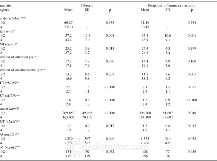
Demographic, epidemiological and biochemical data analyzed according to the different degrees of fibrosis and periportal inflammatory activity are shown in table
III. Among the demographic variables, older age (p =
0.004) and BMI (p = 0.013) were significantly associated with severe fibrosis. Otherwise, the gender, duration of infection and duration of alcohol intake were not associ-ated with severe fibrosis. The serum ALT (p < 0.001), AST (p < 0.001), GGT (p = 0.012) and IgG (p = 0.040) values were significantly higher in severe fibrosis com-pared to no or mild fibrosis. Additionally, the platelet count was significantly lower (p < 0.001) in severe fibro-sis compared to no or mild fibrofibro-sis. The periportal in-flammatory activity was shown to be significantly more severe in older age (p = 0.001) and duration of alcohol in-take (p = 0.001), compared to no or mild periportal activ-ity. The ALT (p = 0.031), AST (p < 0.001) and GGT (p = 0.013) values were significantly higher in severe peripor-tal inflammatory activity (grades 3 and 4), whereas the platelet count was significantly (p = 0.049) lower. On the other hand, gender, BMI, duration of infection, IgG, IgM, did not differentiate between mild and severe periportal inflammatory activity.
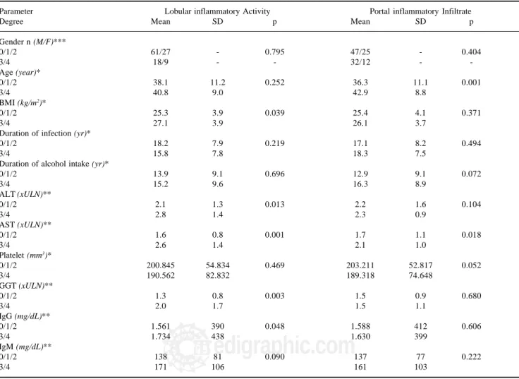
Demographic, epidemiological and biochemical data analyzed according to the different degrees of lobular in-flammatory activity and portal inin-flammatory infiltrate are shown in table IV. There was a significant association be-tween BMI (P = 0.039), higher levels of ALT (p = 0.013), AST (p = 0.001) and GGT (p = 0.003) and severe lobular
Table I. Comparative analysis of the demographic data, epidemiological and biochemical characterization of the patients at the time of liver biopsy
Parameters Group A Group B Group C “p”
Number of patients 41 36 43
Gender n (M/F)**** 17/24 25/11 41/2 < 0.001
Age (year)* 40.8 ± 11.8 36.7 ± 10.4 38.8 ± 9.9 0.248
Alcohol Intake (mean)
Amount (g/d)*** 0 18.1 ± 11.1 149.9 ± 119.2 < 0.001
Duration (yr)*** 0 12.9 ± 9.7 15.2 ± 8.4 0.137
Abstinence (days)*** 0 375.8 ± 918.6 413.3 ± 853.1 0.911
BMI (kg/m2)* 25.3 ± 3.7 25.8 ± 3.9 26.1 ± 4.1 0.672
Contamination factors****
Transfusion n (%) 17 (41.40) 6 (16.70) 12 (27.90) 0.056
Drug addicted n (%) 1 (2.50) 6 (16.70) 17 (39.50) < 0.001
Surgery n (%) 24 (58.50) 17 (47.20) 29 (67.40) 0.192
Others n (%) 8 (19.50) 6 (16.70) 10 (23.30) 0.763
Duration of contamination (yr)*
Laboratory data 19.8 ± 7.9 15.9 ± 7.4 16.5 ± 7.7 0.108
ALT (xULN)** 2.1 ± 1.5 2.3 ± 1.4 2.4 ± 1.4 0.751
AST (xULN)** 1.8 ± 1.0 1.7 ± 0.8 2.2 ± 1.4 0.365
GGT (xULN)** 1.3 ± 1.0 1.5 ± 0.9 1.7 ± 0.1 0.194
Platelet (mm3)* 204.541 ± 57.281 197.583 ± 56.052 190.813 ± 71.173 0.333
AST/ALT (mean)** 0.8 ± 0.3 0.6 ± 0.2 0.7 ± 0.2 0.106
Abbreviations: Group A: abstainers; Group B: light drinkers; Group C: heavy drinkers. M, male; F, female; xULN, x upper limit of normal; BMI: body mass index Values in mean and SD
MF Gomes de Sá Ribeiro et al. Alcoholic intake predisposes to more interface hepatitis in chronic hepatitis C 179
edigraphic.com
Table II. Histopathological results in the three studied groups.
Groups
Abstainers Light drinkers Heavy drinkers Total p*
Fibrosis 0.868
0/1/2 30 73.20% 27 75.00% 30 69.80% 87 72.50%
3/4 11 26.80% 9 25.00% 13 30.20% 33 27.50%
Periportal activity 0.033
0/1/2 26 63.40% 26 72.20% 18 43.90% 70 59.30%
3/4 15 36.60% 10 27.80% 23 56.10% 48 40.70%
Portal inflammatory infiltrate 0.675
0/1/2 27 67.50% 21 58.30% 24 60.00% 72 62.10%
3/4 13 32.50% 15 41.70% 16 40.00% 44 37.90%
Lobular activity 0.481
0/1/2 31 79.50% 29 80.60% 28 70.00% 88 76.50%
3/4 8 20.50% 7 19.40% 12 30.00% 27 23.50%
Abstinence 0.272
< 6 months - - 29 80.60% 30 69.80% 59 74.70%
> 6 months - - 7 19.40% 13 30.20% 20 25.30%
* Chi-Square of Pearson
Table III. Demographic, epidemiological and biochemical data, analyzed according to different degrees of fibrosis and periportal inflammatory activity.
Parameter Fibrosis Periportal inflammatory activity
Degrees Mean SD p Mean SD p
Gender n (M/F)***
0/1/2 60/27 - 0.938 51/19 - 0.234
3/4 23/10 - - 30/18 -
-Age (year)*
0/1/2 37.2 11.3 0.004 35.4 10.6 0.001
3/4 43.4 7.9 43.9 9.1
BMI (kg/m2)*
0/1/2 25.2 3.9 0.013 25.4 4.3 0.299
3/4 27.2 3.7 26.2 3.4
Duration of infection (yr)*
0/1/2 17.3 7.9 0.790 16.4 7.9 0.100
3/4 17.8 7.9 19.1 7.6
Duration of alcohol intake (yr)**
0/1/2 13.3 8.6 0.207 11.1 7.6 0.001
3/4 16.6 9.8 18.2 9.5
ALT (xULN)**
0/1/2 2.1 1.5 < 0.001 2.1 1.5 0.031
3/4 2.7 1.1 2.4 1.1
AST (xULN)**
0/1/2 1.6 0.8 < 0.001 1.6 0.9 < 0.001
3/4 2.8 1.3 2.4 1.2
Platelet (mm3)*
0/1/2 209.850 49.965 < 0.001 206.800 51.607 0.049
3/4 165.006 78.109 184.108 72.607
GGT (xULN)**
0/1/2 1.3 0.9 0.012 1.3 0.8 0.013
3/4 1.9 1.2 1.7 1.1
IgG (mg/dL)**
0/1/2 1.539 307 0.040 1.533 314 0.078
3/4 1.771 567 1.706 497
IgM (mg/dL)**
0/1/2 134 70 0.052 138 77 0.456
3/4 176 119 156 102
M, male, F, female; xULN, x upper limit of normal * Student t-test
MG
180
edigraphic.com
inflammation. The other studied variables did not show differences between the groups. There was a significant association between older age (p = 0.001) and AST (p = 0.018) with severe portal inflammatory infiltrate. For the other variables there were no significant correlations.
Logistic regression analyses: Considering the two levels of the architectural alterations in relation to the different clinical variables, it was shown that the only independent predictive factor of severe fibrosis were AST (odds ratio = 2.734 and CI 95% 1.687–4.431) and platelet count (odds ratio = 0.862 and CI 95% 0.782– 0.950). Considering these variables, the best sensitivity and specificity cut-off was reached as respectively 78.8 and 71.3 (see figure 1a). Considering the two levels of periportal inflammation activity in relation to the dif-ferent clinical parameters of alcoholic groups B and C it was shown that the only independent predictive fac-tors of severe periportal inflammation were AST (odds ratio = 2.684 and CI 95% 1.437–5.013) and duration of
alcohol intake (odds ratio 1.112 CI 95% 1.041–1.188). Considering the studied parameters, the best cut-off for the sensitivity was 75.8% and for specificity 70.5%
(see figure 1b).
Considering the two levels of lobular inflammation ac-tivity in relation to the different clinical parameters, it was shown that the only independent predictive factors of severe lobular inflammation were AST (odds ratio 2.049 CI 95% 1.304-3.221) and GGT (odds ratio 1.866 CI 95% 1.134-3.069). Considering these variables, the best cut-off for sensitivity was 66.7 and specificity 70.5 as shown in figure 1c.
Considering the two levels of portal inflammatory in-filtrate in relation to the different clinical parameters, it was shown that the only independent predictive factor of severe portal inflammatory infiltrate was age (odds ratio 1.071 CI 95% 1.028-1.115). Considering this variable, the best cut-off for sensitivity was 63.6 and specificity 67.6 as shown in figure 1d.
Table IV. Demographic, epidemiological and biochemical data analyzed according to different degree of lobular inflammatory activity and portal inflammatory infiltrate.
Parameter Lobular inflammatory Activity Portal inflammatory Infiltrate
Degree Mean SD p Mean SD p
Gender n (M/F)***
0/1/2 61/27 - 0.795 47/25 - 0.404
3/4 18/9 - - 32/12 -
-Age (year)*
0/1/2 38.1 11.2 0.252 36.3 11.1 0.001
3/4 40.8 9.0 42.9 8.8
BMI (kg/m2)*
0/1/2 25.3 3.9 0.039 25.4 4.1 0.371
3/4 27.1 3.9 26.1 3.7
Duration of infection (yr)*
0/1/2 18.2 7.9 0.219 17.1 8.2 0.494
3/4 15.8 7.8 18.3 7.5
Duration of alcohol intake (yr)*
0/1/2 13.9 9.1 0.696 12.9 9.1 0.072
3/4 15.2 9.6 16.3 8.9
ALT (xULN)**
0/1/2 2.1 1.3 0.013 2.2 1.6 0.104
3/4 2.8 1.4 2.3 0.9
AST (xULN)**
0/1/2 1.6 0.8 0.001 1.7 1.1 0.018
3/4 2.6 1.4 2.1 1.0
Platelet (mm3)*
0/1/2 200.845 54.834 0.469 203.211 52.817 0.052
3/4 190.562 82.832 189.318 74.648
GGT (xULN)**
0/1/2 1.3 0.8 0.003 1.5 0.9 0.680
3/4 2.0 1.7 1.5 1.1
IgG (mg/dL)**
0/1/2 1.561 390 0.048 1.588 412 0.606
3/4 1.734 438 1.630 399
IgM (mg/dL)**
0/1/2 138 81 0.090 137 77 0.222
3/4 171 106 161 103
M, male; F, Female ULN, x upper limit of normal * Student’s t-test
MF Gomes de Sá Ribeiro et al. Alcoholic intake predisposes to more interface hepatitis in chronic hepatitis C 181
edigraphic.com
DiscussionThis study shows that histopathological parameters like fibrosis, lobular inflammation and portal infiltrate in candidates for blood donation with hepatitis C did not differ when alcohol abstainers, light drinkers and heavy drinkers were compared. Only periportal inflam-mation was more severe in heavy drinkers. The most commonly used parameter to evaluate the progression of
liver disease is fibrosis,18 and many studies have
indicat-ed an increasindicat-ed risk of progression of chronic hepatitis
C with increasing levels of alcohol consumption.14,19 As
some of these studies have included higher percentages of cirrhotic patients than ours, our results may reflect an
earlier phase of disease progression.20 As the
inflamma-tory process precedes fibrosis development, it is under-standable that there was a statistically significant differ-ence for inflammation but not for fibrosis. The histo-pathological finding of periportal inflammation cannot be associated with isolated alcohol abuse. In fact, the most common histopathological alcoholic alterations
predominate in zone 3 of the Rappaport acinus.21 On the
other hand, we are dealing with an association of hepati-tis C and alcohol, and the histopathological parameters, suggestive of alcoholic damage, are different from those
of viral hepatitis.22 Another possibility which explains
no differences in fibrosis, when abstainers were com-pared to light or heavy drinkers, is a beta error. In fact, the greater degree of histological architectural alter-ations was found in only 13 patients, the majority of them (61.5%) being heavy drinkers. One can speculate that, in a larger population, a significant difference
would appear, as shown by other authors.6,14
The comparison of demographic and clinical parame-ters between groups have shown interesting differences,
namely, more female are abstainers (58.5%).23 In relation
to contamination factors, the two major risk factors for HCV infection were prevalent in different groups: blood transfusion was more frequently found in abstainers whereas drug addiction was significantly higher in heavy drinkers. It is uncertain why heavy drinkers are more like-ly to be infected with HCV, although alcoholics are more prone to have used injection drugs or have had other
ex-posures to the virus.24,25 Interaction of alcohol and drugs
are considered to enhance susceptibility to infections,26
probably due to alterations in the immune system.27
ALT is commonly used as a marker of hepatic
inflam-mation and damage, especially in HCV infection,28
al-though it can be more pronounced in patients with both
HCV infection and alcoholic liver disease.29 In contrast,
some studies have found persistently normal ALT levels in alcoholic patients HCV infected with a slow
progres-sion rate of fibrosis.30
Differently from other studies,31 we have analyzed
his-topathological variables separately and in relation to epi-demiological and laboratory parameters. An independent risk factor associated with fibrosis is older age, as shown
in several epidemiological studies.32,33 Like others,14 we
have found older age related to fibrosis and periportal in-flammation. The amount of portal inflammatory infiltrate is not usually taken into consideration in the various
clas-sifications of chronic hepatitis,31,34 but is considered in
our classification.17 The only parameter that differentiated
slight from severe portal infiltrate was age. Younger peo-ple more frequently had lower grades of portal inflamma-tory infiltrate. The strong relationship of age with
severi-Figure 1. The cut-off values near the crossing between the sensitivi-ty and specificisensitivi-ty are shown in the figure above.
They correspond to the probability of grades 3 or 4 of each histopatho-logical variable be detected, by cli-nic and biochemical results, accor-ding to the logistic regression analysis.
Concerning fibrosis, the sensitivity was 79.30 and specificity 76.50 for AST and platelets. Concerning pe-riportal inflammation, the sensiti-vity was 69.70 and specificity 71.10 for AST and duration of al-cohol intake. Concerning lobular inflammation, the sensitivity was 69.20 and specificity 64.40 for AST and GGT.
Concerning portal inflammatory infiltrate, the sensitivity was 65.10 and specificity 63.00 for age.
0 10 20 30 40 50 60 70 80 90 100
0 0.1 0.2 0.3 0.4 0.5 0.6 0.7 0.8 0.9 1
Fibrosis (3 or 4) Probability
Sensiti
vity/Specif
icity
Sensitivity Specificity
Periportal Inflammation (3 or 4) Probability
0 10 20 30 40 50 60 70 80 90 100
0 0.1 0.2 0.3 0.4 0.5 0.6 0.7 0.8 0.9 1
Lobular Inflammation (3 or 4) Probability
0 10 20 30 40 50 60 70 80 90 100
0 0.1 0.2 0.3 0.4 0.5 0.6 0.7 0.8 0.9 1
Portal Infiltrate (3 or 4) Probability 0 10 20 30 40 50 60 70 80 90 100 0 10 20 30 40 50 60 70 80 90 100
0 0.1 0.2 0.3 0.4 0.5 0.6 0.7 0.8 0.9 1
MG
182
edigraphic.com
ty of chronic hepatitis may reflect qualitative and quanti-tative changes in the immune response which occur with increasing age, or more likely indicate the longer disease
duration.35 To our knowledge this is the first report of
re-lation between older age and greater portal infiltrate in hepatitis C.
Duration of infection is an important parameter in the progression of the disease, but it has been described in com-munity-based studies that HCV infection may persist for
de-cades without causing clinically apparent liver disease.36,37
Lack of differences regarding duration of infection in our study may be related to the blood donor population, in which a mild liver disease was more frequently found.
Elevated ALT, AST and GGT levels are associated
with fibrosis38,39 and were correlated also with periportal
and lobular inflammation in our study although only AST was an independent predictor of severe fibrosis. One of the possible reasons to explain higher AST values could be HCV-induced liver injury in the mitochondrial fraction of hepatocytes where it is present, and/or due to
the presence of alcohol.40 On the other hand, decreased
platelet count has been used to non-invasively assess the
severity of disease in patients with chronic liver disease.41
Platelet counts clearly separated early from late stages of fibrosis as well as the degrees of periportal inflammation. Although the hepatitis C virus by itself may be related to low platelet counts, many authors have had similar
re-sults.16,42,43 To amplify the opposite relationship between
AST levels and platelets count in determining the stage of fibrosis, a novel index, called “AST to platelet ratio
in-dex” (APRI) has recently been proposed.44 Our results are
in keeping with this study.
Obesity has also been associated with more advanced
degrees of fibrosis in CHC28 and an association between
BMI and fibrosis has been demonstrated.45 In our series,
39.3% of patients presented steatosis, similarly to other histopathological studies of CHC, which have reported microvesicular and macrovesicular fatty change in 31%
to 72% of patients.46-48 Lately, even low alcohol intake
has been shown to play a synergistic effect on HCV
in-fection leading to steatosis.49
Using logistic regression analysis it was remarkable to see that AST was an independent predictor of severity not only for fibrosis and periportal inflammation but also for lobular inflammation. On the other hand, platelet was an independent predictor only for fibrosis. The levels of sen-sitivity varying from 65.1 up to 79.3, and specificity ranging from 63 up to 76.5, were unexpectedly high. There are no doubts about the clinical usefulness of fibro-sis, recognized as the most important factor in the pro-gression of liver disease. So, it was rewarding to find the best sensitivity and specificity levels for the detection of fibrosis, by the use of AST values and platelets counts.
In conclusion, epidemiological and biochemical data can predict both fibrosis and periportal inflammation with acceptable levels of sensitivity and specificity.
References
1. Choo QL, Kuo G, Weiner AJ, Overby LR, Bradley DW, Houghton M. Isolation of a cDNA clone derived from a bloodborne non-A, non-B Hepatitis genome. Science 1989; 44: 359-62.
2. Seeff LB. Natural history of chronic hepatitis C. Hepatology 2002; 36: S35-S46.
3. Mohsen AH. Trent HCV Study Group. The epidemiology of hepati-tis C in a UK health regional population of 5.12 million. GUT 2001; 48: 707-13.
4. Smith BC, Chapman CE, Burt AD, Toms GL, Bassendine MF. Outcome of post-transfusion hepatitis C: disease severity in blood-component recipients and their implicated donors. QJMed 1997; 90: 587-92.
5. Tassopoulos NC, Papatheodoridis GV, Katsoulidou A, Delladetsima JK, Sypsa V, Touloumi G, Nikandros M, et al. Factors associated with severity and disease progression in chronic hepatitis C. Hepatogastroenterol 1998; 45: 1678-83.
6. Pessione F, Degos F, Marcellin P, Duchatelle V, Njapoum C, Martinot-Peignoux M, Degott C, et al. Effect of alcohol consumption on se-rum hepatitis C virus RNA and histological lesions in chronic hepa-titis C. Hepatology 1998; 27(6): 1717-22.
7. Donato F, Tagger A, Gelatti U, Parrinello G, Boffetta P, Albertini A, et al. Alcohol and hepatocellular carcinoma: the effect of lifetime intake and hepatitis C infections in men and women. Am J Epidemiol 2002; 155(4): 323-31.
8. Harris HE, Ramsay ME, Andrews N, Eldridge KP on behalf of the HCV National Register Steering Group. Clinical course of hepatitis C virus during the first decade of infection: cohort study. BMJ 2002; 324: 450-3.
9. Ostapowicz G, Watson KJR, Locarnini SA, Desmond PV. Role of alcohol in the progression of liver disease caused by hepatitis C vi-rus infection. Hepatology 1998; 27(6): 1730-5.
10. Corrao G, Aricò S. Independent and combined action of hepatitis C virus infection and alcohol consumption on the risk of symptomatic liver cirrhosis. Hepatology 1998; 27: 914-9.
11. Patek AJ, Toth IG, Saunders MG, Castro GAH, Emgel JI. Alcohol and dietary factors in cirrhosis. An epidemiological study of 304 alcoholic patients. Arch Intern Med 1975; 135: 1053-7.
12. Monto A, Alonzo J, Watson JJ, Grunfeld C, Wright TL. Steatosis in chronic hepatitis C: relative contributions of obesity, diabetes melli-tus and alcohol. Hepatology 2002; 36(3): 729-36.
13. Marcellin P, Asselah T, Boyer N. Fibrosis and disease progression in hepatitis C. Hepatology 2002; 36: S47-S56.
14. Poynard T, Bedossa P, Opolon P, for the OBSVIRC, METAVIR, CLINIVIR and DOSVIRC groups. Natural history of liver fibrosis progression in patients with chronic hepatitis C. Lancet 1997; 349: 825-32.
15. Alter MJ, Marcolis HS, Krawczyski K, Judson FN, Mares A, Alexander WJ, Hu PY, et al. The natural history of community-ac-quired hepatitis C in the United States. N Engl J Med 1992; 327: 1899-1905.
16. Poynard T, Bedossa P. Age and platelet count: a simple index for predicting the presence of histological lesions in patients with anti-bodies to hepatitis C virus. METAVIR and CLINIVIR Cooperative Study Groups. J Viral Hepatol 1997; 4(3): 199-208.
17. Gayotto LCC e Comitê SBP/SBH. Visão histórica e consenso nacional sobre a classificação das hepatites crônicas. Projeto do Clube de Patologia Hepática da Sociedade Brasileira de Patologia aprovado pela Sociedade Brasileira de Hepatologia. GED 2000; 19(3): 137-40.
18. Poynard T, Ratziu V, Benmanov Y, Martino V, Bedossa P, Opolon P. Fibrosis in patients with chronic hepatitis C: detection and signifi-cance. Semin Liver Dis 2000; 20(1): 47-55.
19. Peters MG, Terrault NA. Alcohol use and hepatitis C. Hepatology 2002; 36: S220-S5.
MF Gomes de Sá Ribeiro et al. Alcoholic intake predisposes to more interface hepatitis in chronic hepatitis C 183
edigraphic.com
21. Maddrey WC. Alcoholic hepatitis: clinicopathologic features and therapy. Semin Liver Dis 1988; 8: 91-102.
22. Hall P de la M. Alcoholic liver disease. In: Pathology of the liver. Mac Sween RNM, Anthony PP, Portmann BC, Burt AD, reds., 3nd ed. Churchill Livingstone, Edinburgh, 1994.
23. Strauss E, Nemoto TC, Borges GT, Cunha AA, Freitas GC, Parrado MAR, et al. Chronic alcoholism: evaluation epidemiologic clinic in 300 cases. GED, Gastroenterol Endoscop Dig 1998; 17: S75. 24. Mendenhall CL, Moritz T, Rouster S, Roselle G, Polito A, Quan S,
Dinelle RK. Epidemiology of hepatitis C among veterans with alco-holic liver disease. The VA Cooperative Study Group 275. Am J Gastroenterol 1993; 88: 1022-6.
25. Verbaan H, Anderson K, Eriksson S. Intravenous drug abuse-the major route of hepatitis C virus transmission among alcohol-dependent individuals? Scand J Gastroenterol 1993; 28: 714-8.
26. MacGregor RR. Alcohol and immune defense. JAMA 1986; 256: 1474-9. 27. Cook RT. Alcohol abuse, alcoholism, and damage to the immune
system-a review. Alcohol: Clin Exp Res 1998; 22: 1927-42. 28. Ortiz V, Berenguer M, Rayón JM, Carrasco D, Berenguer J.
Contri-bution of obesity to hepatitis C-related fibrosis progression. Am J Gastroenterol 2002; 97(9): 2408-2414.
29. Fong TL, Kanel GC, Conrad A, Valinluck B, Charboneau F, Adkins RH. Clinical significance of concomitant hepatitis C infection in pa-tients with alcoholic liver disease. Hepatology 1994; 19(3): 554-557. 30. Mathurin P, Moussalli J, Cadranel JF, Thibault V, Charlotte F, Dumouchel P, Cazier A, et al. Slow progression rate of fibrosis in hepatitis C virus patients with persistently normal alanine transami-nase activity. Hepatology 1998; 27: 868-72.
31. Bedossa P, Poynard T. An algorithm for the grading of activity in chronic hepatitis C. Hepatology 1996; 24: 289-93.
32. Iacono OL, Decastro M, Garcia-Buey L, Garcia-Monzon C, Borque MJ, Sanz P, Garcia-Sanchez A, et al. Epidemiological risk factors and clinic-pathological presentation in chronic hepatitis C. Hepato-Gastroenterol 1998; 45: 1715-1721.
33. Poynard T, Ratziu V, Charlotte F, Goodman Z, McHutchisonJ, Albrecht J. Rates and risk factors of liver fibrosis progression in pa-tients with chronic hepatitis C. J Hepatol 2001; 34: 730-739. 34. Desmet VJ, Gerber M, Hoofnagle JH, Manns M, Scheuer PJ.
Classi-fication of chronic hepatitis: Diagnosis, grading and staging. Hepatology 1994; 19: 1513-1520.
35. Delladetsima JK, Rassidakis G, Tassopoulos NC, Papatheodoridis GV, Smyrnoff T, Vafiadis I. Histopathology of chronic hepatitis C in relation to epidemiological factors. J Hepatol 1996; 24: 27-32. 36. Thomas DL, Astemborski J, Rai RM, Anania FA, Schaeffer M, Galai
N, Nolt K, et al. The natural history of hepatitis C virus infection. Host, viral, and environmental factors. JAMA 2000; 284(4): 450-456.
37. Rodger AJ, Roberts S, Lanigan A, Bowden S, Brown T, Crofts N. Assessment of long-term outcomes of community-acquired hepati-tis C infection in a cohort with sera stored from 1971 to 1975. Hepatology 2000; 32: 582-587.
38. Ghany MG, Kleiner DE, Alter H, Doo E, Khokar F, Promrat K, Herion D, et al. Progression of fibrosis in chronic hepatitis C. Gastroenter-ology 2003; 124: 97-104.
39. Silva IS, Ferraz ML, Perez RM, Lanzoni VP, Figueiredo VM, Silva AE. Role of gamma-glutamyl transferase activity in patients with chronic hepatitis C virus infection. J Gastroenterol Hepatol 2004; 19(3): 314-318.
40. Assy N, Minuk GY. Serum aspartate but not alanine aminotrans-ferase levels help to predict the histological features of chronic hepatitis C viral infections in adults. Am J Gastroenterol 2000; 95(6): 1545-50.
41. Pohl A, Behling C, Oliver D, Kilani M, Monson P, Hassanein T. Serum aminotransferase levels and platelet counts as predictors of degree of fibrosis in chronic hepatitis C virus infection. Am J Gastroenterol 2001; 96(11): 3142-3146.
42. Streiff MB, Mehta S, Thomas DL. Peripheral blood count abnor-malities among patients with hepatitis C in the United States. Hepatology 2002; 35(4): 947-952.
43. Bauduer F, Marty F, Larrouy M, Ducout L. Immunologic thromb-ocytopenic purpura as presenting symptom of hepatitis C infection. Am J Hematol 1998; 57(4): 338-340.
44. Wai CT, Greenson JK, Fontana RJ, Kalbfleisch JD, Marrero JA, Conjeevaram HS, Lok AS. A simple noninvasive index can predict both significant fibrosis and cirrhosis in patients with chronic hepa-titis C. Hepatology 2003; 38: 518-526.
45. Hourigan LF, Macdonald GA, Purdie D, Whitehall VH, Shorthouse C, Clouston A, Powell EE. Fibrosis in chronic hepatitis C correlates significantly with body mass index and steatosis. Hepatology 1999; 29: 1215-9.
46. Scheuer PJ, Ashrafzadeh P, Sherlock S, Brown D, Dusheiko GM. The pathology of hepatitis C. Hepatology 1992; 15: 567-571. 47. Bach N, Thung SN, Schaffner F. The histological features of chronic
hepatitis C and autoimmune chronic hepatitis C: a comparative analy-sis. Hepatology 1992; 15: 572-577.
48. Lefkowitch JH, Schiff ER, Davis RL, Perrillo RP, Lindsay K, Bodenheimer HC Jr., Balart LA, et al. Pathological diagnosis of chronic hepatitis C: a multicenter comparative study with chronic hepatitis B. Gastroenterology 1993; 104: 595-603.