Otras secciones de este sitio:
☞ ☞ ☞ ☞
☞ Índice de este número
☞ ☞ ☞ ☞
☞ Más revistas
☞ ☞ ☞ ☞
☞ Búsqueda
Others sections in this web site:
☞ ☞ ☞ ☞
☞ Contents of this number ☞
☞ ☞ ☞
☞ More journals ☞
☞ ☞ ☞ ☞ Search Artículo:
Evaluation of a third generation anti-HCV assay in predicting viremia in patients with positive HCV antibodies
Copyright © 2002: Mexican Association of Hepatology
ANNALS OF HEPATOLOGY
Number 4 October-December 2002
Volume 1
edigraphic.com
Original ArticleEvaluation of a third generation anti-HCV
assay in predicting viremia in patients with
positive HCV antibodies
Silvia Sookoian MD,1 Gustavo Castaño MD1
Annals of Hepatology
Abstract
The most practical screening test for hepatitis C virus antibodies are second and third- generation enzyme immunoassays. We evaluated the usefulness of the third generation microparticle enzyme immunoassay (MEIA) in predicting HCV viraemia in anti-HCV pos-itive patients. Serum samples from 106 patients with positive anti-HCV were obtained. To evaluate the di-agnostic value of the MEIA test in predicting HCV vi-raemia, anti-HCV positive patients were categorized in two groups according to the presence or absence of serum HCV-RNA. Among the 106 patients, 26 had non detectable serum HCV-RNA and 80 had detect-able HCV-RNA by PCR. The assay automatically cal-culates a result based on the ratio of sample rate to the cut-of rate for each sample and control (S/CO). When the means of S/CO values for patients with detectable and non detectable HCV-RNA were analyzed, a statis-tically significant difference was found, (79.3 SD 22.2 vs. 8.2 SD 6.4, respectively) (p 0.0001). We further an-alyzed the best cut-off value of the S/CO in differenti-ating viremic from non viremic patients. The S/CO value of 26 showed a sensitivity of 99% and a specific-ity off 96% in discriminating both categories of HCV infected patients. In conclusion, our data demonstrate that viremic HCV patients had higher S/CO values in the MEIA test in comparison with non viremic pa-tients. Hence, this assay may be used to predict HCV viraemia in anti-HCV positive individuals.
Key words: Anti-HCV, MEIA, HCV-RNA, microparticle enzyme immunoassay, diagnostic application, AxSYM.
Hepatitis C is a major cause of chronic liver disease, cir-rhosis, and hepatocellular carcinoma world-wide.1 Only a
minority of infected people (less than 30%) resolves acute infection, with most developing chronic infection.2
Diagnosis of HCV infection can be established readily by sensitive and specific serological assays which incor-porates a mixture of viral polypeptides on the solid phase.3 These antigens include both structural proteins,
such as the putative nucleocapside protein, and non-struc-tural region polypeptides such as NS3, NS4, and NS5.
Although successful specific antibody response, the presence of antibodies directed against most viral anti-gens hardly differentiates between patients who devel-oped spontaneous clearance of the virus from those with persistent infection. The gold standard for the accurate evaluation of these two possibilities is by the use of mo-lecular detection of RNA of the virus.
However, training of quality-control measures tech-nologies as well as proficient testing are necessary to en-sure accuracy.4,5 Furthermore, polymerase chain reaction
is not easily available in clinical laboratories.
Recent report have suggested that it is possible to eval-uate antiviral response in HCV treated patients, using a third generation anti-HCV assay.6
In the present study, we aimed to determine the useful-ness of the above mentioned test in predicting HCV vi-raemia in anti-HCV positive patients, since we have pre-viously observed that the ratio of the sample rate to the cut-off rate (S/CO) of third generation anti-HCV assay was substantially different when we compare viremic with non viremic patients.
Patients and methods
Between January 2000 to January 2001, 106 anti-HCV positive patients confirmed by a third-generation supple-mental test, were included in this study.
Patients were evaluated at the Liver Unit of the Arger-ich County Hospital, and were 57 male and 49 female, mean age 43 years, range 21 to 68.
1 Unidad de Hepatología, Departamento de Medicina Interna,
Hospital Dr. Cosme Argerich
Address for correspondence: Silvia Sookoian, MD. Alem 963. Burzaco. (1852) Buenos Aires.
Argentina.
Annals of Hepatology 1(4) 2002: 179-182
180
No patient had a previous history of autoimmune dis-ease, alcohol intake, current intravenous drug use or oth-er chronic livoth-er disease. All patients woth-ere negative for hepatitis B surface antigen and anti-HIV and patients with immunodeficiency conditions were excluded from the study.
Serum was assayed for anti-HCV using HCV version
3.0, AxSYM® (Abbott Diagnostics, Chicago, IL, USA).
The third generation microparticle enzyme immunoassay (MEIA), contains four recombinant proteins: HCr43, a fusion protein consisting of parts of the structural core re-gion and the NS3 rere-gion; c200, containing parts of the NS3 and NS4 regions; c1003, containing a shorter se-quence of the NS3 region and the same part of the NS4 region; and parts of the NS5 region.
The assay 3.0 automatically calculates a result based on the ratio of the sample rate to the cut-off rate for each sample and control (S/CO). In the anti-HCV test, an S/CO equal to or grader than 1.00 is considered reactive.
Anti-HCV reactivity was confirmed by an independent assay in all samples by a line immunoassay, LIA TEK III®, Organon Teknika, according to the manufacturer’s
instructions.
Detection of serum HCV-RNA was performed by a home made reverse-transcription polymerase chain reac-tion (RT-PCR) in all the samples in at least two different samples using primers from the 5’-non coding region of the HCV genome.
Total RNA was extracted by the acid guanidium-phe-nol-chloroform method as previously described.7 Briefly,
150 µl serum were mixed with 500 µL of denaturing solu-tion (4 M guanidium thiocyanate, 25 mM sodium citrate pH 7, 0.5% sarcosyl, 0.1M 2- mercaptoethanol, 50 µl of 2 M NaAc (pH 4.0), 500 µl of phenol and 100 µl of chloro-form. After centrifugation, aqueous phase was recovered and precipitated over night at -20ºC with 650 µL of iso-propanol and 20 µg Dextran T500. The resulting pellet was washed with 70% ethanol and re-suspended in 9 µL of water. RNA obtained was denatured at 78 ºC for 5 min
and primed with 0.4 µg of random hexamers. Reverse
transcription conditions were: 50 mM TrisHCl (pH 8.3), 25 mM KCl, 3 mM MgCl2), 0.1 mM DTT, 1 mM dNTPs, 18 U of ribonuclease inhibitor (Promega) and 100 U of M-MLV reverse transcriptase (Gibco), reaction was per-formed for 90 min at 37 ºC. After heat inactivation at 95ºC for 5 min and chilling on ice, the cDNA was ampli-fied. The 50 ul PCR reaction contained: 20 mM TrisHCl, 50 mM KCl, 50 pmoles of each primer for the 5UT region of HCV genome, 5UT1 (5´ CCTGTGAGGAACTACT-GTCTTCACGC 3´) and 5UT2 ( 5´ AGGTCTCGTAGA CCGTGCACC 3´) and 1.25 U of Taq. The PCR reaction consisted of 40 cycles each with denaturing at 94°C for 30 sec, annealing at 55°C 30 sec, and polymerization at 72°C for 45 sec. Nested PCR was done with 2 µL of PCR product as template, using internal primers 5UT3 (5´ TCT AGC CAT GGC GTT AGT GCG AGT GT 3) and 5UT4 (
5´ CAC TCG CAA GCA CCC TAT CAG GCA GT 3), in the same conditions of the first round. PCR products were analyzed by ultraviolet fluorescence after ethidium bro-mide staining.
The lower limit of HCV-RNA detection was 200 ge-nome copies/mL.
To evaluate the diagnostic value of the MEIA test in predicting HCV viraemia, anti-HCV positive patients were categorized in two groups according to presence or absence of serum HCV-RNA.
S/CO values were analyzed in each group; sensitivity, specificity, positive and negative predictive values of dif-ferent S/CO values in detecting viremic patients were cal-culated.
Receiver-operating characteristic curves, in which the sensitivity is plotted against the false-positive rate (1–the value of specificity) was generated to evaluate the best cut-off point of the S/CO value of the assay.8
All patients provided informed consent, and the study was approved by the Institutional review board of our Hospital.
Statistic analysis
Results were expressed as mean + standard deviation. A p value < 0.05 was considered statistically significant. Sensitivity, specificity, positive and negative predictive values were calculated for different values of S/CO of the MEIA test. Receiver operating characteristic (ROC) curve was performed for S/CO values. The area under the ROC curve and its standard error was calculated using the non parametric method.8 Student’s T test was used for
comparisons.
Results
Among the 106 patients, 26 had non detectable serum HCV-RNA and 80 had detectable HCV-RNA by PCR.
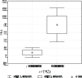
When the means of S/CO values for patients with de-tectable and non dede-tectable HCV-RNA were analyzed, a statistically significant difference was found, (79.3 SD 22.2 vs 8.2 SD 6.4, respectively) (p 0.0001). Figure 1
shows S/CO values according to the HCV-RNA status. We further analyzed the best cut-off value of the S/CO in differentiating viremic from non viremic patients. The S/CO value of 26 showed a sensitivity of 99% and a spec-ificity of 96% in discriminating both categories of HCV infected patients (Table I).
Receiver operating characteristic (ROC) curves dem-onstrates the relationship between true positive ratio (sen-sitivity) and false positive ratio (1- specificity) as one modifies the definition of a positive test.
edigraphic.com
curves: one plotting different S/CO values and the otherplotting the presence of HCV-RNA. As the presence of HCV-RNA represents the gold standard, the area under its curve was 1 and the standard error was 0. The area un-der the curve of S/CO values was 0.9991 and the standard error was 0.0013. No significant difference was found be-tween the areas under the two curves.
Discussion
The most practical screening test for hepatitis C virus antibodies are second and third- generation enzyme im-munoassays.3 The specificity of currently available EIAs
for anti-HCV antibodies is higher than 99 percent. How-ever, a positive result does not differentiate between viremic and non viremic patients, thus patients should be tested for HCV RNA by PCR to confirm viraemia.
In this study, we evaluated the performance of differ-ent MEIA S/CO values in the iddiffer-entification of viremic
from non viremic anti-HCV positive patients. Our data demonstrate that viremic HCV patients had higher S/CO values in the MEIA test in comparison with non viremic patients. Hence, this assay may be used to predict HCV viraemia in anti-HCV positive individuals.
Immunoassay tests analyze diseases and other medical conditions by measuring the body’s antigen/antibody re-action.
Antibodies against HCV has been reported to be present in the serum of patients with chronic hepatitis as well as in the serum of patients who have cleared the vi-rus.9 However, evidence that titre of anti-HCV antibodies
decrease in subjects with spontaneous resolution of the infection comes from several studies,10,11 suggesting that
the presence of HCV antigens plays an important role in the maintenance of a continuos antigenic stimulation of the humoral response.
On the other hand, Baumert et al., recently described that chronic HCV patients who successfully cleared the virus after interferon therapy, had a gradual decline of anti-HCV titres.12 Additionally, Tung et al., reported that
they can differentiate sustained virological responders from non responders to antiviral therapy using the S/CO of the MEIA test, since it decreases significantly in re-sponders patients.6
In conclusion, by establishing 26 as cut-off value of the S/CO in the third generation anti-HCV assay, it is pos-sible to differentiate viremic from non viremic patients.
This assay has the advantages of the enzyme immu-noassay – it is simple to use, allows to process a variety of immune diagnostic tests simultaneously at conventionally settings, has low variability and relatively low expense (3)-, and subsequently, may predict HCV viraemia. In this regard, clinicians may be informed not only about the an-tibody existence against HCV, but also they may infer that patients having a high S/CO value in the MEIA test may be viremic.
Figure 2. Receiver operating curve (ROC) to discriminate sensibility and specificity for different ratio of the sample rate to the cut-off rate S/CO values.
ROC curve for S/CO values
False positive ratio 0.0
0.2 0.4 0.6 0.8 1.0
0.0 0.2 0.4 0.6 0.8 1.0
T
rue
positive
ratio
Figure 1. Ratio of the sample rate to the cut-off rate (S/CO) of the third generation anti-HCV assay according to the HCV-RNA status. S/CO: Ratio of the sample rate to the cut-off rate.
Std. Dev.: standard deviation.
±1.96*Std. Dev. ±1.00*Std. Dev. Mean
RNA HCV -20
0 20 40 60 80 100 120 140
Undetectable Detectable
S/CO
Table 1. Ratio of sample rate to the cut-off (S/CO) value in predicting HCV viraemia in anti-HCV positive patients.
Detectable Non detectable HCV-RNA HCV-RNA
S/CO value > or equal 26 79 1 S/CO value < or equal 25 1 25
Annals of Hepatology 1(4) 2002: 179-182
182
However, the gold standard for detecting HCV vi-raemia is by qualitative polymerase chain reaction, which is indeed recommended by all the guidelines.
References
1. Seeff LB. Natural history of hepatitis C. Hepatology 1997; 26(Suppl 1): 21S-8.
2. Tong MJ, El-Farra NS., Reikes AR, Co RL. Clinical outcomes after transfusion-associated hepatitis C. N Engl J Med 1995; 332: 1436-66.
3. Gretch D. National Institutes of Health Consensus Development Conference Panel Statement: Diagnostic test for Hepatitis C.
Hepatology 1997; 26: 43S-47S.
4. Zaaijer HL, Cuypers HT, Reesink HW, Winkel IN, Gerken G, Lelie PN. Reliability of polymerase chain reaction for detection of hepati-tis C virus. Lancet 1993; 34: 722-724.
5. Busch M, Wilber J, Johnson P, Tobler L, Evans C. Impact of speci-men handling and storage on detection of hepatitis C virus RNA.
Transfusion 1992; 32: 420-425.
6. Tung HD, Lu SN, Lee CM, Wang JH, Chen TM, Hung CH, Huang WS, et al. Antiviral treatment responses in patients with chronic
hepa-titis C virus infection evaluated by a third generation anti-hepahepa-titis C virus assay. J Viral Hepat 2002; 9: 304-308.
7. Chomczynski SP, Sacchi N. Single step method of RNA isolation by guanidium thiocyanate-phenol-chloroform extraction. Analytical Biochemistry 1987; 162: 156-159.
8. Hanley JA, McNeil BJ. The meaning and use of the area under a receiver operating characteristic (ROC) curve. Radiology 1982; 143: 29-36.
9. Perniola R, De Rinaldis C, Leo G. Third-generation assays for hepati-tis C antibodies: a four-year study of pattern changes in patients with chronic and past infection. Panminerva Med 1999; 41(4): 291-4. 10. Lanotte P, Dubois F, Le Pogam S, Guerois C, Fimbel B, Bacq Y,
Gruel Y, et al. The kinetics of antibodies against hepatitis C virus may predict viral clearance in exposed haemophiliacs. J Infect Dis
1998; 178(2): 556-9.
11. Kondili LA, Chionne P, Costantino A, Villano U, Lo Nove C, Pannozzo F, Mele A, et al. Infection rate and spontaneous seroreversion of anti-hepatitis C virus during the natural course of anti-hepatitis C virus infec-tion in the general populainfec-tion. Gut 2002; 50(5): 693-6.