Long-term follow-up of hepatitis C virus-positive
patients with persistently normal serum transaminases
Emanuele La Spada,* Maurizio Soresi,* Lydia Giannitrapani,* Monica La Spada,* Elisa Campagna,* Antonino Terranova,* Fabio Cartabellotta,** Giuseppe Montalto*
* Cattedra di Medicina Interna ed Epatologia. Department of Internal Medicine and Specialties, University of Palermo, Palermo, Italy. ** Divisione di Medicina Interna, Ospedale Buccheri La Ferla, Palermo, Italy.
ABSTRACT
Material and methods. This study prospectively evaluated the progression of liver disease in a group of anti-HCV-positive patients with persistently normal ALT levels (PNALT) who were HCV-RNA positive. Patients selected for this study were those who presented with PNALT according to the Italian Association for the Study of the Liver (AISF) criteria in the year 1995/96 and underwent liver biopsy. They were divided into two groups according to their ALT evolution. Forty-five patients were included in this study. Results. After a median follow-up time of 180 months twenty-five of them maintained PNALT, but two of these developed liver cirrhosis (LC) in a mean time of 174 and 202 months, respectively. Twenty patients had flares of ALT and three of them developed LC in a mean time of 162-178 months. Twelve of these patients underwent current antiviral treatment; six patients were SVR. At baseline, the 5 patients who progressed to LC had age and BMI significantly higher than patients without LC (P < 0.005 and P < 0.01, respectively). Grading (P < 0.006) and staging (P < 0.003) were also more severe at histology, while serum HDL-C levels were statis-tically lower (P < 0.002). Comparing patients with flares of transaminases with and without LC, we found a significant difference at baseline for age, BMI, HDL-C, grading and staging (P < 0.05; P < 0.01 and P < 0.003, respectively). Conclusion. In HCV-RNA positive patients associated with PNALT the grade of disease activity increased over the years in only half of patients and a higher degree of liver fibrosis at baseline was the major relevant factor for progression.
Key words. Chronic hepatitis C. Persistently normal transaminases. Liver histology. Progression of disease. Interferon treatment.
Correspondence and reprint request: Prof. Giuseppe Montalto Department of Internal Medicine and Specialties, University of Palermo Via del Vespro 121; 90127 Palermo, Italy
Tel.: ++ 39091 6552991. Fax: ++ 39091 6552948 E-mail: [email protected]
Manuscript received: April 20, 2012. Manuscript accepted: June 28, 2012.
INTRODUCTION
30-40% of patients with chronic hepatitis C virus (HCV) infection show persistently normal alanine aminotransferase (PNALT) levels.1,2 Although these
were formerly referred to as ‘healthy’ or ‘asympto-matic’ HCV carriers, the natural history of HCV in-fection is not so clear-cut, as the evolution of liver disease in some of these subjects is less benign than previously thought. It has now become clear that the majority of these patients have some degree of histological liver damage, which may be significant
in up to 20% of patients and might progress toward the more severe degrees.4-19
A critical problem to be considered in these sub-jects is the definition of the “persistent” normality of serum ALT levels. In fact, during HCV infection it is not uncommon to observe wide fluctuations in ALT. Levels may remain normal for months or years only to rise quickly in rare cases, in association with a worsening histological picture.2 The Italian
Associa-tion for the Study of the Liver (AISF) some years ago proposed that the time length required to define a PNALT carrier should be 18 months, with a 2-month time lapse between serum assays of ALT levels (a to-tal of 9 assays). This means that a single increase in ALT values above normal in one out of the nine as-says excludes patients from the PNALT category.9
In the past, HCV carriers with PNALT were ex-cluded from antiviral treatment.2 More recently,
37 Follow-up of PNALT. , 2013; 12 (1): 36-43
As a result, antiviral treatment is also currently in-dicated for PNALT carriers, although with some li-mitations.24,25 However, at present it is not clear
which of these patients are at risk for disease pro-gression and, therefore, whether it is worth treating all of them with antiviral therapy.24
The aim of this study was to report the natural history of a group of subjects labeled as PNALT carriers, in accordance with the literature at that time,26,27 who had undergone liver biopsy in the
years 1995-1996 and been followed up for more than 15 years at our centre. In addition, they were divi-ded into two groups according to transaminase le-vels: those with persistently normal ALT and those with increased ALT values.
The aims were to evaluate:
• How many patients remained PNALT for this long period of time and whether they eventually developed liver disease.
• The stage of liver disease in the patients with ele-vated serum ALT, and
• What factors could have influenced the progres-sion of liver disease.
MATERIAL AND METHODS
Patients
This study included subjects extrapolated from our previous studies on patients with PNALT,28,29 who
continued to be followed-up in our outpatient clinic for liver diseases. They all had serum transaminase levels persistently within normal limits (i.e. 40 IU/dL)and had undergone liver biopsy in the years 1995/96. Se-venty patients met these criteria, but 18 of them were excluded because they were HCV-RNA negative at ba-seline. The remaining subjects were carefully followed up by monitoring transaminase levels every three months afterliver biopsy. To date 7 have dropped out, while 45 continue to be followed up and have been in-cluded in this study. In addition, they were divided into two groups according to their serum transamina-se levels following liver biopsy: thotransamina-se with PNALT and those with increased ALT levels. The current assess-ment of patients included a median follow-up of 180 months (range 162-206 months).
Methods
At enrolment to the present study patients un-derwent a general examination, including the
eva-luation of body weight, height, body mass index (BMI), blood pressure and heart rate. The main pa-rameters of liver function and lipidemic patterns were evaluated using commercial kits. Markers of hepatitis B virus and qualitative HCV-RNA were also re-assayed and, as previously described,28,29
pa-tients with a history of alcohol consumption were excluded. To help evaluate their current liver sta-tus, diagnostic imaging techniques were also used i.e. ultrasound of the upper abdomen. Furthermore, two current non-invasive markers of liver fibrosis were used i.e. transient elastography and the APRI score, which was compared to the same score calcu-lated at the time of liver biopsy.
Cirrhosis was diagnosed on the basis of the pre-sence and concordance of unequivocal clinical, bio-chemical and instrumental signs described above.
Ultrasound and color Doppler
Ultrasound of the liver was performed in the morning, after fasting for at least 10 h, by two opera-tors, originally using a real-time Toshiba SSA 270 A apparatus with 3.75 MHz convex and 5 MHz linear probes. As from 2001 a real-time Philips 5000 HDI apparatus was used with 2-5 MHz convex multi-fre-quency and 12-5 MHz linear multi-fremulti-fre-quency probes. The linear probe was used to assess the liver surface. The abilities of the two ultrasound observers (GM, MS) were homogeneous: they had the same professio-nal background, having been trained in this specific field, and both had over a decade of experience.
We considered ultrasound signs of cirrhosis: irre-gular liver surface associated with signs of portal hypertension (portal vein diameter > 1.2 cm or lon-gitudinal diameter of spleen > 12 cm).30,31
Non-invasive markers of liver fibrosis
• Transient elastography. TE was assessed by a single certified operator, using TE (FibroScan®; EchoSens, Paris, France). TE provides an asses-sment of liver stiffness expressed in KPa units as previously described.32 In brief, an ultrasound
transducer probe is mounted on the axis of a vi-brator. Vibrations of mild amplitude and low fre-quency are transmitted by the transducer, inducing an elastic shear wave that propagates through the underlying tissues. The speed of pro-pagation of this vibration across the liver is di-rectly related to tissue stiffness.
liver. Only patients with 10 valid elastometric measures, interquartile ranges (IQR) > 30% and ≥ 60% success rate (the number of valida-ted measurements divided by the total number of measurements) were considered to be relia-ble. A cut-off of 8.3 kPa was used to correctly diagnose subjects with significant fibrosis and a cut-off of 14 kPa to correctly assess liver cirrhosis.33
• AST-to-Platelet-Ratio Index (APRI). Liver fi-brosis was also assessed using a well-validated index, the AST platelet ratio index (APRI), which is calculated as follows: AST/upper limit of nor-mal (ULN) x 100/platelet count (109/L). The
pre-valence of advanced fibrosis was estimated using an APRI index > 1.5 as a reference value.34
Liver biopsy at entry to the study was obtained percutaneously with a Menghini needle and the Histology Activity Index (HAI) evaluated according to Knodell.35 Genotyping was
perfor-med as previously described.8
Patients who had increased serum transaminases were treated according to the current therapies and referred to as SVR or NR according to Euro-pean guidelines.26
Arterial hypertension (AH) was diagnosed in ac-cordance with the WHO/ISH criteria.36 Diabetes
Mellitus or Impaired Fasting Glucose (IFG) were diagnosed according to the Expert Committee on the Diagnosis and Classification of Diabetes Mellitus criteria.37
Statistical analysis
When data distribution was Gaussian, values were expressed as mean ± standard deviation and their differences were calculated using Student’s t-test. Otherwise, data were expressed
as median and range and analyzed with the Mann-Whitney U test. Fisher’s exact test, χ2 test
and Spearman’s rank (ρ) correlations were used when appropriate.
To assess which variables measured at baseline were predictive of progression to LC, the univariate Cox proportional hazards (Hr) model was fitted to each variable. All variables with a P-value < 0.05 were subjected to multivariate analysis to assess their value as independent predictors.
P < 0.05 was considered significant. Statistical analysis was performed using the SPSS for Windows version 16.0.
RESULTS
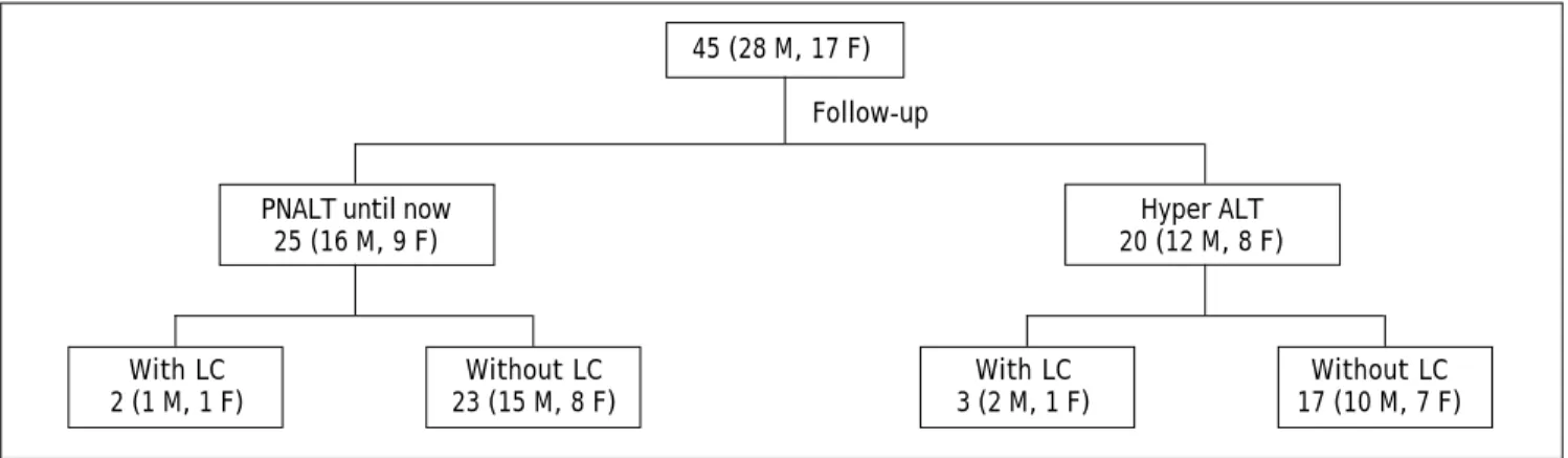
As mentioned above, 45 patients (28M, 17F) were included in this study. They continued to be follo-wed up at our Center: every 3 months to evaluate routine hematochemical parameters, and every 12 months for an abdominal ultrasound scan. Patients with US signs of evolution were followed up with a US every 6 months.
Figure 1 shows the evolution of serum ALT and liver disease in the patients during follow-up. Twenty-five patients maintained persistently nor-mal ALT values during the whole follow-up, but two of them developed frank liver cirrhosis. The genotypes of these patients were the following: five had genotype 2, three genotype 3 and seven-teen genotype 1b. Among the non-cirrhotic sub-jects, only two patients (one genotype 2 and one genotype 1b) received antiviral treatment (Peg-IFN + Ribavirin). The decision to treat was pri-marily based on the determination of the patients to receive treatment, even though there were no signs of progression of liver disease. At the third month of therapy HCV-RNA was negative in both
45 (28 M, 17 F)
Follow-up
PNALT until now Hyper ALT
25 (16 M, 9 F) 20 (12 M, 8 F)
With LC Without LC With LC Without LC
2 (1 M, 1 F) 23 (15 M, 8 F) 3 (2 M, 1 F) 17 (10 M, 7 F)
39 Follow-up of PNALT. , 2013; 12 (1): 36-43
and at the end they were SVRs. In contrast, twen-ty patients presented increased serum transami-nase levels during the follow-up and three of them developed frank liver cirrhosis. Twelve of these patients underwent current antiviral treatment. Six patients were SVR, while 6 were NR. Among the SVR there were three genotype 1b, two genoty-pe 2 and one genotygenoty-pe 3, whereas all the NR pa-tients had genotype 1b. Two of the three cirrhosis patients were included in the treated patients and one of the two was an SVR.
The remaining 8 patients did not receive treat-ment because of fluctuating serum ALT levels < 1.5 N in three cases, presence of co-morbidity in 3 cases and due to a brief, temporary increase in ALT in the remaining two, likely related to the assumption of other drugs.
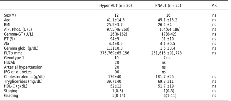
Table 1 compares some baseline demographic, bio-chemical and histological characteristics of the 45 patients divided according to ALT evolution. This comparison did not reveal any statistically signifi-cant differences.
Table 2 shows some demographic, biochemical and histological characteristics at entry to the study of the patients who developed liver cirrhosis, compared to the remaining patients. On univariate analysis, factors associated at baseline with evolution in cirr-hosis were: age, hypergammaglobulinemia, arterial hypertension, staging (P < 0.02), BMI, HBcAb positi-vity, grading (P < 0.05). On multivariate analysis,
staging (P < 0.04) and age (P < 0.05) were found to be independent predictors of liver cirrhosis.
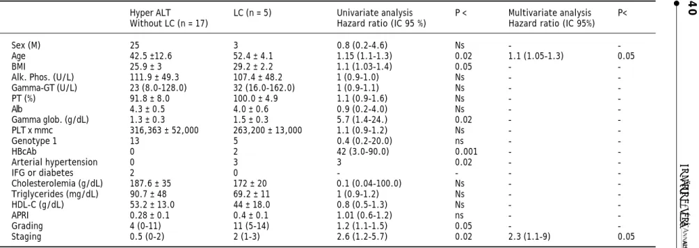
Table 3 shows the same characteristics quoted in tables 1 and 2 at baseline in patients who had flares of ALT, divided into those with or without LC. Com-paring the data, it emerges that on univariate analysis, factors associated with evolution in cirrho-sis were: age, arterial hypertension, grading (P < 0.05), staging (P < 0.02). On multivariate analysis, staging (P < 0.04) and age (P < 0.05) were found as independent predictors of liver cirrhosis.
At gastroesophageal endoscopy 3/5 LC patients showed variable degrees of esophageal varices.
• Elastography. Twenty out of 28 patients un-derwent TE. 6 overweight patients, one case of Parkinson’s disease and one pregnant woman were excluded. Two patients showed values above 8.3 kPa (8.6 in a patient with PNALT and LC and 9.6 in a non-cirrhotic patient with high ALT values) and in only one case was TE above 14 kPa (33.8 in a cirrhotic patient with high ALT values).
• APRI. Table 4 shows APRI scores at baseline and at the end of follow-up. Patients evaluated for liver cirrhosis diagnosis showed a significant increase in the score at the end of follow up (ρ = 0.3; P < 0.04), whereas when they were eva-luated for the presence of fibrosis there was no significant difference.
Table 1. Comparison of some baseline demographic, biochemical and histological characteristics of 28 patients divided according to their ALT evolution.
Hyper ALT (n = 20) PNALT (n = 25) P <
Sex(M) 12 16 ns
Age 41.1±14.5 45.1 ±15.2 ns
BMI 25.5±3.7 26.2 ±4 ns
Alk. Phos. (U/L) 97.5(46-268) 104(64-186) ns
Gamma-GT (U/L) 26(8-162) 17(8-42) ns
PT (%) 94±5 91 ±10 ns
Alb 4.4±0.5 4.1 ±0.5 ns
Gamma glob. (g/dL) 1.31±0.3 1.5 ±0.4 ns
PLT x mmc 375,769±65,156 251,615 ±91,773 ns
Genotype 1 10 7 ns
HBcAb 20 ns
Arterial hypertension 20 ns
IFG or diabetes 00 ns
Cholesterolemia (g/dL) 176±40 181.7 ±25 ns
Tryglicerides (mg/dL) 89.7±40 69.2 ±11 ns
HDL-C (g/dL) 52±12 51.7 ±19 ns
Staging 1(0-3) 1(0-3) ns
La Spada E, et al.
, 2013; 12 (1): 36-43
Table 3. Comparison between characteristics at baseline of patients with liver cirrhosis (LC) vs. patients with hyper ALT without LC.
Baseline Univariate analysis Multivariate analysis Hyper ALT LC (n = 5) Hazard ratio (IC 95%) P < Hazard ratio (IC 95%) P < Without LC (n = 17)
Sex (M) 10 3 0.7 (0.2-4) ns -
-Age 41.1 ± 14.5 52.4 ± 4.1 1.1 (1.05-1.2) 0.05 2 (1.05-7) 0.05
BMI 25.5 ± 3.7 29.2 ± 2.2 1.1 (0.8-1.6) ns -
-Alk. Phos. (U/L) 97.5 (46.0-268.0) 107.4 ± 48.2 1 (0.9-1.0) ns -
-Gamma-GT (U/L) 26 (8.0 ± 162.0) 32 (16.0-162.0) 1 (0.9-1.1) ns -
-PT (%) 94 ± 5 100 ± 4.9 1 (0.9-1.0) ns -
-Alb 4.4 ± 0,5 4.0 ± 0.6 0.6 (0.1-2.6) ns -
-Gamma glob. (g/dL) 1.1 ± 0.3 1.5 ± 0.3 3.8 (0.2-62.0) ns -
-PLT x mmc 429,500 ± 77,000 263,200 ± 13,000 1 (1.0-1.1) ns -
-Genotype 1 3 5 0.3 (0.1-42.0) ns -
-HbcAb 0 2 4.6 (0.8-27.0) ns -
-Arterial hypertension 0 3 6.4 (1.1-38.8) 0.05 -
-IFG or diabetes 0 0 - - -
-Cholesterolemia (g/dL) 176.0 ± 40.1 172.0 ± 20.2 0.9 (0.9-1.1) ns -
-Triglycerides (mg/dL) 89.7 ± 40.0 69.2 ± 11.0 1 (0.9-1.2) ns -
-HDL-C (g/dL) 52.2 ± 12.3 44.0 ± 18.1 0.8 (0.5-1.3) ns -
-APRI 0.3 ± 0.1 0.4 ± 0.1 1.1 (0.8-1.3) ns -
-Grading 5 (0-14) 11 (5-14) 1.2 (1.1-1.8) 0.05 -
-Staging 1 (0-3) 2 (1-3) 1.8 (1.2-3.7) 0.02 2.8 (1.1-9) 0.04
Without LC (n = 17) Hazard ratio (IC 95 %) Hazard ratio (IC 95%)
Sex (M) 25 3 0.8 (0.2-4.6) Ns -
-Age 42.5 ±12.6 52.4 ± 4.1 1.15 (1.1-1.3) 0.02 1.1 (1.05-1.3) 0.05
BMI 25.9 ± 3 29.2 ± 2.2 1.1 (1.03-1.4) 0.05 -
-Alk. Phos. (U/L) 111.9 ± 49.3 107.4 ± 48.2 1 (0.9-1.0) Ns -
-Gamma-GT (U/L) 23 (8.0-128.0) 32 (16.0-162.0) 1 (0.9-1.1) Ns -
-PT (%) 91.8 ± 8.0 100.0 ± 4.9 1.1 (0.9-1.6) Ns -
-Alb 4.3 ± 0.5 4.0 ± 0.6 0.9 (0.2-4.0) Ns -
-Gamma glob. (g/dL) 1.3 ± 0.3 1.5 ± 0.3 5.7 (1.4-24.) 0.02 -
-PLT x mmc 316,363 ± 52,000 263,200 ± 13,000 1.1 (0.9-1.2) Ns -
-Genotype 1 13 5 0.4 (0.2-20.0) ns -
-HBcAb 0 2 42 (3.0-90.0) 0.001 -
-Arterial hypertension 0 3 3 0.02 -
-IFG or diabetes 2 0 - - -
-Cholesterolemia (g/dL) 187.6 ± 35 172 ± 20 0.1 (0.04-100.0) Ns -
-Triglycerides (mg/dL) 90.7 ± 48 69.2 ± 11 1 (0.9-1.2) Ns -
-HDL-C (g/dL) 53.2 ± 13.0 44 ± 18.0 0.8 (0.5-1.3) Ns -
-APRI 0.28 ± 0.1 0.4 ± 0.1 1.01 (0.6-1.2) ns -
-Grading 4 (0-11) 11 (5-14) 1.2 (1.1-1.5) 0.05 -
41 Follow-up of PNALT. , 2013; 12 (1): 36-43
DISCUSSION
In our study population 5/45 HCV-RNA positive patients developed frank liver cirrhosis. This result is not so relevant, as it is lower than the figures al-ready reported in various studies in the literatu-re12,14 and especially in some Italian studies.13,15
Nevertheless it is higher than in other Italian re-ports, where the progression to liver cirrhosis was reported to be slow or absent.18,38 In any case, it
confirms that PNALT patients need to be monitored in the same way as patients presenting high ALT le-vels, since it is difficult to distinguish in which of the patients presenting with PNALT liver disease may progress. The prevalence of subjects with nor-mal liver at biopsy (true “carriers”) is lower than 20% of all PNALT patients.2,6-10 In most cases there
are variable degrees of liver damage, fibrosis is usually mild or absent10 and histology is generally
less severe than in patients with high or fluctuating serum ALT levels.2,7,10,11 Recent studies, however,
have shown more severe liver damage (fibrosis ≥ F2) in at least 20% of cases and liver cirrhosis in 3-5% of patients. There have also been rare cases of hepa-tocellular carcinoma in patients with normal ALT,15,16 even with a histologically normal liver
structure.17
Although limited by the small number of patients, our data would appear to confirm that subjects ol-der in age and presenting a higher degree of liver fi-brosis at baseline are candidates for liver disease progression. Other factors which, in our opinion, may contribute to the progression are the classical co-factors of liver disease i.e. elevated BMI, presence of diabetes mellitus, arterial hypertension, low le-vels of HDL-Chol, or even only an association with anti-HBc positivity. Therefore, it is of paramount importance to monitor this particular category of patients and to correct any eventual metabolic dis-turbances(i.e. insulin resistance)to prevent the
de-velopment of a metabolic syndrome, which will worsen the evolution of liver disease.
Similar data were reported by Persico M, et al. who studied the natural history at ten years of a group of 24 patients with PNALT, compared with a group of 40 patients with high levels of transami-nases, using liver biopsy at baseline and after 5 and 10 years. They did not report any significant histo-logical differences in the three liver biopsies in the PNALT patients, thus suggesting that cirrhosis pro-gression is low or absent in these patients. Liver steatosis was significantly higher in the group with high ALT, confirming that steatosis is a co-factor of disease progression. In this study neither sex nor BMI were significantly different in the two patient groups.18
The small number of our sample patients, in com-mon with other reports in the literature, can be jus-tified by the fact that in order to obtain accurate information a liver biopsy should be performed at both the beginning and the end of the study, which is not justifiable ethically, or only in controlled stu-dies dedicated to this purpose. One limitation of our study is certainly that a second liver biopsy was not performed after all these years, although as a surro-gate we used liver function tests, ultrasound, elasto-graphy and clinical examination to avoid resorting to invasive techniques in the majority of the pa-tients, in whom disease did not progress. However, it is mandatory to continue monitoring these pa-tients, because as we now know from studies of the natural history of HCV disease, it may take decades and sometimes longer before HCV infection progres-ses to cirrhosis. Consequently, patients with normal livers today could later develop evidence of hepatic impairment. These considerations, together with the evidence that combination therapy is equally effecti-ve in subjects with normal ALT, once again indicate that the two groups of patients are not substantially different, consequently, the same criteria for follow-Table 4. APRI values at baseline and at the end of follow up.
APRI Interpretation Baseline End of follow-up
< 0.5 Absence of significant fibrosis 25 22 ρ = 0.15; P = ns
0.5-1.5 Unclassified to significant fibrosis 3 1
> 1.5 Presence of significant fibrosis 0 4
< 1 Absence of cirrhosis 28 24 ρ = 0.31; P < 0.04
1-2 Unclassified to cirrhosis 0 1
> 2 Presence of cirrhosis 0 4
up and treatment recommended for patients with elevated ALT should be applied to subjects with nor-mal ALT.39
Carriers of HCV with PNALT have traditionally been excluded from antiviral treatment, both in trials and in clinical practice.2 A first major
thera-peutic development occurred with the use of IFN-ri-bavirin combination therapy: virologic response rates were obtained that did not differ from those seen in patients with elevated transaminases, as evidenced by a study by Mangia, et al. and later confirmed by other works.20-23 In Italy the treatment
of hepatitis C with pegylated interferon alfa-2a and ribavirin is currently allowed in HCV carriers inde-pendent of their transaminase levels.40 However, in
view of the economic costs, it is essential to carefully select the patients to be referred for treatment. Fundamental criteria for defining optimal treatment protocols may be a patient’s age, his/her motivations, the possibility of eradication (viral genotype), life expectancy, duration of disease, the presence of co-factors of liver disease, adherence to treatment, contraindications, considerations about infective-ness (if the subject is promiscuous or has a stable partner), type of employment (potential infection of others). On the contrary, in patients where the cost-benefit ratio is not favorable (age over 50, relative contraindications, poor motivation, genotype 1, high viral load, presence of co-factors, risk of side effects, etc.) the opportunity of a treatment should be asses-sed case by case, depending on the severity of liver histology. Therapy should thus be reserved for only patients with a high grade of fibrosis (> F2) sub-jects with or without moderate fibrosis need to be closely monitored. Finally, in subjects aged over 60-65 years and with a long duration of disease, it would seem reasonable to carry out a regular clini-cal follow up, thus avoiding both liver biopsy and treatment.
CONCLUSION
In conclusion, our data show that almost half of HCV-RNA positive subjects defined as PNALT, according to the definition of The Italian Associa-tion for the Study of the Liver, i.e. 18 months of follow-up with at least 9 determinations, will have flares of ALT in the long term follow-up but few of them will develop LC. In the other half, serum transaminases will remain within normal limits, but in this group the risk of developing LC, al-though lower, is also present. As well as the tran-saminase levels, other factors related to age at the
start of infection, grading and staging at histolo-gy and the concurrence of some metabolic factors, seem to play an important role in the progression of liver disease. Therefore, as well as ensuring a close clinical, biochemical and instrumental follow-up of liver disease, it is our duty to at least address these modifiable factors, which could make the difference in preventing the progression of liver disease.
REFERENCES
1. Puoti C, Bellis L, Galossi A, Guarisco R, Nicodemo S, Spila-botti L, Unto OD. Antiviral treatment of HCV carriers with persistently normal ALT levels. Mini Rev Med Chem 2008; 8: 150-2.
2. Puoti C. HCV carriers with persistently normal amino-transferase levels: normal does not always mean healthy. J Hepatol 2003; 38: 529-32.
3. Alberti A, Morsica G, Chemello L, Cavalletto D, Noventa F, Pontisso P, Ruol A. Hepatitis C viraemia and liver disease in symptom-free individuals with anti-HCV. Lancet 1992; 340: 697-8.
4. Puoti C, Magrini A, Stati T, Rigato P, Montagnese F, Rossi P, Aldegheri L, et al Clinical, histological, and virological features of hepatitis C virus carriers with persistently normal or abnormal alanine transaminase level. Hepatology
1997; 26: 1393-8.
5. Gholson CF, Morgan K, Catinis G, Favrot D, Taylor B, Gon-zalez E, Balart L. Chronic hepatitis C with normal amino-transferase levels: a clinical histologic study. Am J Gastroenterol 1997; 92: 1788-92.
6. Puoti C, Castellacci R, Montagnese F, Zaltron S, Stornaiuo-lo G, Bergami N, Bellis L. HistoStornaiuo-logical and viroStornaiuo-logical featu-res and follow up of hepatitis C virus carriers with normal amínotransferase levels: the Italian prospective study of the asymptomatic C carriers (ISACC). J Hepatol 2002; 37: 117-23.
7. Pradat P, Alberti A, Poynard T, Esteban Jl, Welland O, Mar-cellin P, Badalamenti S, et al. Predictive value of ALT levels for histologic findings in chronic Hepatitis C: a European Collaborative Study. Hepatology 2002; 36: 973-7.
8. Montalto G, Zignego L, Ruggeri MI, Giannini C, Soresi M, Monti M, Carroccio A, et al. Serum HCV-RNA and liver his-tologic findings in patients with long-term normal transa-minases. Dig Dis Sci 1997; 42: 1703-7.
9. Puoti C, Guido M, Mangia A, Persico M, Prati D. Clinical ma-nagement of HCV carriers with normal aminotransferase levels. Dig Liver Dis 2003; 35: 362-9.
10. Bacon BR. Treatment of patients with Hepatitis C and nor-mal serum aminotransferase levels Proc. of the NIH Con-sensus Conference Management of Hepatitis C.
Hepatology 2002; 36(Suppl. 1). 5179-84.
11. Strader DB, Wright T, Thomas DL, Seeff LB. Diagnosis, ma-nagement and treatment of hepatitis C. AASLD Practice Guideline Hepatology 2004; 39: 1147-71.
12. Huí CK, Belaye T, Montegrande K, Wright TL. A comparison in the progression of liver fibrosis in chronic hepatitis C between persistently normal and elevated transaminase. J Hepatol 2003; 38: 511-7.
43 Follow-up of PNALT. , 2013; 12 (1): 36-43
14. Ghany MG, Kleiner DE, Alter H, Doo E, Khokar F, Promrat K, Herion D, et al. Progression of fibrosis in chronic hepatitis C. Gastroenterology 2003; 124: 97-104.
15. Cividini A, Rebucci C, Silini E, Mondelli MU. Is the natural history of HCV carriers with normal aminotransferase le-vels really benign? Gastroenterology 2001; 121: 1526-7. 16. Persico M, Palmentieri B, Coppola L, Di Giacomo Russo G,
De Marino F, De Sio I, Torella L. Occurrence of HCC in asymptomatic HCV related chronic hepatitis. Dig Dis Sci
2002; 11: 2407-10.
17. Puoti C, Bellis L, Martellino F, Durola L, Spilabotti L, Dell’Unto O, Galossi A, et al. Occurrence of HCC in an apparently healthy HCV carrier. Eur J Gastroenterol He-patol 2005; 17: 1263-4.
18. Persico M, Perrotta S, Persico E, Terraciano L, Folgori A, Ruggeri L, Nicosia A, et al. Hepatitis C virus carriers with persistently normal ALT levels: biological peculiarities and update of the natural history of liver disease at 10 years.
J Viral Hep 2006; 13: 290-6.
19. Dienstag JL, McHutchison JG. American Gastroenterologi-cal Association [AGA] MediGastroenterologi-cal position statement on the management of hepatitis C. Gastroenterology 2006; 130: 225-64.
20. Mangia A, Spinzi G, Vuturo O, Pazienza V, Iacobellis A, Piattelli M, Giacobbe A, et al. Viral clearance in HCV virae-mic patients with normal alanine aminotransferase after combination therapy: a controlled, open-labelled study.
Aliment Pharmacol Ther 2004; 19: 331-7.
21. Lee SS, Sherman M. Pilot Study of interferon-alpha and ri-bavirin treatment in patients with chronic hepatitis C and normal transaminase values. J Viral Hepat 2001; 8: 202-5.
22. Hui CK, Monto A, Belaye T, Lau E, Wirght TL. Outcomes of interferon and ribavirin treatment for chronic hepatitis C in patients with normal serum aminotransferase. Gut
2003; 52: 1644-8.
23. Jacobson IM, Ahmed F, Russo MW, Lebovics E, Dieterich DT, Esposito SP, Bach N, et al. Interferon alpha-2b and ri-bavirin for patients with chronic hepatitis C and normal ALT. Am J Gastroenterol 2004; 99: 1700-5.
24. Alberti A, Bonino F, Bortolotti F, per la Commissione Tera-pia antivirale della Associazione Italiana per lo Studio del fegato (AISF). Trattamento della epatite da HCV. Available from: www.webaisf.org
25. Puoti C, Pellicelli AM, Romano M, Mecenate F, Guarisco R, Barbarini G, Mazzoni E, et al. Treatment of hepatitis C vi-rus carriers with persistently normal alanine aminotrans-ferase levels with peginterferon alpha-2a and ribavirn: a multicentric study. Liver Int 2009; 29: 1479-84.
26. EASL International Consensus Conference on Hepatitis C: Consensus Statement. J Hepatol 1999; 30: 956-61. 27. National Institutes of Health Consensus Development
Con-ference Panel Statement: Management of Hepatitis C. He-patology 1997; 26 (Suppl.): 1335-65.
28. Montalto G, Mazzola A, Soresi M, Consiglio P, Ruggeri M I, Ingrassia G, Cartabellotta A, et al. Antibodies to hepatitis C virus and histological pattern in Sicilian blood donors.
Eur J Int Med 1994; 5: 299-304.
29. Soresi M, Carroccio A, Bonfissuto G, Agate V, Magliarisi C, Aragona F, Levrero M, et al. Ultrasound detection of ab-dominal lymphadenomegaly in subjects with hepatitis C vi-rus infection and persistently normal transaminases: a predictive index of liver histology severity. J Hepatol
1998; 28: 544-9.
30. Gaiani S, Gramantieri L, Venturoli N, Piscaglia F, Siringo S, D’Errico A, Zironi G, et al. What is the criterion for differentiating chronic hepatitis from compensated ci-rrhosis? A prospective study comparing ultrasonogra-phy and percutaneous liver biopsy. J Hepatol 1997; 27: 679-85.
31. Piscaglia F, Donati G, Serra C, Muratori R, Solmi L, Gaiani S, Gramantieri L, et al. Value of splanchnic Doppler ultra-sound in the diagnosis of portal hypertension. Ultrasound Med Biol 2001; 27:893-9.
32. Li Vecchi V, Soresi M, Colomba C, Mazzola G, Colletti P, Mi-neo M, Di Carlo P, et al. Transient elastography: a non-in-vasive tool for assessing liver fibrosis in HIV/HCV patients. World J Gastroenterol 2010; 16: 5225-32. 33. Coco B, Oliveri F, Maina AM, Ciccorossi P, Sacco R,
Co-lombatto P, Bonino F, et al. Transient elastography: a new surrogate marker of liver fibrosis influenced by ma-jor changes of transaminases. J Viral Hepat 2007; 14: 360-9.
34. Wai CT, Greenson JK, Fontana RJ, Kalbfleisch JD, Marrero JA, Conjeevaram HS, Conjeevaram HS, et al. A Simple No-ninvasive Index can predict both significant fibrosis and cirrhosis in patients with chronic hepatitis C. Hepatology
2003; 38: 518-26.
35. Knodell RG, Ishak KG, Black WC, Chen TS, Craig R, Kaplo-witz N, Kiernan TW, et al. Formulation and application of a numerical scoring system for assessing histological activi-ty in asymptomatic chronic active hepatitis. Hepatology
1991; 1: 431-5.
36. Guidelines Sub-Committee. 1999 World Health Organiza-tion-International Society of Hypertension guidelines for the management of hypertension. J Hypert 1999; 17: 151-83.
37. Alberti KG, Zimmet PZ. Definition, diagnosis and classifica-tion of diabetes mellitus and its complicaclassifica-tions. Part 1:diagnosis and classification of diabetes mellitus. Provi-sional report of a WHO consultation. Diabet Med 1998; 15:539-53.
38. Persico M, Persico E, Suozzo R, Conte S, De Seta M, Co-ppola L, et al. Natural history of Hepatitis C virus carriers with persistently normal aminotransferase levels. Gas-troenterology 2000; 118: 760-4.
39. Deuffic-Burban S, Babany G, Lonjon-Domanec I, Deltenre P, Canva-Delcambre V, Dharancy S, Louvet A, et al. Im-pact of pegylated interferon and ribavirirn on morbidity and mortality in patients with chronic hepatitis C and normal aminotransferases in France. Hepatology 2009; 50: 1351-599.