Effect of nitric oxide on the somatostatinergic system in the rat exocrine pancreas
7
0
0
Texto completo
(2) E. Rodr|¨guez-Mart|¨n et al. / Biochimica et Biophysica Acta 1450 (1999) 61^67. 62. tion of NOS from isolated pancreatic acini demonstrates the presence of this enzyme in acinar cells and supports a direct role for NO as a regulator of pancreatic exocrine function at the level of the acinar cells [5,7]. In this regard, L-Arg has been shown to induce an increase in amylase [5] and insulin release [8^10] in the rat pancreas. Inhibitors of the enzymatic conversion of L-Arg to NO inhibited insulin secretion induced by L-Arg in the presence of D-glucose [8]. Colocalization of neuronal NOS and SS has also been found in the pancreatic islets, where strong NOS immunoreactivity appeared in scattered cells which were peripheral in rat and mouse islets and more randomly distributed in human [11,12]. On the other hand, SS is a tetradecapeptide initially isolated from the hypothalamus as an inhibitor of pituitary growth hormone secretion [13]. SS acts as a negative regulator of pancreatic exocrine and endocrine secretions in both pancreas and gut. It has been previously reported that pancreatic acini possess speci¢c binding sites for SS, the binding of which results in the triggering of some biological actions of SS in the pancreas [14,15]. In pancreatic acinar cells, SS receptors are negatively coupled to the adenylyl cyclase (AC) enzyme system via a guanine nucleotide-binding inhibitory protein (Gi-protein) [16], leading to a decrease in adenosine 3P-5P cyclic monophosphate (cAMP) levels. Among its various inhibitory functions, SS decreases pancreatic amylase release [17]. An imbalance between NO and SS may contribute to alterations in pancreatic acinar cell secretion. The aim of this study was to determine whether NO regulates the SS receptor-effector system in rat pancreatic acinar membranes. In this regard, we evaluated the e¡ects of the NOS substrate L-Arg and the NOS inhibitor Ng -L-arginine methyl ester (L-NAME) on the somatostatin receptors, SS-mediated inhibition of AC activity in rat pancreatic acinar membranes. Pancreatic somatostatin-like immunoreactivity (SSLI) content was also determined. 2. Materials and methods 2.1. Materials The stable SS analogue SMS 201-995 and its tyro-. sine analogue Tyr3 -SMS or SMS 204-090 were kindly donated by Sandoz (Basel, Switzerland). Collagenase (from Clostridium histolyticum) was obtained from Serva Fine Chemicals (Tebu, France). L-NAME, L-Arg, bacitracin, phenylmethylsulfonyl£uoride (PMSF), guanosine triphosphate (GTP), 3-isobutyl1-methylxanthine (IBMX) and bovine serum albumin (BSA) were purchased from Sigma (St. Louis, MO, USA). Carrier-free Na[125 I] (IMS 300, 100 mCi/ml) and the rabbit antibody used in the radioimmunoassay technique were purchased from the Radiochemical Center (Amersham, UK). This antiserum was raised in rabbits against SS-14 conjugated to BSA and is speci¢c for SS, but since SS-14 constitutes the C-terminal portion of both SS-25 and SS28, the antiserum does not distinguish between these three forms. In the rat pancreas, SS-14 is the predominant molecular species, constituting 95% of total SS, whereas SS-28 seems to comprise less than 5% of the immunoreactivity [18]. All other agents were of the highest purity commercially available. 2.2. Experimental animals The animal experiments performed in the present study were conducted under the guidelines of the Animal Care Committee of Alcalä University and the experimental protocols have been approved. Male Wistar rats weighing 200^250 g were injected intraperitoneally (i.p.) twice daily at 8 h intervals for 8 days with the following agents: L-NAME (50 mg/ kg) [19,20], L-Arg (150 mg/kg) [21^23], L-NAME plus L-Arg or saline in a volume (400 Wl) equal to that used for the other compounds. The animals were sacri¢ced 14 h after the last injection and the pancreas was removed and trimmed free of fat, connective tissues and lymph nodes. 2.3. Preparation of rat pancreatic acinar membranes Dispersed pancreatic acini were obtained from male Wistar rats after enzymatic degradation of the organ with 0.2 units of collagenase/ml in an oxygenated Krebs-Ringer medium as described by Amsterdam et al. [24]. After thorough washing by sedimentation, acini were transferred to 0.3 M sucrose and homogenized at 4³C using of a Potter homogenizer following the method of Meldolesi et al. [25]. After. BBAMCR 14465 21-4-99.
(3) E. Rodr|¨guez-Mart|¨n et al. / Biochimica et Biophysica Acta 1450 (1999) 61^67. sedimentation at 1500Ug for 12 min, the homogenized membranes were resuspended in 1.56 M sucrose. This suspension was overlaid with 0.3 M sucrose and centrifuged at 105 000Ug for 150 min. The plasma membrane-enriched fraction collected from the interphase was diluted with distilled water and centrifuged at 15 000Ug for 30 min. The supernatant was discarded and the pellet was resuspended in 50 mM Tris-HCl pH 7.4, 0.01 mg/ml bacitracin, 0.2 mM CaCl2 and stored at 370³C. An aliquot was taken for protein determination by the method of Lowry et al. [26]. 2.4. [125 I-Tyr3 ]SMS binding [125 I-Tyr3 ]SMS binding was assayed on pancreatic acinar membranes from Wistar rats by a modi¢ed method [27]. The stable SS-14 analogue SMS 204090 or [Tyr3 ]SMS, which shares the biological properties of SMS 201-995 and has proven valuable in the characterization of SS-14 binding to receptors in rat pancreatic acinar [28] and brain [29] membranes, was radioiodinated by the chloramine-T method and puri¢ed by HPLC according to Antoniotti et al. [30]. Its speci¢c radioactivity was found to be 900 Ci/ mmol. Binding of [125 I-Tyr3 ]SMS to pancreatic acinar membranes was carried out in a total volume of 250 Wl in 50 mM Tris-HCl bu¡er (pH 7.4) containing 0.5 mM MgCl2 , 3 mM NaCl, 0.2 mM CaCl2 , 0.2% (w/v) BSA, 0.5 mg/ml bacitracin and 0.3 mg/ml soybean trypsin inhibitor (binding bu¡er). Plasma membranes (36 Wg protein/ml) were incubated for 90 min at 20³C with 35 pM [125 I-Tyr3 ]SMS in the absence or presence of 0.001^10 nM unlabeled SMS 201-995. Bound and free ligand were separated by centrifugation at 11 000Ug for 4 min at 4³C in a microcentrifuge. Radioactivity in the pellet was measured with a gamma scintillation counter. Non-speci¢c binding was estimated as membrane-associated radioactivity in the presence of 1 WM SMS and speci¢c binding was calculated as the di¡erence between total and non-speci¢c membrane-associated radioactivity. 2.5. Adenylate cyclase assay Adenylate cyclase activity was measured as previously reported [31] with minor modi¢cations. Brie£y,. 63. rat pancreatic acinar membranes (0.12 mg protein/ ml) were incubated with 1.5 mM ATP, 5 mM MgSO4 , 1 mM GTP and an ATP-regenerating system (7.5 mg/ml creatine phosphate and 1 mg/ml creatine kinase), 1 mM IBMX, 0.1 mM PMSF, 1 mg/ml bacitracin, 1 mM EDTA, and tested substances (1039 M SMS 201-995 or 1035 M forskolin (FK)) in 0.1 ml of 0.025 M triethanolamine-HCl bu¡er (pH 7.4). After a 30 min incubation at 30³C, the reaction was stopped by heating the mixture for 3 min. After cooling, 0.2 ml of an alumina slurry (0.75 g/ml in triethanolamine-HCl bu¡er, pH 7.4) was added and the suspension centrifuged. The supernatant was taken for assay of cyclic AMP by the method of Gilman [32]. 2.6. Tissue extraction and SS radioimmunoassay [Tyr11 ]SS was radioiodinated by chloramine-T iodination according to the method of Greenwood [33]. Separation of iodinated SS from unincorporated iodine was carried out on a Sephadex G-25 (¢ne) column equilibrated and eluted with 0.1 M acetic acid in BSA (0.1% w/v). Its speci¢c activity was found to be 900 Ci/mmol. For SSLI measurement, the pancreas was rapidly homogenized in 1 ml of 2 M acetic acid using a Brinkman polytron (setting 5, 30 s). The extracts were boiled for 5 min, cooled in an ice-chilled water bath, and aliquots (100 Wl) were removed for protein determination [26]. The homogenates were subsequently centrifuged at 15 000Ug for 15 min at 4³C and the supernatant was neutralized with 2 M NaOH. The extracts were stored at 370³C until assay. The SS concentration was determined in tissue extracts by a modi¢ed radioimmunoassay method [34], with a sensitivity limit of 10 pg/ml. Incubation tubes prepared in duplicate contained 100 Wl samples of tissue extracts or standard solutions of 0^500 pg of cyclic SS-14 diluted in phosphate bu¡er (0.05 M, pH 7.2 containing 0.3% BSA, 0.01 M EDTA), 200 Wl of appropriately diluted anti-SS serum and 100 Wl of freshly prepared [125 I-Tyr11 ]SS diluted in bu¡er to give 6000 cpm (equivalent to 5^10 pg), in a ¢nal volume of 0.8 ml. All reagents as well as the assay tubes were kept chilled on ice before their incubation at 4³C for 48 h. Bound hormone was separated from free hormone by the addition of 1 ml of dextran-. BBAMCR 14465 21-4-99.
(4) 64. E. Rodr|¨guez-Mart|¨n et al. / Biochimica et Biophysica Acta 1450 (1999) 61^67. coated charcoal (dextran T-70, 0.2% w/v, Pharmacia, Uppsala, Sweden; charcoal: Norit A, 2% w/v, Serva, Feinbiochemica, Heidelberg, Germany). Serial dilution curves for the samples were parallel to the standard curve. The intra- and inter-assay variation coe¤cients were 6.0 and 8.8% respectively. 2.7. Data analysis The LIGAND computer program [35] was used to analyze the binding data. The use of this program made it possible to select the models of receptors which best ¢t a given set of binding data. The same program was also used to present data in the form of Scatchard plots [36] and to compute the values for receptor a¤nity (Kd ) and density (Bmax ) that best ¢t the sets of binding data for each rat. Statistical comparisons of all the data were analyzed by ANOVA and the Newman-Keuls t-test. Means among groups were considered signi¢cantly di¡erent when the P value was less than 0.05. Each individual experiment was performed in duplicate.. Table 2 Equilibrium parameters for [125 I-Tyr3 ]SMS binding to pancreatic acinar membranes from rats treated for 8 days with saline, L-Arg, L-NAME or L-NAME plus L-Arg Groups Saline Bmax Kd L-Arg Bmax Kd L-NAME Bmax Kd L-NAME+L-Arg Bmax Kd. SS receptors 4739 þ 143 0.62 þ 0.03 3718 þ 152** 0.53 þ 0.05 6254 þ 298** 0.57 þ 0.06 4374 þ 132 0.50 þ 0.07. Binding parameters were calculated from Scatchard plots by linear regression. Units for Kd are nM and units for Bmax are femtomoles of SMS bound per mg of protein. Values are the means þ S.E.M. of the data from ¢ve rats in each group. Determinations were made in duplicate for each experiment. Statistical comparison versus controls: **P 6 0.01.. 3. Results. 3.2. [I125 -Tyr3 ]SMS binding. 3.1. Pancreatic somatostatin-like immunoreactivity content. Speci¢c [I125 -Tyr3 ]SMS binding to pancreatic acinar membranes changed linearly with protein concentration and was time-dependent in all experimental groups. An apparent equilibrium was observed between 60 and 120 min at 20³C (data not shown). All subsequent binding experiments were therefore conducted at 20³C for 90 min. Speci¢c [125 I-Tyr3 ]SMS binding to rat pancreatic acinar cell membranes was signi¢cantly decreased in L-Arg-treated rats as compared to control rats (Fig. 1, left panel, Table 2). This decrease was due to a decrease in the maximal number of SS receptors with no changes in the dissociation constant, as revealed by Scatchard plots of the binding data (Fig. 1, right panel, Table 2). The administration of L-NAME+L-Arg reverted the number of SS receptors to control values whereas L-NAME alone increased the SS receptor density. The addition of 1034 ^10311 M of L-Arg and/or L-NAME to the incubation medium changed neither the number nor the a¤nity of the SS receptors in pancreatic acinar cell membranes from normal rats (data not shown).. The administration of L-Arg induced a signi¢cant decrease in SSLI content in the rat pancreas, whereas L-NAME or L-NAME plus L-Arg increased its content (Table 1).. Table 1 Pancreatic somatostatin-like immunoreactive (SSLI) content from rats treated for 8 days with saline, L-Arg, L-NAME or L-NAME plus L-Arg Group. SSLI. Saline L-Arg L-NAME L-NAME+L-Arg. 4.48 þ 0.38 3.6 þ 0.32* 9.8 þ 0.83*** 7.69 þ 10.4*. Units for SSLI are ng of SS per mg pf protein. Values are the means þ S.E.M. of the data from ¢ve rats in each group. Determinations were made in duplicate for each experiment. Statistical comparison versus controls: *P 6 0.05, ***P 6 0.001.. BBAMCR 14465 21-4-99.
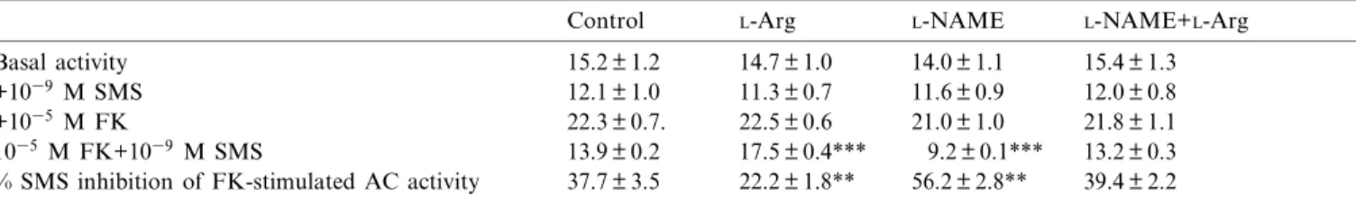
(5) E. Rodr|¨guez-Mart|¨n et al. / Biochimica et Biophysica Acta 1450 (1999) 61^67 L-NAME+L-Arg. ues.. 65. reverted this e¡ect to control val-. 4. Discussion. Fig. 1. (Left panel) Competitive inhibition of speci¢c 125 I-Tyr3 SMS (SMS 204-090) binding to rat pancreatic acinar membranes by unlabeled SMS 201-995. Points correspond to control rats (a), rats treated for 8 days with L-Arg (R), L-NAME (b) or L-NAME combined with L-Arg (O). Each point is the mean of ¢ve separate experiments, each performed in duplicate. (Right panel) Scatchard analysis of the same data. The kinetic constants calculated by Scatchard analysis are given in Table 2.. 3.3. Adenylyl cyclase assay The functional coupling of the SS receptors to the AC system was also investigated. To study the SMSmodulated AC activity, pancreatic acinar cell membranes were incubated with SMS (1039 M) either alone or together with FK (1035 M), a direct AC activator. As shown in Table 3, no signi¢cant di¡erences were seen for either basal or FK-stimulated AC enzyme activity in pancreatic acinar cell membranes from control, L-Arg- and/or L-NAME-treated rats. The e¡ect of SMS 201-995 on FK-stimulated AC activity was markedly decreased in pancreatic acinar membranes from L-Arg-treated rats as compared to control animals (Table 3). This e¡ect, however, was increased in pancreatic acinar membranes from LNAME-treated rats, whereas the administration of. The present study shows that L-Arg, a substrate for NO production, decreases pancreatic SSLI levels and the number of speci¢c SS receptors, without altering their a¤nity, in rat pancreatic acinar cell membranes. In addition, L-Arg induces a decrease in the inhibitory e¡ect of SMS 201-995 on AC activity in these membranes. The L-Arg concentration used in the present study was in agreement with the dose reported by others and has been demonstrated to induce NO production [21^23]. The choice of L-NAME was dictated by its previous characterization as a NOS inhibitor [37^39]. In addition, Bannerman et al. [20] and Dwyer et al. [19] have shown that the level of NOS inhibition actually increases with repeated injections. The pancreatic SSLI levels in the control rats were similar to those previously reported by others [14,40]. L-Arg has been described to produce a dose-related increase of SS release from the isolated perfused rat pancreas [41]. On the other hand, the intracellular concentration of Ca2 , which is known to be a potent stimulator of SS secretion [42,43], is increased by NO. Therefore, it is possible that the increase of SS release by the D-cells of the islets of Langerhans could lead to a decrease of pancreatic SSLI levels found in L-Arg-treated rats. Although ¢ve SS receptors subtypes have thus far been cloned [44], the rat exocrine pancreas appears to express only SS receptor subtype 2 (sst2) [45]. Therefore, the present results suggest that the changes in the SS receptor number observed after L-Arg or. Table 3 The e¡ect of SMS 201-995 (1039 M) and FK (1035 M) on AC activity (pmol cAMP/min/mg protein) in pancreatic acinar membranes from rats treated for 8 days with saline, L-Arg, L-NAME or L-NAME plus L-Arg Basal activity +1039 M SMS +1035 M FK 1035 M FK+1039 M SMS % SMS inhibition of FK-stimulated AC activity. Control. L-Arg. L-NAME. L-NAME+L-Arg. 15.2 þ 1.2 12.1 þ 1.0 22.3 þ 0.7. 13.9 þ 0.2 37.7 þ 3.5. 14.7 þ 1.0 11.3 þ 0.7 22.5 þ 0.6 17.5 þ 0.4*** 22.2 þ 1.8**. 14.0 þ 1.1 11.6 þ 0.9 21.0 þ 1.0 9.2 þ 0.1*** 56.2 þ 2.8**. 15.4 þ 1.3 12.0 þ 0.8 21.8 þ 1.1 13.2 þ 0.3 39.4 þ 2.2. Experiments were performed as described in Section 2. Values represent the means þ S.E.M. of ¢ve separate experiments, each performed in duplicate. Statistical comparison versus control: **P 6 0.01, ***P 6 0.001.. BBAMCR 14465 21-4-99.
(6) 66 L-NAME. E. Rodr|¨guez-Mart|¨n et al. / Biochimica et Biophysica Acta 1450 (1999) 61^67. administration could result, at least in part, from changes of the sst2 subtype. The binding parameters of the SS receptors in the control rats were similar to those previously reported by others [14,15]. The changes in [125 I-Tyr3 ]SMS binding were not a result of a direct e¡ect of L-Arg or L-NAME on the SS receptors, since no change in tracer binding was detected following incubation of fresh pancreatic acinar cell membranes with L-Arg or L-NAME. In addition, NO seems to mediate the action of L-Arg on the SS receptor-e¡ector system because the changes in SS binding and the inhibitory e¡ect of this neuropeptide on AC activity induced by L-Arg were prevented by pretreatment with the NOS inhibitor L-NAME which, when administered alone, increased both parameters. At present, we can only speculate on the identity of the postreceptor mechanisms responsible for the regulation of the SS receptors by NO in rat pancreatic acinar cell membranes. Although most of the NO actions have been reported to be mediated by elevation of cGMP synthesis through activation of soluble guanylate cyclase [1], cGMP has been shown to have no e¡ect on SS binding [46]. Recently, NO has been shown to be involved in Ca2 transport in pancreatic acinar cells [42,43]. Willmott et al. [43] suggest an action of NO in mobilizing intracellular Ca2 from microsomal stores via a signaling pathway involving cGMP and cyclic adenosine diphosphate ribose (cADP). As reported previously, the activation of Ca2 -activated phospholipid-dependent protein kinase C (PKC) and intracellular Ca2 mobilization can inhibit the binding of SS to its receptors [47]. Therefore, it is possible that the increase of intracellular Ca2 induced by endogenous NO could decrease the number of SS receptors in pancreatic acinar cell membranes. On the other hand, pancreatic acinar cells, which do not synthesize SS but possess functional SS receptors [48], may respond to SS produced in the pancreas by the D-cells in the islets of Langerhans as well as to circulating SS. Therefore, it is possible that an increased pancreatic SS release induced by L-Arg could cause a decrease in the number of SS receptors in pancreatic acinar cell membranes. SMS 201-995 did not modify basal AC activity and was a partial antagonist of FK-stimulated pan-. creatic AC activity in agreement with other authors [49]. The ability of SMS 201-995 to inhibit FKstimulated AC was decreased in pancreatic acinar membranes from L-Arg-treated animals when compared with controls. However, this does not appear to be due to any defect in the catalytic subunit of AC itself. Indeed, similar levels of activities were noted in membranes from both control and L-Arg-treated animals when the enzyme was stimulated directly by the diterpene FK. In this regard, it has been recently reported that NO and L-NAME do not modify basal cAMP levels nor that stimulated by human chorionic gonadotrophin hormone (hCG) in the MA-10 murine Leydig tumor cell line [50] and mouse brain [51], respectively. The physiological signi¢cance of the L-Arg-induced down-regulation of the SS receptor-e¡ector system is presently unknown. NO has been demonstrated to increase amylase release [5], whereas SS has been shown to decrease it [52]. In the present study we have demonstrated that L-Arg, the substrate of NOS, induces a decrease in the number of SS receptors and in SMS-mediated inhibition of AC activity. Therefore, it is possible the increase of amylase secretion induced by NO might be due to this L-Arg-induced decrease of the somatostatinergic system. Moreover, these results suggest that the reduced ability of SMS to inhibit AC activity is most probably due to the decrease in the number of SS receptors alone. On the other hand, we have shown that L-Arg reduces SSLI content in the pancreas. It is well established that SS inhibits insulin secretion [53]. NO, however, has a stimulatory e¡ect on insulin secretion [8^10]. Since L-Arg is the substrate for NO production, it is tempting to speculate that the decrease of SSLI caused by L-Arg is responsible for the NOmediated increase of insulin release. Altogether, these results indicate that the NO system may contribute to the regulation of the pancreatic somatostatinergic system. Acknowledgements This study was supported by a grant from the Direcciön General de Investigaciön Cient|¨¢ca y Tëcnica of Spain (PM95-0041).. BBAMCR 14465 21-4-99.
(7) E. Rodr|¨guez-Mart|¨n et al. / Biochimica et Biophysica Acta 1450 (1999) 61^67. References [1] S. Moncada, R.M. Palmer, E.A. Higgs, Pharmacol. Rev. 43 (1991) 109^142. [2] S.H. Snyder, D.S. Bredt, Sci. Am. 266 (1992) 68^77. [3] R.M.J. Palmer, S. Moncada, Biochem. Biophys. Res. Commun. 158 (1989) 348^353. [4] T. Shimosegawa, T. Abe, A. Satoh, R. Abe, Y. Kikuchi, M. Koizumi, T. Toyota, Gastroenterology 105 (1993) 999^1008. [5] R.N. Wrenn, M.G. Currie, L.E. Herman, Life Sci. 55 (1994) 511^518. [6] T. Shimosegawa, T. Abe, A. Satoh, T. Asakura, K. Yoshida, M. Koizumi, T. Toyota, Neurosci. Lett. 148 (1992) 67^ 70. [7] X. Molero, F. Guarner, A. Salas, M. Mourelle, V. Puig, J.R. Malagelada, Gastroenterology 108 (1995) 1855^1862. [8] H.H.H.W. Schimdt, T.D. Warner, K. Ishii, H. Sheng, F. Murad, Science 255 (1992) 721^723. [9] C. Southern, D. Schulster, I.C. Green, FEBS Lett. 276 (1990) 42^44. [10] R. La¡ranchi, V. Gogvadze, C. Richter, G.A. Spinas, Biochem. Biophys. Res. Commun. 217 (1995) 584^591. [11] M.A. Burrell, L.M. Montuenga, M. Garcia, A.C. Villaro, J. Histochem. Cytochem. 44 (1996) 339^346. [12] N.J. Dun, S.L. Dun, R.K.S. Wong, U. Fo«rstermann, Proc. Natl. Acad. Sci. USA 91 (1994) 2955^2959. [13] P. Brazeau, W.W. Vale, R. Burgus, N. Ling, M. Butcher, J. Rivier, R. Guillemin, Science 179 (1973) 77^79. [14] C.B. Srikant, Y.C. Patel, J. Biol. Chem. 261 (1986) 7690^ 7696. [15] S. Knuhtsen, J.-P. Este©ve, C. Cambillau, B. Coläs, C. Susini, N. Vaysse, J. Biol. Chem. 265 (1990) 1129^1133. [16] C. Sakamoto, T. Matozaki, M. Nagao, S. Baba, Am. J. Physiol. 253 (1987) G308^G314. [17] H. Ohnishi, T. Mine, I. Kojima, Biochem. J. 304 (1994) 531^ 536. [18] Y.C. Patel, T. Wheatley, C. Ning, Endocrinology 109 (1981) 1943^1949. [19] M.A. Dwyer, D.S. Bredt, S.H. Snyder, Biochem. Biophys. Res. Commun. 176 (1991) 1136^1141. [20] D.M. Bannerman, P.F. Chapman, P.A.T. Kelly, S.P. Butcher, R.G.M. Morris, J. Neurosci. 14 (1994) 7415^7425. [21] X. Liu, I. Nakano, H. Yamaguchi, T. Ito, M. Goto, S. Koyanagi, M. Kinjoh, H. Nawata, Dig. Dis. Sci. 40 (1995) 2162^2169. [22] Z. Warzecha, A. Dembinski, A. Szlachcic, J. Jaworek, S.J. Konturek, Gastroenterology 104 (1993) A342. [23] A. Buisson, N. Lakhmeche, C. Verrecchia, M. Plotkine, R.G. Boulu, Neuroreport 4 (1993) 444^446. [24] A. Amsterdam, T.E. Salomon, J.D. Jamieson, Methods Cell Biol. 20 (1978) 361^378. [25] J. Meldolesi, J.D. Jamieson, G.E. Palade, J. Cell Biol. 49 (1971) 109^129. [26] O.H. Lowry, N.J. Rosebrough, A.L. Farr, R.J. Randall, J. Neurosci. 193 (1951) 265^275.. 67. [27] B. Coläs, C. Cambillau, L. Buscail, M. Zeggari, J.-P. Este©ve, V. Lautre, F. Thomas, N. Vaysse, C. Susini, Eur. J. Biochem. 207 (1992) 1017^1024. [28] S. Knuhtsen, J.-P. Este©ve, B. Bernadet, N. Vaysse, C. Susini, Biochem. J. 254 (1988) 641^647. [29] J.C. Reubi, Life Sci. 36 (1985) 1829^1836. [30] H. Antoniotti, P. Fagot-Revurat, J.-P. Este©ve, D. Fourmy, L. Pradayrol, A. Ribet, J. Chromatogr. 296 (1984) 181^188. [31] M.D. Houslay, J.C. Metcalfe, G.B. Warren, T.R. Hesketh, G.A. Smith, Biochim. Biophys. Acta 436 (1976) 489^494. [32] A.G. Gilman, Proc. Natl. Acad. Sci. USA 67 (1970) 305^ 312. [33] F.C. Greenwood, W.M. Hunter, J.S. Glover, Biochem. J. 89 (1963) 114^123. [34] Y.C. Patel, S. Reichlin, Endocrinology 102 (1978) 523^530. [35] P.J. Munson, D. Rodbard, Anal. Biochem. 107 (1980) 220^ 239. [36] G. Scatchard, Ann. NY Acad. Sci. 51 (1949) 660^671. [37] D.D. Rees, R.M.J. Palmer, R. Schultz, H.F. Hodson, S. Moncada, Br. J. Pharmacol. 101 (1990) 746^752. [38] S.S. Gross, D.J. Stuehr, K. Aisaka, E.A. Ja¡e, R. Levi, O.W. Gri¤th, Biochem. Biophys. Res. Commun. 170 (1990) 96^ 103. [39] K. Ishii, J.F. Chang, Kerwin, Jr., Z. Huang, F. Murad, Eur. J. Pharmacol. 176 (1990) 219^223. è lvaro-Alonso, G. Mun¬oz-Acedo, E. Arilla, Regul. Pept. [40] I. A 54 (1994) 479^487. [41] S. Seino, Y. Seino, S. Sakurai, K. Tsuda, M. Usami, H. Kuzuya, H. Imura, Regul. Pept. 3 (1982) 271^279. [42] A. Gukovskaya, S. Pandol, Am. J. Physiol. 266 (1994) G350^G356. [43] N. Willmott, J.K. Sethi, T.F. Walseth, H. Lee, A.M. White, A. Galione, J. Biol. Chem. 271 (1996) 3699^3705. [44] D. Hoyer, H. Lu«bbert, C. Bruns, Naunyn-Schmiedeberg's Arch. Pharmacol. 350 (1994) 441^453. [45] J.F. Bruno, Y. Xu, J. Song, M. Berelowitz, Endocrinology 133 (1993) 2561^2567. [46] A. Enjalbert, R. Rasolonjanahari, E. Moyse, C. Kordon, J. Epelbaum, Endocrinology 113 (1983) 822^824. [47] T. Matozaki, C. Sakamoto, M. Nagao, S. Baba, Biomed. Res. 8 (1987) 39^44. [48] C. Sakamoto, I.D. Gold¢ne, J.A. Williams, J. Biol. Chem. 259 (1984) 9623^9627. [49] S. Heisler, Can. J. Physiol. Pharmacol. 61 (1983) 1168^1176. [50] K.O. Punta, E.H. Charreau, O.P. Pignataro, Endocrinology 137 (1996) 5337^5343. [51] K. Yamada, M. Hiramatsu, Y. Noda, T. Mamiya, M. Murai, T. Kameyama, Y. Komori, T. Nikai, H. Sugihara, T. Nabeshima, Neuroscience 74 (1996) 365^374. [52] K. Matsushita, Y. Okabayashi, H. Hasegawa, M. Koide, Y. Kido, T. Okutani, Y. Sugimoto, M. Kasuga, Gastroenterology 104 (1993) 1146^1152. [53] K.G.M.M. Alberti, N.J. Christensen, S.E. Christensen, A. Pange-Hansen, J. Iversen, K. Lundback, K. Seyer-Hansen, M. Orskow, Lancet 2 (1973) 1299^1301.. BBAMCR 14465 21-4-99.
(8)
Figure
Documento similar
Paternal plasma ADMA and SDMA concentrations were associated with increased risk of maternal pregnancy-induced hypertension but not with impaired placentation..
Ovariectomy increases the participation of hyperpolarizing mechanisms in the relaxation of rat aorta
The results showed that ovariectomy did not alter ACh-induced relaxation; incubation with L-NAME, a NO synthase inhibitor, decreased the ACh-induced response to a lesser extent in
In Chapter 2, our aim is to develop the regularity theory of Holder spaces adapted to L and to study estimates of operators like fractional integrals L −σ/ 2 , and fractional powers
Indeed, the pre-incubation of rat microvessels with the NADPH oxidase inhibitor apocynin (10 m mol/L) totally restored to control levels the impaired relaxation to ACh (100 pmol/L to
Thus, the aim of this study was to (i) determine the effect of the cover management techniques (grazing, mowing and tillage) on the floristic composition in the cover vegetation
Regarding the FS results, the experimental unfilled adhesives yielded significantly higher values than the rest of the adhesive tested, followed by the 0.5% Arg@MSN and 1%
No obstante, como esta enfermedad afecta a cada persona de manera diferente, no todas las opciones de cuidado y tratamiento pueden ser apropiadas para cada individuo.. La forma
The Dwellers in the Garden of Allah 109... The Dwellers in the Garden of Allah
