Otras secciones de este sitio:
☞ ☞ ☞ ☞
☞ Índice de este número ☞
☞ ☞ ☞
☞ Más revistas ☞
☞ ☞ ☞
☞ Búsqueda
Others sections in this web site: ☞
☞ ☞ ☞
☞ Contents of this number ☞
☞ ☞ ☞
☞ More journals ☞
☞ ☞ ☞ ☞ Search Artículo:
Versatility of the Forearm Flap in Head and Neck Cancer S u r g e r y
Derechos reservados, Copyright © 2002: Academia Mexicana de Cirugía Cirugía y Cirujanos
Número
Number 2
Marzo-Abril
March-April 2002
Volumen
* Department of Otolaryngology, Head and Neck and Maxillo-Facial Surgery. (Dir. Prof. Ph. Monnier), Centre Hospitalier Universitaire Vaudois, Lausanne, Switzerland.
Solicitud de sobretiros: Dr. Kuauhyama Luna-Ortiz Dept. of Head and Neck Surgery Instituto Nacional de Cancerología.
Av. San Fernando No. 22, Tlalpan, 14080 México, D.F., México. E-mail: [email protected]
Recibido para publicación: 23-11-2001. Aceptado para publicación: 26-02-2002.
Versatility of the Forearm Flap in Head
and Neck Cancer Surgery
Kuauhyama Luna-Ortiz M.D,* Bertrand Jaques M.D, D.D.S.,* Philippe Monnier M.D.,* Philippe Pasche M.D.*
Summary
Background: This study demonstrates our experience in the
use of the radial forearm flap in head and neck surgery and describes the complications that may arise from this procedure.
Methods: A prospective analysis of 55 radial forearm flap
reconstructions in patients undergoing surgery for head and neck cancers was undertaken between February 1993 and December 1996. There were 53 cases of squamous cell car-cinoma (96%), one rhabdomyosarcoma, and one mucoepi-dermoid carcinoma.
Results: Sites of surgical defects were oropharynx in 38
cases (69%), oral cavity in 10 (18%), face in four (7%), and hypopharynx in three (6%). Two flaps could not be left in place, one due to an anatomic abnormality demonstrated by ab-sence of radial artery fasciocutaneous proximal perforations and the other to absence of venous outflow. Arterial and venous thrombosis occurred in eight (15%) patients, with a flap salvage rate of 63% (5/8). Flap loss rate was 9%. Other perioperative local complications included partial necrosis of three flaps, suture dehiscence in five cases, fistula in five cases, and hematoma in four. Eleven patients experienced systemic complications, all with pneumonia, and one died due to septic shock. Long-term complications were trismus in nine patients and local infection in two.
The forearm flap allows one-step reconstruction of large tis-sue defects in the head and neck region. Morbidity at donor site is minimal and easily managed. Flap loss rate is low but continues to be the most important complication.
Key words: Forearm flap, Head and neck, Oral cancer,
Com-plications.
Introduction
In 1981, Yang(1) et al. first described the Chinese or radial
forearm flap. Since that time, it has been widely used for recon-struction of soft tissue defects resulting from surgical excision of head and neck cancers. Tumors of this anatomical region require aggressive surgical treatments to assure local control of the neoplasm. The goals of reconstruction should not be limit-ed to re-establishing the cosmetic appearance but should in-clude restoring the functional state as close to normal as possi-ble. Traditional reconstruction techniques based on pedicle flaps
Resumen
Objetivo: este estudio analiza nuestra experiencia con la
utilización del colgajo antebraquial en la cirugía de cabeza y cuello, así como sus complicaciones.
Método: es un análisis prospectivo de 55 colgajos
antebra-quiales para reconstrucción en pacientes tratados por cán-cer de cabeza y cuello, que se llevó a cabo entre febrero de 1993 a diciembre de 1996. Cincuenta y cinco (96%) de estos casos fueron diagnosticados como carcinoma de células es-camosas, un caso de rabdomiosarcoma y otro como carci-noma mucoepidermoide.
Resultados: los sitios de los defectos quirúrgicos fueron la
oro-faringe en 69%, la cavidad oral en 18%, la cara en 7% y la hipofaringe en 6%. Dos colgajos no pudieron ser colocados durante la cirugía; uno por encontrarse anormalidad anatómica que consistió en la ausencia de perforantes al colgajo fasciocu-táneo de la arteria radial y la ausencia de flujo venoso en otro caso. La trombosis arterial o venosa se presentó en ocho pa-cientes (15%) con salvamento del colgajo de 63% (5 de 8); la pérdida de colgajo fue de 9%. Otras complicaciones locales perioperatorias fueron necrosis parcial de tres colgajos, dehis-cencia de la sutura en cinco casos, fístula en cinco y hemato-ma en cuatro casos. Se presentaron complicaciones sistémi-cas en 11 pacientes (neumonía), uno de los cuales falleció por choque séptico. Las complicaciones a largo plazo fueron tris-mus en nueve pacientes e infección local en dos pacientes. El colgajo antebraquial permite reconstrucción en un solo tiempo en defectos de tejidos extenso en la región de cabe-za y cuello. La morbilidad en el sitio donador es mínima y fácilmente manejado. La pérdida del colgajo es baja pero continúa siendo la complicación más importante.
Palabras clave: colgajo antebraquial, cabeza y cuello,
Luna-Ortiz K y cols.
(latissimus dorsalis, pectoralis major) have the disadvantage of not fitting exactly to the surgical defect because of their thick-ness and the rather short vascular pedicle; additionally, only certain parts of the head and neck can be reached with these flaps(2). The Chinese flap overcomes these difficulties and
al-lows sensitive reinnervation for easy functional rehabilitation(3).
Another advantage is being able to harvest adjacent structures such as bone or tendon, which helps restore cosmesis(4-6).
Po-tential application for this type of flap includes oral cavity and hypopharynx defects, partial or circular defects of oropharynx, and facial and cranio-facial defects(7,8).
The purpose of this study is to report our experience with the use of forearm flaps in head and neck surgery as well as to elucidate complications in a series of patients treated for head and neck cancer.
Materials and Methods
From February 1993 to December 1996, 55 patients who underwent radial forearm flap reconstruction after radical sur-gery for head and neck cancer were studied prospectively. Mean age of patients was 56 years (range: 11-85 years) and mean follow-up time, 20 months (range: 0-45 months). Tumor sites are listed in Table I. Histopathologic diagnosis in 53 patients (96%) was squamous cell carcinoma (15 well differentiated, 26 moderately differentiated, and 11 poorly differentiated), one (2%) rhabdomyosarcoma, and one (2%) mucoepidermoid car-cinoma. Fifty two patients could be staged according to AJCC. Of these, 10 (18%) patients were in stages I and II and 42 (76%) patients were in stages III and IV. In three cases, this system could not be used because of previous surgery. Eleven patients (20%) had relapses, two cases after previous surgery and nine after combined surgery and radiotherapy. Anesthetic risk of our patients (ASA) was as follows: ASA 1 in two (4%) patients; ASA 2 in 33 (60%) patients, and ASA 3 in 20 (36%) patients. Fifty seven forearm flaps were used in reconstruction of these defects. Preoperative evaluation of ulnar artery was by Allen test, to avoid ischemic complications of the hand. Usu-ally, the non-dominant arm was selected as donor site.
All patients received a prophylactic dose of 1.2 g of cla-vulanic acid and amoxicillin and one dose of nadroparin cal-culated according to patient weight the day prior to surgical intervention and 6 h following surgery to prevent vascular thrombosis.
A daily b.i.d. dose of 100 mg of acetylsalicylic acid be-ginning 4 h postoperatively was used for 1 month. Visual check-up of the flap took place every 30 min for the first 24 to 48 h to detect early vascular complications. However, there are some regions, such as oropharynx, where assessment of the flap viability may be difficult due to postoperative ede-ma and difficult access to the region.
Patients who developed arterial or venous thrombosis were immediately re-operated on and thrombus was removed with a
Fogarty® catheter. The flap was perfused with 5,000 IU
uroki-nase in 100 cc of normal saline as thrombolytic treatment after dismanteling arterial and venous anastomosis to prevent sys-temic complication due to thrombolytic agent. All patients were heparinized after thrombolytic treatment with (i.v.) heparin at doses of 1,000 IU/h. Thirty one patients (58%) received adju-vant radiotherapy within 6 weeks postoperatively, and there were no complications with the flap after radiotherapy.
Results
Of 57 forearm flaps that were harvested, a total of 55 flaps could be used in 53 patients. Two patients were withdrawn from the study following surgery; in one case, venous outflow of the flap could not be re-established at time of surgery and the de-fect was reconstructed with a pectoralis major pedicle flap. In the second patient, we encountered anatomic malformation of vascular supply at time of harvesting the forearm flap. In this case, we decided to use a microvascular latissimus dorsi flap. Two successive forearm flaps were used in two patients; in the first, it was used for recurrence of carcinoma of upper lip and in the second, following necrosis of the first flap.
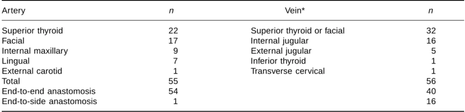
We performed 55 arterial and 56 venous anastomoses be-cause one patient required a double venous anastomosis. Ex-act sites are shown in Table II. Mean primary ischemic time of the flap was 63 min (range: 40-130 minutes). Eight (15%) patients presented vascular thrombosis; blood flow was re-established in four cases without losing the flap, and final loss rate was three flaps (5%). Date of presentation, treat-ment, and complications are listed in Table III.
Reinnervation of the forearm flap was performed in 23 (42%) patients. Of these, 20 (87%) were to lingual nerve, two (9%) to great auricular nerve, and one (4%) to superior laryngeal nerve.
Three (5%) patients had partial forearm flap necrosis af-fecting an area of 10 to 30% of flap size. One had a bilobal
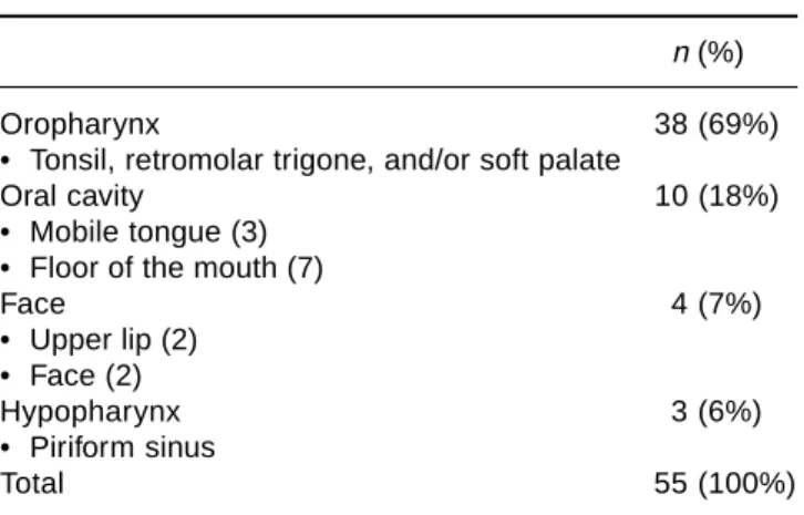
Table I. Localization of tumors
n (%)
Oropharynx 38 (69%)
• Tonsil, retromolar trigone, and/or soft palate
Oral cavity 10 (18%)
• Mobile tongue (3) • Floor of the mouth (7)
Face 4 (7%)
• Upper lip (2) • Face (2)
Hypopharynx 3 (6%)
• Piriform sinus
edigraphic.com
flap in oral cavity covering tongue and floor of the mouth and required local excision of approximately 30% of nonvi-able tissue several weeks later. The remaining two patients had necrosis of 10 to 20% of the flap in the superior part of oropharynx and developed granulation tissue in 10 to 20 days, allowing secondary healing with no consequences. Partial dehiscence of suture was seen in five (9%) patients. Five had a fistula that was treated with Thibloc® occlusive emulsion
(Johnson & Johnson) as well as endoscopic suture of the region; four (8%) patients had a postsurgical hematoma that required cervical drainage. Pneumonia was seen in 11 (21%) patients and one patient died of septic shock.
Fourteen (27%) patients had complications at donor site of the forearm flap. Of these, 10 patients had hyposensitiv-ity affecting thumb, two patients had partial loss of skin graft, one patient presented paresthesia, and another pa-tient presented pain. Functional limitation of the forearm was not observed. The most frequent long-term complica-tions were trismus, which affected nine patients, followed by infection, which affected only two patients. Periopera-tive and postoperaPeriopera-tive complications of recipient and do-nor site are listed in Table IV.
Discussion
Due to its characteristics, the forearm flap is considered the most versatile flap in head and neck surgery. Its thickness and adaptability make it ideal for bridging mucous membrane defects, especially in oropharynx and in oral cavity.
Functional reconstruction of soft palate and tonsillar region still remains a challenge. Because of its pliability, radial forearm flap has a clear advantage over traditional reconstructions with bulky myocutaneous pedicled flaps. Oropharynx reconstruc-tion has become the most frequent indicareconstruc-tion for this flap. In this area, it is essential to leave only a very small opening of the rhinopharynx to prevent rhinolalia and nasal reflux. In case of bilateral soft palate resection, the flap is folded to replace mu-cosa of both rhinopharyngeal and oral sides. The flap is locally de-epidermized and fixed to posterior pharyngeal wall, leaving two very small lumens laterally. In the case of unilateral de-fects, a local flap is elevated from posterior pharyngeal wall and fixed to the remaining soft palate to restore rhinopharyn-geal lining. The oral defect is then covered with forearm flap fixed to posterior pharyngeal wall with transfixant mattress su-ture to prevent anterior displacement of the flap with scarring. This procedure reduced risk of rhinolalia.
Table III. Vascular complications
N Thrombosis Day Treatment Complication
1 Internal maxillar artery <1 Thrombectomy and thrombolysis Total necrosis of the flap
2 Superior thyroid artery <1 Thrombectomy and thrombolysis None (salvage of flap)
3 Vein of the flap 10 No reoperation, missing flap in place Total necrosis of flap
4 Vein of the flap 2 A new forearm flap Total necrosis of flap
5 Vein of the flap 4 Thrombectomy and thrombolysis None (salvage of flap)
and internal jugular vein
6 Vein of the flap <1 Thrombectomy and thrombolysis, None (salvage of flap)
change anastomosis of vein from superior thyroid to internal jugular vein
7 Vein of the flap 2 Thrombectomy and thrombolysis None (salvage of flap)
8 Vein of the flap <1 Thrombectomy and thrombolysis None (salvage of flap)
Table II. Type of anastomosis (n = 53) patients
Artery n Vein* n
Superior thyroid 22 Superior thyroid or facial 32
Facial 17 Internal jugular 16
Internal maxillary 9 External jugular 5
Lingual 7 Inferior thyroid 1
External carotid 1 Transverse cervical 1
Total 55 56
End-to-end anastomosis 54 40
End-to-side anastomosis 1 16
Luna-Ortiz K y cols.
In oral cavity defects, functional prognosis depends on the possibility of restoring mobility of remaining soft tissues, par-ticularly the tongue. Forearm flap with a bilobal design allows reconstruction of floor of the mouth and lateral mobile tongue. This produces an acceptable degree of mobility to the tongue and restores sulcus for dental prosthetic rehabilitation at a later stage. We use forearm flaps even for T1 cancer of anterior floor of the mouth. In these cases, surgical treatment consists of monobloc pull-through resection with neck dissection that leaves a large defect, to obtain large surgical margins.
Forearm flap may be used for partial hypopharynx resection with larynx preservation when primary closure is not possible. In these cases, preservation of pharyngeal musculature on one side is essential for recovery of deglution without broncho-as-piration. Tubulized forearm flaps are described for reconstruc-tion of circular defects caused by circular total pharyngo-laryn-gectomy(7), but at our institution we prefer to use free jejunal
flap or a gastric pull-up, with satisfactory results(9).
On the face, this flap can be useful for reconstruction of large tissue defects with acceptable cosmetic results, espe-cially when a local flap is not available or when there is pre-viously irradiated tissue where transplantation of fresh tis-sue improves healing.
When large defects must be filled, particularly in middle third of the face, myocutaneous tissue such as rectus abdomi-nis or latissimus dorsi flaps are preferred to forearm flap. In our series, we used forearm for the cheek with good cosmetic results. There are previous reports(10,11) on use of this flap as
skin replacement with satisfactory results, and we consider it an excellent option in cases in which the inner portion of nose
or oral cavity must be reconstructed as well. In one patient, we reconstructed superior lip and maxillary region, according to principles described by Sakai et al.(5) and later described by
Sadove et al.(12) for reconstruction of lower lip with radial
fore-arm flap and palmaris longus vascularized tendon for suspen-sion of the lip. In another case, we used this flap to cover an exposed plate of the chin following mandibular reconstruc-tion with osteocutaneous free iliac crest with secondary ne-crosis of skin flap following radiotherapy.
Anatomic anomalies that prevent harvesting of forearm flaps are rare. The most common vascular anomaly of brachial, radi-al, or ulnar artery is due to a high origin of radial artery, which occurs in 15% of cases. The second most common anomaly is superficial ulnar artery, occurring in 2.5%. Distal take-off of radial artery deep to pronator teres muscle is quite rare as is superficial dorsal antebrachial artery. In our series, we discov-ered one anatomic malformation that consisted of absence of radial artery fasciocutaneous proximal perforator branch due to interposition by pronator teres muscle, as described previously by Small and Millar(13) and Funk et al(14). When this abnormal
vascular anatomy is encountered, the surgeon has two options: to seek another flap for reconstruction, or to arterialize main vein of forearm flap, which consists of anastomosis of main vein of flap, retaining a venous pedicle at the end. Distal vein is anastomosed to an artery, and proximal vein is anastomosed to a vein, thus creating an arterio-venous shunt and avoiding pos-sible problems with venous valves(15).
Although our patients are routinely heparinized to reduce primary critical ischemic time, flap necrosis is the major risk of this technique. In our series, we had a total of three (5%) forearm flap losses caused by thrombosis despite efforts to re-establish flow after secondary critical ischemia time. These patients presented the no-reflow phenomenon even after two patients had been exposed to a longer reperfusion time to reduce critical point of ischemic tissue damage for tissue repair. This time has not been fully established and probably varies according to flap type and to individual resistance to ischemia; however, some authors(16,17) propose it to be
ap-proximately 2 weeks while others18 simply mention that this
time may be shorter but do not specify the length. Etiology of this phenomenon is multifactorial and involves vasospasm, endothelial edema, arterio-venous shunting, stagnation, flu-id loss, local acflu-idosis, sludging of red blood cells, develop-ment of microthrombi, altered fibrinolysis, and endothelial cell changes that cause increased platelet adhesion and fi-brin deposits with microthrombus formation(19-23).
This implies that treatment should be use of multiple drugs to prevent thrombosis as well as damage caused by free rad-icals during reperfusion; therefore, it may be necessary to use a thrombolytic agent at time of complication, perhaps a free radical scavenger such as superoxide dismutase or even hyperbaric oxygen(24), to reduce endothelial cell activity, neu-Table IV. Frequency and type of complications
n (%)
I. Perioperative 2 (4)
Anatomical anomaly 1 (2)
No venous outflow of flap 1 (2)
II. Postoperative 39 (71)
Pneumonia (one patient died of septic shock) 11 (21)
Thrombosis 8 (15)
Fistula 5 (9)
Dehiscence of suture of flap 5 (9)
Cervical hematoma 4 (8)
Partial necrosis of flap 3 (5)
Necrosis of flap 3 (5)
III. Donor site 14 (27)
Hyposthesia and paresthesia 11 (21)
Partial necrosis of skin flap 2 (4)
Pain 1 (2)
IV. Long-term 11 (21)
Trismus 9 (17)
edigraphic.com
trophil adherence, or vasoconstriction. There are several stud-ies, most involving animal models(25), which demonstrate that
the complex mechanism of ischemia and events occurring after reperfusion can only be partially reproduced. They do not mention microcirculatory change caused by diseases such as hypertension, diabetes, and atherosclerosis, which are not usually present in animal models and could make the differ-ence in evolution of a particular flap.
Success rate of forearm flap rescue is 65% in our series. In the event of a suspicious vascular commitment, we per-formed surgical exploration that increased the success rate of forearm flap rescue.
Due to homogeneous vascularization, partial necrosis of the flap was infrequent and did not represent a major com-plication in our series. This occurred mainly in oropharynx and affected distal part of flap that covered superior border of the defect, where fistulas are uncommon. If this compli-cation were more common in hypopharynx in which risk of fistula is higher, this flap would not be an ideal option for reconstruction in this area and results would be similar to those reported by Coleman(2) with pedicle flaps, which
fre-quently present with partial marginal necrosis, suture dehis-cences, and fistulas. These represent important complications and may cause some delay in subsequent radiotherapy.
Dehiscence in flap suture lines is usually managed with conservative treatment or with local sutures; however, sec-ondary suture at edges of a fistula yields poor results. Small dehiscences are better treated with debridement to stimulate granulation in this area with complete healing, which usual-ly occurs within 10 to 20 days. In all our cases, fistulas were treated with in situ injection of a fast hardening amino acid solution (Ethibloc®). We have never had to use a new flap to
close a fistula. Complications at donor site are usually toler-ated and the symptomatology tends to diminish with time.
The forearm flap allows one-step reconstruction of large tissue defects with good cosmetic results in head and neck region. Use of this flap should encourage surgeons to per-form large excisions to have a wider surgical margin and prevent local relapses.
Morbidity at donor site is minimal and easily managed. The most important complication continues to be flap loss caused by vascular commitment. Flap loss rate was 9% in this series, which we consider an acceptable figure.
References
1. Yang G, Chen B, Gao Y, et al. Forearm free skin flap transplantation. Natl Med J China 1981;61:139-141.
2. Coleman JJ. Reconstruction of the pharynx after resection for can-cer. A comparison of methods. Ann Surg 1989;209:554-561. 3. Urken ML, Vickery C, Weinberg H, Biller HF. The
neurofasciocuta-neous radial forearm flap in head and neck reconstruction- a
prelimi-nary report. Presented at the Annual Meeting of the Trilogical Socie-ty. San Francisco, CA, USA, April. Laryngoscope 1990;100:161-173. 4. Soutar D, Widdowson WP. Immediate reconstruction of the mandible using a vascularized segment of radius. Head Neck 1986;8:232-246. 5. Sakai S, Soeda S, Endo T, Ishii M, Uchiumi E. A compound radial
artery forearm flap for the reconstruction of lip and chin defect. Br J Plast Surg 1989;42:337-338.
6. Sadove R, Luce EA, McGrath PC. Reconstruction of the lower lip and chin with the composite radial forearm-palmaris longus free flaps. Plast Reconst Surg 1991;88:209-214.
7. Kelly KE, Anthony JP, Singer M. Pharyngoesophageal reconstruc-tion using the radial forearm fasciocutaneous free flap: preliminary results. Otolaryngol Head Neck Surg 1994;111:16-24.
8. Funk GF, Laurenzo JF, Valentino J, McCulloch TM, Frodel JL, Hoffman HT. Free-tissue transfer reconstruction of midfacial and cranio-orbito-facial defect. Arch Otolaryngol Head Neck Surg 1995;121:293-303. 9. Luna-Ortiz K, Brossard E, Jaques B, Monnier P, Pasche P. Jejunum
free graft for reconstruction of circular defects after total pharyngo-laryngectomy. Cir Ciruj 1999;67:11-16.
10. Savant DN, Patel SG, Deshmukh SP, Guajariti R, Bhathena H, Kava-rana N. Folded free radial forearm flap for reconstruction of full-thickness defects of the cheek. Head Neck 1995;17:293-296. 11. Dhairyasheel N, Niranja NS, Watson DP. Reconstruction of the cheek using
a suspended radial forearm free flap. Br Plast Surg 1990;43:365-366. 12. Sadove RC, Luce EA, McGrath PC. Reconstruction of the lower lip
and chin with the composite radial forearm-palmaris longus free flap. Plast Recosnt Surg 1991;88:209-214.
13. Small JO, Millar R. The radial artery forearm flap: an anomaly of the radial artery. Br J Plast Surg 1985;38:501-503.
14. Funk GF, Valentino J, McCulloch TM, Graham SM, Hoffman HT. Anomalies of forearm vascular anatomy encountered during eleva-tion of the radial forearm flap. Head Neck 1995;17:284-292. 15. Klein C, Kovacs A, Stuckensen T. Free arterialized venous forearm
flaps for intraoral reconstruction. Br J Plast Surg 1997;50:166-171. 16. Kerrigan CL, Zelt RG, Daniel RK. Secondary critical ischemia time
of experimental skin flaps. Plast Reconst Surg 1984;74:522-526. 17. Milton SH. Experimental studies on island flaps. II: Ischemia and
delay. Plast Reconst Surg 1972;49:444-447.
18. Babajanian M, Zhang WX, Turk JB, Weinberg H, Biller HF, Urken ML. Temporal factors affecting the secondary critical ischemia of normothermic experimental skin flaps. Arch Otolaryngol Head Neck Surg 1991;117:1360-1364.
19. Zdeblick TA, Sahffer JW, Fiels GA. An ischemia-induced model of revas-cularization failure of replanted limbs. J Hand Surg 1985;10:125-131. 20. Zdeblick TA, Shaffer JW, Fiels GA. The use of urukinase in ischemic
replanted extremities in rats. J Bone Joint Surg Am 1987;69:442-449. 21. Amaes A III, Wright L, Kowada M, Thurston JM, Majno G. Cerebral ischemia. II: The no-reflow phenomenon. Am J Pathol 1968;52:437-447. 22. Chait LA, May JW, O’Brien BM, Hurley JV. The effect of the perfu-sion of various solutions on the no-reflow phenomenon in experi-mental free flaps. Plast Reconstr Surg 1978;61:421-430.
23. May JW, Chait LA, O’Brien BM, Hurley JV. The no-reflow pheno-menon in experimental free flaps. Plast Reconst Surg 1978;256-267. 24. Stevens DW, Weiss DD, Koller WA, Bianchi DA. Survival of normo-thermic microvascular flaps after prolonged secondary ischemia: effects of hyperbaric oxygen. Otolaryngol Head Neck Surg 1996;155:360-364.