CorSalud 2014 Ene-Mar;6(1):110-118
RNPS 2235-145 © 2009-2014 Cardiocentro “Ernesto Che Guevara”, Villa Clara, Cuba. Todos los derechos reservados.
110
Sociedad Cubana de Cardiología
________________
Caso Clínico
Enfermedad coronaria multivaso, disfunción endotelial y
angioplastia en la diabetes mellitus. A propósito de un caso
Dr. Suilbert Rodríguez Blanco
a, Dr.C. Javier Almeida Gómez
by Lic. Juan C. Pérez Guerra
ba Policlínico Docente “Nguyen Van Troi”. La Habana, Cuba.
b Laboratorio de Hemodinámica y Cardiología Intervencionista. Hospital “Hermanos Ameijeiras”. La Habana, Cuba.
Full English text of this article is also available
INFORMACIÓN DEL ARTÍCULO
Recibido: 12 de marzo de 2013 Modificado: 11 de junio de 2013 Aceptado: 18 de julio de 2013
Conflictos de intereses
Los autores declaran que no existen conflictos de intereses
Abreviaturas
DE: disfunción endotelial
DM: diabetes mellitus
HTA: hipertensión arterial
ICP: intervencion coronaria percutánea
TCI: tronco coronario izquierdo
Versiones On-Line:
Español - Inglés
S Rodríguez Blanco
Calle 17 Nº 1470 e/ 28 y 30. Vedado. La Habana, Cuba.
Correo electrónico:
suilbert@infomed.sld.cu
RESUMEN
La enfermedad coronaria es la principal causa de morbilidad y mortalidad en los pacientes con diabetes mellitus, la cual produce alteraciones en el endotelio y en el músculo liso vascular. Esta disfunción endotelial es precursora de lesiones aterogé-nicas. En este artículo se presenta el caso de una paciente diabética con enfermedad de tronco que fue tratada con éxito mediante intervencionismo coronario percutáneo y presentó progresión rápida de la enfermedad aterosclerótica en otros vasos, por lo que necesitó nueva revascularización percutánea. Se presentan las imágenes angio-gráficas y se comentan aspectos de la disfunción endotelial en la diabetes mellitus y su tratamiento percutáneo. Es importante identificar y tratar la disfunción endotelial tempranamente en los pacientes diabéticos. La elección del método de revasculari-zación debe ser individualizado.
Palabras clave: Diabetes mellitus, Disfunción endotelial, Enfermedad coronaria
multi-vaso, Angioplastia coronaria
Multivessel coronary artery disease, angioplasty and endothelial
dysfunction in diabetes mellitus. Case Report
ABSTRACT
Coronary heart disease is the leading cause of morbidity and mortality in patients with diabetes mellitus, and causes changes in the endothelium and vascular smooth muscle. This endothelial dysfunction is a precursor of atherogenic lesions. This article describes the case of a diabetic patient with left main trunk disease who was success-fully treated with percutaneous coronary intervention and showed rapid progression of atherosclerotic disease in other vessels, so she needed new percutaneous revascu-larization. Angiographic images are presented and aspects of endothelial dysfunction in diabetes mellitus and its percutaneous treatment are commented. It is important to early identify and treat endothelial dysfunction in diabetic patients. The choice of the revascularization method should be individualized.
Key words: Diabetes mellitus, Endothelial dysfunction, Multivessel coronary artery
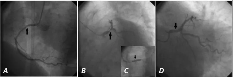
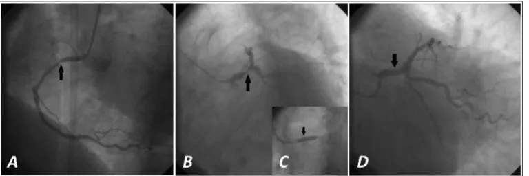
Figura 1. Coronariografía y ACTP. A. Lesión no significativa de CD (flecha). Vista oblicua anterior izquierda. B. Lesión grave
del TCI (flecha). Vista oblicua anterior izquierda con angulación caudal. C. ACTP del TCI (la flecha señala el momento de la implantación del stent). D. Resultado del procedimiento.
INTRODUCCIÓN
La enfermedad coronaria es la causa individual más frecuente de muerte en el mundo. Más de 7 millones de personas mueren cada año como consecuencia de la cardiopatía isquémica, lo que corresponde a un 12,8 % de todas las muertes1.
Estaenfermedadeslaprincipalcausademorbilidad y mortalidad en los pacientes con diabetes mellitus (DM). En los Estados Unidos se realizan aproximada-mente un millón y medio de intervenciones coronarias por año entre cirugías de revascularización miocárdica (CRM) e intervenciones coronarias percutáneas (ICP), y se estima que el 25 % de esos pacientes son diabéti-cos. Debido al impacto de la DM en el sistema cardio-vascular, esta población precisa un tratamiento especí-fico no solo de la diabetes como enfermedad de base, sino de la cardiopatía isquémica asociada2.
En este artículo se presenta el caso de una paciente diabética que fue tratada con éxito mediante ICP y se comentan aspectos fundamentales de la literatura al respecto.
CASO CLÍNICO
Mujer de 69 años,exfumadora(fumócercade40años, 1 cajetilla diaria), con antecedentes de hipertensión arterial (HTA) desde hace 25 años, en tratamiento con enalapril y clortalidona, y de DM no insulinodepen-diente desde hace 15 años, tratada con 2 tabletas diarias de metformina.
Acude en busca de atención médica por presentar cansancio fácil y dolor en el cuello, de carácter
opresi-vo que aparece a los esfuerzos físicos.
Su índice de masa corporal era de 29,6 kg/m2 y los complementarios realizados mostraban: colesterol 6,74 mmol/L, triglicéridos 3,09 mmol/L, glucemia 8,74 mmol/L y creatinina 91 mmol/L.
El electrocardiograma basal de 12 derivaciones mostraba una onda T aplanada de V1-V6. Se realizó coronariografía en el Laboratorio de Hemodinámica del Hospital “Hermanos Ameijeiras” (Figura 1), donde se encontró una estenosis de 85 % en el cuerpo del tronco coronario izquierdo (TCI), una lesión de 50 % en la porción proximal de la primera obtusa marginal, y el resto de los vasos no tenían lesiones significativas.
Previa dilatación de la lesión del TCI, se colocó un
stent liberador de fármaco en cuerpo de TCI. Se utilizó
la vía radial derecha, y se logró un éxito angiográfico (Figura 1), clínico y del procedimiento.
La paciente fue egresada a las 24 horas del proce-dimiento, se impuso tratamiento con aspirina, clopido-grel (doble antiagregación durante 1 año), atorvasta-tina, enalapril, atenolol y clortalidona.
A los 5 meses la paciente refiere presentar opre-sión precordial que dura alrededor de 5 minutos y se alivia con nitroglicerina sublingual; se interroga y se constata poco control metabólico y transgresiones medicamentosas. Se realizó nueva coronariografía (Figura 2), donde se confirma el buen resultado del
stent implantado en el TCI y la progresión de la
perma-Enfermedad coronaria multivaso, disfunción endotelial y angioplastia en la diabetes mellitus…
CorSalud 2014 Ene-Mar;6(1):110-118
112
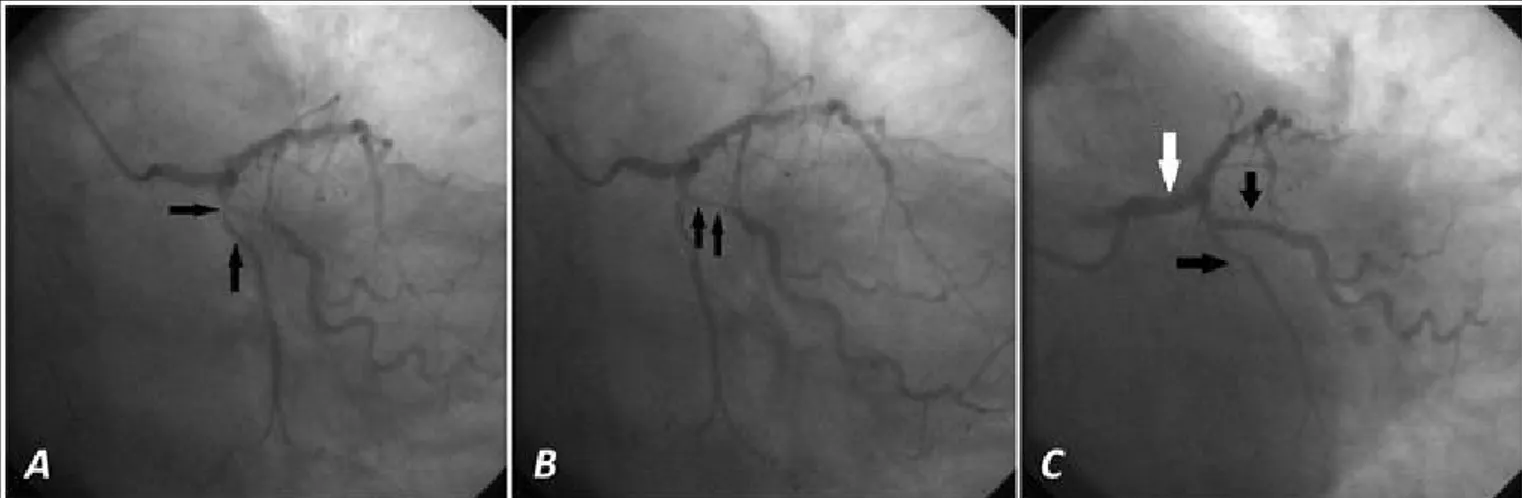
Figura 3. Control angiográfico a los 7 meses del segundo procedimiento. A. Persiste buen resultado de todos los stents
(flechas). B y C. Cuantificación angiográfica digital en el TCI.
necía invariable.
Se realizó ICP con stent convencional en ambas le-siones (Figura 2), se mantuvo el tratamiento fármaco-lógico y se insistió en ajustar el control metabólico e iniciar un programa de rehabilitación cardiovascular.
A los 7 meses la paciente se mantenía asintomáti-ca, con buen control metabólico y cumpliendo la reha-bilitación. Se realizó coronariografía de seguimiento (Figura 3), según el protocolo del centro para la enfer-medad del TCI y se demostró la persistencia del buen resultado de todos los stents implantados, sin otras alteraciones.
COMENTARIO
La DM produce alteraciones en el endotelio y en el músculo liso vascular, disfunción plaquetaria, vaso-constricción y respuesta proliferativa en los sitios de lesión3. El endotelio vascular no se debe considerar
como el recubrimiento pasivo interpuesto entre la sangre y el árbol vascular, sino como un órgano muy extenso del organismo humano, que cumple disímiles e importantes funciones4-5.
El óxido nítrico derivado del endotelio, constituye el compuesto vasodilatador natural más importante del organismo5-7. Otra sustancia producida por las
cé-lulas endoteliales es la prostaciclina, que provoca
Figura 2. Coronariografía y ACTP a los 5 meses. A y B. Progresión de la enfermedad aterosclerótica en Cx y OM (flechas). Vista
relajación del músculo liso vascular, y, por el contrario, también sintetiza moléculas vasoconstrictoras, como la angiotensina II, la endotelina-1 y el tromboxano A2, que se oponen a la acción vasorrelajante del óxido nítrico y promueven, además, la agregación plaqueta-ria y la proliferación de las células musculares lisas6-9.
En el endotelio también se produce trombomoduli-na, activador tisular del plasminógeno y glucosamino-glucanos del tipo heparán sulfato, la que garantiza una hemorreología normal (concepto que incluye, entre otros aspectos, la capacidad de mantenerse la sangre en estado líquido aun cuando esta tiene un contacto prolongado con la pared vascular); y, con efectos con-trarios, sustancias trombogénicas, como el inhibidor del activador tisular del plasminógeno, el factor de necrosis tumoral alfa, la interleuquina-1 y el factor tisular o hístico4-6,8.
Disfunción endotelial (DE)
Se puede definir como la serie de alteraciones que afectan la síntesis, liberación, difusión o degradación de los factores que se sintetizan por el endotelio. En otra definición se reconoce a la DE como la pérdida de la capacidad del endotelio de modular las funciones fisiológicas del lecho vascular. La DE no es homogénea en sus características ni en su distribución, estos aspectos varían en dependencia de la enfermedad que esté presente, así como del lecho vascular afectado. Entre los mecanismos inductores de daño vascular, y en consecuencia, de DE y las enfermedades que se asocian con su aparición, se encuentran: el estrés oxidativo, la hiperhomocisteinemia, la dislipidemia, la HTA, la obesidad, el hiperinsulinismo y la DM. Por su parte, la DE se ha detectado en prácticamente todas las enfermedades vasculares, y se presenta en muchos de los casos, incluso antes de que aparezcan las mani-festaciones clínicas5,8,10-12.
Disfunción endotelial y diabetes mellitus
La hiperglucemia crónica se asocia con un aumento de la formación de productos avanzados de la glicosila-ción y una hiperactividad del complejo aldosa reduc-tasa-proteína quinasa C, lo cual provoca, por mecanis-mos complejos, un incremento del estrés oxidativo, fenómeno que está íntimamente ligado a la aparición de DE en los individuos que padecen DM13,14.
La DE es un suceso temprano en el curso de la DM tipo 2, incluso, existen evidencias de que los marcado-res de DE están elevados en este tipo de pacientes
diabéticos, años antes de que la enfermedad se ma-nifieste clínicamente. En la DM tipo 2 se sabe que, además de la hiperglucemia, también influyen en la aparición de DE, la resistencia a la insulina y el hiper-insulinismo resultante13,14. Por su parte, el 60 % de los
individuos con DM tipo 2 son hipertensos y el 90 %, obeso.
La causa más común de muerte entre los europeos adultos con DM es la enfermedad coronaria, en diver-sos estudios se ha demostrado que este grupo tiene un riesgo 2 o 3 veces mayor que las personas sin diabetes15, los sujetos diabéticos mueren 10 a 15 años
antes que los que pertenecen a la población general, y, sobre todo, por enfermedades vasculares; además está demostrado que un diabético tiene igual riesgo de sufrir un infarto de miocardio, que un individuo que ha tenido un primer episodio coronario16-18.
Marcadores de DE en la DM
En la DM está afectada la síntesis de óxido nítrico, su biodisponibilidad y viabilidad, así como la respuesta relajante del endotelio19,20.
Se ha demostrado que la hemoglobina glucosilada no es solo un evaluador del grado de control metabó-lico, sino que también puede participar en la génesis de la DE. Una hemoglobina glucosilada elevada circu-lando libremente en el plasma, puede inducir la dis-minución de la relajación mediada por óxido nítrico mediante la generación de radicales superóxido20-22.
Otro marcador de DE elevado en los individuos diabéticos, es la endotelina-1. Se considera que su au-mento se relaciona con la aparición de la HTA y la aterosclerosis más precoz y grave, que generalmente acompaña a la DM, sobre todo, a la de tipo 24-7.
Dislipidemia diabética y DE
La dislipidemia diabética se caracteriza por hipertrigli-ceridemia moderada, lipoproteínas de alta densidad disminuidas y presencia de lipoproteínas de baja den-sidad pequeñas y densas, que son muy aterogénicas; y si bien el colesterol total generalmente es normal, elevaciones de su concentración sanguínea que no tienen repercusión clínica en el sujeto sin DM, sí incre-mentan el riesgo cardiovascular de 2 a 3 veces en el diabético22,23.
Enfermedad coronaria multivaso, disfunción endotelial y angioplastia en la diabetes mellitus…
CorSalud 2014 Ene-Mar;6(1):110-118
114
relacionado con la gravedad de la aterosclerosis coro-naria, que con mucha frecuencia se aprecia en estos pacientes24.Se conoce que las alteraciones diabéticas
lipoproteícas dependientes de triglicéridos, se mag-nifican en el estado posprandial, y que se relacionan también con la aparición de la DE y la cardiopatía isquémica, de ahí la importancia del estudio lipídico posprandial en el diabético. La resistencia insulínica es probablemente el núcleo de los mecanismos fisiopa-tológicos de la dislipidemia diabética25-27.
HTA, obesidad, DM y DE
La prevalencia de HTA en los diabéticos es aproxima-damente el doble que en la población no diabética, y cuando la HTA no está controlada, se duplica el riesgo de padecer enfermedad coronaria28-30.
Desde el punto de vista fisiopatológico, se postula que, en ausencia de disfunción renal, la resistencia in-sulínica y la hiperinsulinemia compensadora ocupan un lugar central en la etiopatogenia de la HTA en la DM, aunque se conoce que es multifactorial29.
La obesidad, asociada frecuentemente con la DM tipo 2 (diabesidad) y la resistencia a la insulina, se ha relacionado con un aumento de la frecuencia de la enfermedad coronaria en los diabéticos tipo 2. En los diabéticos obesos se han encontrado niveles aumenta-dos de E-selectina, endotelina-1, resistina, leptina y resistencia a la acción de esta hormona peptídica; así como una disminución de la adiponectina, la produc-ción de óxido nítrico dependiente de leptina y la vaso-dilatación dependiente del endotelio29,30.
Puede decirse que en los individuos con diabetes se presentan con mayor frecuencia todas las enfermeda-des vasculares relacionadas con el fenómeno ateros-clerótico y se sabe que la DE está asociada de forma importante con la aparición de la aterosclerosis31,32.
Tratamiento de la DE en la DM
Existen evidencias que permiten afirmar que no hay mejor medida terapéutica para evitar la aparición de DE o disminuir sus efectos adversos en los diabéticos, que lograr un control metabólico óptimo33,34, con o sin
tratamiento farmacológico.
Existe gran controversia en relación con la utilidad de compuestos antioxidantes en las enfermedades en las cuales se ha demostrado la presencia de un au-mento del estrés oxidativo y una disminución de las defensas antioxidantes, incluida la DM. Sin embargo, se han usado múltiples compuestos antioxidantes para
tratar el estrés oxidativo y la DE asociada con la DM 31-34.
Tratamiento de las lesiones ateroscleróticas en el diabético
Datos de autopsias demuestran que la aterosclerosis coronaria del diabético es más grave, con afectación de un mayor número de vasos, una distribución más difusa y con un mayor número de placas complicadas, ulceradas y con trombo, que en la población no dia-bética35. Los estudios coronariográficos confirman
le-siones más graves y difusas, tanto proximales como distales, con menor desarrollo de circulación colateral y una mayor presencia de placas de riesgo. Los diabé-ticos muestran un más rápido crecimiento de las lesiones cuando se comparan estudios repetidos en un mismo paciente. Los nuevos procedimientos de explo-ración intracoronaria (ultrasonido intravascular y tomografía de coherencia óptica) confirman la presen-cia de un mayor número de placas calientes y mayor tasa de complicaciones. Como ocurrió en el caso que se presenta, la respuesta de los vasos coronarios a los procedimientos intervencionistas es menos favora-ble35,36.
Varios estudios37-41 han demostrado que el
trata-miento médico óptimo es tan efectivo como la CRM o la ICP, en los pacientes con angina estable crónica y enfermedad coronaria leve. Mientras que en los pa-cientes con enfermedad coronaria moderada o grave estos procedimientos, combinados con tratamiento médico óptimo, producen mayor supervivencia y alivio sintomático que el tratamiento médico solo.
Revisión de ensayos comparativos
En el análisis de subgrupos de los estudios ERACI-II42 y
ARTS43,44, los pacientes diabéticos tratados con CRM
El estudio CARDia46 tiene como objetivo la
compa-ración entre la angioplastia coronaria con implante de
stents y la cirugía cardíaca en pacientes diabéticos con
enfermedad coronaria multivaso sintomática. Se in-cluyeron 510 pacientes diabéticos con enfermedad multivaso o enfermedad de un único vaso pero con gran complejidad, y se aleatorizaron a CRM o ICP (ini-cialmente con stents metálicos y posteriormente far-macoactivos), y utilización rutinaria de abciximab. El objetivo primario fue el combinado de mortalidad por cualquier causa, infarto de miocardio y accidente cere-brovascular. Como objetivo secundario, la combina-ción del objetivo primario y la necesidad de una nueva revascularización. Se utilizó un diseño de no inferiori-dad, de tal forma que para considerar a la angioplastia no inferior a la cirugía, el límite superior del intervalo de confianza del 95 % (IC 95 %) debía ser inferior a 1,346.
Tras un año de seguimiento, el objetivo primario se alcanzó en el 15 % del grupo quirúrgico y en el 13 % del grupo de ICP. Las tasas de mortalidad total fueron iguales y la combinación de muerte, infarto, accidente cerebrovascular o necesidad de nueva revasculari-zación (objetivo secundario) fue del 11,3 y 19,3 %. Cuando se compararon los pacientes quirúrgicos con el subgrupo de pacientes que habían recibido stents
farmacoactivos (69 % del total), el objetivo primario se alcanzó en el 12,4 y 11,6 %, respectivamente46,47.
Sus resultados al año indicaron que aunque la an-gioplastia es una técnica que se puede llevar a cabo con seguridad en estos pacientes, a largo plazo no se ha demostrado la no inferioridad. Respecto al objetivo secundario, la cirugía es significativamente mejor, a expensas sobre todo de una menor necesidad de nue-va renue-vascularización. En relación al objetivo primario, el límite superior del IC 95 % supera el límite marcado para la no inferioridad, tanto en el grupo global como si consideramos únicamente a los pacientes tratados con stents farmacoactivos de nueva generación46,47.
ElDr. Eric Bates (Universidad de Michigan, Ann Ar-bor), en sus comentarios a los artículos de Farkouh48 y
Hlatky49, dijo a Heartwire50 que si se analiza
estric-tamente el estudio clínico y la evidencia previamente publicada, FREEDOM respalda la superioridad de la CRM con respecto a la ICP50. Sin embargo, señaló que
desde el punto de vista del ejercicio clínico muchos intervencionistas pueden identificar a los pacientes con alto y bajo riesgo, y en consecuencia pueden diri-girlos al tratamiento de revascularización más
apro-piado. Por este motivo, algunos datos de registros han mostrado que los episodios clínicos han sido similares en diabéticos sometidos a CRM o ICP48-50.
«No es ICP contra CRM », dijo Bates50. «Estos son
procedimientos de revascularización complementarios y estos estudios muestran que la CRM debiera ser una parte importante del debate, pero a nivel del paciente individual, hay factores como el riesgo de accidente cerebrovascular, su estado frágil, la función renal, la función pulmonar, la preferencia del paciente, la experiencia del médico y otras variables que influyen en la decisión para cada caso».
CONSIDERACIONES FINALES
La DE se presenta con frecuencia en los sujetos con DM, incluso, puede detectarse en algunos de estos in-dividuos al comienzo de la enfermedad metabólica. En los diabéticos, la hiperglucemia crónica y la existencia frecuente de comorbilidades asociadas con la DM, fa-vorecen la aparición de la DE, su presencia demuestra que existen condiciones metabólicas para la aparición de la microangiopatía y la macroangiopatía (ateroscle-rosis) diabéticas. En cuanto al tratamiento, se le reco-noce una acción antidisfunción endotelial a disímiles medidas terapéuticas medicamentosas y no medica-mentosas, y entre las últimas es esencial el alcance de un control metabólico óptimo.
En los casos en que se decida revascularización miocárdica, le evidencia apunta al tratamiento quirúr-gico de los pacientes diabéticos con enfermedad coro-naria multivaso. Aunque el ICP no deja de ser una opción en aquellos pacientes con contraindicaciones específicas para la cirugía. Y se deben ver ambos métodos como procedimientos de revascularización complementarios.
En este caso se realizó tratamiento con ICP y se lo-gró un éxito angiográfico, clínico y del procedimiento.
REFERENCIAS BIBLIOGRÁFICAS
1. WHO. The top 10 causes of death. Fact sheet Nº 310. [Internet]. WHO; Updated June 2013. [citado 2013 Feb 6] Disponible en:
http://www.who.int/mediacentre/factsheets/fs310 /en/index.html
Enfermedad coronaria multivaso, disfunción endotelial y angioplastia en la diabetes mellitus…
CorSalud 2014 Ene-Mar;6(1):110-118
116
3. Pandolfi A, Cetrullo D, Polishuck R, Alberta MM, Calafiore A, Pellegrini G, et al. Plasminogen acti-vator inhibitor type 1 is increased in the arterial walloftypeIIdiabeticsubjects.ArteriosclerThromb Vasc Biol 2001;21(8):1378-82.
4. Arpa Gámez A, González Sotolongo O, Roldós Cuza E, Borges Helps A, Acosta Vaillant R. El síndrome metabólico como factor de riesgo para la disfun-ción endotelial. Rev Cubana Med Milit [Internet]. 2007 [citado 2013 Feb 12];36(1):[aprox. 10 p.]. Disponible en:
http://www.bvs.sld.cu/revistas/mil/vol36_01_07/ mil02107.htm
5. Esteller Pérez A. Biología de la pared vascular y síndrome metabólico. Nutr Hosp. 2005;XX(1):5-17. 6. López A. Disfunción endotelial y metabolismo del corazón en la insuficiencia cardíaca. Haematolo-gica/Edición española. 2008;93(Extra 1):333-6. 7. Acosta AG, Añez J, Andara CV, Bermúdez V,
Bermú-dez F. Mecanismos moleculares de la disfunción endotelial: de la síntesis a la acción del óxido ní-trico. Arch Venez Farmacol Terap. 2006;25(2):54-9. 8. Cohen RA. Role of nitric oxide in diabetic
complica-tions. Am J Ther. 2005;12(6):499-502.
9. Esper RJ, Nordaby RA, Vilariño JO, Paragano A, Cacharrón JL, Machado RA. Endothelial dysfunc-tion: a comprehensive appraisal. Cardiovasc Diabe-tol. 2006;5:4.
10.Huijberts MS, Becker A, Stehouwer CD. Homocys-teine and vascular disease in diabetes: a double hit? Clin Chem Lab Med. 2005;43(10):993-1000. 11.Tellez J. Adiponectina y disfunción endotelial.
RESPYN [Internet]. 2005 [citado 2013 Feb 12];16 (Edición Especial): [aprox. 6 p.]. Disponible en:
http://www.respyn.uanl.mx/especiales/2005/ee-16-2005/documentos/12.htm
12.Cachofeiro V, Miana M, Martín-Fernández B, de las Heras N, Lahera V. Obesidad, inflamación y disfun-ción endotelial. Rev Esp Obes. 2006;4(4):195-204. 13.Woodman RJ, Chew GT, Watts GF. Mechanisms,
significance and treatment of vascular dysfunction in type 2 diabetes mellitus: focus on lipid-regulating therapy. Drugs. 2005;65(1):31-74.
14.Ceriello A, Motz E. Is oxidative stress the patho-genic mechanism underlying insulin resistance, dia-betes, and cardiovascular disease? The common soil hypothesis revisited. Arterioscler Thromb Vasc Biol. 2004;24(5):816-23.
15.Laakso M. Hyperglycemia and cardiovascular
dis-ease in type 2 diabetes. Diabetes. 1999;48(5):937-42.
16. Moldoveanu E, Tanaseanu C, Tanaseanu S, Kosaka T, Manea G, Marta DS, et al. Plasma markers of endothelial dysfunction in type 2 diabetics. Eur J Intern Med. 2006;17(1):38-42.
17.Charvát J, Michalova K, Chlumský J, Valenta Z, Kva-pil M. The association between left ventricle dias-tolic dysfunction and endothelial dysfunction and the results of stress myocardial SPECT in asympto-matic patients with type 2 diabetes. J Int Med Res. 2005;33(5):473-82.
18.Karasik A. Glycaemic control is essential for effect-ive cardiovascular risk reduction across the type 2 diabetes continuum. Ann Med. 2005;37(4):250-8. 19.Rodríguez L, López P, Petidier R, Neira M. Solís J,
Pavón I, et al. Effect of glycaemic control on the vascular nitric oxide system in patients with type 1 diabetes. J Hypertens. 2003;21(6):1137-43.
20.Endemann DH, Schiffrin EL. Nitric oxide, oxidative excess, and vascular complications of diabetes me-llitus. Curr Hypertens Rep. 2004;6(2):85-9.
21.Home P. Contributions of basal and post-prandial hyperglycaemia to micro- and macrovascular com-plications in people with type 2 diabetes. Curr Med Res Opin. 2005;21(7):989-98.
22.Woodman RJ, Chew GT, Watts GF. Mechanisms, significance and treatment of vascular dysfunction in type 2 diabetes mellitus: focus on lipid-regulating therapy. Drugs. 2005;65(1):31-74.
23.Wägner AM, Sánchez JL, Pérez A. Diabetes mellitus y lipemia posprandial. Endocrinol Nutr. 2000; 47(10):311-21.
24.Lee IK, Kim HS, Bae JH. Endothelial dysfunction: its relationship with acute hyperglycaemia and hyper-lipidemia. Int J Clin Pract. 2002;129(Suppl):59-64. 25.Heine RJ, Balkau B, Ceriello A, Del Prato S, Horton
ES, Taskinen MR. What does postprandial hypergly-caemia mean? Diabet Med. 2004;21(3):208-13. 26.Tushuizen ME, Diamant M, Heine RJ. Postprandial
dysmetabolism and cardiovascular disease in type 2 diabetes. Postgrad Med J. 2005;81(951):1-6. 27.Saxena R, Madhu SV, Shukla R, Prabhu KM,
Gamb-hir JK. Postprandial hypertriglyceridemia and oxid-ative stress in patients of type 2 diabetes mellitus with macrovascular complications. Clin Chim Acta. 2009;359(1-2):101-8.
display enhanced basal platelet activation and decreased antioxidant status. Diabetes. 2004;53(4): 1046-51.
29.García JA, Gonseski VC, González TP, Franco FF. Renoprotección en diabetes e hipertensión: Revi-sión bibliográfica de la conducta actual. Rev Postgr VIa Cáted Med. 2005;144:11-5.
30.Dixon LJ, Hughes SM, Rooney K, Madden A, Devine A, Leahey W, et al. Increased superoxide produc-tion in hypertensive patients with diabetes melli-tus: role of nitric oxide synthase. Am J Hypertens. 2005;18(6):839-43.
31.Huidobroa A, Cuevas A, Chamorro G, Maiz A, Ro-sowski J, Villarroel L, et al. Resistencia insulínica y cardiopatía coronaria. Clin Invest Arterioscl. 2000; 12(3):153-9.
32.Botla CE. Insuficiencia cardíaca y diabetes. Una combinación de alto riesgo. Rev Insuf Cardíaca. 2009;4(3):107-13.
33.Esposito K, Giugliano D, Nappo F, Marfella R; Cam-panian Postprandial Hyperglycemia Study Group. Regression of carotid atherosclerosis by control of postprandial hyperglycemia in type 2 diabetes mellitus. Circulation. 2004;110(2):214-9.
34.Richards RJ. Postprandial hyperglycemia. J La State Med Soc. 2003;155(5):260-5.
35.González-Maqueda I. De la disfunción endotelial a la formación de la placa de ateroma. En: Rio A, De Pablo C, Editores. Manual de Medicina Preventiva. Publicación Oficial de la Sociedad Española de Cardiología. Sección de Cardiopatía Preventiva y Rehabilitación. Madrid: Scientific Communication Management; 2005. p. 25-41.
36.González-Maqueda I. La enfermedad coronaria del diabético. Diagnóstico, pronóstico y tratamiento.
Rev Esp Cardiol. 2007;7(Supl. H):29-41. 37.Hlatky MA, Boothroyd DB, Bravata DM, Boersma E,
Booth J, Brooks MM, et al. Coronary artery bypass surgery compared with percutaneous coronary in-terventions for multivessel disease: a collaborative analysis of individual patient data from ten ran-domised trials. Lancet. 2009;373(9670):1190-7. 38.Smith SC, Faxon D, Cascio W, Schaff H, Gardner T,
Jacobs A, et al. Prevention Conference VI: Diabetes and Cardiovascular Disease: Writing Group VI: re-vascularization in diabetic patients. Circulation. 2002;105(18):e165-9.
39.Hillis LD, Smith PK, Anderson JL, Bittl JA, Bridges CR, Byrne JG, et al. 2011 ACCF/AHA Guideline for
Coro-nary Artery Bypass Graft Surgery: executive sum-mary: a report of the American College of Car-diology Foundation/American Heart Association Task Force on Practice Guidelines. Circulation. 2011;124(23):2610-42. [Erratum, Circulation. 2011; 124(25):e956].
40.Brown ML, Sund TM III, Gersh BJ. Indications for revascularization. En: Cohn LH, Editor. Cardiac sur-gery in the adult. 3 ed. New York: McGraw Hill Edu-cation; 2007. p. 551.
41.Hannan EL, Wu C, Walford G, Culliford AT, Gold JP, Smith CR, et al. Drug-eluting stents vs. coronary-artery bypass grafting in multivessel coronary disease. N Engl J Med. 2008;358(4):331-41.
42.Rodriguez AE, Baldi J, Fernández-Pereira C, Navia J, Rodriguez Alemparte M, Delacasa A, et al. Five-year follow-up of the Argentine randomized trial of coronary angioplasty with stenting versus coronary bypass surgery in patients with multiple vessel disease (ERACI II). J Am Coll Cardiol. 2005;46(4): 582-8.
43.Serruys PW, Ong AT, van Herwerden LA, Sousa JE, Jatene A, Bonnier JJ, et al. Five-year outcomes after coronary stenting versus bypass surgery for the treatment of multivessel disease: the final analysis of the Arterial Revascularization Therapies Study (ARTS) randomized trial. J Am Coll Cardiol. 2005; 46(4):575-81.
44. Ix JH, Mercado N, Shlipak MG, Lemos PA, Boersma E, Lindeboom W, et al. Association of chronic kidney disease with clinical outcomes after coro-nary revascularization: the Arterial Revasculari-zation Therapies Study (ARTS). Am Heart J. 2005; 149(3):512-9.
45.Stone GW, Midei M, Newman W, Sanz M, Hermiller JB, Williams J, et al; SPIRIT III Investigators. Com-parison of an everolimus-eluting stent and a pacli-taxel-eluting stent in patients with coronary artery disease: a randomized trial. JAMA. 2008;299(16): 1903-13.
46.Kapur A, Hall RJ, Malik IS, Qureshi AC, Butts J, de Belder M, et al. Randomized comparison of per-cutaneous coronary intervention with coronary artery bypass grafting in diabetic patients: 1-year results of the CARDia (Coronary Artery Revas-cularization in Diabetes) trial. J Am Coll Cardiol. 2010;55(5):432-40.
CorSalud 2014 Jan-Mar;6(1):110-118
RNPS 2235-145 © 2009-2014 Cardiocentro “Ernesto Che Guevara”, Villa Clara, Cuba. All rights reserved.
110
Cuban Society of Cardiology
________________
Case Report
Multivessel coronary artery disease, angioplasty and endothelial
dysfunction in diabetes mellitus. Case Report
Suilbert Rodríguez Blanco
a, MD; Javier Almeida Gómez
b, PhD; and Juan C. Pérez
Guerra
b, BN
a Nguyen Van Troi Teaching Polyclinic. Havana, Cuba.
b bInterventional Cardiology and Catheterization Laboratory. Hermanos Ameijeiras Hospital. Havana, Cuba.
Este artículo también está disponible en español
ARTICLE INFORMATION
Received: March 12, 2013 Modified: June 11, 2013 Accepted: July 18, 2013
Competing interests
The authors declare no competing interests
Acronyms
CABG: coronary artery bypass graft
DM: diabetes mellitus
ED: endothelial dysfunction
HT: hypertension
LMCA: left main coronary artery
PCI: percutaneous coronary intervention
On-Line Versions:
Spanish - English
S Rodríguez Blanco
Calle 17 Nº 1470 e/ 28 y 30. Vedado. La Habana, Cuba.
E-mail address:
suilbert@infomed.sld.cu
ABSTRACT
Coronary heart disease is the leading cause of morbidity and mortality in patients with diabetes mellitus, and causes changes in the endothelium and vascular smooth muscle. This endothelial dysfunction is a precursor of atherogenic lesions. This article describes the case of a diabetic patient with left main trunk disease who was success-fully treated with percutaneous coronary intervention and showed rapid progression of atherosclerotic disease in other vessels, so she needed new percutaneous revascu-larization. Angiographic images are presented and aspects of endothelial dysfunction in diabetes mellitus and its percutaneous treatment are commented. It is important to early identify and treat endothelial dysfunction in diabetic patients. The choice of the revascularization method should be individualized.
Key words: Diabetes mellitus, Endothelial dysfunction, Multivessel coronary artery
disease, Coronary angioplasty
Enfermedad coronaria multivaso, disfunción endotelial y angioplastia
en la diabetes mellitus. A propósito de un caso
RESUMEN
La enfermedad coronaria es la principal causa de morbilidad y mortalidad en los pacientes con diabetes mellitus, la cual produce alteraciones en el endotelio y en el músculo liso vascular. Esta disfunción endotelial es precursora de lesiones aterogé-nicas. En este artículo se presenta el caso de una paciente diabética con enfermedad de tronco que fue tratada con éxito mediante intervencionismo coronario percutáneo y presentó progresión rápida de la enfermedad aterosclerótica en otros vasos, por lo que necesitó nueva revascularización percutánea. Se presentan las imágenes angio-gráficas y se comentan aspectos de la disfunción endotelial en la diabetes mellitus y su tratamiento percutáneo. Es importante identificar y tratar la disfunción endotelial tempranamente en los pacientes diabéticos. La elección del método de revasculari-zación debe ser individualizado.
Palabras clave: Diabetes mellitus, Disfunción endotelial, Enfermedad coronaria
Figura 1. Coronary angiography and PTCA. A. No significant lesion in RC (arrow). Left anterior oblique view. B. Severe LMCA
lesion (arrow). Left anterior oblique view with caudal angulation. C. PTCA of the LMCA (arrow indicates the time of stent implantation). D. Outcome of the procedure.
INTRODUCTION
Coronary heart disease is the most common single cause of death worldwide. More than 7 million people die each year as a result of ischemic heart disease, which corresponds to 12.8 % of all deaths1.
This disease is the main cause of morbidity and mortality in patients with diabetes mellitus (DM). In the United States about one and half million coronary interventions are performed annually between coro-nary artery bypass graft surgeries (CABG) and percuta-neous coronary intervention (PCI), and it is estimated that 25% of these patients are diabetics. Due to the impact of DM on the cardiovascular system, this po-pulation requires not only specific treatment for dia-betes as the underlying disease, but also for the associated ischemic heart disease2.
This article describes the case of a diabetic patient who was successfully treated with PCI, and funda-mental aspects of the literature about it are discussed.
CASE REPORT
69-year-old woman, ex-smoker (smoked for about 40 years, one pack per day), with a history of hyper-tension (HT) for 25 years, treated with enalapril and chlorthalidone, and non-insulin dependent DM for 15 years, treated 2 daily tablets of metformin.
She seeks medical care foreasy fatigue and
op-pressive pain in the neck on physical exertion.
Her BMI was 29.6 kg/m2 and the tests performed showed cholesterol 6.74 mmol/L, Triglycerides 3.09 mmol/L, glucose 8.74 mmol/L and creatinine 91 mmol/L.
The baseline 12-lead electrocardiogram showed a flattened T wave in V1-V6. Coronary angiography was performed in the Catheterization Laboratory of Her-manos Ameijeiras Hospital (Figure 1), where a stenosis of 85 % in the body of the left main coronary artery (LMCA), and an injury of 50 % in the proximal portion of the first obtuse margin were found; the rest of the vessels had no significant lesions.
After dilation of the LMCA lesion, a drug eluting stent was placed in the body of LMCA. Right radial approach was used, and an angiographic (Figure 1), clinical and procedural success was achieved.
The patient was discharged 24 hours after the pro-cedure, with the following treatment: aspirin, clopido-grel (dual antiplatelet therapy for 1 year), atorvastatin, enalapril, atenolol and chlorthalidone.
non-Multivessel coronary artery disease, angioplasty and endothelial dysfunction in diabetes mellitus. Case Report
CorSalud 2014 Ene-Mar;6(1):110-118
112
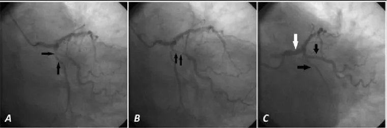
Figura 3. Angiographic control at 7 months of the second procedure. A. Persists success of all stents (arrows). B and C. Digital
angiographic quantification in LMCA.
significant lesion of the right coronary remained un-changed.
PCIwasperformedwithconventionalstentsinboth lesions (Figure 2), drug treatment was maintained and stress was made onadjusting metabolic control and starting a cardiac rehabilitation program.
At 7 months the patient remained asymptomatic with good metabolic control and doing rehabilitation. Follow-up coronary angiography (Figure 3) was per-formed according to the protocol of the Center for LMCA disease and the success of all implanted stents
was demonstrated, with no other abnormalities.
COMMENTS
DM produces changes in the endothelium and vas-cular smooth muscle, platelet dysfunction, vasocons-triction and proliferative response at sites of injury3.
The vascular endothelium should not be considered as the passive coating interposed between the blood and the vascular tree, but as a very large organ of the
Figura 2. Coronary angiography and PTCA at 5 months. A and B. Progression of atherosclerotic disease in Cx and MO (arrows).
human body, which fulfills important and dissimilar functions4-5.
The endothelium-derived nitric oxide is the most important natural vasodilator compound in the body 5-7. Another substance produced by endothelial cells is
prostacyclin that causes relaxation of vascular smooth muscle, and, conversely, also synthesizes vasocons-trictor molecules such as angiotensin II, endothelin-1 and thromboxane A2, which oppose the vasorelaxing action of nitric oxide, and also promote platelet aggregation and proliferation of smooth muscle cells6-9.
Thrombomodulin is also produced in the endo-thelium, and is a tissue activator of plasminogen and glycosaminoglycan of heparan sulfate type, which ensures a normal hemorheology (a concept that in-cludes, among other things, the ability to keep the blood in liquid state even when it has a long contact with the vessel wall) and, with the opposite effects, thrombogenic substances such as the inhibitor of tissue plasminogen activator, tumor necrosis factor alpha, interleukin-1 , and the tissue factor4-6,8.
Endothelial dysfunction (ED)
It can be defined as the number of conditions affecting the synthesis, release, diffusion, or degradation of factors synthesized by the endothelium. In another definition ED is recognized as the endothelium’s loss of the ability to modulate physiological functions of the vascular bed. ED is not homogeneous in its charac-teristics anddistribution; these aspects vary depending on the diseasethat is present, and the affected cular bed. Among the triggering mechanisms of vas-cular damage, and consequently, of ED and diseases that are associated with its appearance are: oxidative stress, hyperhomocysteinemia, dyslipidemia, hyper-tension, obesity, hyperinsulinemia and diabetes. Meanwhile, ED has been detected in virtually all vas-cular diseases and occurs in many cases, even before the clinical symptoms appear5,8,10-12.
Endothelial dysfunction and diabetes mellitus
Chronic hyperglycemia is associated with increased formation of advanced glycosylation products and hyperactivity of aldose reductase-protein kinase C
complex, which leads, by complex mechanisms, to an increased oxidative stress, a phenomenon that is closely linked to the occurrence of ED in individuals with DM13,14.
ED is an early event in the course of type 2 dia-betes, there is even evidence that ED markers are elevated in this type of diabetic patients, years before the disease manifests clinically. It is known thatin type 2 DM, in addition to hyperglycemia, the onset of ED is also influenced by insulin resistance and the resulting hyperinsulinemia13,14. Meanwhile, 60 % of individuals
with type 2 diabetes are hypertensive and 90% obese. The most common cause of death among European adults with diabetes is coronary artery disease. Several studies have shown that this group has a risk 2 to 3 times higher than people without diabetes15, diabetic
subjects die 10-15 years earlier than those belonging to the general population, and especially due to vascular diseases; besides it has been demonstrated that a diabetic patient has the same risk of having a heart attack, than an individual who has had a first coronary event16-18.
ED Markers in DM
In DM, the synthesis of nitric oxide, its bioavailability and viability, as well as the relaxing response of the endothelium are affected19,20.
It has been demonstrated that glycosylated he-moglobin is not only an assessor of metabolic control, but it may also participate in the genesis of the ED. Elevated glycated hemoglobin circulating freely in the plasma, can induce the reduction of nitric oxide-mediated relaxation through the generation of super-oxide radicals20-22.
Another elevated marker of ED in diabetic indi-viduals is endothelin-1. Its increase is considered to be related to the onset of hypertension and the earlier and more severe atherosclerosis, which usually ac-companies DM, especially type 244-7.
Diabetic dyslipidemia and ED
Multivessel coronary artery disease, angioplasty and endothelial dysfunction in diabetes mellitus. Case Report
CorSalud 2014 Ene-Mar;6(1):110-118
114
cholesterol is usually normal, elevated blood con-centration that have no clinical impact in subjects without DM, do increase2-3 times cardiovascular risk in diabetic patients22,23.
Hypertriglyceridemia is currently regarded as a predictor of cardiovascular disease, and the elevation in the plasma of triglyceride-rich lipoproteins in dia-betic subjects has been related to the severity of co-ronary atherosclerosis, which is very often seen in these patients24. It is known that
triglyceride-depen-dent lipoprotein diabetic disorders, are magnified in the postprandial state, and are also associated with the onset of ED and ischemic heart disease, hence the importance of postprandial lipid study in diabetics. Insulin resistance is probably the core of the patho-physiological mechanisms of diabetic dyslipidemia25-27.
Hypertension, obesity, DM and ED
The prevalence of hypertension in diabetics is about twice that in the non-diabetic population, and when hypertension is not controlled, the risk of coronary heart disease doubles 28-30.
It is postulated, from the pathophysiological point of view, that in the absence of renal dysfunction, insulin resistance and compensatory hyperinsulinemia are central in the pathogenesis of hypertension in DM, although it is known to be multifactorial29.
Obesity, frequently associated with type 2 diabetes (diabesity) and insulin resistance, has been related with increased frequency of coronary artery disease in type 2 diabetics. In obese diabetics increased levels of E -selectin, endothelin-1, resistin, leptin, and resis-tance to the action of this peptide hormone has been found, as well as a decrease of adiponectin, leptin-dependent nitric oxide production and endothelium-dependent vasodilation29,30.
Arguably, in individuals with diabetes all vascular diseases related to the atherosclerotic phenomenon occur more frequentlyand it is known that ED is sig-nificantly associated with the development of atheros-clerosis31,32.
Treatment of ED in DM
There is evidence to support the conclusion that the best therapeutic measure to prevent the onset of ED
or reduce its adverse effects in diabetics, is to achieve an optimal metabolic control33,34, with or without drug
treatment.
There is considerable controversy regarding the usefulness of antioxidant compounds in diseases, in-cluding DM, in which the presence of increased oxidative stress and decreased antioxidant defenses has been demonstrated. However, multiple antioxi-dant compounds have been used to treat oxidative stress and ED associated with DM31-34.
Treatment of atherosclerotic lesions in diabetics
Autopsy data show that coronary atherosclerosis in diabetics is more severe, with involvement of a greater number of vessels, a more diffuse distribution and a greater number of complicated, ulcerated plaques and with thrombus than in the non-diabetic population35.
Angiographic studies confirm more severe and diffuse lesions, both proximal and distal, less collateral cir-culation and increased presence of risk plaques. Diabetics show a faster growth of the lesions when repeated studies in the same patient are compared. New exploration intracoronary procedures (intravas-cular ultrasound and optical coherence tomography) confirm the presence of a greater number of hot plaques and higher complication rate. As in the case presented, the response of coronary vessels to inter-ventional procedures is less favorable35,36.
Several studies37-41 have shown that optimal
me-dical therapy is as effective as CABG or PCI in patients with chronic stable angina and mild heart disease. While these procedures in patients with moderate or severe coronary disease, combined with optimal me-dical treatment, produce longer survival and better symptomatic relief than medical treatment alone.
Review of comparative trials
participated in the study had an average annual volume of only 57 CABG and use of thebridge with internal mammary artery was only 89%, both per-centages are modest by today's standards and may have contributed to increased mortality at 30 days42,45.
The CARDia46 trial aims at comparing coronary
an-gioplasty with stent implantation and cardiac surgery in diabetic patients with symptomatic multivessel coronary disease. 510 diabetic patients with multi-vessel disease or single multi-vessel disease but with great complexity were included and randomized to CABG or ICP (initially with metal and then with drug-eluting stents), and routine use of abciximab. The primary endpoint was a composite of death from any cause, myocardial infarction and stroke; and the secondary endpoint, the combination of the primary endpoint and the need for repeat revascularization. A non-inferiority design was used, so that to consider angio-plasty not inferior to surgery, the upper limit of the confidence interval of 95% (95 % CI) had to be less than 1.346.
After a year of follow-up, the primary endpoint was achieved in 15% of the surgical group and 13% in the PCI group. The total mortality rates were equal and the combination of death, myocardial infarction, stroke, or repeat revascularization (secondary end-point) was 11.3 and 19.3 %. When surgical patients were compared with the subgroup of patients who received drug-eluting stents (69% of total), the prima-ry endpoint was achieved in 12.4 and 11.6 %, res-pectively46,47.
Their results at one year indicated that although angioplasty is a technique that can be performed safely in these patients, in the long term noninferiority has not been demonstrated. Regarding the secondary endpoint, surgery is significantly better, especially at the expense of a reduced need for repeat revas-cularization. Regarding the primary endpoint, the upper limit of 95 % exceeds the limit determined for non-inferiority both in the overall group and also if only patients treated with new-generation drug-eluting stents are considered46,47.
Dr. Eric Bates ( University of Michigan, Ann Arbor ), in his comments to the articles of Farkouh48 and
Hlatky49 told Heartwire50that if the clinical trial and the
previously published evidence are strictly analyzed, FREEDOM supports the superiority of CABG with respect to ICP50. However, he noted that from the
clinical practice point of view many interventionists
can identify patients with high and low risk, and there-fore direct them to the most appropriate revasculari-zation treatment. For this reason, some data of re-gistries have shown that clinical events were similar in diabetics undergoing CABG or PCI48-50.
«It's not PCI vs CABG», said Bates50. «These are
complementary revascularization procedures, and these trials show that CABG should be an important part of the discussion, but on an individual patient level, there are factors such as the risk of stroke, frailty, renal function, pulmonary function, patient preference, operator experience, and other variables that go into making an individualized patient deci-sion».
FINAL CONSIDERATIONS
ED occurs frequently in subjects with DM, it can even be detected in some of these individuals at the beginning of the metabolic disease. In diabetics, chro-nic hyperglycemia and the frequent presence of comorbidities associated with DM, favor the develop-ment of ED, its presence shows that there are metabolic conditions for the occurrence of diabetic microangiopathy and macroangiopathy (atheros-clerosis). As for treatment, several drug and non-drug therapeutic measures are known to have an endo-thelial anti-dysfunction action, and among the latter it is essential to reach optimal metabolic control.
In cases when myocardial revascularization is de-cided, evidence points to the surgical treatment of diabetic patients with multivessel coronary disease; although ICP is still an option for patients with specific contraindications to surgery. And both methods should be seen as complementary revascularization procedures.
In this case treatment was performed with ICP and an angiographic, clinical and procedural success was achieved.
REFERENCES
1. WHO. The top 10 causes of death. Fact sheet Nº 310. [Internet]. WHO; Updated June 2013. [citado 2013 Feb 6] Disponible en:
Multivessel coronary artery disease, angioplasty and endothelial dysfunction in diabetes mellitus. Case Report
CorSalud 2014 Ene-Mar;6(1):110-118
116
2. Roger VL, Go AS, Lloyd-Jones DM, Benjamin EJ, Be-rry JD, Borden WB, et al. Heart disease and stroke statistics – 2012 update: a report from the Amer-ican Heart Association. Circulation 2012;125(1)e2-e220. [Erratum, Circulation 2012;125(22):e1002]. 3. Pandolfi A, Cetrullo D, Polishuck R, Alberta MM,
Calafiore A, Pellegrini G, et al. Plasminogen acti-vator inhibitor type 1 is increased in the arterial walloftypeIIdiabeticsubjects.ArteriosclerThromb Vasc Biol 2001;21(8):1378-82.
4. Arpa Gámez A, González Sotolongo O, Roldós Cuza E, Borges Helps A, Acosta Vaillant R. El síndrome metabólico como factor de riesgo para la disfun-ción endotelial. Rev Cubana Med Milit [Internet]. 2007 [citado 2013 Feb 12];36(1):[aprox. 10 p.]. Disponible en:
http://www.bvs.sld.cu/revistas/mil/vol36_01_07/ mil02107.htm
5. Esteller Pérez A. Biología de la pared vascular y síndrome metabólico. Nutr Hosp. 2005;XX(1):5-17. 6. López A. Disfunción endotelial y metabolismo del corazón en la insuficiencia cardíaca. Haematolo-gica/Edición española. 2008;93(Extra 1):333-6. 7. Acosta AG, Añez J, Andara CV, Bermúdez V,
Bermú-dez F. Mecanismos moleculares de la disfunción endotelial: de la síntesis a la acción del óxido ní-trico. Arch Venez Farmacol Terap. 2006;25(2):54-9. 8. Cohen RA. Role of nitric oxide in diabetic
complica-tions. Am J Ther. 2005;12(6):499-502.
9. Esper RJ, Nordaby RA, Vilariño JO, Paragano A, Cacharrón JL, Machado RA. Endothelial dysfunc-tion: a comprehensive appraisal. Cardiovasc Diabe-tol. 2006;5:4.
10.Huijberts MS, Becker A, Stehouwer CD. Homocys-teine and vascular disease in diabetes: a double hit? Clin Chem Lab Med. 2005;43(10):993-1000. 11.Tellez J. Adiponectina y disfunción endotelial.
RESPYN [Internet]. 2005 [citado 2013 Feb 12];16 (Edición Especial): [aprox. 6 p.]. Disponible en:
http://www.respyn.uanl.mx/especiales/2005/ee-16-2005/documentos/12.htm
12.Cachofeiro V, Miana M, Martín-Fernández B, de las Heras N, Lahera V. Obesidad, inflamación y disfun-ción endotelial. Rev Esp Obes. 2006;4(4):195-204. 13.Woodman RJ, Chew GT, Watts GF. Mechanisms,
significance and treatment of vascular dysfunction in type 2 diabetes mellitus: focus on lipid-regulating therapy. Drugs. 2005;65(1):31-74.
14.Ceriello A, Motz E. Is oxidative stress the
patho-genic mechanism underlying insulin resistance, dia-betes, and cardiovascular disease? The common soil hypothesis revisited. Arterioscler Thromb Vasc Biol. 2004;24(5):816-23.
15.Laakso M. Hyperglycemia and cardiovascular dis-ease in type 2 diabetes. Diabetes. 1999;48(5):937-42.
16. Moldoveanu E, Tanaseanu C, Tanaseanu S, Kosaka T, Manea G, Marta DS, et al. Plasma markers of endothelial dysfunction in type 2 diabetics. Eur J Intern Med. 2006;17(1):38-42.
17.Charvát J, Michalova K, Chlumský J, Valenta Z, Kva-pil M. The association between left ventricle dias-tolic dysfunction and endothelial dysfunction and the results of stress myocardial SPECT in asympto-matic patients with type 2 diabetes. J Int Med Res. 2005;33(5):473-82.
18.Karasik A. Glycaemic control is essential for effect-ive cardiovascular risk reduction across the type 2 diabetes continuum. Ann Med. 2005;37(4):250-8. 19.Rodríguez L, López P, Petidier R, Neira M. Solís J,
Pavón I, et al. Effect of glycaemic control on the vascular nitric oxide system in patients with type 1 diabetes. J Hypertens. 2003;21(6):1137-43.
20.Endemann DH, Schiffrin EL. Nitric oxide, oxidative excess, and vascular complications of diabetes me-llitus. Curr Hypertens Rep. 2004;6(2):85-9.
21.Home P. Contributions of basal and post-prandial hyperglycaemia to micro- and macrovascular com-plications in people with type 2 diabetes. Curr Med Res Opin. 2005;21(7):989-98.
22.Woodman RJ, Chew GT, Watts GF. Mechanisms, significance and treatment of vascular dysfunction in type 2 diabetes mellitus: focus on lipid-regulating therapy. Drugs. 2005;65(1):31-74.
23.Wägner AM, Sánchez JL, Pérez A. Diabetes mellitus y lipemia posprandial. Endocrinol Nutr. 2000; 47(10):311-21.
24.Lee IK, Kim HS, Bae JH. Endothelial dysfunction: its relationship with acute hyperglycaemia and hyper-lipidemia. Int J Clin Pract. 2002;129(Suppl):59-64. 25.Heine RJ, Balkau B, Ceriello A, Del Prato S, Horton
ES, Taskinen MR. What does postprandial hypergly-caemia mean? Diabet Med. 2004;21(3):208-13. 26.Tushuizen ME, Diamant M, Heine RJ. Postprandial
dysmetabolism and cardiovascular disease in type 2 diabetes. Postgrad Med J. 2005;81(951):1-6. 27.Saxena R, Madhu SV, Shukla R, Prabhu KM,
oxid-ative stress in patients of type 2 diabetes mellitus with macrovascular complications. Clin Chim Acta. 2009;359(1-2):101-8.
28.Véricel E, Januel C, Carreras M, Moulin P, Lagarde M. Diabetic patients without vascular complications display enhanced basal platelet activation and decreased antioxidant status. Diabetes. 2004;53(4): 1046-51.
29.García JA, Gonseski VC, González TP, Franco FF. Renoprotección en diabetes e hipertensión: Revi-sión bibliográfica de la conducta actual. Rev Postgr VIa Cáted Med. 2005;144:11-5.
30.Dixon LJ, Hughes SM, Rooney K, Madden A, Devine A, Leahey W, et al. Increased superoxide produc-tion in hypertensive patients with diabetes melli-tus: role of nitric oxide synthase. Am J Hypertens. 2005;18(6):839-43.
31.Huidobroa A, Cuevas A, Chamorro G, Maiz A, Ro-sowski J, Villarroel L, et al. Resistencia insulínica y cardiopatía coronaria. Clin Invest Arterioscl. 2000; 12(3):153-9.
32.Botla CE. Insuficiencia cardíaca y diabetes. Una combinación de alto riesgo. Rev Insuf Cardíaca. 2009;4(3):107-13.
33.Esposito K, Giugliano D, Nappo F, Marfella R; Cam-panian Postprandial Hyperglycemia Study Group. Regression of carotid atherosclerosis by control of postprandial hyperglycemia in type 2 diabetes mellitus. Circulation. 2004;110(2):214-9.
34.Richards RJ. Postprandial hyperglycemia. J La State Med Soc. 2003;155(5):260-5.
35.González-Maqueda I. De la disfunción endotelial a la formación de la placa de ateroma. En: Rio A, De Pablo C, Editores. Manual de Medicina Preventiva. Publicación Oficial de la Sociedad Española de Cardiología. Sección de Cardiopatía Preventiva y Rehabilitación. Madrid: Scientific Communication Management; 2005. p. 25-41.
36.González-Maqueda I. La enfermedad coronaria del diabético. Diagnóstico, pronóstico y tratamiento.
Rev Esp Cardiol. 2007;7(Supl. H):29-41. 37.Hlatky MA, Boothroyd DB, Bravata DM, Boersma E,
Booth J, Brooks MM, et al. Coronary artery bypass surgery compared with percutaneous coronary in-terventions for multivessel disease: a collaborative analysis of individual patient data from ten ran-domised trials. Lancet. 2009;373(9670):1190-7. 38.Smith SC, Faxon D, Cascio W, Schaff H, Gardner T,
Jacobs A, et al. Prevention Conference VI: Diabetes
and Cardiovascular Disease: Writing Group VI: re-vascularization in diabetic patients. Circulation. 2002;105(18):e165-9.
39.Hillis LD, Smith PK, Anderson JL, Bittl JA, Bridges CR, Byrne JG, et al. 2011 ACCF/AHA Guideline for Coro-nary Artery Bypass Graft Surgery: executive sum-mary: a report of the American College of Car-diology Foundation/American Heart Association Task Force on Practice Guidelines. Circulation. 2011;124(23):2610-42. [Erratum, Circulation. 2011; 124(25):e956].
40.Brown ML, Sund TM III, Gersh BJ. Indications for revascularization. En: Cohn LH, Editor. Cardiac sur-gery in the adult. 3 ed. New York: McGraw Hill Edu-cation; 2007. p. 551.
41.Hannan EL, Wu C, Walford G, Culliford AT, Gold JP, Smith CR, et al. Drug-eluting stents vs. coronary-artery bypass grafting in multivessel coronary disease. N Engl J Med. 2008;358(4):331-41.
42.Rodriguez AE, Baldi J, Fernández-Pereira C, Navia J, Rodriguez Alemparte M, Delacasa A, et al. Five-year follow-up of the Argentine randomized trial of coronary angioplasty with stenting versus coronary bypass surgery in patients with multiple vessel disease (ERACI II). J Am Coll Cardiol. 2005;46(4): 582-8.
43.Serruys PW, Ong AT, van Herwerden LA, Sousa JE, Jatene A, Bonnier JJ, et al. Five-year outcomes after coronary stenting versus bypass surgery for the treatment of multivessel disease: the final analysis of the Arterial Revascularization Therapies Study (ARTS) randomized trial. J Am Coll Cardiol. 2005; 46(4):575-81.
44. Ix JH, Mercado N, Shlipak MG, Lemos PA, Boersma E, Lindeboom W, et al. Association of chronic kidney disease with clinical outcomes after coro-nary revascularization: the Arterial Revasculari-zation Therapies Study (ARTS). Am Heart J. 2005; 149(3):512-9.
45.Stone GW, Midei M, Newman W, Sanz M, Hermiller JB, Williams J, et al; SPIRIT III Investigators. Com-parison of an everolimus-eluting stent and a pacli-taxel-eluting stent in patients with coronary artery disease: a randomized trial. JAMA. 2008;299(16): 1903-13.
Multivessel coronary artery disease, angioplasty and endothelial dysfunction in diabetes mellitus. Case Report
CorSalud 2014 Ene-Mar;6(1):110-118
118
results of the CARDia (Coronary Artery Revas-cularization in Diabetes) trial. J Am Coll Cardiol. 2010;55(5):432-40.
47.Groot MW, Head SJ, Bogers AJ, Kappetein AP. Co-ronary revascularization in diabetic patients. A focus on the 3-year SYNTAX trial outcomes. Herz. 2012;37(3):281-6.
48.Farkouh ME, Domanski M, Sleeper LA, Siami FS, Dangas G, Mack M, et al. Strategies for multivessel
revascularization in patients with diabetes. N Engl J Med. 2012;367(25):2375-84.
49.Hlatky MA. Compelling evidence for coronary-by-pass surgery in patients with diabetes. N Engl J Med. 2012;367(25):2437-8.
50.O’Riordan M. FREEDOM: CABG superior to PCI in diabetic patients with coronary disease. [Artículo en Internet]. [citado 2013 Feb 19]. Disponible en:
focus on the 3-year SYNTAX trial outcomes. Herz. 2012;37(3):281-6.
48.Farkouh ME, Domanski M, Sleeper LA, Siami FS, Dangas G, Mack M, et al. Strategies for multivessel revascularization in patients with diabetes. N Engl J Med. 2012;367(25):2375-84.
49.Hlatky MA. Compelling evidence for
coronary-by-pass surgery in patients with diabetes. N Engl J Med. 2012;367(25):2437-8.
50.O’Riordan M. FREEDOM: CABG superior to PCI in diabetic patients with coronary disease. [Artículo en Internet]. [citado 2013 Feb 19]. Disponible en: