Human adipose derived stem cells are superior to human osteoblasts
(HOB) in bone tissue engineering on a collagen-
fi
broin-ELR blend
Esen Sayin
a,b, Rosti Hama Rashid
c, Jos
e Carlos Rodríguez-Cabello
d, Ahmed Elsheikh
c,
Erkan Türker Baran
b, Vasif Hasirci
a,b,e,*aMETU, Department of Biotechnology, Ankara, Turkey
bBIOMATEN, METU Center of Excellence in Biomaterials and Tissue Engineering, Dumlupinar Blvd No: 1, 06800 Ankara, Turkey cUniversity of Liverpool, School of Engineering, L69 3GH Liverpool, UK
dBIOFORGE, CIBER-BBN, Campus“Miguel Delibes”Edificio LUCIA, Universidad de Valladolid, Paseo Belen 19, 47011 Valladolid, Spain eMETU, Department of Biological Sciences, Ankara, 06800, Turkey
a r t i c l e i n f o
Article history:
Received 19 January 2017 Received in revised form 10 April 2017
Accepted 12 April 2017 Available online 27 April 2017
Keywords:
Adipose-derived stem cells Human osteoblasts Tissue engineering Bone
Mechanical properties
a b s t r a c t
The ultrastructure of the bone provides a unique mechanical strength against compressive, torsional and tensional stresses. An elastin-like recombinamer (ELR) with a nucleation sequence for hydroxyapatite was incorporated intofilms prepared from a collagenesilkfibroin blend carrying microchannel patterns to stimulate anisotropic osteogenesis. SEM and fluorescence microscopy showed the alignment of adipose-derived stem cells (ADSCs) and the human osteoblasts (HOBs) on the ridges and in the grooves of microchannel patterned collagen-fibroin-ELR blendfilms. The Young's modulus and the ultimate tensile strength (UTS) of untreatedfilms were 0.58±0.13 MPa and 0.18±0.05 MPa, respectively. After 28 days of cell culture, ADSC seeded film had a Young's modulus of 1.21 ± 0.42 MPa and UTS of 0.32±0.15 MPa which were about 3 fold higher than HOB seededfilms. The difference in Young's modulus was statistically significant (p: 0.02). ADSCs attached, proliferated and mineralized better than the HOBs. In the light of these results, ADSCs served as a better cell source than HOBs for bone tissue engineering of collagen-fibroin-ELR based constructs used in this study. We have thus shown the enhancement in the tensile mechanical properties of the bone tissue engineered scaffolds by using ADSCs.
©2017 The Authors. Production and hosting by Elsevier B.V. on behalf of KeAi Communications Co., Ltd. This is an open access article under the CC BY-NC-ND license (http://creativecommons.org/licenses/by-nc-nd/4.0/).
1. Introduction
Musculoskeletal disorders are the second biggest reason for long term disability of patients in the world after the mental and
behavioral disorders[1]. Fractures or diseases such as osteoporosis
lead to a decrease in bone quality and mechanical integrity. Auto-grafts and alloAuto-grafts are used in the conventional therapy of bone defects. Bone autografts are highly preferred over the allografts in order to eliminate infection and immune rejection risks. Avail-ability of vasculature and mechanical properties adequate for
cortical bone[2]are important properties of autografts, but their
scarcity, site morbidity, infection and bleeding at the extraction site are serious problems. In order to have mechanically strong grafts,
ceramics such as hydroxyapatite and tricalcium phosphate [3],
which can directly bond to the bone surface, were tested but not preferred because they are brittle and have a high density. Other
synthetic options such as bioactive glass[4]were also unsuitable
due to their brittleness and tendency to fracture upon cyclic loading or when drilling during implant surgery. On the other hand, bone tissue engineering using synthetic or biological polymers can overcome most of these drawbacks (except mechanical weakness). Besides, degradation products of polyesters such as poly(lactic acid)
(PLA)[5], poly(glycolic acid) (PGA)[6]and their copolymers were
shown to create an acidic environment. In addition, the synthetic polymers do not possess cell signaling sequences that are naturally
present in the structure of biological polymers such asfibronectin,
collagen, vitronectin andfibrinogen.
Tissue engineering (TE) can still be an appropriate alternative
*Corresponding author. METU, Department of Biological Sciences, Ankara, Turkey. Tel.:þ90 312 2105180.
E-mail address:[email protected](V. Hasirci).
Peer review under responsibility of KeAi Communications Co., Ltd.
Contents lists available atScienceDirect
Bioactive Materials
j o u rn a l h o m e p a g e :h t t p : / / w w w . k e a i p u b l i s h in g . c o m / e n / j o u r n a l s / b i o a c t i v e - m a t e r i a ls /
http://dx.doi.org/10.1016/j.bioactmat.2017.04.001
for small and medium sized bone defects because TE can introduce to the scaffolds properties like osteoconduction, osteoinduction and osteointegration. The main constituents of bone tissue engi-neering are biocompatible and biodegradable porous scaffolds and autologous cells to be seeded into the scaffolds to obtain
biologi-cally and mechanibiologi-cally adequate tissue substitutes [7]. Recently,
protein based scaffolds gained importance in load bearing sites in the body such as bone, cartilage, tendon, meniscus, vessels, skin,
bladder and cornea [8], but since they lacked the required
me-chanical strength, additional materials such as hydroxyapatite for
bone needed to be added to improve their mechanical strength[9].
Collagen type I has a special place among the natural polymers because it is secreted by a variety of cells including osteoblasts and constitutes the main organic phase of extracellular matrix (ECM)
[10]and contributes significantly to the viscoelasticity of bone[11].
In bone, collagenfibrils are reinforced by plate-shaped HAp
nano-crystals 10e20 nm in length and 2e3 nm width [12]. However,
reconstituted collagen has a much lower mechanical strength than
the bone to be substituted[13]. The reason for this is partly the lack
of fibrillar arrangement due to hydrolysis during reconstitution.
Additionally, collagen denatures during sterilization and this
de-creases its resistance to enzymes and mechanical strength[14]. As
an alternative to collagen, silkfibroin has been proposed as protein
biomaterial for load bearing TE applications due to its unique
me-chanical properties. Self assembledBombyx morifibroin molecules
have a crystalline
b
-sheet structure which gives its high tensilestrength and toughness, and the remaining amorphous region
provides the elasticity needed[15]. However, handling of pure silk
fibroin scaffolds is an issue and therefore, it is advantageous to use
it as a blend with more flexible materials such as collagen, to
achieve optimum mechanical strength and ease of use.
Recently, recombinant proteins have become protein mate-rials of choice due to their tailor-made properties. Elastin-like recombinamers (ELRs) are such recombinant proteins coded in a synthetic DNA and expressed via the use of high yield vectors.
VPGXG repeating sequences, where X is a natural or modified
amino acid except L-proline originates from elastin which is an ECM protein found in many tissues including bone and is responsible for their elasticity. The amino acid sequences are
combined in repeating fashion to form the backbone of ELRs[16].
Most ELRs are thermoresponsive materials and aggregate at temperatures higher than their inverse transition temperature
(ITT). This feature aids purification of ELRs via
solubilization-precipitation [17]. ELRs have earlier been used in bone tissue
engineering and shown to be biocompatible [18]. ELRs with
special sequences were reported for bone tissue engineering in
order to increase cell adhesion[19], to form an antifouling coat
on titanium implants [20] or to enhance nucleation of HAp on
the implant surfaces[21].
In this paper, swelling and crystallinity offilms of pure collagen,
fibroin and their blends were studied. ELR was added to the blend
composition to compare the suitability of the stem cells and pri-mary osteoblasts for bone tissue engineering. In this study, an ELR
with [(VPGIG)2(VPGKG) (VPGIG)2]2DDDEEKFLRRIGRFG [(VPGIG)2
(VPGKG) (VPGIG)2]2repeats was used (Fig. 1a). The
DDDEEKFLR-RIGRFG region is responsible for nucleation of hydroxyapatite. This analog fragment was adapted from statherin, a salivary protein,
that has high affinity for calcium and phosphate ions and helps
maintain calcification dynamics of tooth enamel [22]. This
frag-ment was shown to induce mineralization on ELR containing sur-faces in a supersaturated solution of divalent ions such as simulated
bodyfluid (SBF)[21]. We aimed anisotropic cell growth to mimic
the osteon organization. Microchannel pattern decorated films
were seeded with human osteoblasts (HOBs) and human adipose-derived stem cells (ADSCs) to align the cells and expected them
to secrete ECM in a parallel fashion as in cortical bone. It was hy-pothesized that alignment could lead to anisotropic mechanical strength to resist the compressive, tensional and torsional forces
[23]. In this study the organization of bone lamellae was mimicked
with a collagen-fibroin-ELR blendfilm carrying oriented collagen
fibers and the influence of cell type on the mechanical properties of
the resultant scaffolds were studied using HOBs and ADSCs (Fig. 1b). This is the first study in the literature that compares mature and stem cells with respect to their proliferation potential
and influence on the mechanical strength of the scaffold for their
potential use in bone tissue engineering.
2. Materials and methods
2.1. ELR expression and purification
ELR ([(VPGIG)2 (VPGKG) (VPGIG)2]2 DDDEEKFLRRIGRFG
[(VPGIG)2 (VPGKG) (VPGIG)2]2) carrying an HAp nucleating
sequence was produced and characterized by Prof. Jose Carlos
Rodríguez-Cabello (Universidad de Valladolid, Spain). The theo-retical mass of the ELR was calculated according to recombinamer
design and found as 31,877 Da[24]. Briefly,E. colisystem was used
for the oligopeptide synthesis. Cells were lysed by ultrasonication
and protein was purified by applying a series of cold and warm
centrifugation steps and dialysis. Purification was carried out by
using aggregation of the recombinamer with a lower critical solu-tion temperature (LCST) above its transisolu-tion temperature. ITT was
found to be 32C at pH¼7.36 as determined by size measurement
between 20C and 40C by using Nano-ZS (Malvern,
Worcester-shire, UK). Molecular weight and purity of protein were confirmed
by matrix-assisted laser desorption/ionization time-of-flight
(MALDI-TOF) with a sharp peak at 31,857.21 Da and sodium dodecyl sulfate polyacrylamide gel electrophoresis (SDS-PAGE) with a distinctive band around 30 kDa that was close to the theo-retical mass.
2.2. Template preparation
The chemical etching method was applied for the production of patterned silicon wafer at Bilkent University (Ankara, Turkey).
Microchannel dimensions were 5
m
m, 10m
m, and 5m
m, for ridgewidth, groove width and depth, respectively (Fig. 1c). Negative
replicas were produced from poly(dimethylsiloxane) (PDMS) by mixing PDMS prepolymer and curing agent (Sylgard 184 Elastomer Kit, Dow Corning, Midland, Michigan, USA) in 10:1 ratio and
heating at 70C for 3 h[25](Fig. 1d).
2.3. Film preparation
Collagen type I was isolated from SpragueeDawley rat tails and
fibroin was purified from silkfibers ofBombyx moriaccording to
previously published methods[26].Bombyx morisilk threads were
gift from Prof. Esra Karaca, Uludag University (Bursa, Turkey). In brief, for collagen type I isolation, tendons were removed and dis-solved in cold acetic acid (0.5 M). Filtered solution was dialyzed against phosphate buffer (24 mM, pH 7.2) and centrifuged. Pellets were dissolved in acetic acid (0.15 M) and NaCl (5%) was added to solution. Next day, precipitated collagen was separated via centri-fugation and dissolved in acetic acid (0.15 M). After a week of dialysis, solution was centrifuged and collagen was sterilized in ethanol (70%) for 2 days. Following the centrifugation step, collagen
was lyophilized for long term storage. For silkfibroin isolation, silk
threads (12.5 g) were washed in boiled Na2CO3(0.02 M) for 30 min
and dried at 37C. Fibroin was dissolved in 9.3 M LiBr (60C) and
Pure collagen (1.6% (w/v)), purefibroin (8% (w/v)) and blend of
the two (1.6% (w/v) collagen and 0.8% (w/v) fibroin) films were
prepared. In order to use forin vitrostudies, 2.6% (w/v)
collagen-fibroin-ELR blend films were produced in a ratio of collagen:fi
-broin:ELR 6:3:1. For the production of all types offilms, materials
were dissolved in 0.5 M acetic acid. Solution (250
m
L/cm2) was caston patterned PDMS replicas (2.55 cm2) and dried at room
tem-perature. Afterwards, the collagen bearingfilms were crosslinked
with 1-ethyl-3-[3-dimethylaminopropyl]carbodiimide hydrochlo-ride (EDC) (12.5 mM) and N-hydroxysulfosuccinimide (NHS)
(5.2 mM) (Sigma, St. Louis, Missouri, USA) andfibroinfilms were
stabilized in methanol solution (90% v/v) at room temperature as
performed at the previous study (Fig. 1d) [26]. After the
crosslinking procedure, pattern dimensions (without and with ELR
films) were measured with a profilometer (NewView™ 73003D
Optical Surface Profiler, Zygo, Middlefield, Connecticut, USA) and
compared.
2.4. Swelling test
The swelling degrees of pure collagen, purefibroin and
collagen-fibroin blendfilms were determined via gravimetric method.
Pre-weighed, crosslinked films were incubated in 10 mM PBS for
24 h at 37 C, rinsed with distilled water and blotted lightly.
Swelling was calculated as follows:
Swellingð%Þ ¼wSwD wD
100
where wSis the swollen weight and wDis the dry weight.
2.5. Differential scanning calorimetry
Glass transition temperatures of crosslinked collagen, fibroin
and collagen-fibroin blendfilms were determined by differential
scanning calorimetry (DSC) (Perkin Elmer Diamond, Waltham, Massachusetts, USA). Samples were heated under nitrogen gas at a
rate of 10C/min in the range 0e250C.
2.6. ADSC isolation and characterization
Human fat tissue was obtained from consenting patients at Cag Hospital (Ankara, Turkey) and the project protocols were approved by Middle East Technical University Ethical Committee
(B.30.2.ODT.0.AH.00.00/126/95-1585). Lipoaspirate tissue was
processed according to the literature[27]. In brief, the tissue was
washed with PBS (pH 7.4, 10 mM) and digested with collagenase
type I (150
m
g/mL, Gibco, Waltham, Massachusetts, USA) in HBSS(Lonza, Basel, Switzerland) on a shaker at 250 rpm for 1 h (37C).
10% FBS was added and 160 mM NH4Cl (pH 7.2) was used to lyse the
red blood cells. Bicoll separating solution (Biochrom, Darmstadt, Germany) was used to separate cell layer. Cells were resuspended
in PBS andfiltered through cell strainers (100 and 40
m
m, BDBio-sciences, Franklin Lakes, New Jersey, USA). As culture medium, low glucose DMEM (Biochrom, Darmstadt, Germany) supplemented with 40% FBS, 1% penicillin-streptomycin, 250 ng/mL amphotericin B and 10 ng/mL epidermal growth factor (EGF) was used for 1 day.
Thenfloating cells were discarded and FBS ratio was changed to
10%.
For phenotype characterization cells were detached with 0.25%
trypsineEDTA (5 min, at 37C) and centrifuged (5 min, 3000 rpm).
ADSCs were counted with NucleoCounter (ChemoMetec A/S,
Allerod, Denmark). ADSC phenotype was confirmed as reported
earlier[26].
2.7. HOB isolation and characterization
Bone samples were obtained from the Gulhane Medical Military Academy (GATA) (Ankara, Turkey) with the approval of GATA (50687469-1491-262-14/1648.4-553) and the Middle East Tech-nical University (28620816/203-598) Ethical Committees and consent of the patient. HOBs were isolated from the healthy bone tissue by the surgeon during the elective joint replacement surgery. Bone were cut into pieces and transferred to sterile transport me-dium (DMEM high glucose supplemented with 2% penicillin-streptomycin, 500 ng/mL amphotericin B). Fragments were placed in medium (DMEM high glucose (Sigma, St. Louis, Missouri, USA) supplemented with 10% FBS, 1% penicillin-streptomycin, 250 ng/mL
amphotericin B). After thefirst week, the medium was refreshed
twice a week. Cells migrating out of the explants, reached 90%
confluency at the end of 34 days and were trypsinized with 0.25%
trypsineEDTA for 2 min at 37C.
For proliferation, harvested cells were transferred into McCoy's 5A (Lonza, Basel, Switzerland) containing 10% FBS, 1% penicillin-streptomycin, 0.5% L-glutamine, 250 ng/mL amphotericin B and
24
m
g/mL L-ascorbic acid.For HOB characterization, the isolated cells were immuno-stained with anti-collagen type I (Catalog no: ab23446, Abcam, Cambridge, UK) and anti-osteopontin (Catalog no: ab8448, Abcam, Cambridge, UK) antibodies and their nuclei were stained with DAPI
(Invitrogen, Waltham, Massachusetts, USA). HOBs werefixed with
4% (w/v) paraformaldehyde for 30 min at room temperature. Cells were washed twice and permeabilized with 1% (v/v) Triton X-100 for 12 min at room temperature. After washing, samples were
incubated at 37C for 30 min in the blocking solution (1% BSA, 0.1%
Tween 20, 0.1% FCS, 0.1% sodium azide in PBS) and samples were incubated with anti-collagen type I and anti-osteopontin antibody
solutions (final antibody concentration 10
m
g mL1in 0.1% BSA inPBS) for 2 h at 37C. Then, cells were incubated with Alexa Fluor
488-conjugated goat anti-mouse IgG (Catalog no: A11029, Invi-trogen, Waltham, Massachusetts, USA) and Alexa Fluor 488-conjugated goat anti-rabbit IgG (Catalog no: A11034, Invitrogen,
Waltham, Massachusetts, USA) as secondary antibody (final
anti-body concentration 20
m
g mL1in 0.1% BSA in PBS) for 1 h at 37Cto complete the immunostaining of collagen type I and osteopontin markers, respectively. Cell nuclei were stained with DAPI. Samples
were visualized with fluorescence microscopy (Zeiss, Jena,
Ger-many). HOBs with passage numbers up to 5 were used inin vitro
studies.
2.8. ADSC and HOB proliferation
Films were sterilized in 70% (v/v) ethanol for 2 h and then air
dried inside the laminarflow hood. 10,000 cells were seeded on the
films and after one day in growth medium,films were placed into
new wells. ADSCs were cultured in the growth medium for 1 week and then the medium was changed with osteogenic medium (high glucose DMEM supplemented with 10% FBS, 1% L-glutamine, 1% penicillin-streptomycin, 250 ng/mL amphotericin B, 100 nM
dexa-methasone, 10 mM
b
-glycerophosphate and 50m
M L-ascorbic acid)in which ADSCs were cultured for the next 3 weeks. Since HOBs are originally bone cells no osteogenic medium was used. Proliferation of ADSCs and HOBs were measured with 10% Alamar Blue test (Invitrogen, Waltham, Massachusetts, USA) in colorless DMEM (HyClone, Logan, Utah, USA) on Days 1, 7, 14, 21 and 28. Cells were
incubated with Alamar Blue solution for 1 h at 37C and optical
densities were determined at 570 and 595 nm in a multiwell plate reader (Molecular Devices, Sunnyvale, CA, USA). Absorbances were converted into percent reduction values and cell numbers were determined from a calibration curve.
2.9. Scanning electron microscopy (SEM)
Morphologies of ADSCs and HOBs onfilm surfaces were
exam-ined with SEM on Day 28. Additionally, surfaces of unseededfilms
were examined on Days 1 and 28. Films were washed with PBS and
0.1 M cacodylate buffer (pH 7.4) and then, cells werefixed with
glutaraldehyde solution (2.5% in cacodylate buffer) for 2 h at room temperature. Films were stained in 1% osmium tetraoxide (in cacodylate buffer) for 1 h. Samples were dehydrated in graded
se-ries of ethanol (50e95%). Afterfinal incubation in pure ethanol for
15 min, dehydratedfilms were freeze dried (Sanyo MDF-U53865,
Osaka, Japan), coated with Au-Pd and analyzed with SEM and EDX (FEI, Quanta 400F, Eindhoven, Holland).
2.10. Cell alignment
On Day 28, cell seeded samples were stained with Phalloidin Alexa Fluor 532 (Invitrogen, Waltham, Massachusetts, USA). Cells
were fixed and permeabilized as explained earlier, incubated in
blocking solution (1% BSA in PBS) at 37C for 30 min and in
Phal-loidin solution (118
m
g mL1final concentration prepared in PBSwith 0.1% BSA) at 37C for 1 h. Images were obtained with Confocal
2.11. Mineralization
Quantity of calcium in samples was measured with a colori-metric assay based on o-cresol phthalein complexone that forms a violet colored complex with calcium. Films of Day 28 were washed with PBS and calcium was extracted from them by immersing in
0.6 N HCl overnight at 4C. Supernatant (10
m
L) was added to asolution (190
m
L) containing equal volumes of calcium bindingre-agent (0.024% o-cresol phthalein complexone and 0.25% 8-hydroxyquinone in water) and calcium buffer (500 mmol/L 2-amino-2-methyl-1,3 propanediol in water). All reagents were pur-chased from Sigma-Aldrich (St. Louis, Missouri, USA). Absorbance was measured at 570 nm with a multiwell plate reader. The amount of calcium in each sample was determined with a standard curve. Normalized calcium calculations were performed by subtracting
the background calcium reading, unseededfilm of Day 28, from
each value and then dividing with the cell numbers measured with Alamar Blue assay.
2.12. Tensile testing of ADSC and HOB seededfilms
ADSC and HOB seeded collagen-fibroin-ELR blend films were
tested after 28 days of culture to investigate the effect of ECM
secretion of cells on the tensile behavior of blendfilms. None of the
samples werefixed. Films (n¼3e5) were cut into 4 mm10 mm
strips and tested at room temperature by Instron 3366 Uniaxial Testing Machine (Instron, Norwood, MA, USA) with a 10 N load cell. Films were placed in a longitudinal direction parallel to the microchannel axis with custom made clamps in a Perspex chamber,
which wasfilled with PBS. Preload (0.01 N) was applied in uniaxial
tension mode to prevent loose layout of films between clamps.
Preload rate was 0.4 mm/min and no data was recorded during preload. After preload, uniaxial tension was applied up to a maximum load of 10 N at an elongation rate of 0.04 mm/min which corresponded to 1.0%/min strain rate. Testing was continued until failure.
2.13. Statistical analysis
Significant differences between groups were examined with
one-way ANOVA followed by Tukey's test andp0.05 was taken
as statistically significant. Two-way ANOVA was performed for
the cell proliferation results. Student'st-test was employed for
the statistical analysis of the normalized mineralization assay results.
3. Results and discussion
3.1. Swelling test
ECM is known for its ability to carry water needed for the
metabolism[28] and therefore, scaffolds also need to contain a
comparable amount of water to create an appropriate microenvi-ronment for the cells. Swelling ratio (water uptake capacity of the
films per unit sample weight) was determined for collagen,fibroin
and collagen-fibroin blendfilms. Collagenfilm gained significantly
more water thanfibroin film (163% vs 47%) (Fig. 2a and c). It is
known that the amorphous regions of the scaffold swell more than crystalline regions due to fewer contact points between the
poly-meric chains allowing liquid influx [29]. The blend swelled less
than the pure collagenfilm because of thefibroin in the
composi-tion of the blend. The presence of collagen twice as much asfibroin
led to a significantly higher swelling capacity (139%) than pure
fibroin.
3.2. Thermal analysis
Glass transition temperature (Tg) provides information about
the organization of a material. The thermal properties of the
collagen andfibroin reported by several studies. Lu et al. found
higher Tgvalue with the increase in the
b
-sheet structure of thefibroin after stabilization by the use of water annealing process
[30]. Additionally, formation of crystalline structure was stimulated
by the application of EDC crosslinking to collagen-fibroin blend
hydrogels which in turn elevated the Tgof the blend material[31].
DSC revealed that Tgof pure collagen (55.5C) was significantly
lower than that of purefibroin (81.5C) (Fig. 2b and c) due to the
b
-sheet rich content offibroin[32]. The Tgof the blend (58.8C) on
the other hand is closer to the collagen due to the high collagen
content in the blend and much more smaller thanfibroin because
the crystallinity offibroin is decreased due to the more amorphous
collagen[33]. In the rest of the study, collagen-fibroin-ELR blend
was employed since collagen-fibroin blend has superior properties
such as optimum water uptake due to collagen and enhanced
mechanical strength owing to presence offibroin.
3.3. Pattern dimensions
The depth and width of the grooves and ridges of the
micro-channels were determined as 4.3 ± 0.1
m
m, 10.0± 0.3m
m and7.4 ±0.3
m
m, respectively for the collagen-fibroin blendfilm byusing a profilometer (Fig. 2d). For the ELR-addedfilms, dimensions
were 4.6±0.2
m
m, 10.3±0.4m
m and 6.8±0.3m
m (Fig. 2e) showingthat the presence of ELR does not lead to a distinct change in the
dimensions of micropatterns. In addition, bothfilms had the same
topography with the template showing thefidelity of the process of
microchannel patternedfilm production.
3.4. Characterization of HOB
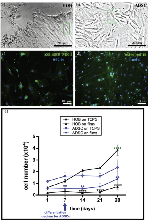
Human osteoblasts (HOBs) were isolated from the tissues of patients who underwent joint replacement surgery. HOBs were smaller in size in comparison to ADSCs as the phase contrast
mi-croscopy images show (Fig. 3a and b). According to literature, HOBs
can be half the size of ADSCs but a definite comparison is not
possible due to the heterogeneity of the size of ADSCs [34,35].
Additionally, HOB phenotype was confirmed by immunostaining
for common human osteoblast markers secreted by HOB: anti-collagen type I for anti-collagen type I and anti-osteopontin for
osteo-pontin[36,37]. These proteins constitute different components of
the extracellular matrix in bone. The isolation of HOBs in this study is considered successful because no cells were observed with
DAPI-only staining; this indicates that contaminating cells likefibroblasts
originating from the isolation step are not present (Fig. 3c and d).
Immunostaining also indicated that HOBs preserved their pheno-type during passaging up to 5 which was the highest number of passage used in this study.
3.5. Proliferation of ADSC and HOB on collagen-fibroin-ELR blend films
Alamar Blue assay was performed to study cell proliferation on Days 1, 7, 14, 21 and 28. The osteogenic differentiation of the ADSCs was induced with the introduction in differentiation medium to drive the ADSCs into osteoblast phenotype. The attachment and proliferation rate of the ADSCs and HOBs were compared for their
potential to form an engineered bone tissue on biodegradablefilms.
Collagen-fibroin-ELR blend films supported ADSC adhesion and
growth (Fig. 3e). Application of osteogenic medium on Day 7
in a plateau (on Days 14 and 21) on both TCPS and collagen-fi
broin-ELR blend surfaces possibly due to differentiation of stem cells[38].
No osteogenic medium was used for HOBs since they already
possessed osteoblast phenotype (Fig. 3c and d) and they showed a
proliferation trend as the ADSCs onfilm. HOBs tested on the same
surfaces showed 2-fold lower adhesion on both surfaces than ADSCs (Day 1). As the incubation time went by, the cell number of
both HOBs and ADSCs were higher on TCPS (3.9 cm2) than onfilm
(2.55 cm2) surfaces. The larger surface area of the TCPS along with
its cell adhesive chemistry can explain the higher cell attachment and proliferation extent. It can be suggested that wider surface of TCPS provided a space for cell attachment and division which in turn might have enhanced Day 1 results of HOBs and ADSCs.
Interestingly, number of ADSC onfilms was approximately 2-fold
higher than that of the HOB at each time point. It can therefore
be stated that ADSCs proliferate on collagen-fibroin-ELR blendfilms
more than HOBs, despite the low rate due to differentiation.
Pre-viously, HOBs were shown to proliferate more on silkfibroin coated
PCL - biphasic calcium phosphate (BCP) scaffolds than uncoated
ones[39]. Additionally, Gronthos et al. showed that HOBs adhered
on the collagen type I surface as well as surfaces coated with other
ECM components (collagen types IV, V,fibronectin, laminin, and
vitronectin)[40]. According to these studies, HOBs perform well on
both collagen andfibroin materials and this is an advantage of the
blendfilm. Similarly, another study pointed out that while ADSCs
attached on collagen scaffolds at a higher rate than silk fibroin
scaffolds, bone marrow stem cells (BMSCs) attached on both
scaf-folds without any particular preference for either surface[41].
3.6. Surface characterization and morphology of ADSCs and HOBs on collagen-fibroin-ELR blendfilms
Collagen-fibroin-ELR blendfilms patterned with microchannels
were produced with predetermined groove and ridge topographies as SEM micrographs showed and crosslinking did not cause any swelling in the physical cues which can be the case in a water based
crosslinking medium (Fig. 4a). Features were mainly preserved
after crosslinking with EDC/NHS in methanol. Blend films could
withstand 27 days of cell culture conditions (Fig. 4b). However,
some holes in the films could be seen due to film degradation
during culture.
HOBs were lower in number than ADSCs and both HOBs (Fig. 4c)
and ADSCs (Fig. 4d) were guided on collagen-fibroin-ELR blendfilm surfaces after 28 days of cell culture. Energy Dispersive X-Ray Spectrometry (EDX) showed no Ca and P atoms on the unseeded
(Days 1 and 28) and HOB seeded (Day 28)films. ADSCs, on the other
hand, deposited calcium and phosphorus containing compounds
on the collagen-fibroin-ELR blendfilms with a Ca/P ratio of 0.47
after 28 days of cell culture. This value is lower than that of HAp
(1.67); itsin vivoprecursor octacalcium phosphate (OCP) has this
value as 1.33[9]and tricalcium phosphate as 1.5. The reason for the
0.47 needs to be further investigated.
3.7. ADSC and HOB alignment
Guidance of bone cells is important because it helps an
aniso-tropic ECM deposition by aligned cells. On Day 28, actinfilament
staining of seeded ADSCs and HOBs showed that cytoskeletons of these cells were aligned along the microchannel direction on
patterned collagen-fibroinfilms (Fig. 5a and b). It can be stated that
ADSCs and HOBs were aligned on ridges and grooves smaller than the cells as seen in different examples involving other cell types and
materials[43e45]. BMSCs were also a supporting example for cell
alignment on a similar sized microchannels composed of
ther-moresponsive poly(N-isopropylacrylamide)films with ELR
adsor-bed on the surface[46]. Additionally, the guidance of HOBs was
reported by Biggs et al. with the best alignment on microchannels
of poly(methylmethacrylate) (PMMA) with the grooves of 10
m
m insize amongst the ones that had 25 and 100
m
m width[47].Previ-ously, ADSCs were also reported to align on graphene oxide (ridge:
30
m
m, groove: 15m
m) and collagen-fibroin (ridge: 7.4m
m, groove:10
m
m) microchannels with increased osteogenic differentiation inregard to smooth surfaces[26,48].
3.8. Quantification of mineralization
The amount of mineral deposited on the HOB and ADSC seeded
collagen-fibroin-ELR blend films were quantified with o-cresol
phthalein assay on Day 28 in order to get a chemical measurement.
Unseeded collagen-fibroin-ELR blendfilm, incubated for 28 days,
was employed as control. Calcium was totally extracted from the
film structure via HCl treatment; controls were used for
back-ground level check. There was no statistically significant difference
between unseededfilm of Day 28 (14
m
g) and HOB seededfilm onDay 28 (24
m
g) (Fig. 5c). The low levels of calcium was alsosup-ported by EDX analysis. When HOB seededfilm was compared with
unseededfilm on Day 28, it can be said that HOBs deposited
cal-cium however, the amount was not significantly higher. The
cal-cium content of ADSC seeded ELR blendfilms on Day 28 (274
m
g)was significantly higher than on similarfilms seeded with HOB.
Calcium amount was normalized to cell number and ADSCs were
shown to deposite significantly higher amounts of mineral than
HOBs (Fig. 5d). This result indicated that ADSCs produced more
minerals than HOBs.
3.9. Mechanical testing
Films that were used in this study were designed to mimic bone lamellae and were tested along the microchannel direction to investigate the contribution of ECM synthesized by the seeded HOBs and ADSCs to the tensile properties. Amruthwar et al. studied the effect of ELR addition to collagen hydrogels for bone tissue engineering. Improvement in the UTS and E were observed from 0.34 MPa to 0.99 MPa and 4.06 MPa to 11.41 MPa with the addition of ELR (25 mg) to collagen (8 mg) hydrogel due to the more
concentrated protein content in the scaffold[18]. These values are
higher than the UTS and E values measured in this study because of
the lower polymer concentration in collagen-fibroin-ELR blend
film. Causa et al. reported that addition of 13% (v/v) HAp to PCL
scaffolds enhanced the UTS from 0.93 MPa to 2.19 MPa[42]. As far
as we know the mechanical properties of bone tissue engineered scaffolds has been enhanced by concentrated solutions of synthetic polymer or with the addition of ceramics. These results in our study showed the importance of seeded cell type on the mechanical
strength of bone tissue engineered scaffolds for thefirst time.
Tensile properties of unseeded collagen-fibroin-ELR blendfilms
on Days 1 and 28 and ADSC and HOB seeded collagen-fibroin-ELR
blendfilms on Day 28 were determined. UTS, E (Fig. 5e) and EB
were calculated and the average values are presented inFig. 5f. No
significant difference could be observed for UTS and EB for all
groups. On Day 28, unseeded collagen-fibroin-ELR blendfilm had
higher UTS, E and EB than unseededfilm on Day 1. This result could
be due to collagen degradation being faster thanfibroin[26]. This
selective degradation could expose a stronger and crystalline
structure offibroin (
b
-sheet) after 28 days of cell culture. The higherb
-sheet level offibroin was supported by its low swelling degreeand high Tgwhich are indications of the superior crystallinity of
fibroin over collagenfilm. Similar to our results, the positive
rela-tionship between higher crystallinity level of more crosslinked polycaprolactone fumarate and increased tensile modulus was
re-ported by another study[49]. Hu et al. employedfilms formulated
with recombinant human like collagen andfibroin for hepatic
tis-sue engineering purposes. They showed EB elevated from 28.7% to
30.9% with the increase offibroin content by 10% (w/w) infilms
[50]. In our work, on Day 1, E was 0.58±0.13 MPa for unseeded
collagen-fibroin-ELR blend film. This value is higher than the
literature value of 502 ± 575 kPa which was measured from
crosslinked and microchannel patterned HAp nucleated ELR
membrane after 7 days of incubation in simulated bodyfluid[51].
By taking this result into account, it can be suggested that collagen
andfibroin promoted the mechanical strength of thefilm.
ADSC seeding contributed to the UTS and E which indicated ECM secretion however, HOB seeding decreased these properties
substantially and led to highest EB. Significantly enhanced E in
ADSC seededfilm (1.21 MPa) when compared to HOB seededfilm
(0.41 MPa) could be explained by matrix metalloproteinase-2
(MMP-2) secretion, by HOBs in vitro[52]. A relevant work also
showed that MMP-2 is downregulated during osteogenic
differ-entiation of ADSCs [53] and therefore, degradation by MMP-2
might have lowered E significantly for HOB seeded film by
breaking the protein chains and leaving them more stretchable. In return, this effect could lead to increase in the EB up to 2-fold
(71.32%) when compared to other films. Additionally, Ascenzi
et al. applied tensile test in longitudinal direction for wet human
fully calcified osteon and measured EB as 6.84%[54]which isfi
ve-fold lower than that of Day 28film on which ADSCs were seeded
(35.53±19.18%).
4. Conclusions
In this study, swelling test and DSC experiments showed that
the reason of higher mechanical strength of unseededfilm
incu-bated for 28 days when compared to Day 1film was the increase in
fibroin fraction relative to collagen as a result of the polymer
degradation. Guided HOBs and ADSCs on microchannel patterned
collagen-fibroin-ELR blendfilms mimicked the naturally aligned
bone tissue. Furthermore, higher rates of cell adhesion, prolifera-tion, mineralization and mechanical properties were obtained by ADSC seeding when compared to HOB due to enhanced ECM secretion. Potential of ADSC over HOB was proven by these means are vital for bone tissue engineering substrates. Osteogenically differentiated ADSCs improved the stiffness and tensile strength of
the collagen-fibroin-ELRfilms after 28 days of incubation.
Conflict of interests
We declare that authors have no competingfinancial interest.
Acknowledgements
We acknowledge Prof. Muharrem Demirogullari from Cag Hos-pital (Ankara, Turkey) and Prof. Cemil Yildiz from GATA (Ankara, Turkey) for providing the lipoaspirate material and bone sections, respectively. We would like to acknowledge Prof. Esra Karaca from
Uludag University (Bursa, Turkey) for kindly providing theBombyx
morisilk threads. We would like to acknowledge METU Central
Laboratory for DSC and SEM analysis. The authors would like to
thank METU (BAP-07.02.2013.101) for thefinancial support of the
study by E.S., and the Scientific and Technological Research Council
of Turkey (TUBITAK) for the scholarship to E.S. through BIDEB 2211C. We are grateful to Ministry of Development of Turkey for funding BIOMATEN through Grant DPT2011K120350. J.C.R.C. ac-knowledges the funding from the EC (HEALTH-F4-2011-278557, PITN-GA-2012-317306, MSCA-ITN-2014-642687 and NMP-2014-646075), MINECO (MAT2013-42473-R and MAT2015-68901R) and JCyL (VA244U13, VA313U14 and VA015U16).
References
[1] T. Vos, et al., Years lived with disability (YLDs) for 1160 sequelae of 289 dis-eases and injuries 1990-2010: a systematic analysis for the Global Burden of Disease Study 2010, Lancet 380 (2012) 2163e2196,http://dx.doi.org/10.1016/ S0140-6736(12)61729-2.
[2] P.V. Giannoudis, H. Dinopoulos, E. Tsiridis, Bone substitutes: an update, Injury 36 (Suppl 3) (2005) S20eS27,http://dx.doi.org/10.1016/j.injury.2005.07.029. [3] S. Tarafder, V.K. Balla, N.M. Davies, A. Bandyopadhyay, S. Bose, Microwave-sintered 3D printed tricalcium phosphate scaffolds for bone tissue engineer-ing, J. Tissue Eng. Regen. Med. 7 (2013) 631e641,http://dx.doi.org/10.1002/ term.555.
[4] M.N. Rahaman, D.E. Day, B. Sonny Bal, Q. Fu, S.B. Jung, L.F. Bonewald, A.P. Tomsia, Bioactive glass in tissue engineering, Acta Biomater. 7 (2011) 2355e2373,http://dx.doi.org/10.1016/j.actbio.2011.03.016.
[5] A.S. Badami, M.R. Kreke, M.S. Thompson, J.S. Riffle, A.S. Goldstein, Effect of fiber diameter on spreading, proliferation, and differentiation of osteoblastic cells on electrospun poly(lactic acid) substrates, Biomaterials 27 (2006) 596e606,http://dx.doi.org/10.1016/j.biomaterials.2005.05.084.
[6] E.D. Boland, G.E. Wnek, D.G. Simpson, K.J. Pawlowski, G.L. Bowlin, Tailoring tissue engineering scaffolds using electrostatic processing techniques: a study of poly(glycolic acid), J. Macromol. Sci. Part A 38 (2001) 1231e1243,http:// dx.doi.org/10.1081/MA-100108380.
[7] I. Rajzer, E. Menaszek, R. Kwiatkowski, J.A. Planell, O. Castano, Electrospun gelatin/poly(ε-caprolactone) fibrous scaffold modified with calcium phos-phate for bone tissue engineering, Mater. Sci. Eng. C 44 (2014) 183e190, http://dx.doi.org/10.1016/j.msec.2014.08.017.
[8] E. Sayin, E.T. Baran, V. Hasirci, Protein-based materials in load-bearing tissue-engineering applications, Regen. Med. 9 (2014) 687e701,http://dx.doi.org/ 10.2217/rme.14.52.
[9] M.T. Arafat, C.X.F. Lam, A.K. Ekaputra, S.Y. Wong, X. Li, I. Gibson, Biomimetic composite coating on rapid prototyped scaffolds for bone tissue engineering, Acta Biomater. 7 (2011) 809e820, http://dx.doi.org/10.1016/ j.actbio.2010.09.010.
[10] R. Murugan, S. Ramakrishna, Development of nanocomposites for bone grafting, Compos. Sci. Technol. 65 (2005) 2385e2406, http://dx.doi.org/ 10.1016/j.compscitech.2005.07.022.
[11] R. Puxkandl, I. Zizak, O. Paris, J. Keckes, W. Tesch, S. Bernstorff, P. Purslow, P. Fratzl, Viscoelastic properties of collagen: synchrotron radiation in-vestigations and structural model, Philos. Trans. R. Soc. B Biol. Sci. 357 (2002) 191e197,http://dx.doi.org/10.1098/rstb.2001.1033.
[12] R. Kane, P.X. Ma, Mimicking the nanostructure of bone matrix to regenerate bone, Mater. Today 16 (2013) 418e423, http://dx.doi.org/10.1016/ j.mattod.2013.11.001.
[13] K.J. Burg, S. Porter, J.F. Kellam, Biomaterial developments for bone tissue en-gineering, Biomaterials 21 (2000) 2347e2359, http://dx.doi.org/10.1016/ S0142-9612(00)00102-2.
[14] R. Parenteau-Bareil, R. Gauvin, F. Berthod, Collagen-based biomaterials for tissue engineering applications, Materials 3 (2010) 1863e1887, http:// dx.doi.org/10.3390/ma3031863.
[15] C.M. Agrawal, J.L. Ong, M.R. Appleford, G. Mani, Introduction to Biomaterials:
Basic Theory with Engineering Applications, Cambridge University Press, New York, NY, USA, 2013.
[16] K. Trabbic-Carlson, L.A. Setton, A. Chilkoti, Swelling and mechanical behaviors of chemically cross-linked hydrogels of elastin-like polypeptides, Bio-macromolecules 4 (2003) 572e580,http://dx.doi.org/10.1021/bm025671z. [17] J.C. Rodríguez-Cabello, L. Martín, A. Girotti, C. García-Arevalo, F.J. Arias,
M. Alonso, Emerging applications of multifunctional elastin-like recombi-namers, Nanomedicine 6 (2011) 111e122, http://dx.doi.org/10.2217/ nnm.10.141.
[18] S.S. Amruthwar, A.V. Janorkar, In vitro evaluation of elastin-like polypeptide-collagen composite scaffold for bone tissue engineering, Dent. Mater. 29 (2013) 211e220,http://dx.doi.org/10.1016/j.dental.2012.10.003.
[19] N. Ozturk, A. Girotti, G.T. Kose, J.C. Rodríguez-Cabello, V. Hasirci, Dynamic cell culturing and its application to micropatterned, elastin-like protein-modified poly(N-isopropylacrylamide) scaffolds, Biomaterials 30 (2009) 5417e5426, http://dx.doi.org/10.1016/j.biomaterials.2009.06.044.
[20] E. Salvagni, G. Berguig, E. Engel, J.C. Rodriguez-Cabello, G. Coullerez, M. Textor, J.A. Planell, F.J. Gil, C. Aparicio, A bioactive elastin-like recombinamer reduces unspecific protein adsorption and enhances cell response on titanium sur-faces, Colloids Surf. B Biointerfaces 114 (2014) 225e233,http://dx.doi.org/ 10.1016/j.colsurfb.2013.10.008.
[21] S. Prieto, A. Shkilnyy, C. Rumplasch, A. Ribeiro, F.J. Arias, J.C. Rodríguez-Cabello, A. Taubert, Biomimetic calcium phosphate mineralization with multifunctional elastin-like recombinamers, Biomacromolecules 12 (2011) 1480e1486,http://dx.doi.org/10.1021/bm200287c.
[22] P.A. Raj, M. Johnsson, M.J. Levine, G.H. Nancollas, Salivary statherin:
depen-dence on sequence, charge, hydrogen bonding potency, and helical confor-mation for adsorption to hydroxyapatite and inhibition of mineralization, J.
Biol. Chem. 267 (1992) 5968e5976.
[23] D. Carnelli, P. Vena, M. Dao, C. Ortiz, R. Contro, Orientation and size-dependent mechanical modulation within individual secondary osteons in cortical bone tissue, J. R. Soc. Interface 10 (2013) 1e12,http://dx.doi.org/ 10.1098/rsif.2012.0953.
[24] J.S. Barbosa, R.R. Costa, A.M. Testera, M. Alonso, J.C. Rodríguez-Cabello, J.F. Mano, Multi-layered films containing a biomimetic stimuli-responsive recombinant protein, Nanoscale Res. Lett. 4 (2009) 1247e1253, http:// dx.doi.org/10.1007/s11671-009-9388-5.
[25] H. Kenar, A. Kocabas, A. Aydinli, V. Hasirci, Chemical and topographical modification of PHBV surface to promote osteoblast alignment and confine-ment, J. Biomed. Mater. Res. A 85 (2008) 1001e1010, http://dx.doi.org/ 10.1002/jbm.a.31638.
[26] E. Sayin, E. Türker Baran, V. Hasirci, Osteogenic differentiation of adipose derived stem cells on high and low aspect ratio micropatterns, J. Biomater. Sci. Polym. Ed. 5063 (2015) 1e44, http://dx.doi.org/10.1080/09205063. 2015.1100494.
[27] M.P. Francis, P.C. Sachs, L.W. Elmore, S.E. Holt, Isolating adipose-derived mesenchymal stem cells from lipoaspirate blood and saline fraction, Organ-ogenesis 6 (2010) 11e14.http://www.pubmedcentral.nih.gov/articlerender.
fcgi?artid¼2861738&tool¼pmcentrez&rendertype¼abstract.
[28] G.M. Cooper, The Cell - A Molecular Approach, second ed., Sinauer Associates,
Sunderland (MA), 2000 citeulike-article-id:10266975.
[29] I. Sakurada, Polyvinyl Alcohol Fibers, CRC Press, 1985.
mechanism and control of silk fibroin, Biomacromolecules 12 (2011) 1080e1086,http://dx.doi.org/10.1021/bm101422j.
[31] Q. Lv, K. Hu, Q. Feng, F. Cui, Fibroin/collagen hybrid hydrogels with cross-linking method: preparation, properties, and cytocompatibility, J. Biomed. Mater. Res. Part A 84 (1) (2008) 198e207, http://dx.doi.org/10.1002/ jbm.a.31366.
[32] S. Das, F. Pati, Y.J. Choi, G. Rijal, J.H. Shim, S.W. Kim, A.R. Ray, D.W. Cho, S. Ghosh, Bioprintable, cell-laden silk fibroin-gelatin hydrogel supporting multilineage differentiation of stem cells for fabrication of three-dimensional tissue constructs, Acta Biomater. 11 (2015) 233e246, http://dx.doi.org/ 10.1016/j.actbio.2014.09.023.
[33] M.D. Shoulders, R.T. Raines, Collagen structure and stability, Annu. Rev. Bio-chem. 78 (2009) 929e958, http://dx.doi.org/10.1146/annurev.biochem. 77.032207.120833.
[34] C.R. Wheeless, J.A. Nunley, J.R. Urbaniak, in: D.U.M.C.D. of O. Surgery,
Wheeless' Textbook of Orthopaedics, 2016.
[35] Y.J. Ryu, T.J. Cho, D.S. Lee, J.Y. Choi, J. Cho, Phenotypic characterization and In vivo localization of human adipose-derived mesenchymal stem cells, Mol. Cells 35 (2013) 557e564,http://dx.doi.org/10.1007/s10059-013-0112-z. [36] S.F. El-Amin, E. Botchwey, R. Tuli, M.D. Kofron, A. Mesfin, S. Sethuraman,
R.S. Tuan, C.T. Laurencin, Human osteoblast cells: isolation, characterization, and growth on polymers for musculoskeletal tissue engineering, J. Biomed. Mater. Res. Part A 76 (2006) 439e449,http://dx.doi.org/10.1002/jbm.a.30411. [37] J.E. Aubin, F. Lui, L. Malaval, A.K. Gupta, Osteoblast and chondroblast differ-entiation, Bone 17 (1995),http://dx.doi.org/10.1016/8756-3282(95)00183-E. [38] M.J. Coelho, M.H. Fernandes, Human bone cell cultures in biocompatibility
testing. Part II: effect of ascorbic acid,b-glycerophosphate and dexametha-sone on osteoblastic differentiation, Biomaterials 21 (2000) 1095e1102, http://dx.doi.org/10.1016/S0142-9612(99)00192-1.
[39] S.I. Roohani-Esfahani, Z.F. Lu, J.J. Li, R. Ellis-Behnke, D.L. Kaplan, H. Zreiqat, Effect of self-assembled nanofibrous silk/polycaprolactone layer on the osteoconductivity and mechanical properties of biphasic calcium phosphate scaffolds, Acta Biomater. 8 (2012) 302e312, http://dx.doi.org/10.1016/ j.actbio.2011.10.009.
[40] S. Gronthos, K. Stewart, S.E. Graves, S. Hay, P.J. Simmons, Integrin expression and function on human, J. Bone Min. Res. 12 (1997) 1189e1197,http:// dx.doi.org/10.1359/jbmr.1997.12.8.1189.
[41] J.R. Mauney, T. Nguyen, K. Gillen, C. Kirker-Head, J.M. Gimble, D.L. Kaplan, Engineering adipose-like tissuein vitroandin vivoutilizing human bone marrow and adipose-derived mesenchymal stem cells with silkfibroin 3D scaffolds, Biomaterials 28 (2007) 5280e5290, http://dx.doi.org/10.1016/ j.biomaterials.2007.08.017.
[42] F. Causa, P.A. Netti, L. Ambrosio, G. Ciapetti, N. Baldini, S. Pagani, D. Martini, A. Giunti, Poly-epsilon-caprolactone/hydroxyapatite composites for bone regeneration: in vitro characterization and human osteoblast response, J. Biomed. Mater. Res. Part A 76 (2006) 151e162,http://dx.doi.org/10.1002/
jbm.a.30528.
[43] H. Kenar, G.T. K€ose, V. Hasirci, Tissue engineering of bone on micropatterned biodegradable polyester films, Biomaterials 27 (2006) 885e895, http:// dx.doi.org/10.1016/j.biomaterials.2005.07.001.
[44] N.E. Vrana, A. Elsheikh, N. Builles, O. Damour, V. Hasirci, Effect of human corneal keratocytes and retinal pigment epithelial cells on the mechanical properties of micropatterned collagen films, Biomaterials 28 (2007) 4303e4310,http://dx.doi.org/10.1016/j.biomaterials.2007.06.013.
[45] P. Zorlutuna, N. Builles, O. Damour, A. Elsheikh, V. Hasirci, Influence of kera-tocytes and retinal pigment epithelial cells on the mechanical properties of polyester-based tissue engineering micropatterned films, Biomaterials 28 (2007) 3489e3496,http://dx.doi.org/10.1016/j.biomaterials.2007.04.013. [46] N. Ozturk, A. Girotti, G.T. Kose, J.C. Rodríguez-Cabello, V. Hasirci, Dynamic cell
culturing and its application to micropatterned, elastin-like protein-modified poly(N-isopropylacrylamide) scaffolds, Biomaterials 30 (2009) 5417e5426, http://dx.doi.org/10.1016/j.biomaterials.2009.06.044.
[47] M.J.P. Biggs, R.G. Richards, S. McFarlane, C.D.W. Wilkinson, R.O.C. Oreffo, M.J. Dalby, Adhesion formation of primary human osteoblasts and the func-tional response of mesenchymal stem cells to 330nm deep microgrooves, J. R. Soc. Interface 5 (2008) 1231e1242,http://dx.doi.org/10.1098/rsif.2008.0035. [48] T. Kim, S. Shah, L. Yang, P.T. Yin, K. Hossain, B. Conley, J. Choi, K. Lee, Con-trolling differentiation of adipose-derived stem cells using combinatorial graphene hybrid-pattern arrays, ACS Nano 9 (2015) 3780e3790, http:// dx.doi.org/10.1021/nn5066028.
[49] S. Wang, M.J. Yaszemski, J.A. Gruetzmacher, L. Lu, Photo-crosslinked poly(3 -caprolactone fumarate) networks: roles of crystallinity and crosslinking density in determining mechanical properties, Polymer 49 (2008) 5692e5699, http://dx.doi.org/10.1016/j.polymer.2008.10.021.
[50] K. Hu, Biocompatible fibroin blendedfilms with recombinant human-like collagen for hepatic tissue engineering, J. Bioact. Compat. Polym. 21 (2006) 23e37,http://dx.doi.org/10.1177/0883911506060455.
[51] E. Tejeda-Montes, A. Klymov, M.R. Nejadnik, M. Alonso, J.C. Rodriguez-Cabello, X.F. Walboomers, A. Mata, Mineralization and bone regeneration using a bioactive elastin-like recombinamer membrane, Biomaterials 35 (2014) 8339e8347,http://dx.doi.org/10.1016/j.biomaterials.2014.05.095.
[52] L. Rifas, L.R. Halstead, W.A. Peck, L.V. Avioli, H.G. Welgus, Human osteoblasts
in vitrosecrete tissue inhibitor of metalloproteinases and gelatinase but not interstitial collagenase as major cellular products, J. Clin. Investig. 84 (1989) 686e694,http://dx.doi.org/10.1172/JCI114216.
[53] H. Egusa, K. Iida, M. Kobayashi, T.Y. Lin, M. Zhu, P.A. Zuk, C.J. Wang, D.K. Thakor, M.H. Hedrick, I. Nishimura, Downregulation of extracellular matrix-related gene clusters during osteogenic differentiation of human bone marrow- and adipose tissue-derived stromal cells, Tissue Eng. 13 (2007) 2589e2600,http://dx.doi.org/10.1089/ten.2007.0080.