Determination of non protein amino acids and betaines in vegetable oils by flow injection triple quadrupole tandem mass spectrometry: a screening method for the detection of adulterations of olive oils
32
0
0
Texto completo
(2) 24. ABSTRACT. 25 26. A novel screening method using an automated flow-injection electrospray. 27. ionization tandem mass spectrometry system is proposed for the simultaneous. 28. determination of five non-protein amino acids (β-alanine, alloisoleucine, ornithine,. 29. citrulline, pyroglutamic acid) and three betaines (glycine betaine, trigonelline, proline. 30. betaine) after derivatization with butanolic HCl. MS/MS experiments were carried out. 31. in a triple quadrupole instrument using a multiple reaction monitoring mode in less than. 32. 2 min. The proposed method provided high fingerprinting power to identify the. 33. presence of five of the studied compounds in different types of vegetable oils (soybean,. 34. sunflower, corn and olive) with LODs at ppb levels. The method was validated and. 35. different mixtures of extra virgin olive oil with seed oils were analyzed achieving the. 36. typification for the detection of adulterations in extra virgin olive oils up to 2% w/w.. 37. The non-protein amino acid ornithine was confirmed as marker for adulterations in the. 38. olive oils analyzed.. 39. 40. 41. 42. 43. Keywords: Adulteration; Betaines; Non-protein amino acids; Screening; Tandem Mass. 44. Spectrometry; Vegetable oils.. 45. 2.
(3) 46. INTRODUCTION. 47 48. Olive oil authentication is an issue of great interest in the manufacturing. 49. countries of this food product. Authenticity covers many aspects, including adulteration,. 50. mislabelling, characterization, and misleading origin (1). Owing to its higher price,. 51. extra virgin olive oil is susceptible of undergoing fraudulent practices concerning its. 52. authenticity being a frequent problem for regulatory agencies, oil suppliers and. 53. consumers. The most common process is the addition of cheaper vegetable oils with. 54. less commercial value such as seed oils, which is not allowed by European regulations. 55. (2). For this reason, there is a need for detecting lower adulteration levels in the food. 56. industry which requires the development of new analytical methodologies with suitable. 57. sensitivity, accuracy, and straightforward automation.. 58. A wide number of powerful techniques have been employed for the. 59. authentication of olive oils and can be classified into chromatographic and. 60. spectrometric techniques. Separative methods for identifying adulterated olive oils. 61. based on HPLC or GC (1, 3-5) and, more recently on capillary electrophoresis (CE) (6-. 62. 8) with and without mass spectrometry (MS) detection, have widely been employed.. 63. Spectrometric methods without separation, such as MS, Nuclear Magnetic Resonance,. 64. Raman Spectroscopy, Infrared Spectroscopy, Fluorescence and Chemiluminiscence,. 65. among others, have also broadly been reported (5, 9).. 66. Among the spectrometric techniques, MS has become a powerful technique to. 67. characterize vegetable oils. This is because soft ionization methods, such as electrospray. 68. ionization mass spectrometry (ESI-MS), combine sensitivity, selectivity and versatility. 69. being capable of gently ionize a wide range of molecules, especially polar ones (10, 11).. 70. In fact, they allow the fingerprinting in complex food matrices, as vegetable oils, and 3.
(4) 71. provide structural information on their components. In addition, the injection of the. 72. sample can easily be automated particularly when using robotized sample injection. 73. systems such as direct infusion or Flow-Injection Analysis (FIA) permitting very high. 74. throughput. The most commonly used mass analyzers for this purpose are tandem mass. 75. analyzers. Thus, for the analysis of several vegetable oils, the use of direct infusion in. 76. MS has been reported employing Ion Trap (IT) (12-16) or quadrupole time-of-flight. 77. (QTOF) (17, 18) mass spectrometers, while FIA has been used with triple quadrupole. 78. (QqQ) (19) or QTOF (20, 21). The target compounds studied in these works were free. 79. fatty acids, triacylglycerols, diacylglycerols and monoacylglycerols as major. 80. components, and phenolic compounds as minor components. The minimum adulteration. 81. limits found in blended vegetable oils were: 10% w/w for mixtures of olive oil with. 82. soybean oil (18), 10% w/w for olive oil with hazelnut oil (21), and 20% w/w (14) or. 83. even 1% w/w (16) for extra virgin olive oils with ordinary olive oils. To these works,. 84. the need to use complex chemometric techniques and/or multivariate analysis was. 85. described. Thus, a chemometric treatment of the spectral information by principal. 86. component analysis (16-18, 20, 21), linear discriminant analysis (12-15, 21) and/or. 87. hierarchical clustering analysis (16) were reported.. 88. On the other hand, screening methods present the capability of a high sample. 89. throughput and are used to sift a large number of samples, being specifically designed to. 90. avoid false compliant results. Accordingly, the objective of this work was the. 91. development of a new sensitive screening methodology by FIA-ESI-MS/MS with a. 92. QqQ mass spectrometer based on multiple reaction monitoring (MRM) mode to detect. 93. adulterations of olive oils with other vegetable oils using free-minor components as. 94. markers. Thus, the simultaneous determination of three betaines and five non-protein. 95. amino acids in vegetable oils (soybean, sunflower, corn and olive oils) was carried out 4.
(5) 96. to find at least one of them that can serve as screening marker for the adulteration of. 97. olive oils.. 98 99 100. MATERIALS AND METHODS. 101 102. Reagents and standards. Figure 1 shows the five non-protein amino acids (β-. 103. alanine, pyroglutamic acid, alloisoleucine, ornithine and citrulline) and the three. 104. betaines (glycine betaine, trigonelline and proline betaine) studied in this work and. 105. selected according to the possibilities shown with previous methodologies (6-8).. 106. Trigonelline, β-alanine and glycine betaine were supplied from Sigma (St.. 107. Louis, MO, USA). Proline betaine was from Hallochem Pharma (Chongqing, China).. 108. Pyroglutamic acid, alloisoleucine, ornithine, citrulline and hydrogen chloride/1-butanol. 109. solution were from Fluka (Buchs, Switzerland). Methanol, chloroform and acetonitrile. 110. were supplied from Scharlau Chemie (Barcelona, Spain). Formic acid was from Riedel-. 111. de Häen (Seelze, Germany). Distilled water was deionized by using a Milli-Q system. 112. (Millipore, Bedford, MA, USA).. 113. Stock standard solutions (1 mg/mL) were prepared in acetonitrile/water (40:60,. 114. v/v). Standard solutions were further obtained by serial dilutions of each stock standard. 115. solution with the same acetonitrile/water solution. The standard solutions were. 116. evaporated to dryness with vacuum at 15 mbar and 80 ºC using a concentrator. 117. (Labconco, Barcelona, Spain). Finally, the derivatization with butanolic HCl was. 118. accomplished before injection in the MS system.. 119. 5.
(6) 120. FIA-ESI-MS analysis. Analyses were accomplished by using a liquid. 121. chromatography system (model 1200 from Agilent Technologies, Palo Alto, CA, USA). 122. coupled through an orthogonal electrospray interface (model G1948B from Agilent. 123. Technologies, Palo Alto, CA, USA) to a triple quadrupole mass spectrometer (model. 124. 6410A from Agilent Technologies, Palo Alto, CA, USA). Ultra-pure nitrogen. 125. (99.999%) was used as collision gas, while pure nitrogen (97.0%) was used as nebulizer. 126. gas. Data acquisition was performed using the Mass Hunter software (version B.01.03. 127. from Agilent Technologies, Palo Alto, CA, USA). Flow-injection analyses were. 128. achieved using a binary pump and introducing as the carrier solvent 60% of component. 129. A (0.1% v/v formic acid in water) and 40% of component B (0.1% v/v formic acid in. 130. acetonitrile) at a flow rate of 0.1 mL/min. A 40 cm long PEEK capillary of 1/16 in.. 131. outer diameter and 0.13 mm inner diameter (Agilent Technologies, Palo Alto, CA,. 132. USA) was used to connect the flow from the autosampler to the ion source. Injection. 133. volumes of 20 µL were used for samples and standards, and injections of 20 µL of. 134. solvent blanks were performed between samples for injector needle rinsing. The. 135. samples and standards were maintained at 4 ºC in the thermostated autosampler during. 136. analysis.. 137. The ESI source conditions were: positive mode, gas temperature 325 ºC, gas. 138. flow 10 L/min, nebulizer pressure 20 psi and capillary voltage 3500 V. The tandem. 139. mass spectrometric detection was operated in the MRM mode for the highest possible. 140. selectivity and sensitivity. The dwell time was adjusted to a time of 100 ms for each. 141. mass transition between the precursor and product ion in order to obtain maximum. 142. sensitivity with a sufficient number of points to define the peak to quantify (≈ 15 points. 143. per peak). The collision induced dissociation was performed using ultra-pure nitrogen. 144. as the collision gas in the second quadrupole (Q2) collision cell. First quadrupole (Q1) 6.
(7) 145. and third quadrupole (Q3) were tuned at unit mass resolution. A summary of the. 146. optimized mass spectrometric conditions for MRM mode is provided in Table 1.. 147 148. Vegetable oils. Nine extra virgin olive oils, three Arbequina (AEVOO), three. 149. Picual (PEVOO), and three Hojiblanca (HEVOO), and nine seed oils, three refined. 150. sunflower oils (RSO), three refined corn oils (RCO), and three refined soybean oils. 151. (RSYO) were acquired in different supermarkets (Madrid, Spain). For each variety and. 152. botanical origin, the trademarks were different, selecting the most consumed and. 153. marketed in Spain.. 154. Sample preparation was carried out according to the method described by. 155. Sánchez-Hernández et al. (6). 40 g of each oil were placed in a centrifuge tube,. 156. dissolved with 160 mL of methanol/chloroform (2/1, v/v) and left at -20 ºC overnight.. 157. Then, the tubes were centrifuged (4000g, 15 min, 4 ºC). The upper phase obtained was. 158. collected in a new tube and, the rest of the sample was dissolved with 100 mL of. 159. methanol/chloroform/water (2/1/0.8, v/v/v). After its centrifugation (4000g, 15 min, 4. 160. ºC), the new upper phase obtained was combined with the previous one. The two. 161. combined fractions were mixed with 40 mL of chloroform and 100 mL of water, and. 162. after centrifugation (4000g, 15 min, 4 ºC) an aqueous and an organic phase were. 163. obtained. The aqueous phase, containing the compounds of interest, was collected and. 164. evaporated to dryness with vacuum at 15 mbar and 80 ºC in a concentrator. Finally, the. 165. derivatization with butanolic HCl was accomplished before injection in the MS system.. 166 167. Derivatization. The procedure consisted in a butyl ester derivatization using 3. 168. M HCl in butanol as derivatizing agent (7, 8). Thus, 0.5 mL of derivatizing agent were. 169. added to the evaporated extract of standards, while up to 1 mL was added to the 7.
(8) 170. evaporated extract of samples in order to have enough agent excess. After shake in a. 171. vortex, the reaction was performed in an oven at 80 ºC during 30 min. The. 172. derivatization process was stopped keeping the solution in the freezer during 5 min.. 173. Then, the solutions were evaporated to dryness with vacuum at 15 mbar and 80 ºC in a. 174. concentrator. Finally, the analytes were reconstituted in 500 µl of acetonitrile/water. 175. (40/60, v/v).. 176 177. Validation Study. The MS/MS screening method for the quantitation of non-. 178. protein amino acids and betaines was validated for vegetable oils analysis by. 179. determining the performance characteristics required by the Commission Decision. 180. 2002/657/EC concerning the performance of analytical methods and the interpretation. 181. of results (22). During method validation the considered characteristics were:. 182. selectivity, linearity, accuracy, precision and detection capability (CCβ) also known as. 183. limit of detection (LOD).. 184. The transitions from precursor to product ions of the target compounds were. 185. used to verify the selectivity. Thus, the identification of the compounds was possible. 186. using at least two MS/MS transitions to achieve the four identification points required in. 187. accordance with the Commission Decision 2002/657/EC (22).. 188. Linearity was tested in a range of 5-500 ng/mL by analyzing six concentration. 189. levels of standard solutions. Each point of the calibration graph corresponded to the. 190. mean value from three individual injections of the standard mixture. The obtained data. 191. were used to build the calibration curves by taking the peak area for each compound. 192. versus standard concentrations using a least squares linear regression model.. 193 194. CCβ was determined by injecting low amounts of each standard compound and considering six times the signal to noise ratio as the LOD. 8.
(9) 195. Precision of the method (repeatability and intermediate precision) was assessed. 196. for the quantification transition of each compound by analyzing the oil samples.. 197. Instrumental repeatability was determined from six replicate injections in the same day;. 198. the method repeatability was established from three individual samples injected by. 199. triplicate in the same day; and the intermediate precision of the method was evaluated. 200. from three individual samples injected in triplicate in two consecutive days. Precision. 201. was expressed as relative standard deviations (RSD in %) of the peak area.. 202. The accuracy of the method was evaluated as the recovery obtained for each. 203. compound when samples of HEVOO were spiked with standard solutions at. 204. concentration levels covering low (0.1 ng/g of each compound) and high concentrations. 205. (10 ng/g of each compound). Three injections of each sample were analyzed. The. 206. percentage of recovery was determined from: Recovery (%) =. 207. C. −C Spiked Initial × 100 C Added. 208 209. where “CSpiked” is the obtained concentration of the spiked samples, “CInitial” is the. 210. concentration of the sample without spiking, and “CAdded” is the concentration of the. 211. added standards.. 212 213. Quantitative method. Since the presence of matrix interferences in vegetable. 214. oils was demonstrated previously by CE–UV (6), the quantification of the samples was. 215. carried out using single-point standard addition method calibration. With this aim, two. 216. solutions were injected for each vegetable oil: (i) the sample solution and (ii) the spiked. 217. sample with a known amount of non-protein amino acids and betaines. Seed oils. 218. samples were spiked with 100 ng/mL of each non-protein amino acid (except in the case 9.
(10) 219. of citrulline for which 50 ng/mL was added) and 50 ng/mL of glycine betaine, 25. 220. ng/mL of trigonelline and 25 ng/mL of proline betaine. Extra virgin olive oils samples. 221. were spiked with 20 ng/mL of each non-protein amino acid (except for citrulline for. 222. which 10 ng/mL was added), 10 ng/mL of glycine betaine, 5 ng/mL of trigonelline and. 223. 5 ng/mL of proline betaine.. 224 225. Statistical Analysis. The data set for ANOVA test and multivariate statistical. 226. analysis were processed using Statgraphics 5.1 software. ANOVA test allows evaluate. 227. the linearity of the experimental data. It was carried out from the data of calibration. 228. curves of each studied compound. Multivariate statistical analysis was used for the. 229. statistical classification of the botanical oil origin where the variability of data values. 230. was tested by discriminant function analysis. With this aim, the data employed were the. 231. type of each sample according with its botanical origin (RSYO, RSO, RCO, HEVOO,. 232. AEVOO, PEVOO or adulterated olive oils) and the concentration, in ng/g, of the. 233. identified compounds (citrulline, ornithine, pyroglutamic acid, glycine betaine and. 234. trigonelline).. 235 236 237. RESULTS AND DISCUSSION. 238 239. Development of the screening method. A method based on a strategy of. 240. screening and quantitation was developed in this work. In this sense, the screening. 241. method provides full information enabling the analyte to be identified at the level of. 242. interest (22).. 10.
(11) 243. In conformity with Directive 96/23/EC (23), only those analytical techniques. 244. having a false compliant rate of < 5% (β-error) at the level of interest shall be used for. 245. screening purposes. If no permitted limit has been established for a substance. 246. (substances with “zero tolerance”), as in the case of the compounds to be considered as. 247. markers of adulteration, it is necessary to develop methods enabling the identification of. 248. these compounds at the lowest levels possible. For this reason, we have decided to. 249. develop a quantitative screening method based on a of FIA-ESI-MS/MS system with. 250. QqQ working in MRM mode, as it is possibly one of the most sensitive and selective. 251. analytical methods.. 252. MS experiments were firstly achieved in order to obtain the maximum intensity. 253. of the precursor ions to work in the MRM mode. Standard solutions of each compound. 254. (1 ng/mL) were individually injected to optimize some MS parameters in MS mode.. 255. Thus, dry gas temperature and the capillary voltage were modified between 250 and 325. 256. ºC, and between 2500 and 4000 V, respectively. With higher values of temperature and. 257. voltage better ionizations were achieved being 325 ºC and 3500 V the optimized values. 258. as a compromise among the different compounds studied. Second, the fragmentor. 259. voltage (between 60 V and 140 V in steps of 10 V) was adjusted for each compound.. 260. All the full-scan mass spectra were dominated by the protonated molecule (MH+) of. 261. each derivatized amino acid (amino acid butyl ester) or molecular ion (M+) of each. 262. derivatized betaine (betaine butyl ester). Third, the collision energies (between 5 V and. 263. 25 V in steps of 5 V) were optimized for each precursor/product transition from product. 264. ions spectra using the MS/MS mode.. 265. Figure 2 shows the product ions mass spectra of the studied compounds for the. 266. optimal values of fragmentor voltage and the most representative values of collision. 267. energy in order to illustrate a characteristic product ions mass spectrum for each 11.
(12) 268. compound. Note that although the precursor ions were the same as those obtained in our. 269. previous works using an IT as analyzer (7, 8), the product ions were in some cases. 270. different. Thus, for β-alanine, three transitions were obtained with a new product ion of. 271. m/z 57. For proline betaine and citrulline, better selectivity was reached because two. 272. product ions (m/z 58 and 84) were new for proline betaine and three product ions (m/z. 273. 70, 113 and 159) were new for citrulline. In addition, with this methodology, the most. 274. abundant ion for citrulline corresponded to m/z 70 instead of m/z 215. Similar results. 275. were observed for ornithine and pyroglutamic acid where, the most abundant transitions. 276. were those with the lowest product ions (at m/z 70 for ornithine instead of m/z 172, and. 277. at m/z 84 for pyroglutamic acid instead of m/z 186). Also, although pyroglutamic acid. 278. showed again five product ions, the product ion m/z 102 was obtained instead of m/z. 279. 242. Finally, in the case of glycine betaine, trigonelline and alloisoleucine two. 280. transitions were obtained being the product ions of m/z 59 and m/z 92 new for glycine. 281. betaine and trigonelline, respectively. In conclusion, all MS/MS spectra obtained with. 282. the QqQ were richer in product ions than those obtained by IT, providing more. 283. information of the studied compounds. This could be explained because the energy of. 284. fragmentation in a QqQ MS is higher than in an IT MS.. 285. From these results, three transitions precursor ion to product ion were selected. 286. and monitored for each compound in order to optimize the analysis by MRM and to. 287. verify the presence of the compounds. The exception was alloisoleucine, trigonelline. 288. and glycine betaine for which only two transitions could be monitored. In this sense, the. 289. transition with the most abundant product ion was designated as quantifier transition. 290. and was used to calculate the concentration of the compounds. Meanwhile, the other. 291. transitions corresponding to the other product ions were used to verify the presence of. 292. the compounds (qualifier transitions). Thus, the identification of compounds in the 12.
(13) 293. samples was achieved only when the two aspects were corroborated. First, both. 294. transitions (quantifiers and qualifiers) were detected. Second, between the transitions,. 295. the same relative intensities of ions obtained by standard solutions analyzed on the same. 296. day were shown, within a margin of tolerance in accordance with the requirements set. 297. by Commission Decision 2002/657/EC for mass spectrometric techniques (22). The. 298. optimized mass parameters in MRM mode for each transition monitored are reported in. 299. Table 1.. 300. In the validation study, as described in Materials and Methods, excellent. 301. selectivity was obtained in MRM mode for all compounds because, their identification. 302. was possible using at least two MS/MS transitions to achieve the four identification. 303. points required in accordance with the Commission Decision 2002/657/EC (22).. 304. Taking the peak area for each compound versus standard concentrations, good. 305. linearity for calibration curves was observed for all studied compounds at the. 306. concentrations checked with adequate determination coefficients (R2 > 0.99) as can be. 307. seen in Table 2. ANOVA test enabled to confirm that experimental data fit properly to. 308. linear models for each compound. Thus, the test obtained, from the calibration curves. 309. by the Statgraphics software, showed that the lack of fit was always statistically smaller. 310. than the pure error with p-values ≤ 0.05. Therefore, the intercept was considered not. 311. different from 0 with a confidence level of 95%.. 312. Excellent values of LOD were reached at ng/mL levels in the measured. 313. solutions and at pg/g levels in the samples. The results for each compound are reported. 314. in Table 2. Thus, LODs ranging from 1.0-5.0 ng/mL or 12.5-62.5 pg/g were achieved.. 315. According to the LOD definitions, if the concentrations of the studied compounds are. 316. lower than their LODs, this method has a remaining risk of making a false decision. 13.
(14) 317. (false compliant or β-error) < 5%, which is a requirement that must meet any screening. 318. method in conformity with the Council Directive 96/23/EC (23).. 319. Good precision was obtained with values of relative standard deviations (RSDs. 320. in %) for peak areas which were lower than 3% for those compounds that were detected. 321. in the oils (Table 3). The values of β-alanine, alloisoleucine and proline betaine were. 322. not considered because the relative ion intensities of their qualifier ions were out of the. 323. confidence intervals set by Commission Decision 2002/657/EC for mass spectrometric. 324. techniques (22).. 325. Finally, values of recovery ranging from 81% to 95% with standard deviations. 326. ≤ 1% were obtained for samples of olive oil spiked at low (0.1 ng/g of each compound). 327. and high level (10 ng/g of each compound) as can be seen in Table 3 for HEVOO-1.. 328. Again the values of β-alanine, alloisoleucine and proline betaine were not included in. 329. the table because their qualifier ions were out of limit.. 330 331. Quantitation of non-protein amino acids and betaines in vegetable oils. The. 332. developed method in MRM mode was applied to the quantitation of the selected non-. 333. protein amino acids and betaines in vegetable oils. Once analyzed all the samples by the. 334. screening method it was observed that the first qualifier ion for β-alanine, the qualifier. 335. ion for alloisoleucine and both qualifier ions for proline betaine were not verifiable. 336. because they were present but their relative ion intensities were out of the confidence. 337. interval limits. Therefore, only the verified compounds (citrulline, ornithine,. 338. pyroglutamic acid, glycine betaine and trigonelline) were quantified in the samples. 339. (Table 4) as indicated in Materials and Methods.. 14.
(15) 340. The highest contents were obtained for pyroglutamic acid and glycine betaine. 341. and the lowest for citrulline, being this about 40 times lower. Citrulline was first. 342. detected in these oils due to the excellent sensitivity of the screening method developed.. 343. This results allowed to confirm the traceability of citrulline from seeds to their. 344. corresponding oils which was not possible previously (7). For all the other compounds,. 345. their traceability was reiterated, except for alloisoleucine and β-alanine whose presence. 346. was not verified.. 347. In extra virgin olive oils, pyroglutamic acid, glycine betaine and trigonelline. 348. were determined obtaining much lower amounts than in seeds oils (about 18, 55, 40. 349. times lower, respectively). Moreover, the amount of trigonelline was significantly lower. 350. than the other two compounds. Based on the results obtained from seed oils, it is. 351. expected that the presence of these compounds in extra virgin olive oils come from. 352. olives. However, arguably they are in very low concentration despite the fact that. 353. HEVOO extraction is performed by mechanical processes rather than by refining as. 354. seeds. Note that, the contents of ornithine and citrulline were lower than the LOD in the. 355. analyzed olive oils. Therefore, the presence of one of these two compounds could serve. 356. as a marker of adulteration of olive oil with seed oils, but taking into account the higher. 357. concentration of ornithine in the seed oil (about 10 times higher than citrulline), this. 358. non-protein amino acid could be an ideal candidate to detect smaller amounts of. 359. adulteration.. 360. Thus, the usefulness of the developed method was evaluated by analyzing. 361. mixtures of olive oil samples with seed oil as soybean oil. These samples were selected. 362. as representative samples in order to compare the results with those achieved by a. 363. previous separation step (7, 8). Percentages of 2%, 5% and 10% w/w corroborated the. 364. presence of ornithine in the olive oil mixtures and the increase of pyroglutamic acid, 15.
(16) 365. glycine betaine and trigonelline concentrations (Table 4). However, citrulline was not. 366. detected in the mixtures due to its low concentration in the samples being necessary a. 367. higher percentage of mixture (around 50% w/w) to be quantified. According to the. 368. different contents of ornithine in the seed oils studied (between 1.1 and 2.9 ng/g), the. 369. minimum amount of seed oil that it is possible to detect could be between about 2% and. 370. 6%. These percentages could even be lower if it is considered that the content of. 371. ornithine in olive oils may not be zero, just below the LOD, and therefore could. 372. contribute to the total content of ornithine when an adulteration exists. Thus, note that. 373. the concentrations of ornithine in the blended samples (between HEVOO-1 and RSYO-. 374. 3 in Table 4) were higher than expected. This higher concentration than the expected. 375. one by simple dilution may be due to the contribution of ornithine in the oil at a. 376. concentration below the LOD.. 377. In any case, ornithine was corroborated as marker for olive oils adulterations. 378. with other vegetable oils at percentage below 10%, percentage enough because in case. 379. of fraud, adulterations over 10% would be made to obtain some profit. In addition,. 380. identification of ornithine was confirmed through its three precursor/product transitions. 381. in real samples (Figure 3). From these results, it can be stated that the developed. 382. method can be considered a sensitive method for the quality evaluation of extra virgin. 383. olive oils permitting their authentication using a unique compound, as ornithine, without. 384. the necessity of analyzing a high number of samples to use chemometric tools.. 385 386. Comparison with the results obtained using a previous separation step.. 387. Results obtained by FIA-MS/MS method with QqQ were in excellent agreement with. 388. those obtained by CE-MS/MS with IT for the samples analyzed in this work (7, 8). 16.
(17) 389. carrying out the same sample treatment in both cases. This corroborates the accuracy of. 390. both methods.. 391. Analysis by CE-IT provided fundamental information about the studied non-. 392. protein amino acids and betaines in vegetable oils and about the authenticity of virgin. 393. olive oils analyzed (7, 8). However, the developed strategy by FIA-QqQ was more. 394. rapid, precise and sensitive. Thus, without a separation step, analysis time was only two. 395. minutes for all compounds, while, with a separation step (CE) times about 12 min for. 396. betaines (8) and 15 min for amino acids (7) were attained. A better precision was. 397. obtained with the FIA-QqQ system with values less than 3% compared to almost 11%. 398. with CE-IT. Finally, lower LODs were obtained by FIA-QqQ (between 2 to 8 times) for. 399. all compounds studied except for ornithine and β-alanine for which similar LODs were. 400. achieved. However, since FIA-QqQ is not a separative method, it presents the. 401. disadvantage of the worst selectivity, and some interference could not be avoided. Thus,. 402. in the case of non-protein amino acids, while ornithine and alloisoleucine were. 403. proposed as markers by CE-IT (7), ornithine was corroborated by FIA-QqQ as marker. 404. but alloisoleucine identification was not confirmed.. 405. In conclusion, the screening methodology proposed allows checking the. 406. adulteration of the olive oils with the seed oils analyzed using ornithine as a marker,. 407. while the CE-MS/MS with IT methodology could be used as confirmatory method in. 408. the case of a suspected non-compliant result.. 409 410. Botanical origin classification of vegetable oils. Although the fingerprinting. 411. reported methods without any separation step are fast and simple, some plant oils have. 412. similar fingerprints and the comparison of concentrations for the studied compounds in. 413. pure and adulterated samples is not often sufficient proof for their authentication 17.
(18) 414. because of the complexity of the data matrix being necessary an statistical evaluation. 415. for the discrimination of different samples.. 416. The developed method characterizes the studied compounds in vegetable oil. 417. samples and allows to detect adulterations in extra virgin olive oils without requiring. 418. chemometric tools. However, in order to mathematically test the feasibility and good. 419. possibilities of the developed method, it was evaluated, by multivariate analysis, for the. 420. statistical classification of the botanical origin of oils. Thus, a discriminant function. 421. analysis was applied to the compound data to distinguish the oil samples (seed oils,. 422. olive oils and mixtures of them) obtaining three discriminant functions for the five. 423. variables selected (citrulline, ornithine, pyroglutamic acid, glycine betaine and. 424. trigonelline). As shown in Figure 4 a complete separation of four groups was achieved. 425. by function 2 and 1 and the recognition ability was 100% for each class. Thus, the three. 426. kinds of seed oils (sunflower, corn and soybean) were classified in three different. 427. groups, while the three varieties of olive oils were grouped. On the other hand, the. 428. mixtures of extra virgin olive oil with soybean oil were adequately characterized. 429. between the soybean oils and olive oils groups. Among 21 samples analyzed, none. 430. sample was incorrectly classified confirming the usefulness of the developed. 431. methodology.. 432 433. Conclusions. Results obtained in this work show the potential of tandem mass. 434. spectrometry with automated flow-injection analysis in the quality control of olive oils.. 435. The presence of at least two transitions in MRM mode for each compound allowed the. 436. simultaneous characterization of the studied non-protein amino acids and betaines.. 437. Validation data revealed that the method yielded reliable and reproducible results.. 438. Excellent LODs (at ppb level) were obtained. The developed method was applied to 18.
(19) 439. vegetable oils by determining glycine betaine, ornithine, trigonelline, citrulline and. 440. pyroglutamic acid while proline betaine, β-alanine and alloisoleucine could not be. 441. verified by this method in samples. Concentrations of ornithine and citrulline in olive. 442. oils were below the LOD while the analysis of mixtures of vegetable oils enabled. 443. ornithine to be corroborated as marker for the detection of adulterations in the olive oils. 444. analyzed. Multivariate analysis evaluation showed the statistical reliability of the. 445. method allowing to classify the botanical origin for all sample oils reaching 100% of. 446. differentiation. The proposed method is selective and sensitive, and enables the. 447. elimination of the separation step. Thus, the method proposed in this work has shown to. 448. be an interesting tool for olive oil authentication.. 449 450 451. ACKNOWLEDGEMENT. 452 453. The authors thank Flor Castro and Jose Luis Novella from the Pilot Plant of. 454. Fine Chemistry of the University of Alcalá for technical assistance and for scientific. 455. cooperation, respectively.. 456. 19.
(20) 457. LITERATURED CITED. 458 459. (1). techniques. J. Chromatogr. A 2000, 881, 93-104.. 460 461. Aparicio, R.; Aparicio-Ruíz, R. Authentication of vegetable oils by chromatographic. (2). Council Regulation (EC) 865/2004 of 29 April 2004 on the common organisation of. 462. the market in olive oil and table olives, Official Journal of the European Communities. 463. Commission 2004, L206, 37-50.. 464. (3). constituents in vegetable oils. J. Chromatogr. A 2000, 881, 131-148.. 465 466. Cert, A.; Moreda, W.; Pérez-Camino, M. C. Chromatographic analysis of minor. (4). Cserháti, T.; Forgács, E.; Deyl, Z.; Miksik, I. Chromatography in authenticity and. 467. traceability tests of vegetable oils and dairy products: a review. Biomed. Chromatogr.. 468. 2005, 19, 183-190.. 469. (5). and antioxidants. J. Agric. Food Chem. 2010, 58, 5991-6006.. 470 471. Frankel, E. N. Chemistry of Extra Virgin Olive Oil: adulteration, oxidative stability,. (6). Sánchez-Hernández, L.; Puchalska, P., García-Ruiz, C.; Crego, A. L.; Marina, M. L.. 472. Determination of trigonelline in seeds and vegetable oils by capillary electrophoresis. 473. as a novel marker for the detection of adulterations in olive oils. J. Agric. Food Chem.. 474. 2010, 58, 7489-7496.. 475. (7). Sánchez-Hernández, L.; Marina, M. L.; Crego, A. L. A capillary electrophoresis–. 476. tandem mass spectrometry methodology for the determination of non-protein amino. 477. acids in vegetable oils as novel markers for the detection of adulterations in olive oils.. 478. J. Chromatogr. A 2011, 1218, 4944-4951.. 479 480. (8). Sánchez-Hernández, L.; Castro-Puyana, M.; Marina, M. L.; Crego, A. L. Determination of betaines in vegetable oils by capillary electrophoresis tandem mass. 20.
(21) 481. spectrometry – application to the detection of olive oil adulteration with seed oils.. 482. Electrophoresis 2011, 32, 1394-1401.. 483. (9). Arvanitoyannis, I. S.; Vlachos, A. Implementation of physicochemical and sensory. 484. analysis in conjunction with multivariate analysis towards assessing olive oil. 485. authentication/adulteration. Crit. Rev. Food Sci. Nutr. 2007, 47, 441-498.. 486. (10). Cole, R. B. Fundamental Aspects of Electrospray Ionization (ESI) in: Electrospray. 487. Ionization Mass Spectrometry: Fundamentals, Instrumentation and Applications;. 488. Wiley, John & Sons, New York, 1997, pp 1-175 (chapter 1).. 489. (11). Pramanik, B. N.; Ganguly, A. K.; Gross, M. L. Electrospray Ionization Mass. 490. Spectrometry: History, Theory and Instrumentation in: Applied electrospray mass. 491. spectrometry; Marcel Dekker, New York, 2002 (chapter 1).. 492. (12). Lerma-García, M. J.; Ramis-Ramos, G.; Herrero-Martínez, J. M.; Simó-Alfonso, E. F.. 493. Classification of vegetable oils according to their botanical origin using amino acid. 494. profiles established by direct infusion mass spectrometry. Rapid Commun. Mass. 495. Spectrom. 2007, 21, 3751-3755.. 496. (13). Lerma-García, M. J., Herrero-Martínez, J. M.; Ramis-Ramos, G.; Simó-Alfonso, E. F.. 497. Prediction of the genetic variety of Spanish extra virgin olive oils using fatty acid and. 498. phenolic compound profiles established by direct infusion mass spectrometry. Food. 499. Chem. 2008, 108, 1142-1148.. 500. (14). Lerma-García, M. J.; Herrero-Martínez, J. M.; Ramis-Ramos, G.; Simó-Alfonso, E. F.. 501. Evaluation of the quality of olive oil using fatty acid profiles by direct infusion. 502. electrospray ionization mass spectrometry. Food Chem. 2008, 107, 1307-1313.. 503 504. (15). Lerma-García, M. J.; Ramis-Ramos, G.; Herrero-Martínez, J. M.; Simó-Alfonso, E. F. Classification of vegetable oils according to their botanical origin using sterol profiles. 21.
(22) 505. established by direct infusion mass spectrometry. Rapid Commun. Mass Spectrom.. 506. 2008, 22, 973-978.. 507. (16). Alves, J. O.; Neto, W. B.; Mitsutake, H.; Alves, P. S. P.; Augusti, R. Extra virgin (EV). 508. and ordinary (ON) olive oils: distinction and detection of adulteration (EV with ON). 509. as determined by direct infusion electrospray ionization mass spectrometry and. 510. chemometric approaches. Rapid Commun. Mass Spectrom. 2010, 24, 1875-1880.. 511. (17). Goodacre, R.; Vaidyanathan, S.; Bianchi, G.; Kell, D. B. Metabolic profiling using. 512. direct infusion electrospray ionisation mass spectrometry for the characterisation of. 513. olive oils. Analyst 2002, 127, 1457-1462.. 514. (18). Catharino, R. R.; Haddad, R.; Cabrini, L. G.; Cunha, I. B. S.; Sawaya, A. C. H. F.;. 515. Eberlin, M. N. Characterization of vegetable oils by electrospray ionization mass. 516. spectrometry fingerprinting: Classification, quality, adulteration, and aging. Anal.. 517. Chem. 2005, 77, 7429-7433.. 518. (19). Perri, E.; Raffaelli, A.; Sindona, G. Quantitation of oleuropein in virgin olive oil by. 519. ionspray mass spectrometry - Selected reaction monitoring. J. Agric. Food Chem.. 520. 1999, 47, 4156-4160.. 521. (20). Gómez-Ariza, J. L.; Arias-Borrego, A.; García-Barrera, T. Use of flow injection. 522. atmospheric pressure photoionization quadrupole time-of-flight mass spectrometry for. 523. fast olive oil fingerprinting. Rapid Commun. Mass Spectrom. 2006, 20, 1181-1186.. 524. (21). Gómez-Ariza, J. L.; Arias-Borrego, A.; García-Barrera, T.; Beltran, R. Comparative. 525. study of electrospray and photospray ionization sources coupled to quadrupole time-. 526. of-flight mass spectrometer for olive oil authentication. Talanta 2006, 70, 859-869.. 527. (22). Commission Decision 2002/657/EC of 12 August 2002 concerning the performance of. 528. analytical methods and the interpretation of results, Official Journal of the European. 529. Communities 2002, L221, 8-36. 22.
(23) 530. (23). Council Directive 96/23/EC of 29 April 1996 on measures to monitor certain. 531. substances and residues thereof in live animals and animal products, Official Journal. 532. of the European Communities Commission 1996, L125, 10-32.. 533 534 535. FOOTNOTE. 536 537. The projects involved in this work have been: project CTQ2009-09022 from. 538. the Spanish Ministry of Science and Innovation (Spain), project S2009/AGR-1464 from. 539. the Comunidad Autónoma of Madrid (Spain) and European funding from FEDER. 540. programme (ANALISYC-II), and project CCG10-UAH/AGR-5950 from the University. 541. of Alcalá and the Comunidad Autónoma of Madrid (Spain).. 542 543 544 545 546 547 548 549 550 551 552 553 554 23.
(24) 555. Figure captions. 556 557. Figure 1. Structure of the non-protein amino acids and betaines studied.. 558 559. Figure 2. Product ion mass spectra (positive mode) of individual standard solutions 1. 560. µg/mL of β-alanine (fv, 80; ce, 15 V); proline betaine (fv, 120; ce, 25 V); citrulline (fv,. 561. 90; ce, 15 V); ornithine (fv, 70; ce, 15 V); pyroglutamic acid (fv, 80; ce, 15 V); glycine. 562. betaine (fv, 120; ce, 25 V); trigonelline (fv, 100; ce, 25 V); and alloisoleucine (fv, 80;. 563. ce, 20 V). Other conditions are indicated in the Materials and Methods section.. 564. Fragmentor voltage (fv) and collision energy (ce).. 565 566. Figure 3. MRM spectra for ornithine in a standard solution (0.1 µg/mL) and in the oil. 567. mixtures of HEVOO-1 with a 2%, 5% and 10% w/w of RSYO-3. Other conditions are. 568. indicated in the Materials and Methods section.. 569 570. Figure 4. Discriminant function analysis on ESI-MS/MS data of the oil samples:. 571. sunflower oils, corn oils, soybean oils, olive oils and oil mixtures with 2%, 5% and 10%. 572. w/w of seed oils.. 573 574 575 576 577 578 579 24.
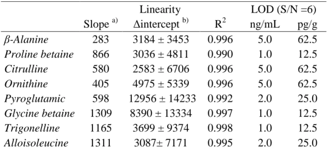
(25) Table 1. Optimal MRM conditions for the studied compounds: transitions, type of transition, percentage of the relative ion abundance, fragmentor voltage (fv) and collision energy (ce).. Compound. Transitions (m/z). β-Alanine. 146.2 > 90.1 146.2 > 72.0. Q q1. Relative ion abundance b) 100 99. Proline betaine. 146.2 > 57.1 200.2 > 144.1 200.2 > 58.0. q2 Q q1. Citrulline. 200.2 > 84.0 232.2 > 70.0 232.2 > 215.2. Ornithine. Pyroglutamic acid Glycine betaine Trigonelline Alloisoleucine. Type a). fv (V). ce (V). 80 80. 5 5. 38 100 20. 80 120 120. 15 20 25. q2 Q q1. 19 100 76. 120 90 90. 25 25 5. 232.2 > 113.1 189.3 > 70.1 189.3 > 172.2. q2 Q q1. 50 100 39. 90 70 70. 15 25 5. 189.3 > 116.1 260.2 > 84.0 260.2 > 186.1. q2 Q q1. 16 100 30. 70 80 80. 10 25 10. 260.2 > 130.1 174.2 > 118.1 174.2 > 59.1 194.2 > 138.1 194.2 > 92.0 188.2 > 86.1 188.2 > 69.1. q2 Q q1 Q q1 Q q1. 23 100 57 100 27 100 17. 80 120 120 100 100 80 80. 15 10 25 15 25 5 20. a). Type of transition: Q = Quantifier transition; q1 = First qualifier transition; q2 = Second qualifier transition. b) Relative ion abundance with respect to the base peak. 25.
(26) Table 2. Linearity and LODs for the MRM developed method.. Slope β-Alanine 283 Proline betaine 866 Citrulline 580 Ornithine 405 Pyroglutamic 598 Glycine betaine 1309 Trigonelline 1165 Alloisoleucine 1311. a). Linearity ∆intercept b) 3184 ± 3453 3036 ± 4811 2583 ± 6706 4975 ± 5339 12956 ± 14233 8390 ± 13334 3699 ± 9374 3087± 7171. 2. R 0.996 0.990 0.996 0.996 0.992 0.997 0.998 0.995. a). Counts mL/ng×min. b). Confidence intervals at 95%: ± t × sintercept. LOD (S/N =6) ng/mL pg/g 5.0 62.5 1.0 12.5 5.0 62.5 5.0 62.5 2.0 25.0 1.0 12.5 1.0 12.5 2.0 25.0. 26.
(27) Table 3. Precision in RSD (%) in peak area for samples of sunflower oil (RSO-1) by the MRM method and recovery (average ± standard deviation, n=3) for samples of olive oil (HEVOO-1) spiked at low level (0.1 ng/g of each compound) and at high level (10 ng/g of each compound).. Citrulline Precision Instrumental repeatability (n = 6) 1.9 Method repeatability (n = 3) 2.0 Intermediate precision (n = 6) 2.7 Recovery at: Low level 84.3 ± 0.7 High level 85.1 ± 0.7. Ornithine. Pyroglutamic acid. Glycine betaine. Trigonelline. 1.9 2.5 2.6. 1.0 1.3 1.6. 1.7 1.5 1.5. 1.3 2.1 2.9. 87.9 ± 0.9 81.4 ± 0.3. 90.1 ± 0.5 85.1 ± 0.5. 85.8 ± 0.9 94.9 ± 0.9. 83.2 ± 0.8 82.8 ± 0.4. 27.
(28) Table 4. Botanical origin, name of the vegetable oil samples analyzed in this work and their quantitation (average in ng/g ± standard deviation, n=3) obtained by FIA-ESIMS/MS. ND not detected. Origin Sunflower oil. Corn oil. Soybean oil Hojiblanca extra virgin olive oil Arbequina extra virgin olive oil Picual extra virgin olive oil Mixtures of HEVOO-1 with RSYO-3. Sample Name RSO-1 RSO-2 RSO-3 RCO-1 RCO-2 RCO-3 RSYO-1 RSYO-2 RSYO-3 HEVOO-1 HEVOO-2 HEVOO-3 AEVOO-1 AEVOO-2 AEVOO-3 PEVOO-1 PEVOO-2 PEVOO-3 10% 5% 2%. Citrulline. Ornithine. Pyroglutamic acid. Glycine betaine. Trigonelline. 0.15 ± 0.01 0.16 ± 0.01 0.16 ± 0.01 0.17 ± 0.01 0.19 ± 0.02 0.20 ± 0.01 0.23 ± 0.01 0.24 ± 0.01 0.23 ± 0.02. 2.9 ± 0.2 1.1 ± 0.1 1.4 ± 0.2 1.4 ± 0.3 1.2 ± 0.8 1.6 ± 0.1 2.2 ± 0.1 1.1 ± 0.2 1.2 ± 0.1. ND. ND. ND. ND. ND. ND. ND. 0.21 ± 0.01 0.15 ± 0.02 0.10 ± 0.06. 5.9 ± 0.3 4.9 ± 0.9 6.8 ± 0.1 5.3 ± 0.4 7.2 ± 0.1 5.4 ± 0.1 13.6 ± 0.7 10.1 ± 0.3 12.7 ± 0.9 0.54 ± 0.02 0.71 ± 0.03 0.18 ± 0.02 0.86 ± 0.03 0.82 ± 0.01 0.15 ± 0.01 0.39 ± 0.01 0.14 ± 0.02 0.18 ± 0.03 1.80 ± 0.08 1.09 ± 0.05 0.68 ± 0.02. 9±1 10 ± 2 13 ± 1 4.1 ± 0.5 8.6 ± 0.2 5.0 ± 0.5 4.7 ± 0.9 10.2 ± 0.3 5.2 ± 0.4 0.13 ± 0.01 0.11 ± 0.01 0.10 ± 0.03 0.14 ± 0.02 0.21 ± 0.02 0.20 ± 0.01 0.11 ± 0.01 0.12 ± 0.07 0.13 ± 0.01 0.65 ± 0.02 0.44 ± 0.02 0.25 ± 0.01. 5±1 6±1 7±1 0.3 ± 0.1 1.2 ± 0.2 0.5 ± 0.1 0.5 ± 0.1 1.0 ± 0.1 0.7 ± 0.1 0.051 ± 0.005 0.049 ± 0.005 0.051 ± 0.005 0.09 ± 0.01 0.059 ± 0.005 0.088 ± 0.001 0.053± 0.001 0.073 ± 0.009 0.055 ± 0.005 0.14 ± 0.01 0.10 ± 0.02 0.067 ± 0.002. 28.
(29) Figure 1.. Non-protein amino acids NH2. H2N. OH. H2N. NH. OH. O. O. O. β-Alanine. Citrulline NH2. NH2 H2N. OH. OH. O. N H. O. Ornithine. OH. H3C H3C. O. Pyroglutamic acid. O. Alloisoleucine. Betaines CH3. OH +. N H3C. CH3 O. Proline betaine. H3C. +. OH. +. H3C N H3C. N. O. O. Glycine betaine. Trigonelline. OH. 29.
(30) Figure 2.. 4 x10 + Product Ion (0,109-0,823 min, 86 scans) (200,2 -> **) 144,10 2,4. 3 x10 + Product Ion (0,156-0,966 min, 100 scans) (146,2 -> **) 4,0 57,10 72,00. β-Alanine. 1,6. 90,10. 1,2. 2,0. 0,8. 1,0. 0,4 60. 80. 100. 120 140 160 180 200 220 240 Counts (%) vs. Mass-to-Charge (m/z). 260. Citrulline. 8 70,00. 80. 100. 120 140 160 180 200 220 240 Counts (%) vs. Mass-to-Charge (m/z). 260. 60. 260. Ornithine. 0,8. 80. 100. 120 140 160 180 200 220 240 Counts (%) vs. Mass-to-Charge (m/z). 260. Glycine betaine. 1,6 59,10 1,2 0,8. 158,10 102,09. 100. 2,0. 130,10. 0,4. 80. 172,19. 4 x10 + Product Ion (0,127-0,804 min, 81 scans) (174,2 -> **) 2,4 118,10. Pyroglutamic acid. 1,2. 186,10. 0,4. 120 140 160 180 200 220 240 Counts (%) vs. Mass-to-Charge (m/z). 260. 4 x10 + Product Ion (0,125-0,810 min, 83 scans) (194,2 -> **) 138,10 1,6. Trigonelline. 0. 60. 80. 100. 120 140 160 180 200 220 240 Counts (%) vs. Mass-to-Charge (m/z). 260. 4 x10 + Product Ion (0,124-0,817 min, 85 scans) (188,2 -> **) 86,10 1 Alloisoleucine. 1,2. 0,8 0,6. 0,8. 92,00. 0,4. 0,4 0. 120 140 160 180 200 220 240 Counts (%) vs. Mass-to-Charge (m/z). 116,10. 0,2. x104+ Product Ion (0,123-0,820 min, 86 scans) (260,2 -> **) 84,00. 60. 100. 0,4. 159,08 60. 80. 0,6. 215,20. 2. 0. 60. 84,00. 0,8. 4. 0. 58,00. 4 x10 + Product Ion (0,124-0,817 min, 85 scans) (189,3 -> **) 70,10 1,0. 3 min, 87 scans) (232,2 -> **) x10 + Product Ion (0,115-0,823 113,10. 6. Proline betaine. 2,0. 3,0. 69,10. 0,2 60. 80. 100. 120 140 160 180 200 220 240 Counts (%) vs. Mass-to-Charge (m/z). 260. 0. 60. 80. 100. 120 140 160 180 200 220 240 Counts (%) vs. Mass-to-Charge (m/z). 30. 260.
(31) Figure 3.. x10. 2 + MRM: (0,196-0,974 min, 19 scans) (189,3 -> **). Ornithine 0.1 µg/mL. 70,10. 1. 172,20. 0,5 116,10 0. 20. 40. 60 80 100 120 140 160 180 Counts vs. Mass-to-Charge (m/z). 200. 220. x10 2 + MRM: (0,164-1,215 min, 30 scans) (189,3 -> **) HEVOO_RSYO_10% 70,10 1 116,10. 172,20. 0 2 + MRM: (0,146-1,233 min, 31 scans) (189,3-> **) HEVOO_RSYO_5%. x10. 70,10. 1. 116,10. 172,20. 0 2 x10 + MRM:(0,164-1,215 min, 30 scans) (189,3 -> **) HEVOO_RSYO_2% 70,10. 1. 172,20 116,10 0 0. 20. 40. 60. 80 100 120 140 160 180 Counts vs. Mass-to-Charge (m/z). 200. 220. 31.
(32) Figure 4.. Discriminant Function Analysis. Function 2. 42. Samples Corn oil Soya oil Sunflower oil mixture olive oil Centroides Centroids. 32 22 12 2. 2% 5% 10%. -8 -16. -6. 4. 14. 24. 34. 44. Function 1. 32.
(33)
Figure

Documento similar