BMC Clinical Pathology (2001) 1:1
Methodology article
A simple method to assess the oxidative susceptibility of low density
lipoproteins
Adriana E Scoccia, María Silvina Molinuevo, Antonio Desmond McCarthy
and Ana María Cortizo*
Address: Cátedra de Bioquímica Patológica, Facultad de Ciencias Exactas, Universidad Nacional de La Plata, La Plata, Argentina
E-mail: Adriana E Scoccia - ascoccia@biol.unlp.edu.ar; María Silvina Molinuevo - silmol@biol.unlp.edu.ar; Antonio Desmond McCarthy - mccarthy@biol.unlp.edu.ar; Ana María Cortizo* - cortizo@biol.unlp.edu.ar
*Corresponding author
Abstract
Background: Oxidative modification of low density lipoproteins (LDL) is recognized as one of the major processes involved in atherogenesis. The in vitro standardized measurement of LDL oxidative susceptibility could thus be of clinical significance. The aim of the present study was to establish a method which would allow the evaluation of oxidative susceptibility of LDL in the general clinical laboratory.
Results: LDL was isolated from human plasma by selective precipitation with amphipathic polymers. The ability of LDL to form peroxides was assessed by measuring thiobarbituric acid reactive substances (TBARS) after incubation with Cu2+ and H2O2. Reaction kinetics showed a three-phase pattern (latency, propagation and decomposition phases) which allowed us to select 150 min as the time point to stop the incubation by cooling and EDTA addition. The mixture Cu2+/ H2O2 yielded more lipoperoxides than each one on its own at the same time end-point. Induced peroxidation was measured in normal subjects and in type 2 diabetic patients. In the control group, results were 21.7 ± 1.5 nmol MDA/mg LDL protein, while in the diabetic group results were significantly increased (39.0 ± 3.0 nmol MDA/mg LDL protein; p < 0.001).
Conclusion: a simple and useful method is presented for the routine determination of LDL susceptibility to peroxidation in a clinical laboratory.
Background
Atherosclerosis is a pathology that affects many people and may cause their death or disability due to myocardial infarction or strokes. Although the clinical manifesta-tions of the disease have been established, the underly-ing mechanism of atherogenesis is still unclear. Recent theory points toward the oxidative modification of LDL (LDL-Ox) as one of the major involved processes [1]. Nevertheless, hardly any of the biological effects of LDL-Ox have been tested in vivo.
Taking into account the potential clinical importance of the oxidative modification of LDL, many studies have been carried out to quantify their in vitro susceptibility to oxidation. This measurement is thought to correlate with the LDL oxidative susceptibility within the arterial wall [2].
Plasmatic LDLs may be isolated by different methods, which include sequential and density-gradient ultracen-trifugation, chromatography, electrophoresis and
selec-Published: 20 June 2001
BMC Clinical Pathology 2001, 1:1
Received: 9 March 2001 Accepted: 20 June 2001
This article is available from: http://www.biomedcentral.com/1472-6890/1/1
tive precipitation [3]. Lipid peroxidation is a very complex process that involves the chain reaction of free radicals with polyunsaturated fatty acids. These reac-tions lead to rearrangements of double bonds in conju-gated dienes, hydroperoxide generation, lipid breakdown into lower molecular weight fragments, as well as chemical modifications in the apo B protein [4,5,6,7,8]. The extent of lipid peroxidation can be esti-mated by measurement of thiobarbituric reactive sub-stances (TBARS). This method, although nonspecific, is of value in purified systems. TBARS determination mainly measures malondialdehyde (MDA) derived from the hydroperoxidation of unsaturated fatty acids with three or more double bonds.
Many studies have been carried out to establish the role of Fe3+, Fe2+ and Cu2+ in the oxidation of LDL [1,8,9]. In
biological systems, the reduction of oxygen yields hydro-gen peroxide and superoxide radical. The reaction be-tween these two species generates a hydroxyl radical, which is the reactive oxygen species with the shortest half life and highest reactivity. This reaction, which is ki-netically slow, can be accelerated by catalytic amounts of iron or copper salts [10].
In the present study we present a simple method which would allow the high-throughput routine evaluation of the oxidative susceptibility of LDLs in the simultaneous
presence of Cu2+ and H
2O2 in the general clinical
labora-tory. LDLs were isolated by selective precipitation and their oxidative susceptibility was evaluated through the quantitation of TBARS.
Results
Optimization of oxidative susceptibility assay
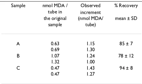
As expressed in Materials and Methods, different vol-umes of solubilizing solution were used to resuspend LDL precipitates. Relatively low volumes (0.4 ml) gave lower intra-assay coefficients of variation (4.8 %) than relatively high volumes of 1.0 ml (CV = 10.8 %). These re-sults correspond to the analysis of 22 samples deter-mined in duplicate. In order to assess the recovery of standard, a fixed amount of 1.44 nmol MDA / tube was added to aliquots of previously assayed duplicated resus-pended LDL samples from three different plasmas. As can be seen in Table 1 the MDA recovery varied between 78.1 and 93.8 % of the true value.
In experiments aimed at adjusting the number of precip-itate washes needed, LDL precipprecip-itate was washed once or twice with precipitating solution, or not washed at all, prior to solubilizing. The protein concentration and cho-lesterol content of the resulting resuspended LDL sam-ples was then determined in duplicate. The first wash diminished the protein content of the resuspended LDL sample by 20 %, whereas the second wash further de-creased protein content by 3 %. On the other hand, the cholesterol content of resuspended LDL samples did not
vary as a consequence of successive washes (99 ± 3 and
[image:2.612.56.293.505.646.2]98 ± 2 % of non washed precipitate, for 1 or 2 washes re-spectively). The samples were also submitted to agarose electrophoresis, and bands revealed with Coomasie bril-liant blue, in order to evaluate the possible presence of contaminating plasma proteins. The unwashed precipi-tate showed a clearly visible band corresponding to albu-min, as well as another band of greater intensity with the electrophoretic mobility of LDL. One and two washes with precipitating reagent greatly diminished - but did not completely eliminate - the albumin band, without provoking any changes in the intensity of the LDL band (data not shown). As the washing procedure eliminates non-apoB co-precipitating plasma proteins without cho-lesterol losses, a single wash was selected as the standard procedure.
Table 1: Recovery of 1.44 nmol MDA/tube added to duplicated re-suspended LDL samples obtained from three independent plas-ma samples.
Sample nmol MDA / tube in
Observed increment
% Recovery
the original sample
(nmol MDA/ tube)
mean ± SD
A 0.63 1.15 85 ± 7
0.69 1.30
B 1.07 1.24 78 ± 12
1.32 1.00
C 0.47 1.43 94 ± 8
[image:2.612.314.554.602.676.2]0.47 1.27
Table 2: Effect of Triton X-100 on the LDL oxidative susceptibility assay.
Solubilizing reagent MDA nmol / mg LDL protein mean ± SD (n = 3)
NaCl 20.3 ± 0.6
NaCl + Triton X-100 21.0 ± 0.7
In other experiments, the effect of the presence of Triton X-100 in the solubilizing solution was evaluated by as-saying the oxidative susceptibility in three independent precipitations of the same sample. Two solubilizing rea-gents were investigated, 50 g/l NaCl and 0.1 % Triton X-100 in 50 g/l NaCl. Table 2 shows that the same results for LDL oxidative susceptibility were obtained with the two procedures. However, since resuspending the pre-cipitate with Triton X-100 was found to be less time-con-suming, it was chosen as the standard method.
The kinetics of Cu2+/H
2O2-induced LDL peroxidation
was monitored by measuring the TBARS levels in aliq-uots of three resuspended LDL samples incubated from
15 to 180 min with Cu2+/H
2O2 (Figure 1). An initial lag
phase could be observed with no increments in the ab-sorbance, followed by another with a maximum slope (propagation phase). A final phase was evident with low-er absorbance increments (decomposition phase). Fig-ure 1 represents three examples of various LDL preparations. In most cases, the propagation phase reached a maximum at about 150 min. Thus, this incuba-tion period was selected for the LDL oxidaincuba-tion reacincuba-tion. In further experiments, EDTA was validated as an effec-tive inhibitor of the basal and induced oxidation reac-tion. After an incubation of 150 min, the results for samples submitted to oxidation in the presence of EDTA (30 nmol/tube) monitored for TBARS formation, did not show significant differences when compared with the
blanks (with Cu2+/H
2O2, without sample). Thus, EDTA
at this concentration was subsequently used to
effective-ly stop the oxidative reaction induced by Cu2+/H
2O2.
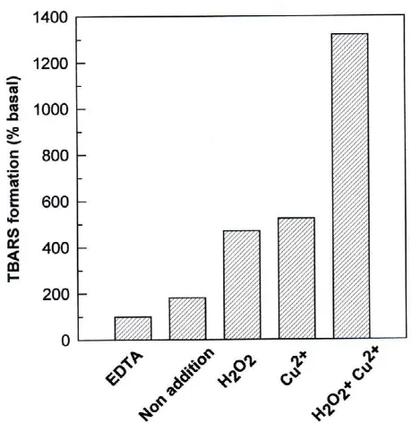
The basal LDL oxidation (as defined in Materials and Methods) was extremely low and was arbitrary assigned 100 % value (Figure 2). In absence of EDTA and oxidat-ing agents, the samples showed an inherent oxidability, with TBARS values of approximately twice that of basal
LDL oxidation. We next analyzed the effect of Cu2+ and/
or H2O2 as inducers of LDL oxidation reaction. When
added separately, Cu2+ and H
2O2 increased TBARS
for-mation by approximately 5- fold. However; the
simulta-neous addition of H2O2 and Cu2+ induced a synergistic
increase in TBARS levels (approximately 13- fold). Dou-bling doses of Cu2+ or H
2O2 did not further increase the
oxidation levels of LDL.
The influence of LDL protein content on the TBARS re-action, was evaluated by increasing the volume of resus-pended LDL samples, under constant TBARS reagent volume and incubation period (Figure 3). The reaction was linear up to an LDL protein content of approximate-ly 300 µg/tube.
In order to characterize the LDL isolated by selective pre-cipitation, and to investigate the possible damage of the inner structure of LDL caused by this method, we per-formed an agarose electrophoresis of the resuspended LDL sample in parallel with the LDL isolated by ultra-centrifugation and the corresponding whole plasma sample. As can be seen in Figure 4, LDL fraction isolated by both methods showed the same electrophoretic mo-bility and no contamination by other lipoprotein frac-tions.
LDL oxidation susceptibility in control and diabetic sam-ples
In order to evaluate the method's usefulness in separat-ing a control population from another with increased risk for cardiovascular disease, a group of 30 normal subjects and 12 type 2 diabetic patients were submitted to this assay. The oxidative susceptibility of LDL was sig-nificantly greater in the diabetic group than in the con-trol population (39.0 ± 3.0 vs. 21.7 ± 1.5 nmol MDA / mg LDL protein; p < 0.001).
Discussion
The oxidative modification of LDL appears to be in-volved in the development of various degenerative dis-Figure 1
Kinetics of in vitro LDL peroxidation assessed by TBARS formation. An aliquot of 100 µl plasma from three different patients (A, B, C) was assayed. All sample precipi-tates were redissolved in 0.4 ml of solubilizing solution, incu-bated with Cu2+/H2O2 for different periods of time, and 100
[image:3.612.322.549.95.309.2]eases such as atherosclerosis, carcinogenesis, aging and diabetes mellitus [11,12]. Standard reference methods to prepare LDL from plasma employ ultracentrifugation. However, the selective precipitation methods, which are more accessible than ultracentrifugation, are widely used in the clinical laboratory for the measurement of the cholesterol content in different lipoprotein fractions. In particular, selective precipitation of LDL may be ap-proached in different ways: by addition of heparin at an exactly controlled pH of 5.12 in the absence of divalent cations; or with polyvinylsulphate in the presence of EDTA and polyethylene glycol methyl ether; with am-phipathic polymers in imidazole buffer at pH 6.10 (bi-oMerieux). An excellent statistical correlation is obtained when these methods are compared with refer-ence ultracentrifugation methods, providing samples with triglyceride concentration above 8 mmol/l and those from patients with hyperlipoproteinemia Type III are excluded [13]. In particular, the precipitating reagent used in the present work (bioMerieux), shows a good correlation coefficient (r = 0.96) when compared with ul-tracentrifugation methods [3]. Its selectivity and the preservation of the immunological properties as well as the lipid composition of the native original LDLs have also been demonstrated [3,9,14,15]. In our present study, we were unable to find differences in agarose
electro-phoretic mobility between LDL fractions obtained by this method of selective precipitation and ultracentrifu-gation. In addition, no contaminating lipoprotein frac-tions were observed by this electrophoretic method. In our standard procedure we washed the LDL precipitate once prior to solubilizing. Thus, it was necessary to es-tablish whether there were changes in LDL cholesterol content, which could invalidate the original method's correlation with ultracentrifugation. However, we were unable to find cholesterol losses as a consequence of one or two washes with precipitating reagent.
Arshad et al. [16] developed a simple method to assess
whole plasma susceptibility to peroxidation by Cu2+/
H2O2 incubation. They used thiobarbituric acid reactivi-ty to evaluate lipid peroxidation, a method which is not entirely specific. However, it proved to be easy to per-form and accessible for the analysis of many samples. In the present work, we measured LDL-associated TBARS after induction of lipid peroxidation with a mixture of
Cu2+ and H
2O2.
Several methodological aspects of our procedure were subsequently addressed, in order to achieve its optimiza-tion. a) The intra-assay precision was found to depend on the volume of solubilizing solution employed. In our standard working conditions, the CV was 4.8 %, which is lower than the precision limit established for the deter-mination of selectively precipitated lipoprotein choles-terol (CV < 5%) [3], and so can be considered acceptable. b) The observed percentage of recovery for exogenously added MDA (Table 1) was comparable to that of the TBARS reaction (82-100 %) [10]. These results suggest that the additioned MDA was still TBA reactive and did not generate any interfering substances, since the ob-served increment in MDA content did not significantly differ from that of the true value. c) It is important to en-sure that the precipitate is not contaminated with non-LDL serum proteins, since results are expressed per non-LDL protein content. This contribution to variability was eliminated by washing the LDL precipitate. d) When the composition of the solubilizing solution was evaluated, precipitate redissolution effectively occurred in 50 g/l NaCl. However, the addition of Triton X-100 was chosen because it shortened the period of LDL redissolution. e) Lipid peroxidation kinetics have been extensively stud-ied [8,10]. It is known that LDL oxidation in the presence
of Cu2+ shows three phases: latency, propagation and
de-composition. This has been established by determina-tion of hydroperoxides, TBARS or other aldehydes, fluorescent products and conjugated dienes. It has been shown that during the latency and propagation phases, as well as during the early stages of the decomposition phase, the time-courses of diene, TBARS and lipid hy-droperoxide formation, are practically coincident [8]. In-Figure 2
[image:4.612.61.291.89.325.2]deed, the corresponding maxima coincide temporally. However, each individual's LDL shows its own particular kinetics so that sample to sample variations could repre-sent a problem when - as in the prerepre-sent study - a single measurement of only one parameter is taken after a long incubation time. This does not allow us to conclusively establish whether the sample is at the end of its propaga-tion phase, or has already begun its decomposipropaga-tion phase. In our preliminary studies of TBARS time-course, we found a lag phase followed by a maximum slope which ended at 150 min, the time point adopted for our standard procedure. A slower increment in absorbance was observed from this point on, a fact that may have been due to the decomposition of accumulated products. f) In the absence of oxidation inhibitors, LDL oxidation may continue throughout the TBA reaction period, thus contributing to the method's variability. This was effec-tively prevented by the addition of EDTA prior to the TBA reaction, which acts as an inhibitor of LDL
oxida-tion by Cu2+ sequestration. g) Our experiments show
that the combination of Cu2+ and H
2O2 is more effective
for the induction of LDL oxidation, than each agent its own. The observed results suggest a synergistic mecha-nism of action between both reagents. Previous studies
have addressed the Cu2+-induced in vitro oxidation of
plasma LDL [17]. These authors found a value of 21 ± 3
nmol MDA / mg LDL protein, obtained from four normal subjects, for LDL isolated by ultracentrifugation. This is practically coincident with the results which we obtained with our control healthy population (21.7 ± 1.5 nmol MDA / mg LDL protein), as would be expected from the reported correlation between LDL obtained by ultracen-trifugation and by the LDL-precipitating method of bi-oMerieux. Recently, Guerci et al.[14] studied the LDL oxidation susceptibility of normolipidemic diabetic and non-diabetic patients. These authors found a significant increase in type 2 diabetic patients vs. healthy subjects, particularly in the group of type 2 diabetic females, in which LDL oxidation susceptibility was highest. In coin-cidence with these reported results, LDL oxidative sus-ceptibility of our type 2 diabetic patients was significantly greater (39.0 ± 3.0 nmol MDA / mg LDL protein) than the control group.
[image:5.612.326.553.93.315.2]The LDL precipitation method which we have used in this study is based on interaction with glycosaminogly-cans (GAG). However, both lipid composition and the content of sialic acid can modulate the interaction with GAG. In this context, particles such as small dense LDL can interact with GAG with high affinity. In addition, the precipitation procedure may increase the susceptibility for oxidation by copper since copper penetrates the LDL particle more easily after precipitation. In consequence, we cannot discard the possibility that our results may re-flect a preselection of LDL with higher susceptibility for oxidation.
Figure 3
Influence of the LDL protein content on TBARS reaction linearity. Increasing doses of a LDL sample were submitted to constant oxidative conditions. Results are expressed as mean of duplicate determinations. y = 4.35 . 10 -3× - 6.57 . 10-3, r2 = 0.994; p < 0.001.
Figure 4
[image:5.612.317.551.411.602.2]Conclusion
A simple method for the in vitro measurement of LDL oxidation susceptibility has been optimized, and applied to a group of healthy subjects and type 2 diabetic pa-tients. This straightforward approach could facilitate the comparison of results obtained from an increased number of general clinical laboratories, and thus allow us to move a step further towards the standardization of a procedure of potential clinical importance.
Materials and methods
Materials
LDL Cholesterol kit (cat. Number 61532) was provided by bioMerieux (Marcy l'Etoile France). Hydragel Lipo+Lp(a) kit for agarose electrophoresis was obtained from Sebia. Thiobarbituric acid was obtained from Mer-ck. 1,1',3,3'- tetra-methoxy-propane or malondialdehyde (MDA) was used as standard and purchased from SIG-MA Co. St. Louis, MO, USA. All chemicals were of analyt-ical grade and used without further purification.
Sample collection
Twelve type 2 diabetic patients of both sexes (37 - 65 years old) were studied. The degree of metabolic control was assessed by the measurement of fasting plasma
glu-cose (mean 7.2 ± 1.1 mmol/l), fasting plasma HbA1c
(mean 6.8 ± 1.3 %; NV 4.8-6.0 %) and they were normo-lipidemic. A series of 30 control non-diabetic subjects of both sexes (age range, 35-60 year old) was processed in parallel. All controls were normolipidemic according to the Alfedian criteria [18], and none of the subjects were taking any drug known to influence lipid or lipoprotein metabolism. Blood samples were obtained on heparin (5 U/ml) by venipuncture from subjects with 12 hours fast-ing. Plasma was separated rapidly and processed imme-diately. Alternatively, the samples were stored at 4 ºC for 24 hours or at -20º C for not more than 2 days.
Method of LDL isolation
LDL was selectively precipitated from 100 µl of plasma by addition of bioMerieux precipitating reagent of LDL-Cholesterol kit and vortex-mixed [14]. The mixture was incubated for 30 min at 2-8 ºC, and centrifuged for 5 min. The supernatant was discarded and the precipitate was washed with precipitating reagent. The washed pre-cipitate was redissolved in different volumes of solubiliz-ing solution (0.01% Triton X100 in 50 g/l NaCl) at 37 ºC, and vortex-mixed (resuspended LDL sample) [15]. Brad-ford's method [19] was used to determine the total pro-tein content of the resuspended LDL sample, using bovine serum albumin as a standard. For selected exper-iments, the LDL fraction was obtained by density gradi-ent ultracgradi-entrifugation as has been previously described [13].
Characterization of LDL isolated by selective precipitation
Representative samples were subjected to ultracentrifu-gation and selective precipitation (0, 1 or 2 washes) in or-der to isolate the LDL fraction. Subsequently, LDL fractions obtained by both methods, as well as the whole plasma, were electrophoresed in agarose according to the manufacturer's instructions. Briefly, electrophoresis was performed at a constant voltage of 130 V and initial intensity of 25 mA, for 80 minutes. The gel was dried and bands were revealed with either Sudan Black or Cooma-sie brillant blue.
LDL-cholesterol determination
LDL was obtained by selective precipitation of represent-ative samples, and the resulting precipitates were washed once, twice, or not at all with the precipitating re-agent, prior to resuspending with the solubilizing solu-tion. Cholesterol content of the resulting resuspended LDL samples was determined by a commercial enzymat-ic kit (Colestat, Wiener Laboratories Argentina).
Basal and induced LDL oxidation
Basal LDL oxidation was determined by incubating an aliquot of 100 µl resuspended LDL sample, containing 50-90 µg protein, with 30 µl of 1 mM EDTA and 45 µl of distilled water. The corresponding blank was determined substituting the resuspended LDL sample by solubilizing solution.
In other experiments, resuspended LDL sample was
mixed with 50 µl of 100 µM Cu2+ (freshly prepared in
phosphate buffer saline solution, PBS, pH 7.4) and 25 µl of H2O2 solution (300 ml/l H2O2 in PBS, stock solution
corresponds to 10 volume commercial H2O2). Blank was
performed with solubilizing solution instead of resus-pended LDL sample. In all cases, sample and blank were incubated at 37 ºC for different periods of time with oc-casional stirring. At the end of the incubation period, the lipid peroxidation was stopped by cooling and addition of 30 µl of 1 mM EDTA.
TBARS determination
Statistical analysis
Results were expressed as mean ± SD and mean ± SEM.
Statistical analysis was performed by Student's t test; a p value < 0.05 was considered statistically significant. Lin-ear regression analysis was used for testing correlations between variables.
Acknowledgements
We thank Dra Susana Etcheverry for the kind revision of the paper. We are also grateful to Dr B. Corsico for the preparation of LDL fraction by ultra-centrifugation. AMC is a member of the Carrera del Investigador, CICPBA. This work was partially supported by grants from Facultad de Ciencias Ex-actas, UNLP, CICPBA. Authors thanks Dr Christian Coppens, Marcy, France and Dr Javier Goodman from bioMerieux Argentina for the provi-sion of the LDL-cholesterol kit. We also thank Wiener Argentina for the donation of the Cholesterol kit.
References
1. Steinbergt D: Low density lipoprotein oxidation and its patho-biological significance.J Biol Chem 1997, 272:20963-20966 2. Schwartz CJ, Valente A: Atherogenesis and coronary heart
dis-ease: cellular mechanism.In International textbook of Diabetes Mel-litus. Edited by Alberti KGMM, DeFronzo RA, Keen H, Zimmet P. Chichester: John Wiley, 19971535-1541
3. Rifai N, Warnick GR, McNamara JR, Belcher JD, Grinstead GF, Frantz ID Jr: Measurement of low- density- lipoprotein cholesterol in serum: a status report.Clin Chem 1992, 38:150-160
4. Esterbauer H, Jürgens G, Quehenberger O, Koller E: Autoxidation of human low density lipoprotein: loss of polyunsaturated fatty acids and vitamin E and generation of aldehides.J Lipid Res 1987, 28:495-509
5. Buege JA, Aust SD: Microsomal lipid peroxidation.Methods Enzy-mol 1978, 52:302-310
6. Wallin B, Camejo G: Lipoprotein oxidation and measurement of hydroperoxide formation in a single microtitre plate.Scand J Clin Lab Invest 1994, 54:341-346
7. El-Saadani M, Esterbauer H, El-Sayed M, Goher M, Nassar AY, Jürgens GA: Spectrophotometric assay for lipid peroxides in serum lipoproteins using a commercially available reagent.J Lipid Res 1989, 30:627-630
8. Esterbauer H, Gebicki J, Puhl H, Jürgens G: The role of lipid perox-idation and antioxidants in oxidative modification of LDL.
Free Radic Biol Med 1992, 13:341-390
9. Taus M, Ferretti G, Dousset N, Moreau J, Battino M, Solera ML, Valdiguie P, Curatola G: Suceptibility to in vitro lipid peroxida-tion of low density lipoproteins and erythrocyte membranes from liver cirrotic patients.Scand J Clin Lab Invest 1994, 54 :147-153
10. Coudray C, Richard MJ, Favier AE: Determination of primary and secondary lipid peroxidation products: plasma lipid hy-droperoxides and thiobarbituric acid reactive substances.In Analysis of free radicals in biological systems. Edited by Favier AE, Cadet J, Kalyanaraman B, FontecaveM, Pierre JL. Base. Boston. Berlin: Birkhäuser Verlag, 1995185-200
11. Lyons TJ: Oxidized low density lipoproteins: a role in the pathogenesis of atherosclerosis in diabetes?Diabetes Med 1991,
8:411-9
12. Steinberg D, Parthasarathy S, Carew TE, Khoo JC, Wilztum JL: Be-yond cholesterol: modifications of low-density lipoprotein that increase its atherogenecity.N Engl J Med 1989, 320:915-24 13. Demacker PN, Hijmans AG, Brenninkmeijer BJ, Jansen AP, van't Laar A: Five methods for determining low -density lipoprotein cholesterol compared.Clin Chem 1994, 30:1797-1800
14. Guerci B, Antebi H, Meyer L, Durlach V, Ziegler O, Nicolas JP, Alcin-dor LG, Drouin P: Increased ability of LDL from normolipi-demic Type 2 diabetic women to generate peroxides.Clin Chem 1999, 45:1439-1448
15. Moss MA, Wong CSY, Tan MH, Pett K, Jacklin CLE: Determination of low density lipoprotein cholesterol (LDL-C) in serum by BioMeriex cholesterol/phospholipids polyanions precipita-tion method and comparison with preparative ultracentrifu-gation.Clin Chem 1986, 32:1096-7
16. Arshad MAQ, Bhadra S, Cohen RM, Subbiah MTR: Plasma lipopro-tein peroxidation potential: a test to evaluate individual sus-ceptibility to peroxidation.Clin Chem 1991, 37:1756-1758 17. Lavy A, Brook GJ, Dankner G, Ben Amotz A, Aviram M: Enhanced
in vitro oxidation of plasma lipoproteins derived from hyper-cholesterolemic patients.Metabolism 1991, 40:794-799 18. Brun JM, Droulin P, Berthezene F, Jacotot B, Pometta D:
Dyslipidé-mies dy patient diabétique.Diabetes Metab 1995, 21:59-62 19. Bradford MM: A rapid and sensitive method for the
quantita-tion of microgram quantities of protein utilizing the princi-ple of protein-dye binding.Anal Biochem 1976, 72:243-254 20. Ohkawa H, Ohishi N, Yagi K: Assay for lipid peroxides in animal
tissues by thiobarbituric acid reaction. Anal Biochem 1979,
95:351-358
Pre-publication history
The pre-publication history for this paper can be ac-cessed here:
http://www.biomedcentral.com/content/backmatter/ 1472-6890-1-1-b1.pdf
Publish with BioMedcentraland every
scientist can read your work free of charge
"BioMedcentral will be the most significant development for disseminating the results of biomedical research in our lifetime."
Paul Nurse, Director-General, Imperial Cancer Research Fund
Publish with BMC and your research papers will be:
available free of charge to the entire biomedical community
peer reviewed and published immediately upon acceptance
cited in PubMed and archived on PubMed Central
yours - you keep the copyright
editorial@biomedcentral.com Submit your manuscript here:
http://www.biomedcentral.com/manuscript/