Supervisory Committee: Prof. Anne-Mieke Vandamme Prof. Kristel Van-Laethem Prof. Arley Gomez-Lopez Prof. Otto Sussmann
Doctoral Thesis in Biomedical Sciences
Andrea-Clemencia Pineda-Peña
-Cl em en cia Pi ne da -Pe ña T H E D YN A M IC S O F L O C A L H IV -1 EPI D EM IC S T he C olo m bia n a nd Be lg ia n C oh or ts Au gu st 20 14Bogotá, Colombia / Leuven, Belgium 2014
Laboratory of Clinical and Epidemiological Virology
THE DYNAMICS OF LOCAL HIV-1 EPIDEMICS
The Colombian and Belgian cohorts
Andrea-Clemencia Pineda-Peña
Double Degree KU Leuven and Universidad del Rosario
Dissertation presented in partial fulfillment of the requirements for the double degree of Doctor in Biomedical Sciences
Leuven and Bogotá, 2014
KU Leuven
Biomedical Sciences Group Faculty of Medicine
Department of Microbiology and Immunology Rega Institute for Medical Research Laboratory of Clinical and Epidemiological Virology
Leuven, Belgium
Universidad del Rosario
Faculty of Sciences and Mathematics Faculty of Medicine
Biomedical Doctoral School
Clinical and Molecular Aspects of Infectious Diseases Group
Bogotá, Colombia
Jury:
Promoter of KU Leuven: Prof. Dr. Anne-Mieke Vandamme Co-promoter of KU Leuven: Prof. Dr. Kristel Van Laethem Promoter of Universidad del Rosario: Prof. Dr. Arley Gomez Lopez Co-promoter of Universidad del Rosario: Prof. Dr. Otto A. Sussmann Jury members: Prof. Dr. Inge Derdelinckx
Prof. Dr. Geert Molenberghs
Prof. Dr. Eduardo Gotuzzo
“Take the first step in faith. You don't have to see the whole staircase, just take the first step”
Martin Luther King, Jr.
ACKNOWLEDGMENTS / AGRADECIMIENTOS
Some traditions consider gratefulness one of the keys of the life. Since I do believe it, I would like to thank you everybody here and there. Thank you for sharing this path and teaching me
how life is worthy every second!
Algunas tradiciones consideran la gratitud como una de las claves de la vida. Como lo creo, quisiera agradecer a todos aquí y allá. Gracias por compartir ese camino y enseñarme como la
vida vale cada segundo!
Primero quiero agradecer a Dios quien ha estado conmigo siempre y me ha dado esta oportunidad, y a toda mi familia en el cielo que me ha acompañado y animado en este camino.
I would like to thank to my promoters Prof. Anne-Mieke Vandamme, Prof. Kristel Van Laethem, Prof. Arley Gomez and Prof. Otto Sussmann for their support in this doctorate. To Annemie and Kristel, thank you for your guidance and fruitful scientific discussions. Your patience and knowledge were valuable for this thesis. To Annemie, I hope you continue with your leadership, open heart, adventurous attitude and free mind. To Kristel, keep your warm-hearted personality, reliability, and structured mind (plus all I’ve told you before). Arley muchas gracias por brindarme tu incondicional apoyo como profesor y amigo, tus palabras siempre fueron inspiradoras y oportunas. Continúa con fe en todos tus propósitos. Otto gracias por introducirme en este medio del VIH, transmitirme tu pasión por las Infecciosas, tu confianza e impulsarme a continuar con mis metas. Continúa con tu buen humor y pasión por tu profesión.
I am grateful with my Jury Members Prof. Inge Derdelinckx, Prof. Eduardo Gotuzzo, Prof. Geert Molenberghs, Prof. Chris Verhofstede and Prof. Ghislain Opdenakker for their suggestions for the improvement of this thesis and their encouragement. I am indebted with the coordinator of my Doctoral training in Colombia, Prof. Luisa Marina Matheus and my administrative supervisor, Prof. Manuel Alfonso Patarroyo.
You and Patrick are very inspiring about love. To Sietse, thank you for your administrative support and help in the last steps.
For my former friends and colleagues at the Rega: To Jurgen, I was impressed for your warm personality, smile and humor in every moment, thank you for your statistical support and your affable mood. To Britta & Gertjan, I wish you all the love in your new adventure; please keep that cheerful (Britta) and amiable mood (Gertjan). To Stijn, thank you for your patience and help with the computers stuff, but more for your trust and your nice words. To Raphael, I hope you keep your lovely smile and positive attitude in this new path. To Fausta, please keep your perseverance and big smile. To Monica, I shared nice moments with you, keep seeing the positive side of the life. To Lia, I hope you keep your determination alive and accomplish your goals. To Laura, I really enjoyed our Italian-Spanish-English talks. I am grateful for little but nice moments with Abbas, Caro Palma, Fabienne, Goedele, Heidi, Joke, Kiyoshi, Lien, Supinya, Tim and Yang. To the future member Tim, good luck with the next steps.
I would like to show my gratitude to the people from the Evolutionary and computational Lab: Bram, Guy, Nidia, Nuno, Filip, Philippe and Tiago. Especially to Nuno, you have been my buddy all these years, thank you for your encouragement, teachings, and your kindness. To Nidia, thank you for the friendship and cheerful mood! Philippe, I am grateful for your suggestions and useful discussions. I also would like to show my gratitude for the people of the 6th and 7th floor!. Especialmente a Alexandra, quien me acompañó en todo momento en esta travesía y terminó siendo parte de nuestra familia :-) Muchos éxitos siempre, que sigas creciendo en todos los aspectos! And Bobby, your positive mood and advices were very helpful! Marc and Elizabeth, keep your nice humor and kindness!
Thank you to Prof. Eric Van Wijngaerden for his guidance in the clinical consultation and his willingness to speak several languages at the same time in order to help me understand. I am also grateful with Dr. Paul de Munter and Dr. Inge Derdelicks from the Internal Medicine group.
Quisiera agradecer a las personas de la Universidad del Rosario, especialmente a las de la Unidad de Microbiología. Maria Antonia, Dora y Maria Cristina, gracias por ser mi apoyo por todos esos años y transmitirme su amor por las enfermedades infecciosas. Giovanni, tú has sido mi colega y amigo por todo este tiempo, gracias por los momentos vividos en Colombia y en Lovaina… espero sigas cumpliendo tus metas junto a Mafe y tu bella familia. Sigue con tu humor y buen trabajo! A todas mis profes de Genética, Claudia Silva, amiga, profesora y colega…gracias por tus dosis de amor todos estos años. Prof. Restrepo, Dorita, Heidi y Norita, gracias por ser mi familia genetista e introducirme en esta área de la investigación. Gracias Luisa por tu apoyo como coordinadora del Doctorado
Agradezco profundamente a mis amigos y colegas de la Fundación Instituto de Inmunología de Colombia. Agradezco al FIDIC por su soporte académico y su apoyo especialmente en el primer semestre del doctorado. Especialmente a Manuel Alfonso, gracias por creer en mí, por darme ánimo y consejo en todo momento, ser ese apoyo y amigo incondicional. Espero logres ese gran sueño que te has propuesto!. A las chicas HPV: Marina, Milena, y Sara. Espero continúen con su excelente trabajo y dedicación. Me alegra que sean ejemplo de amistad!
investigación en la institución y Diana, gracias por tu incondicional ayuda y entusiasmo en ese camino investigativo y de Comités.
Thank you to my friends in Leuven, you have been a great support in these 4 years. To my wonderful and always helpful neighbors: Ona & Uwe (a quienes admiro su dedicación y amabilidad), Isael & Maria (por su generosidad y darnos la bienvenida), Vladimir & Keith (you are very warm-hearted), Herman & Monique (your sympathy and cordiality are valuable). To an excellent teacher and lovely friend: Det. Gracias a la comunidad hispano-parlante de Lovaina por su incondicional apoyo y amistad: A Juan Carlos, gracias por tu apoyo, compañía y buenos momentos; continua con tu misión de servicio y humor. A Paul, gracias por esos momentos valiosos y ejemplo de servicio. Adriana Moreno, continúa con ese espíritu libre y carisma. Cesar, tu alegría y animo son envidiables, sigue adelante con tus metas e irradiando felicidad. Melvin, me alegra verte feliz y con mucho ánimo hacia el futuro. Italo & Oksana, gracias por los momentos compartidos y su espíritu amable. A Wouter & Elizabeth, gracias por los momentos compartidos y el apoyo-amistad que le han brindado a Jorgito. Carme & Xavi, su espontanea sonrisa y calidez han sido invaluables. Carolina & Oscar, ustedes nos recibieron con los brazos abiertos, su corazón es de puertas abiertas igual que su generosidad, realmente muchos éxitos en este camino. Natalia & Eddy, espero sigan ese camino con mucha felicidad y Nata admiro tu perseverancia y chispa. Camilo, te deseo mucha felicidad en esta nueva etapa con Ale, sigue juicioso y de buen humor. Jorge Duitama, gracias por tu amistad y generosidad. Andrea Ávila, a sumercé que le vaya bien con Pole y tus metas académicas, continúa con esa chispa que alegra. Carolina Vallecilla, continúa con ese empuje que te caracteriza y amor por Martino. A Milda & Marc, gracias por su inmensa generosidad y adicionalmente, Milda eres una mujer llena de amor. A Varima & Chris, ustedes tienen una alegria contagiosa y emocionante, continuen con ese amor que los caracteriza. A David & Tara, su apoyo principalmente a Jorge y de paso a mí ha sido importante en este camino. Aunque hemos compartido pocos pero intensos momentos, he estado asombrada de la generosidad y calidez de Constanza & familia, Sergio & Maritza, Vilma &Jimmy, Jenny & familia, Gladys & familia, Myriam & familia, y Jesus & familia.
Tengo una gran familia de amigos de toda la vida: Nicolas & Silvi, Zhyla & Andres, Catalina & Nicolas, Paola & Mauricio. A ustedes gracias por su compañía desde la distancia, sé que cuento en todo momento con ustedes y su incondicional apoyo. A mi familia, Rosarito, Milingos, Linis y Nacho, gracias por cada momento de apoyo, compañía, y aunque lejos… siempre procurando mi bien. Jorge, Glorita e Ilbita, gracias por creer en mí en cada momento y apoyarme en todas mis empresas. Sus palabras positivas son una motivación importante. Mamalita, gracias por tu eterna sonrisa y tu cariño inmenso. Tu amor ha cambiado mucho la vida de nuestra familia…gracias nuevamente. Ana, Jorge, Carolina, Néstor, Diana, Isabella y Gabriel, gracias por sus oraciones, palabras de apoyo, amor, ternura, ánimo y humor. Espero continúen colmados de amor, éxitos, salud y amor.
Mami, gracias por ser una soñadora y lo más importante darte el lujo de disfrutar cada momento de tu vida. Sigue adelante cumpliendo con tu misión, y tu amor hacia la gente que te rodea. Gracias Alfredito por ser esa compañía de paz para mi ma.
Jorgito, gracias por ser mi Luzh! Tu incondicional apoyo y amor han sido mi perfecta compañía durante este viaje, gracias por elegir acompañarme e inspirarme cada día a ser mejor. Admiro tu capacidad de servicio, adaptabilidad, flexibilidad e increíble buen humor. Muchos nuevos retos vendrán, los cuales espero compartir contigo. Además quisiera continuar creciendo junto a ti y escribiendo nuestra historia juntos…
TA BLE O F CON TENTS
Abbreviations………...…v
Summary………....vii
Resumen………..…..ix
Samenvatting………..………....….xiii
CHAPTER 1: GENERAL INTRODUCTION……..….……….….….………....…...1
1.1. History ... 3
1.2. Worldwide statistics ... 3
1.3. The Human Immunodeficiency Virus ... 4
1.4. Molecular epidemiology ... 8
1.5. Stages of HIV-1 infection ... 15
1.6. Antiretroviral therapy ... 19
1.7. Dynamics of HIV epidemic ... 33
1.8. Rationale and objectives ... 36
CHAPTER 2: AUTOMATED SUBTYPING OF HIV-1 GENETIC SEQUENCES FOR CLINICAL AND SURVEILLANCE PURPOSES: PERFORMANCE EVALUATION OF THE NEW REGA VERSION 3 AND SEVEN OTHER TOOLS……….……....………..…….……..…...….51
2.1 Summary ... 53
2.2 Introduction ... 54
2.3 Material and Methods ... 55
2.4 Results ... 59
2.5 Discussion ... 70
2.6 Conclusions and Recommendations ... 74
CHAPTER 3: MOLECULAR EPIDEMIOLOGY AND PHYLODYNAMICS OF THE HIV-1 EPIDEMIC IN COLOMBIA………...……….…………...…...81
3.1 Summary ... 83
3.3 Material and Methods ... 84
3.4 Results ... 89
3.5 Discussion ... 94
CHAPTER 4: HIV-1 TRANSMITTED DRUG RESISTANCE IN LATIN AMERICA AND CARIBBEAN: WHAT DO WE KNOW?.……..……….………..….………..99
4.1 Summary ... 101
4.2 Introduction ... 101
4.3 Material and Methods ... 104
4.4 Results ... 107
4.5 Discussion ... 114
4.6 Conclusions and Recommendations ... 115
CHAPTER 5: TRENDS AND PREDICTORS OF TDR AND CLUSTERS WITH TDR IN A LOCAL BELGIAN HIV-1 EPIDEMIC..………..…...………….………....…121
5.1 Summary ... 123
5.2 Introduction ... 123
5.3 Material and Methods ... 124
5.4 Results ... 127
5.6 Discussion ... 142
5.6 Conclusion ... 145
CHAPTER 6: GENERAL DISCUSSION……….……….………..……...…..…..149
6.1 A tale of two local HIV-1 epidemics ... 151
6.2 Molecular epidemiology research in Colombia and Belgium ... 154
6.3 Perspectives ... 160
6.4 Conclusion ... 163
SUPPLEMENTARY MATERIAL ...………..……….…....171
FIGURE S
Figure 1. 1. Prevalence of HIV worldwide ... 4
Figure 1. 2. Mofphology of HIV ... 5
Figure 1. 3. Genomic structure of HIV ... 6
Figure 1. 4. Replication cycle ... 8
Figure 1. 5. Classification of HIV... 10
Figure 1. 6. Estimated time line of the global spread of HIV ... 11
Figure 1. 7. Epidemiology of subtypes worldwide ... 13
Figure 1. 8. Pathogenesis of HIV-1 ... 18
Figure 2. 1. Methodology of the study ... 60
Figure 2. 2. Frequent problems with the classification of CRFs ... 66
Figure 3. 1. Significant epidemiological links of Colombian Subtype B with other countries and within Colombia ... 86
Figure 3. 2. Phylogenetic relation of Colombian and global subtype HIV-1B diversity ... 90
Figure 3. 3. Bayesian phylogeographic analyses of HIV-1 subtype B in Colombia ... 92
Figure 4. 1. HIV and TDR prevalence in Latin America and Caribbean ... 103
Figure 4. 2. Scaling-up of ART ... 104
Figure 4. 3. Selection of studies ... 106
Figure 4. 4. Prevalence of TDR and mutations in LAC ... 110
Figure 5. 1. Temporal trends and factors associated with TDR ... 133
TA BLES
Table 1.1: Activity and resistance mechanism of antiretroviral drugs approved by United States Food
and Drug Administration ... 20
Table 1.2: When and what to start first line ART for adults ... 23
Table 1.3: Mutations or polymorphisms (PMP) present in non-B subtypes... 32
Table 2.1: Performance of statistical-based subtyping tools. ... 62
Table 2.2: Performance of similarity-based subtyping tools ... 63
Table 2.3: Performance of phylogeny-based subtyping tools. ... 64
Table 2.4: Operational characteristics of subtyping tools ... 76
Table 3.1: The mean of the TMRCA for each cluster and the 95 % HPD intervals ... 91
Table 5.1: Characteristics of the Leuven ND cohort and factors associated with TDR. ... 129
v |
Abbreviations
ABBREVIATIONS
ABC: Abacavir
ACIN: Spanish abbreviation for Asociacin
Colombiana de Infectología
AIDS: Acquired Immunodeficiency
syndrome
aLTR: Approximate likelihood ratio test
ANRS: French abbreviation for Agence
National de Recerche sur le SIDA
APV: Amprenavir
ART: Antiretroviral therapy
ARC: AIDS Reference Center
ATP: Adenosine triphosphate
ATV: Atazanavir
AZT: Zidovudine
BCI: Bayesian Credible interval
BF: Bayes Factor
BLAST: Basic Local Alignment Search Tool
BREACH: Belgium Research on AIDS and HIV
Consortium
BSSVS: Bayesian Stochastic Search Variable
Selection
CXR4: CXC-chemokine receptor 4
CCR5: CC-chemokine receptor 5
&&5¨ 32 base pairs deletion in the allele of CCR5
CD4: Cluster of differentiation 4
CD8: Cluster of differentiation 8
CDC: Center for Disease Control and
Prevention
CI: Confidence interval
COMET: COntext-based Modeling for
Expeditious Typing
CPR: Calibrated Population Resistance
Tool
CRA: Coreceptor antagonists
CRF: Circulating Recombinant Form
d4T: Estavudine
ddC: Zalcitabine
ddI: Didanosine
DHHS: United States Department of Health
and Human Services
DNA: Deoxyribonucleic acid
DLV: Delavirdine
DRV: Darunavir
DTV: Dolutegravir
EACS: European AIDS Clinical Society
EFV: Efavirenz
env: Envelope
ELISA: Enzyme linked immunosorbent
assay
ETV: Etravirine
EWIs: Early warning indicators
FI: Fusion inhibitors
FTC: Emtricitabine
FSV: Fosamprenavir
gag: group specific antigen gene
gp: glycoprotein
GSS: Genotypic susceptibility score
GTR: Generalised time reversible model
GTR+G+I: GTR plus gamma (Ƚ) and proportion
invariant model
HAART: Highly active antiretroviral therapy
HPD: Highest posterior density
HIV: Human immunodeficiency virus
HIVdb: Stanford HIV Drug Resistance
Database
HLA: Human leukocyte antigen
HPTN: HIV Prevention trials network
IDV: Indinavir
Ig: Immunoglobulin
IN: Integrase or p31
INI: Integrase inhibitor
ISP-WIV: Belgian Scientific Institute for Public
Health
IQR: Interquartile range
vi |
jpHMM: Jumping profile Hidden Markov
Model
LAC: Latin America and Caribbean
LANL: Los Alamos database
LTR: Long terminal repeat sequences
LPV: Lopinavir
MA: Matrix or p17
ML: Maximum likelihood method
MPhy: Manual Phylogenetic analysis
mRNA: Messenger RNA
MCC: Maximum clade credibility
MJ: Markov jumps
MSM: Men who have sex with men
MTCT: Mother to child transmission
MVC: Maraviroc
NCBI: National Center for Biotechnology
Information
NFV: Nelfinavir
NGS: Next generation sequencing
NJ: Neighbor joining method
NNRTI: Non-nucleoside reverse
transcriptase inhibitors
NRTI: Nucleoside/nucleotide reverse
transcriptase inhibitors
nts: Nucleotides
NU: Nucleocapsid
NVP: Nevirapine
OAM: Orinoquía y Amazonas regions
OPS: Spanish abbreviation for PAHO
OR: Odds ratio
PAHO: Pan American Health Organization
PBMC: Peripheral Blood Mononuclear Cells
PCR: Polymerase chain reaction
PEP: Post-exposure prophylaxis
PLHIV: People living with HIV
PI: Protease inhibitors
PMP: Polymorphism
pol: polymerase gene
PR: Protease or p11
PrEP: Pre-exposure prophylaxis
RAL: Raltegravir
REGA: Rega Institute for Medical Research
REGAv2: REGA automated subtyping tool
version 2
REGAv3: REGA automated subtyping tool
version 3
RNA: Ribonucleic acid
RPV: Rilpivirine
RT: Reverse Transcriptase or p66/p51
RTV or /r: Ritonavir
SCUEAL: Subtype Classification Using
Evolutionary ALgorithms
SDRM: Surveillance of drug resistance
mutations
SIV: Simian Immunodeficiency Virus
SQV: Saquinavir
STAR: HIV-1 Subtype Analyzer
STD: Sexually transmitted diseases
SU: Surface protein or glycoprotein 120
TAMs: Thymidine analogue mutations
TasP: Treatment as prevention
tat: Transactivator HIV gene expression
TC: Transmission cluster
TDF: Tenofovir
TDR: Transmitted drug resistance
TDR-OT: Onward transmission of resistance
TMRCA: The most recent common ancestor
TPV: Tipranavir
T20: Enfuvirtide
U: Unclassified subtype or recombinant
UK: United Kingdom
UNAIDS: Joint United Nations Programme on
HIV/AIDS
URFs: Unique recombinant forms
USA: United States of America
WHO: World Health Organization
ZDV: Zidovudine
vii |
Summary
SUMMARY
Approximately 35 million of people were living with Human Immunodeficiency Virus (HIV) and 2 million were newly infected in 2012. Prevention measures and a higher coverage of antiretroviral therapy (ART) resulted in a decline of HIV incidence by more than 50% in 26 countries between 2001 and 2012. In addition, the observed impact of ART on community-level HIV incidence mainly on clinical trials has influenced clinical guidelines to start treatment early and to set up test-and-treat strategies or treatment as prevention (TasP) in some countries. However, it is a matter of debate if these policies would be cost-effectively in different settings. It is imperative that local governments become better informed with timely data about the characteristics of the local HIV epidemics, because they could influence the development of effective preventive policies. In this project we wanted to gain a deeper understanding of the dynamics of the HIV-1 epidemic in Belgium and Colombia, using viral sequences, socio-demographic and clinical data, complemented with statistical and phylogenetic approaches. These two countries share an increasing incidence and prevalence of HIV-1 infection in recent years, but differ in the HIV health care and availability of relevant data. Consequently, different research questions were triggered for each country.
Unlike Belgium, there was limited information available about the molecular epidemiology of HIV-1 in Colombia and the spread of subtypes. In chapter 3, we focused on viral diversity and how the virus spread geographically and temporally in Colombia. Subtype B still predominated between 2002 and 2007 in the country. Additionally, phylogenetic analyses showed that multiple introductions occurred in the early 80s and suggested a link between the Colombian and Spanish epidemics. Bogotá, the capital, was the main exporter of the HIV epidemic, and other important cities within Colombia acted as sinks. These findings suggest an important role of tourism in the spread of HIV, but further studies should investigate the complete dynamics of this epidemic.
viii |
imply that surveillance of TDR should be frequently performed in countries such as Brazil or Venezuela or warranted in settings with scarce data such as Colombia, Central America and the Andean region.
We investigated the evolution in TDR prevalence for the different drug classes and its association with other socio- demographic factors at the AIDS Reference Center and AIDS Reference Laboratory in UZ Leuven as a first step in the study of the epidemic in Belgium. The TDR and viral diversity prevalence were stable in Leuven between 1998 and 2012, while a parabolic trend was observed for TDR against non-nucleoside reverse transcriptase inhibitors. We were not able to identify a specific population significantly associated with TDR or with spread of TDR, but men who have sex with men and originating from Belgium seemed to play an important role. The high number of chronically infected individuals in the cohort and the number of patients included in transmission clusters suggest that national collaborative studies should be prioritized to formulate policies that target earlier HIV diagnosis and prevention of transmission.
As a methodological step, we evaluated the performance of automated subtyping tools with the aim to classify quickly the large amount of sequences used in different analyses in the Belgian and Colombian cohorts. Since our laboratory developed REGA version 3 (REGAv3), we compared our tool with the previous version REGA version 2 and six other tools. We found that most of the tools accurately identified subtype C and B, the most frequent clades worldwide. However, the performance was variable for other subtypes. COMET, jpHMM, REGAv3, and SCUEAL had good performance for pure subtypes using the pol region whereas COMET and REGAv3 also had good performance when analyzing most of the CRFs. Consequently, we recommended the use of two subtyping tools for surveillance and clinical purposes, and this was the strategy we also used to classify subtypes in other chapters.
ix |
Resumen
RESUMEN
Aproximadamente 35 millones de personas vivían con el Virus de la Inmunodeficiencia Humana (VIH) y 2 millones de casos de nuevas infecciones ocurrieron en el 2012. Medidas de prevención y una mayor cobertura de la terapia antirretroviral (TAR) permitió una disminución de la incidencia del VIH en más de un 50% en 26 países entre 2001 y 2012. Además, el impacto observado de la TAR en la incidencia del VIH a nivel de salud pública en los ensayos clínicos ha influido en las guías clínicas para iniciar un tratamiento antirretroviral más temprano y la creación de estrategias como “test and treat” (testear-diagnosticar y tratar) o tratamiento como prevención (TasP; del Ingles Treatment as Prevention) en algunos países. Sin embargo, es debatible si estas estrategias serían costo-efectivas en diferentes entornos. Por lo tanto, es imperativo que los gobiernos a nivel local estén mejor informados sobre las características específicas de la epidemia por VIH con datos actualizados, ya que pueden influir en el desarrollo de políticas de prevención eficaces. En este proyecto nuestro objetivo es describir la dinámica de la epidemia del VIH-1 en Bélgica y Colombia, utilizando secuencias virales, datos socio-demográficos y clínicos, complementados con métodos estadísticos y filogenéticos. Estos dos países comparten un incremento de la incidencia y prevalencia de la infección por VIH-1 en los últimos años, pero difieren en la prestación de servicios de salud y la disponibilidad de información pertinente. Dado estas diferencias, las preguntas de investigación fueron diferentes para cada país
A diferencia de Bélgica, había poca información disponible acerca de la epidemiología molecular del VIH-1 en Colombia y la consecuente propagación de los subtipos. En el capítulo 3, nos hemos centrado en la diversidad viral y cómo el virus se propagó temporalmente y geográficamente en Colombia. El subtipo B predominó en el país entre los años 2002 y 2007. Además, los análisis filogenéticos mostraron que múltiples introducciones se produjeron entre finales de los años 70s y principios de los 80s e indicaron que las epidemias de Colombia y España estaban relacionadas. A nivel nacional, Bogotá, la capital, fue el principal exportador de la epidemia del VIH, y otras ciudades importantes actuaron como importadores del virus. Estos hallazgos sugieren un papel importante del turismo en la propagación del VIH, pero nuevos estudios son necesarios para investigar la dinámica de esta epidemia.
x |
de datos públicas que incluyeran información acerca de Colombia y otros países de América Latina y el Caribe. La prevalencia de TDR se mantuvo estable y en algunos países la prevalencia fue mayor al 5%. Para Colombia, la prevalencia de TDR fue alrededor del 6% en Colombia, pero no hubo datos sobre tendencias o factores socioeconómicos o clínicos relacionados. Estos resultados implican que la vigilancia de TDR se debe realizar con frecuencia en países como Brasil o Venezuela o es requerida urgentemente en países con escasez de datos, tales como Colombia, América Central y la región Andina.
Como un primer paso en el estudio de la epidemia en Bélgica, investigamos la evolución de la prevalencia de TDR para las diferentes clases de drogas y su asociación con otros factores socio-demográficos en el Centro de Referencia del SIDA y del Laboratorio de Referencia de SIDA en UZ Leuven. La prevalencia de la diversidad viral y TDR fueron estables en Lovaina entre los años 1998 y 2012, mientras que se observó una tendencia parabólica de resistencia contra los inhibidores no nucleósidos de la transcriptasa reversa. Aunque no se identificó significamente una población específica asociada con TDR o con la propagación de TDR, se evidenció una posible asociación con los hombres que tienen relaciones sexuales con hombres, nacidos en Bélgica. El elevado número de personas con infección crónica en la cohorte y que participan en grupos de transmisión, sugiere que se debe priorizar a nivel nacional estudios colaborativos los cuales ayuden a la formulación de políticas de salud pública para fortalecer el diagnóstico y prevención de la transmisión del VIH.
xi |
Resumen
xiii |
Samenvatting
SAMENVATTING
In 2012 waren er wereldwijd ongeveer 35 miljoen mensen met een HIV besmetting en liepen er 2 miljoen mensen die besmetting op. Preventieve maatregelen en een groter bereik van antiretrovirale behandeling (ART) resulteerde tussen 2001 en 2012 in een daling van de HIV incidentie met meer dan
50% in 26 landen. Het geobserveerde effect van ART op de incidentie van HIV in de bevolking zorgde voor een bijsturing van klinische richtlijnen in de richting van vroegtijdige behandeling en het opzetten van een test-en-behandel strategie of behandelen als preventie (“treatment as prevention”, (TasP)) in sommige landen. Het is echter nog niet duidelijk of deze aanpak in verschillende omstandigheden kosteneffectief zal zijn. Daarom moeten lokale overheden beter geinformeerd worden met actuele gegevens over de kernmerken van de lokale HIV epidemies, omdat zij de ontwikkeling van doeltreffende preventieve maatregelen kunnen beïnvloeden. Het doel van deze studie was een beter begrip van de dynamiek van de HIV-1 epidemie in België en Colombia, met behulp van virale sequentie, socio-demografische en klinische gegevens, aangevuld met statistische en fylogenetische benaderingen. In beide landen is in de voorbije jaren zowel de incidentie als de prevalentie van HIV-1 infecties gestegen, maar zijn er verschillen in desbetreffende gezondheidszorg en de beschikbaarheid van relevante gegevens. Deze verschillen resulteerden in verschillende onderzoeksvragen voor beide landen.
Anders dan in België, was er in Colombia slechts beperkt informatie beschikbaar over de moleculaire epidemiologie van HIV-1 en de verspreiding van subtypes. In hoofdstuk 3 ligt de focus op virale diversiteit in Colombia en de verspreiding van het virus in tijd en ruimte aldaar. Tussen 2002 rn 2007 had subtype B er nog de overhand. Fylogenetische analyse toonde aan dat meerdere introducties plaatsvonden in het begin van de jaren 1980 en suggereerde een verband tussen de epidemies in Colombia en in Spanje. De hoofdstad Bogota was the voornaamste exporteur van de HIV epidemie, terwijl andere belangrijke steden in Colombia als importeur functioneerden. Deze resulaten suggereren dat toerisme een belangrijke rol speelde in de verspreiding van HIV, maar meer onderzoek is nodig om de volledige dynamiek van deze epidemie te ontrafelen.
xiv |
Colombia en andere Latijns-Amerikaanse en Caraïbische landen. De prevalentie van totale TDR was stabiel, maar overtrof in sommige landen de 5%. De gerapporteerde prevalentie van TDR was in Colombia rond de 6%, maar er waren geen gegevens over trends of geassocieerde factoren. Deze vaststellingen betekenen dat monitoring van TDR frequent moet uitgevoerd worden in landen zoals Brazilië of Venezuela, of aanbevolen moet worden op plaatsen met slechts beperkte gegevens, zoals in Colombia, in Centraal Amerika en in het Andesgebied.
We onderzochten de evolutie van de prevalentie van overdracht van resistent HIV-1 (‘Transmission of drug resistance” or TDR) voor de verschillende klassen van remmers en zijn associatie met andere socio-demografische factoren in het AIDS Referentie Centrum and AIDS Referentie Laboratorium van UZ Leuven, als een eerste stap in de studie van de epidemie in België. TDR en virale diversiteit waren tussen 1998 en 2012 stabiel in België, maar voor TDR tegen niet-nucleoside reverse transcriptase inhibitoren observeerden we een parabolische trend. We konden geen specifieke populatie identificeren die significant geassocieerd was met TDR of de verspreiding van TDR, maar de combinatie van MSM en Belgische origine leek een belangrijke rol te spelen. Het grote aantal chronisch geïnfecteerden in de cohorte en het aantal patiënten in transmissie clusters suggereren dat de voorrang moet verleend worden aan collaboratief onderzoek op nationale schaal, om zo maatregelen te formuleren met als doelwit vroegere diagnose en preventie van transmissie van HIV.
In een methodologische studie evalueerden we de prestatie van geautomatiseerde subtyping tools, met als doel een snelle klassificatie van de grote hoeveelheden sequenties die gebruikt zijn in de verschillende analyses in de cohortes uit België en Colombia. Omdat ons laboratorium REGA versie 3 (REGAv3) ontwikkeld heeft, vergeleken we onze tool met de vorige versie, REGA versie 2 en zes andere tools. We zagen dat de meeste tools subtypes C en B, de claden die wereldwijd het meest voorkomen, accuraat konden identificeren. Voor de andere subtypes verschilde de prestatie van tool tot tool. COMET, jpHMM, REGAv3 en SCUEAL presteerden goed voor zuivere subtypes wanneer de
pol regio werd geanalyseerd, terwijl COMET en REGAv3 ook goed resultaat opleverden bij de analyse van de meeste CRFs. We raadden daarom aan om telkens twee subtyping tools te gebruiken in het kader van surveillance studies en klinische monitoring, en dat is ook de strategie die we zelf toepasten in de andere hoofdstukken.
xv |
2 |
CONTENTS
1.1. History ... 3
1.2. Worlwide statistics ... 3
1.3. The Human Immunodeficiency Virus ... 4
1.4. Molecular epidemiology ... 8
1.5. Stages of HIV-1 infection ... 15
1.6. Antiretroviral therapy ... 19
1.7. Dynamics of HIV epidemic ... 33
1.8. Rationale and objectives ... 36
Figure 1. 1. Prevalence of HIV worldwide ... 4 Figure 1. 2. Mofphology of HIV ... 5 Figure 1. 3. Genomic structure of HIV ... 6 Figure 1. 4. Replication cycle ... 8 Figure 1. 5. Classification of HIV... 10 Figure 1. 6. Estimated time line of the global spread of HIV ... 11 Figure 1. 7. Epidemiology of subtypes worldwide ... 13 Figure 1. 8. Pathogenesis of HIV-1 ... 18
3 |
Chapter 1: General Introduction
1.1
HISTORY
The first cases of human immunodeficiency virus (HIV) infections were described in 1981 when
Kaposi’s sarcoma and pneumonia by Pneumocystis carinii (now Pneumocystis jiroveci) were reported
in the United States of America (USA) [1, 2]. With the increasing number of opportunistic infections and
cancers in previous healthy people and the impairment of cell-mediated immunity, the acronym
Acquired Immunodeficiency Syndrome (AIDS) was introduced one year later. Subsequently, a
retrovirus was identified in 1983 by the French group headed by Luc Montagnier [3, 4]. In 1987, the
hope of a treatment became reality with the use of zidovudine (ZDV), which decreased mortality and
opportunistic infections [5]. Subsequently, mono- and bi-therapies were prescribed with the introduction
of didanosine (ddI) and zalcitabine (ddC). The first evidence of resistance was reported in 1989 with the
identification of mutations within reverse transcriptase [6, 7], together with the transient effect of
mono-therapy in preventing disease progression [8]. It was not until 1996 that Highly Active Antiretroviral
Therapy (HAART) was used that eventually resulted into a decrease of mortality and morbidity [9].
1.2
WORLDWIDE STATISTICS
According to the Joint United Nations Programme on HIV/AIDS (UNAIDS) and the World Health
Organization (WHO) an estimated 35.3 million people were living with HIV in 2012 (Figure 1.1), 1.6
million of people died and 2.3 million were infected. The annual number of new infections has
decreased by 50% in 26 countries between 2001 and 2012 [10]. The highest prevalence is still
concentrated in sub-Saharan Africa, which has been the most affected region by the epidemic, followed
by the Caribbean where HIV’s incidence has decreased since the 2000s [10]. However, other
epidemics like the ones in Belgium and Colombia are characterized with an increase in the number of
new infections during the last years. Since these epidemics are the subject of this thesis, they will be
4 |
FIGURE 1. 1. PREVALENCE OF HIV WORLDWIDE
Figure 1.1: Prevalence of HIV worldwide. HIV prevalence is shown in shades of red according to
UNAIDS-WHO report 2013. The estimated number of people living with HIV (*), number of new HIV
infections (^) and number of AIDS-related deaths (†) in each geographical region are displayed.
Adapted from UNAIDS [10].
1.3
THE HUMAN IMMUNODEFICIENCY VIRUS
According to the International Committee on Taxonomy of Viruses, HIV is classified into the genus
Lentivirus, subfamily Orthoretrovirinae, and family Retroviridae [11]. Retroviruses cause chronic
infections with a range of pathogenic manifestations such as cancer, immunodeficiency and central
nervous system diseases. Retroviruses are characterized with a similar morphology, genomic structure
5’LTR-gag-pol-env-3’LTR and replicative cycle [12].
1.3.1 MORPHOLOGY AND GENOMIC STRUCTURE
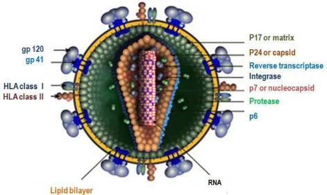
The HIV mature virion is roughly spherical with a diameter of approximately 120 nm (Figure 1.2). The
envelope is a lipid bilayer that is derived from the membrane of the host cell and that contains host
proteins such as major histocompatibility antigens, actin and ubiquitin (yellow). At the viral surface
spikes are formed by glycoproteins gp120 that interact with transmembrane proteins gp41 (blue). The
inner surface of the envelope is lined with about 2,000 copies of matrix protein p17 (dark green). The
5 |
Chapter 1: General Introduction
contains the essential enzymes protease (PR), reverse transcriptase (RT), integrase (IN) and two
identical copies of unspliced positive single stranded ribonucleic acid (RNA) molecules that are bound
to the nucleocapsid proteins p7 (red with white) [13].
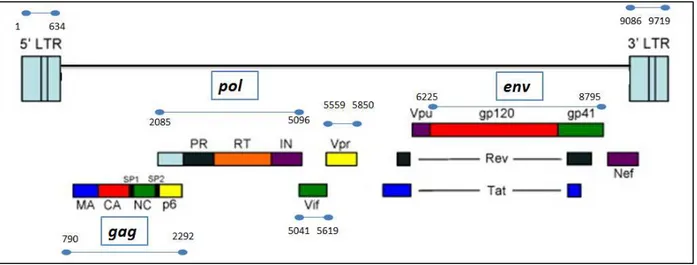
The length of the HIV proviral genome is around 9,700 nucleotides. Multiple reading frames lead to
nine partly overlapping genes. Three genes encode the structural and enzymatic proteins commonly
found in retroviruses. The group specific antigen (gag) gene encodes the precursor p55 that is
subsequently cleaved by PR, into the structural proteins matrix (MA or p17), capsid (CA or p24),
nucleocapsid (NC or p7) and p6. The polymerase (pol) gene encodes PR (p11), RT (p66/p51), and IN
(p31). The env gene encodes surface gp120 and transmembrane gp41 glycoproteins [12].
FIGURE 1. 2. MOFPHOLOGY OF HIV
Figure 1.2: Morphology of HIV. Adapted from http://www.hiv.lanl.gov
Two other genes are essential for virus propagation and replication, for instance tat (transactivator of
HIV gene expression) and rev (regulatory factor). Four accessory or auxiliary genes are not essential in
vitro but are necessary for spread and disease progression in vivo: vif (viral infectivity factor), vpr (viral
protein R in HIV-1 and its homolog vpx in HIV-2), vpu (viral protein U), and nef (before named negative
6 |
FIGURE 1. 3. GENOMIC STRUCTURE OF HIV
Figure 1.3: Genomic structure of HIV. The relative locations of the open reading frames
gag, pol, env, vif, vpr, vpu, nef, tat and rev are shown. The blue lines indicate the positions
according to HXB2 strain. Adapted from [14].
1.3.2 REPLICATION CYCLE
The entry of HIV requires the presence of the cluster of differentiation 4 (CD4), present on
T-lymphocytes, macrophages, dendritic cells and brain microglia. The replication cycle includes ten
important steps that are divided in two phases (Figure 1.4). The early phase includes binding to
integration, whereas the late phase begins with transcription and ends with viral maturation [13].
EARLY PHASE
The interaction between gp120 and the amino-terminal domain of CD4 causes the binding between
virus and target cell. Afterwards, conformational changes allow the binding between gp120 and the
chemokine receptors CXC-chemokine receptor 4 (CXCR4) and the CC-chemokine receptor 5 (CCR5).
The CCR5 co-receptor is generally used in early stages of the infection and CXCR4 in the late stages.
Based on the use of these co-receptors, the tropism of viruses could be classified as X4, R5, R5+X4
and R5X4, which is important for therapeutic options. The interaction between
CD4-gp120-CCR5/CXCR4 triggers conformational changes within gp41, which promotes the physical approach
between viral and cellular membranes and fusion [12-14].
Entry of the HIV core into the cytoplasm causes the disintegration of the capsid with the aim to release
the viral proteins. This process is known as uncoating. RT catalyzes the reverse transcription of two
7 |
Chapter 1: General Introduction
DNA is imported within the nucleus as part of the preintegration complex that additionally includes
viral proteins such as MA, IN, RT and Vpr [13].
Subsequently, the IN catalyzes the integration of proviral DNA into the host cell genome. This provirus
serves as template for the synthesis of RNA that encodes the structural, enzymatic, regulatory and
accessory proteins. The provirus is the basis for viral latency and reservoirs. Latency occurs when the
CD4 cell turns into a resting status and limited transcription occurs. Activation of the resting cell results
into complete transcription and a productive infection [15]. Viral reservoirs are a small pool of cells
within lymphoid tissue and the brain, that provides a long-lived source of rebound viraemia [16].
LATE PHASE
Transcription is initiated at the LTR site, in which Tat is an essential transcriptional activator to
increase viral RNA synthesis. This process generates three types of RNA: (i) unspliced or genomic
RNA that is the messenger RNA (mRNA) for Gag and Gag-Pol precursors; (ii) singly spliced mRNAs
that encodes Env precursor, Vpu, Vif and Vpr; and (iii) multiply spliced mRNAs that are translated into
Tat, Nef and Rev [12, 17]. With the aim to translate all types of mRNA, the unspliced and singly spliced
mRNAs need to be transported to the cytoplasm by Rev. Indeed, Rev acts by binding the rev
responsive element, promoting the nuclear export and cycling between nucleus and cytoplasm of
unspliced and singly spliced mRNA [12, 18].
Following the translation of the proteins, the assembly involves the Gag precursor polyprotein pr55,
which binds to the plasma membrane and promotes the interaction of gag-gag proteins, encapsidation
of RNA and the incorporation of Env proteins in the host membrane. Env precursor is translated in the
rough endoplasmic reticulum and cleaved by host protease in the Golgi apparatus [17]. The budding of
the immature virus includes interactions between host factors, and the p6 domain within Gag to hijack
the endosomal machinery and release it. Viral maturation requires the cleavage of Gag and Pol
polyproteins by PR in order to re-assemble the virion. A single cell can produce thousands of virus
particles until the apoptosis of the host cell. Meanwhile new virus particles could start another
8 |
FIGURE 1. 4. REPLICATION CYCLE
Figure 1.4: Replication cycle. Adapted from [19].
1.4
MOLECULAR EPIDEMIOLOGY
The evolution of HIV is the consequence of several viral intrinsic mechanisms: the lack of proofreading
activity of RT, the high mutation rate of 3.4 x 10⁻⁵ mutations per base pair per replication cycle [20, 21],
the high viral production of 1010 virions per day [22, 23], the process of recombination with 7 to 30
crossovers per genome per round of replication, the flexible conformation of HIV proteins, the short
generation time between 24-48 hours [24] and Apolipoprotein B mRNA editing enzyme, catalytic
polypeptide-like (APOBEC) editing [25].
1.4.1 PHYLOGENETIC ANALYSES ON THE SURVEILLANCE OF HIV
Phylogenetics describes evolution and relationships among genes or genes fragments, by inferring their
common ancestor. Therefore, it is necessary for phylogenetic analyses that sequences under
investigation are from the same gene and without evidence of recombination [26]. Phylogenetic tools
have been used in molecular epidemiology to investigate the origin of HIV and to classify subtypes [27]
9 |
Chapter 1: General Introduction
Phylogenetic analyses include the pre-processing of data from datasets or databases, such as the
retrieval of particular fragments of a gene and an alignment to put homologous residues arranged in
columns with the assistance of software packages such as ClustalW and Muscle [26, 28, 29]. The
reconstruction of a tree could be performed by two different algorithm methods: optimality search
criterion and clustering. Each one can use different kind of data such as discrete characters
(morphological, physiological characteristics, nucleotides or amino acids) or a distance matrix of
pairwise genetic dissimilarities [30]. Usually pairwise distance methods are complemented with
evolutionary models that provide a statistical description of the substitution of nucleotides or amino
acids in the sequence [26]. The most common used methods in phylogenetics of HIV are the distance
pairwise method that uses clustering called Neighbor-joining (NJ) and the character state algorithm that
use optimality criterion called Maximum likelihood (ML). Nonparametric bootstrap is used to determine
the phylogenetic branch support in both methods [31]. However, the approximate likelihood ratio test
(aLRT) is another fast alternative instead of nonparametric bootstrap when ML method is used [32].
The choice of the method depends on the kind of analyses, for instance NJ method works well for
subtyping HIV sequences [33] (see section 1.4.4), whereas ML method is used in cluster transmission
analyses because the exhaustive searching of different tree topologies (see section 1.7) [26, 34-39].
Whereas NJ or ML methods construct one tree, Bayesian methods integrate all plausible trees and
provide confidence intervals (or Bayesian credible intervals) for any evolutionary relationship [26]. This
is summarized in the maximum clade credibility (MCC) tree that also shows different traits, for instance
the posterior distribution or probability. These techniques have several applications, for instance cluster
transmission analyses (see section 1.7) [40-43], time-scaled phylogenies [44-48], and lately,
phylogeographic diffusion models [49-51]. Phylogeography is based on the measurable imprint on the
genome of rapidly evolving viruses that occurs simultaneously with the geographical dispersal over a
specific period [52, 53]. Therefore it can provide insights about the viral introduction and dissemination
in a region or the impact of human mobility on the spread of the HIV epidemic [51, 52].
1.4.2 ORIGIN OF HIV
Based upon clinical records, colonial literature and phylogenetic analyses of primate and human
lentiviruses, the origin of HIV has been related with bushmeat hunting and keeping primates as pets
[54-56]. HIV-1 group M (major) and group N (nonoutlier) originated from Simian Immunodeficiency
Virus (SIV) (Figure 1.5), which was isolated from a subspecies of chimpanzee in West Africa, more
specifically from Pan troglodytes troglodytes [57]. The origin of HIV-1 group O (outlier) and P (putative)
10 |
FIGURE 1. 5. CLASSIFICATION OF HIV
Figure 1.5: Classification of HIV. HIV is classified in types 1 (green) and 2 (blue). HIV-1 is divided in
groups M, N, O and P. The group M includes nine subtypes, subtype A and F are divided in
sub-subtypes e.g. A1 or F1. Some examples of circulating recombinant forms (CRFs) are shown in violet.
HIV-2 is classified in eight groups (A-H), but only the most frequent groups A and B are shown [27, 58].
Tree adapted from [60].
The origin of HIV-1 group M was dated between 1900s and 1930s (Figure 1.6) [59, 61-64]. Two recent
articles support its origin in the early 1900s. One included the phylogenetic analysis including the two
oldest samples obtained from patients in Kinshasa, Democratic Republic of Congo [64]. According to a
study including phylogenetic analysis, computational simulations and review of colonial literature [59],
the main driver of the spread of HIV in the early 20th century was linked to sexual transmitted diseases,
especially ulcerative diseases, in the heterosexual risk transmission group, whereas the increase of city
population size and the low frequency of circumcision at that time had little effect in the initial viral
11 |
Chapter 1: General Introduction
HIV-2 originated from interspecies transmission between Sooty mangabey monkeys (Cercocebus atys
atys) and humans. Each HIV-2 group (A-H) originated from independent cross-species transmission
events from this monkey species infected with SIV [65]. HIV-2 group A originated around 1930s and
group B around 1935s (Figure 1.6) [51, 63, 66, 67].
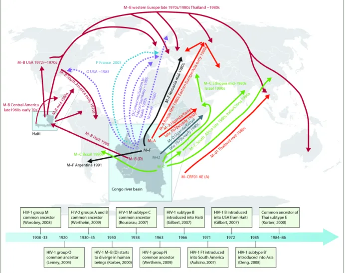
FIGURE 1. 6. ESTIMATED TIME LINE OF THE GLOBAL SPREAD OF HIV
Figure 1.6: Estimated time line of the global spread of HIV. Adapted from [60]. Additional subtype B
12 |
1.4.3 MOLECULAR EPIDEMIOLOGY
Group M is the predominant circulating HIV-1 group, that is classified in subtypes A-D, F-H, J and K
(Figure 1.5) [27]. The circulating recombinant forms (CRFs) are recombinants for which the full length
sequences are fully characterized and specific breakpoints can be found in at least three people
epidemiologically unlinked [27]. These are denominated according to the subtypes that compose the
recombinant, and numbered according to the order of description (Figure 1.5). Up to date, there are 61
CRFs described in HIV-1 and one in HIV-2 (http://www.hiv.lanl.gov, accessed in February 2014).
Additionally, inter-subtype recombination is common resulting into unique recombinant forms (URFs)
within single patients [23].
The rare HIV-1 groups N, O and P have been identified mainly in Cameroon [58, 71, 72]. HIV-2 groups
A and B have spread in West Africa causing 1-2 million of cases, whereas the other HIV-2 groups (C-H)
have been rarely isolated [10, 73, 74].
To estimate HIV diversity large amounts of published and unpublished data were summarized and
weighted according to the number of people living with HIV in each country by the WHO network of HIV
Isolation and Characterization [75]. Subtype C was responsible for about 50% of HIV-1 infections
worldwide between 2000 and 2007 (Figure 1.7). It was dominant in Southern and East Africa, India and
Oceania. Subtype B was the most frequent subtype in Western and Central Europe and America.
Regarding the molecular epidemiology of Belgium and Colombia, subtype B is predominant in both
countries [76-79]. However, the percentage of subtype B is around 50% according to the last
nationwide survey carried out in Belgium until 2006 [78]. In contrast, subtype B accounts for around
99% of the HIV infections in Colombia until 2002 [76, 77, 79]. The epidemiology of these two countries
will be further discussed in chapter 6.
Several studies have reported on the origin of subtypes around the world [45, 61, 80, 81]. The most
studied and prevalent subtype in America and Europe is B. The subtype B epidemic started to spread
from Central Africa to Haiti around 1966 (1962-1970), before dispersing in a single migration to the USA
around 1969 (1966-1972) and to Trinidad and Tobago around 1973 (1970-1976). Phylogenetic analysis
showed that single chance events, ecological interactions and specific population bottlenecks and
founder effects influenced the spread of subtype B in the Americas [48]. A recent study also suggested
that the epidemic in South America was influenced by migration and multiple introductions from
13 |
Chapter 1: General Introduction
North American men who have sex with men (MSM) and intravenous drug users (IVDU) [82-84]. Most
of the migratory pathways of subtype B in Europe are bidirectional. However, some countries are
sources and others are sinks. For instance, tourism could partially explain the viral dispersal from
Greece, Portugal, Serbia and Spain to Central Europe. Similarly, Austria, Belgium and Luxembourg
have an imported epidemic due to the highest immigration from other European countries [83].
Concerning the origin of subtype B in Colombia and migration pathways, there is not information
available (see chapter3). On the other hand, multiple introductions from Africa and Asia were involved
in the spread of non-B subtypes in Europe [60], and links with former colonies in Central-Africa
influenced from the start the relative high prevalence of non-B subtypes in Belgium, Portugal and
France [85].
FIGURE 1. 7. EPIDEMIOLOGY OF SUBTYPES WORLDWIDE
Figure 1.7: Epidemiology of subtypes worldwide. Subtype C (blue), subtype B (orange) and
subtype A (red) are the three most prevalent subtypes worldwide. The prevalence of subtypes varies
with geographical localization. Adapted from [74]. Data for Colombia and Belgium were adapted from
14 |
The currently most prevalent subtype C originated around 1958 (1949-1960) [80]. A rapid spread in
South Africa followed by the introductions to India and China could explain the high prevalence of this
subtype (Figures 1.6 and 1.7) [60]. In the 1990s, it was also introduced to Brazil directly via East Africa
[86], or via an intermediate step in UK [45]. Other multiple introductions have been reported for Italy,
mainly from South America and India [87]. Although other few non-B subtypes have been studied [46,
47, 66, 88, 89], the increasing availability of more data from these subtypes is expected to unravel the
routes of the HIV-1 spread.
1.4.4 METHODS OF SUBTYPING HIV
Although serological and genotypic methods have been used to identify HIV strains, the gold standard
to classify subtypes is based upon the manual phylogenetic analysis of full-length viral sequences.
Subtypes are phylogenetically equidistant, generating a starlike tree (Figure 1.5). The genetic variation
between subtypes is usually 25-35% depending on the genes compared, whereas within a subtype it
can be 15-20% [23]. Some subtypes have sub-subtypes with a smaller genetic distance (e.g. A1-A4
and F1-F2). The classification of HIV is based on phylogenetic analyses, in which the clustering pattern
of viral sequences provides information about genetic similarity and evolutionary rate [27]. This
classification is dynamic because it could change with the availability of new sequences. However, it is
expensive to sequence the full-length viral genome and the analysis is time consuming. Since most
available sequences are obtained within the framework of drug resistance testing, automatic tools have
been developed with the aim to identify subtypes based upon PR and RT fragments [90-92]. In
addition, the automated subtyping tools enable the quick classification of one to hundreds of
sequences.
According to the methodology used to assign an HIV-1 clade to a query sequence, automatic tools can
be divided into three main types. First, similarity-based tools use an alignment between the reference
and the query sequence and calculate the match. Examples include the NCBI subtyping tool [93],
Stanford [94], Geno2pheno [95] and EuResist (http://engine.euresist.org/data_analysis/
viral_sequence/new). Second, statistical-based tools use prediction by partial matching compression
algorithm like COntext-based Modeling for Expeditious Typing COMET [96], position-specific scoring
matrices plus a statistical model such as STAR [97] or jumping profile Hidden Markov Models such as
jpHMM [98]. Third, phylogeny-based tools like REGA [99] and SCUEAL [100]. Currently, it is not clear
15 |
Chapter 1: General Introduction
1.5
STAGES OF HIV-1 INFECTION
HIV is present in blood, genital fluids and breast milk. Transmission events are mainly the consequence
of exposure to HIV at mucosal surfaces (80%) and the remaining transmissions are caused by
percutaneous or intravenous inoculation. The per-act HIV transmission depends on the type of
exposure. For instance, it is around 92.5% for blood transfusion, 22.5% for mother to child transmission
(MTCT), 0.63% for IVDU, and 0.23% for percutaneous needle stick injury. Sexual transmission risk
depends on the type of intercourse. Indeed, the per-act HIV transmission risk for receptive anal
intercourse is 1.38%, for insertive anal intercourse 0.11%, for receptive penile-vaginal intercourse
0.08%, and for insertive penile-vaginal intercourse 0.04% [101]. The sexual transmission risk increases
with genital ulcer diseases and high viral load, and during acute or late state of the disease, and
decreases with circumcision, male condom use, and use of antiretroviral therapy (ART) [101].
Genetic diversity may affect the transmissibility [102, 103]. Subtype C seems to be more transmissible
than subtypes A and D according to studies performed in pregnant women [104, 105] but not in
heterosexual couples [106]. In Kenya, the MTCT rate was higher among women infected with subtype
D than with subtype A [107], whereas in Tanzania subtype A was more likely to be transmitted than
subtype D [104]. A study performed in discordant couples revealed a higher rate of heterosexual
transmission of subtype A than subtype D [108] which seems to be in agreement with the higher
number of infections of subtype A worldwide (Figure 1.7). In another study, CRF01_AE seemed also to
be more transmissible than subtype B in IVDU [109].
After exposure, HIV initially replicates in mucosal, submucosal and lymphoreticular tissues and
therefore it is not detected in plasma yet. The clinical manifestations of acute infection could be
asymptomatic, similar to flu-like syndrome with pharyngitis, non-tender lymphadenopathies or
mucocutaneous ulcerations by Candida spp, Epstein-Barr virus or Herpes simplex virus (acute retroviral
syndrome) [110]. Other less frequent signs or symptoms can be presented such as generalized
maculopapular rash, hematologic disturbances like anemia or thrombocytopenia, neurological disorders
like aseptic meningitis and reactivation of Varicella-zoster virus [111].
The phases of HIV-1 infection can be categorized based on the sequential positivity of diagnostic
assays (Fiebig stages I-VI, Figure 1.8A). The period between infection and the detection of viral RNA is
named eclipse, and can last 7 to 21 days. At the end of the eclipse phase, HIV reaches the
16 |
a decrease of CD4 T cells is observed [112]. In the initial phase of acute infection, the algorithm for the
diagnosis of HIV-1 infection includes detection of viral RNA, p24 antigen or fourth generation combined
HIV antigen/antibody immunoassay [113, 114]. The viral burden is high during acute infection.
Therefore, transmission of HIV is mainly driven by recently infected individuals, who account for 5 to
50% of the transmissions [34, 102, 103, 115-117]. The seroconversion occurs in Fiebig stage III when
viral-specific antibodies are detected in serum [118]. The viral load gradually decreases over 12-20
weeks and reaches a viral set point [119, 120]. Meanwhile the pressure of adaptive immune responses
selects mutants and virus diversification occurs.
The Fiebig scale depends on the used test methods [121]. Ideally, these methods should be able to
detect all subtypes, although some assays have a lower performance [122]. False-negative results
have been reported in fourth-generation immunoassays in subtypes A, C, F, H, CRF01_AE and O.
Similarly, false negatives for subtypes B, C and F have been described for gp41 immunoassays [123,
124]. These results are the consequence of differences in viral epitopes [123, 125]. Rapid HIV tests
have also shown low sensitivity for D, F, H, CRF01_AG, O and HIV-2 [122, 125-128]. In general, viral
load assays perform well for B and non B subtypes, but the assays based on integrase seem to perform
better on viral strains belonging to M, O and N than assays using primers and probes in the gag gene
[122, 129] .
The early chronic infection phase is frequently asymptomatic but some individuals do display clinical
manifestations like persistent generalized lymphadenopathy. The duration of the clinically latent phase
is variable but it could last between 1 and 10 years (Figure 1.8). In the meantime the CD4 count
declines on average with 50 cells /mm3 per year, which is influenced by the viral burden [130]. The final
AIDS stage is reached when there are less than 200 CD4 cells /mm3 or opportunistic infections and
malignancies occur [111, 131]. When the CD4 count reaches 50 cells/mm3 and ART is not provided,
the median survival is 12 to 18 months [132].
Some patients are the exception of the described profile in clinical progression and are named
long-term nonprogressors. They can be infected for more than 10 years without any clinical symptom and
CD4 count above 500 cells/mm3 without receiving ART [133]. Similarly, the “elite controllers” are
characterized with a spontaneous virological control defined as viraemias below 50 copies/mL [134].
The genetic background and differences in immunological response, such as cytotoxic T cell response
17 |
Chapter 1: General Introduction
Despite 30 years of research, the impact of subtypes on disease progression is still a matter of debate
due to conflicting data and the presence of several confounding factors [107, 135]. Several reports from
Uganda, Tanzania, Kenya and London have shown a faster progression in people infected with subtype
D than with other subtypes [136-138]. The dual or X4 tropism of subtype D, the more frequent formation
of syncytium and the higher replication capacity of the pol region are factors that could explain the
faster decrease of CD4, compared with other subtypes [137, 139, 140]. A cohort study performed in
London could not detect differences in disease progression between subtypes A, B, C and CRF02_AG
[141]. However, a systematic review showed that subtype C is also aggressive, followed by G,
CRF01_AE, and CRF02_AG mainly in developing countries [135]. Recent reports on larger study
populations also showed the rapid disease progression associated with subtype C, which may be
related to the increased replication capacity [142, 143]. Similarly, CRF01_AE is associated with X4
tropism and consequently low CD4 count in MSM [144, 145]. The viral load has also been compared
between different cohorts. People infected with CRF01_AE had threefold higher viral load after three
months of seroconversion when compared to subtype B, but no difference was found after one year
[146]. Two reports showed higher viral loads in subtype D infections [137, 139] but other two did not
[141, 147], which raise the question whether other factors such as comorbidities, co-infections,
constitution and socio-economic conditions could impact the different response and disease
progression [135, 147]. A recent systematic review pointed to the heterogeneity of the outcome
measures and suggested to use subtype A as comparator and the inclusion of some typical outcome
measurements such as relative risk, odds ratio (OR) or hazard ratio [135]. The standardization of future
studies might enable the pooling of different datasets and provide valuable information for clinicians in
18 |
FIGURE 1. 8. PATHOGENESIS OF HIV-1
Figure 1.8: Pathogenesis of HIV-1. A) The Fiebig stages are represented by roman numbers and are
classified according to a positive result for viral RNA measured by PCR (I), for p24 antigen (II) and for
antibodies immunoglobulin M (IgM) measured by enzyme linked immunosorbent assay (ELISA) but not
by western blot (III), for antibodies detected by ELISA and by western blot (still indeterminate) (IV), for
antibodies detected by ELISA and by western blot (positive, but still p31 negative) (V), for antibodies
detected by ELISA and by western blot (positive including for p31) (VI). B) The relationship between
viral load (red line) and CD4+T cell (blue line) over time in an untreated patient. R5 virus is usually
found during the early stages of the infection, whereas X4 viruses in the chronic stage. Abbreviation: