RNA Interference in Biology and Medicine
OLLIVIER MILHAVET, DEVIN S. GARY, AND MARK P. MATTSON
Laboratory of Neurosciences (O.M., M.P.M.), National Institute on Aging, Gerontology Research Center, Baltimore, Maryland; and Departments of Neurology (D.S.G.) and Neuroscience (M.P.M.), Johns Hopkins University School of Medicine, Baltimore, Maryland
Abstract. . . 629
I. Introduction . . . 630
II. Principles of RNA interference . . . 630
A. Post-transcriptional gene silencing and the discovery of RNA interference . . . 630
B. Mechanisms of RNA interference . . . 632
C. Other related phenomena . . . 634
III. Technical considerations in the use of RNA interference . . . 634
A. Design and synthesis of small interfering RNAs . . . 634
B. Construction of plasmids and viral vectors for RNA interference . . . 637
C. Transfection methods . . . 638
IV. Applications of RNA interference to establishing gene function. . . 640
A. Signal transduction . . . 640
B. Cell cycle regulation . . . 640
C. Development . . . 641
D. Macromolecular synthesis and degradation. . . 641
E. Cell motility . . . 641
F. Cell death . . . 642
G. Viral invasion/replication . . . 642
V. Therapeutic applications of RNA interference . . . 643
A. Cancer. . . 643
B. Infectious diseases . . . 643
C. Cardiovascular and cerebrovascular diseases . . . 644
D. Neurodegenerative disorders . . . 644
VI. The future of RNA interference in biology and medicine. . . 645
References. . . 646
Abstract——First discovered in plants the nematode
Caenorhabditis elegans, the production of small inter-fering RNAs (siRNAs) that bind to and induce the deg-radation of specific endogenous mRNAs is now recog-nized as a mechanism that is widely employed by eukaryotic cells to inhibit protein production at a post-transcriptional level. The endogenous siRNAs are typi-cally 19- to 23-base double-stranded RNA oligonucleo-tides, produced from much larger RNAs that upon binding to target mRNAs recruit RNases to a protein complexthat degrades the targeted mRNA. Methods for expressing siRNAs in cells in culture and in vivo using
viral vectors, and for transfecting cells with synthetic siRNAs, have been developed and are being used to es-tablish the functions of specific proteins in various cell types and organisms. RNA interference methods pro-vide several major advantages over prior methods (an-tisense DNA or antibody-based techniques) for suppress-ing gene expression. Recent preclinical studies suggest that RNA interference technology holds promise for the treatment of various diseases. Pharmacologists have long dreamed of the ability to selectively antagonize or eliminate the function of individual proteins—RNAi technology may eventually make that dream a reality.
Address correspondence to: Mark P. Mattson, Laboratory of Neurosciences, National Institute on Aging, Gerontology Research Center, 5600 Nathan Shock Drive, Baltimore, MD 21224. E-mail: [email protected]
Article, publication date, and citation information can be found at http://pharmrev.aspetjournals.org. DOI: 10.1124/pr.55.4.1.
PHARMACOLOGICALREVIEWS Vol. 55, No. 4
Copyright © 2003 by The American Society for Pharmacology and Experimental Therapeutics 30403/1106516
Pharmacol Rev55:629–648, 2003 Printed in U.S.A
629
by guest on February 25, 2015
pharmrev.aspetjournals.org
I. Introduction
RNA interference (RNAi1), as commonly defined, is a
phenomenon leading to post-transcriptional gene silenc-ing (PTGS) after endogenous production or artificial in-troduction into a cell of small interfering double strand RNA (siRNA) with sequences complementary to the tar-geted gene (Bosher and Labouesse, 2000). Whereas the transcription of the gene is normal, the translation of the protein is prevented by selective degradation of its encoded mRNA. However, PTGS is not restricted to RNAi and has emerged as a more complex mechanism that involves several different proteins and small RNAs. It is presumed that cells employ RNAi to tightly regulate protein levels in response to various environmental stimuli, although the extent to which this mechanism is employed by specific cell types remains to be discovered. However, the fact that RNAi is operative in cells of organisms ranging from plants, to nematodes and flies, and to mammals attests to its fundamental importance in the selective suppression of protein translation by targeted degradation of the encoding mRNA. Beyond its biological relevance, PTGS is emerging as a powerful tool to study the function of individual proteins or sets of proteins. User-friendly technologies for introducing siRNA into cells, in culture or in vivo, to achieve a selective reduction of single or multiple proteins of in-terest are rapidly evolving. The present article reviews this emerging technology, findings obtained to date us-ing such RNAi methods, and the potential of RNAi-based therapeutics for treating human disease.
II. Principles of RNA Interference
RNA interference most likely evolved as a mechanism for cells to eliminate unwanted foreign genes. Foreign genes are often present in cells at high copy numbers, being present as viral genes, transposable elements, or as plasmids introduced experimentally in cell transfec-tion protocols. It has been known for several decades that the level of expression of transgenes usually de-creases as the number of copies present in the cell in-creases and that endogenous homologous genes can also be suppressed by the presence of the transgene (Napoli et al., 1990). Although such gene silencing can occur at the transcriptional level, it is now recognized that a major mechanism of gene suppression occurs post-tran-scriptionally, and that a major mechanism for this PTGS
is RNAi, the selective degradation of mRNAs targeted by siRNAs (Van Blokland et al., 1994). Such PTGS via RNAi can occur very rapidly with proteins for many genes, being decreased within hours, and completely absent within 24 h (Pruss et al., 1997). Based upon these and other findings initially made in studies of plants (Ratcliff et al., 1997), it seems very likely that RNAi evolved as a mechanism to defend plant cells against viral infections.
A. Post-Transcriptional Gene Silencing and the Discovery of RNA Interference
PTGS and RNAi were discovered in genetic transfor-mation studies of eukaryotic cells, principally plants and worms, wherein it was shown that mRNAs for the en-coded transgene alone, or together with mRNAs for ho-mologous endogenous genes are very low or absent de-spite high levels of transcription (Fire, 1999; Marathe et al., 2000). The ability to manipulate and monitor gene expression in the plant Arabidopsis thaliana and the roundworm Caenorhabditis elegans (the genomes of both species are now complete) revealed the process of RNAi and allowed the relatively rapid identification of several genes that regulate the RNAi process.
Transgenes insert into the genomes of plants by re-combination in an apparently random manner so that the number of inserted copies, their chromosomal loca-tion, and their local arrangement within the chromo-some vary among transformants. The observation of an inverse correlation between copy number and the level of gene expression suggested that an increased copy number of a particular gene results in silencing of that gene (Assaad et al., 1993). It was initially thought that such gene silencing was due to reduced gene transcrip-tion resulting from interactranscrip-tions between closely linked copies that result in the formation of secondary struc-tures that promote methylation and inhibition of tran-scription (Ye and Signer, 1996). Further studies showed that transcriptional gene silencing (TGS) could also oc-cur in trans, such that one transgene can be silenced by another transgene introduced either by crossing or transformation. It was then proposed that a silencing RNA is produced by one locus that somehow effects the silencing of the other gene by a mechanism involving RNA-mediated inhibition of transcription (Mette et al., 2000). Although some data were consistent with such mechanisms of transcriptional silencing, additional data suggested the involvement of PTGS. The presence of double-stranded RNAs (dsRNAs) and their cleavage into siRNAs of approximately 23 nucleotides was demon-strated, and it was then shown that expression of dsRNA with sequences corresponding to open reading frames in plants results in PTGS (Hamilton and Baul-combe, 1999). Similarly, expression of dsRNAs with se-quences complementary to those of endogenous genes 1Abbreviations: RNAi, RNA interference; AIF, apotosis-inducing
fac-tor; dsRNA, double-stranded RNA; IAP, inhibitor of apoptosis protein; PTGS, post-transcriptional gene silencing; RISC, RNA-induced silenc-ing complex; shRNA, short hairpin RNA; siRNA, small interfersilenc-ing RNA; IP3, inositol 1,4,5-trisphosphate; NMD, nonsense-mediated
results in the selective silencing of those genes in C. elegans(Zamore et al., 2000). Collectively, the studies of A. thaliana and C. elegans showed that both TGS and PTGS can be initiated by the same RNA degradation pathway—TGS occurs when the dsRNA includes pro-moter sequences, whereas PTGS occurs when the dsRNA includes coding sequences. Although degrada-tion of dsRNA is common to both mechanisms of gene silencing, the results also indicated that dsRNA-medi-ated TGS and PTGS involve different specific steps.
Although RNAi as a mechanism of PTGS was first discovered in plants and may have evolved as a cellular defense mechanism against foreign DNA and RNA, it is very clear that RNAi is widely employed in most if not all eukaryotic cells as a mechanism to regulate the ex-pression of endogenous genes. In 1998, it was discovered that injection of dsRNA was much more effective for silencing of gene expression inC. elegansthan was sin-gle-stranded antisense RNA (Fire et al., 1998). This experimentally induced PTGS, the first report of the use of RNAi as a tool in biology, was very potent, and re-markably, the PTGS occurred not only in the worms to which the dsRNA was administered, but also in their progeny. It was then demonstrated that the endogenous mRNA was the target of the injected dsRNA by a post-transcriptional mechanism and involving degradation of the targeted mRNA (Montgomery et al., 1998). Surpris-ingly, it was further shown that the dsRNA is effective at very low concentrations, such that the copy numbers of the targeted mRNA are far greater than the number of dsRNAs present in the cell (Fire et al., 1998; Kenner-dell and Carthew, 1998). In addition, the suppression of the protein encoded by the targeted mRNA was found to persist through many rounds of cell division. The latter two observations strongly suggested that cells possess a mechanism for amplifying the RNAi mechanism. Not only can the RNAi process be maintained within cells of a common lineage, but it can also be transferred between cells, as shown inC. eleganswhere injection of dsRNA into the intestine results in silencing of the targeted gene in all cells of the F1 progeny of that worm (Fire et al., 1998). Indeed, dsRNA can enter cells and induce PTGS when worms are soaked in a solution containing the dsRNA or when the worms are fed bacteria express-ing dsRNA (Tabara et al., 1998; Timmons and Fire, 1998). Recently, a transmembrane protein called SID-1 was identified as a possible mediator of intercellular transfer of RNAi (Winston et al., 2002).
Subsequently, other organisms were assayed for their capacity to induce RNAi. Evidence for RNAi in Drosoph-ila was first demonstrated by Kennerdell and coworkers (Kennerdell and Carthew, 1998) who showed the in-volvement of thefrizzledandfrizzled2genes in the wing-less pathway after introduction of dsRNA into embryos. Again, several techniques were developed in order to use dsRNA in this organism leading to the establishment of cell-free (Tuschl et al., 1999) and cell culture models
(Caplen et al., 2000). A system that employed dsRNA as an extended hairpin-loop RNA was developed to induce heritable gene silencing (Kennerdell and Carthew, 2000). TheDrosophila system has allowed the identifi-cation of several endogenous genes that play key roles in the RNAi process. An RNA nuclease activity called RISC (RNA-induced silencing complex) was discovered that is responsible for the degradation of endogenous mRNAs, as well as small nucleotide fragments (⬃25 nucleotides in length), which could be used as guides by RISC (Ham-mond et al., 2000). They later characterized RISC as a ribonucleoproteic complex (Hammond et al., 2001). These results were soon extended by showing that RNAi is an ATP-dependent and translation-independent event where the introduced dsRNA is processed into 21–23 nucleotide fragments that guide the cleavage of endog-enous transcripts (Zamore et al., 2000). The enzyme responsible for the processing of the dsRNA was later discovered as a RNase III family nuclease named Dicer, a protein with high homology to the rde-1 C. elegans gene (Bernstein et al., 2001). To study the functions of RNAi in yeast, Volpe et al. (2002) deleted argonaute, dicer, and RNA-dependent RNA polymerase homologs; deletion resulted in the accumulation of complementary transcripts from centromeric heterochromatic repeats and de-repression of transgenes integrated at the cen-tromere and impairment of cencen-tromere function. The authors of the latter study proposed that dsRNA arising from centromeric repeats targets the formation and maintenance of heterochromatin through RNAi.
RNAi mechanisms may provide explanations for var-ious biological phenomena that were prevvar-iously de-scribed, but without any understanding of the possible underlying mechanism. For example, it was recently proposed that RNAi mechanisms could explain the con-troversial process of RNA-mediated memory transfer in planaria (Smalheiser et al., 2001). It will certainly be of interest to investigate possible roles for RNAi in the many different physiological processes that involve mod-ulation of protein levels at a post-transcriptional level.
B. Mechanism of RNA Interference
A clearer picture of PTGS emerged from several dif-ferent basic observations, including the necessity of transcriptionally active genes and the ability of RNA viruses to silence a homologous endogenous gene (En-glish et al., 1997). Within the last 3 years, a flurry of studies have identified several of the molecules that mediate RNAi, and the mechanism whereby these mol-ecules effect the selective degradation of targeted mR-NAs. It is now clear that the production of dsRNA with sequence complementary to the mRNA being targeted is fundamental to the process of PTGS; single-stranded RNA is not sufficient to induce PTGS. The importance of dsRNA is supported by a wealth of data. Transgenes engineered to synthesize dsRNA require only a few cop-ies of the dsRNA to achieve PTGS and can induce
cosup-pression. There are several ways such transgenes pro-duce dsRNA including the synthesis of long hairpin mRNAs by transcription of an inverted repeat (Kenner-dell and Carthew, 2000; Tavernarakis et al., 2000), and transcription of complementary sense and antisense strands by opposing promoters (Wang et al., 2000). Other studies have shown that although cells may ini-tially produce very long dsRNAs, they are cleaved into smaller dsRNAs, 21–25 nucleotides in length, that actu-ally mediate RNAi (Hamilton and Baulcombe, 1999).
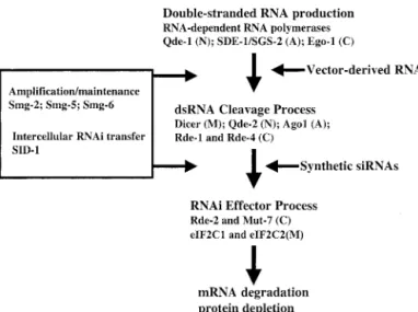
How does a small dsRNA with a sequence complemen-tary to a specific mRNA effect PTGS? The proteins that mediate the RNAi process have been identified using several approaches, most notably genetic screens for mutants resistant to RNAi inC. elegansand resistant to PTGS in Neurospora and Arabidopsis (Fig. 2). These studies have identified homologous genes in each spe-cies, and subsequent identification of mammalian ho-mologs, that encode proteins required for RNAi. Three homologous genes identified in the initial screens are rde-1 inC. elegans(Tabara et al., 1999),qde-2in Neu-rospora (Cogoni et al., 1996) and ago-1 in Arabidopsis (Dalmay et al., 2000; Fagard et al., 2000). The proteins encoded by each of these genes share homologies with the eIF2C translation factors. In C. elegans, rde-1 is required for germline transmission of RNAi, but not for transmission of RNAi among cells of the worm (Grishok
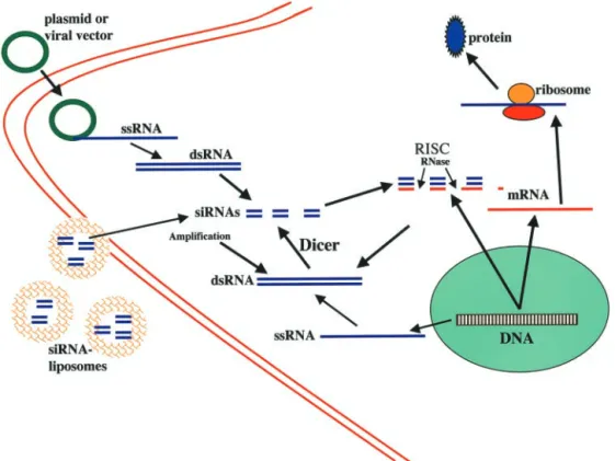
FIG. 1. Pathways of RNAi. Cells produce single-stranded RNA (ssRNA), which provide a template for the formation of dsRNA, which involves the
et al., 2000) suggesting that it plays a role in the pro-duction of the RNAi signal. Another set of homologous genes involved in RNAi, identified in the screens for PTGS/RNAi mutants, include ego-1 in C. elegans (Tabara et al., 1999; Smardon et al., 2000), qde-1 in Neurospora(Cogoni and Macino, 1999) andsgs2/sde1in Arabidopsis(Mourrain et al., 2000). The latter proteins appear to be required for PTGS and may act by cata-lyzing the production of dsRNA in cells because they contain motifs similar to those of RNA-directed RNA polymerases that convert a single-stranded RNA tem-plate into a dsRNA. Further studies inC. eleganshave identified the smg-2, smg-5, and smg-6 genes as being involved in PTGS. However, the products of these genes are not required for the initial silencing by dsRNA, but are required for long-term maintenance of the gene sup-pression (Domeier et al., 2000). Smg-2 apparently am-plifies the RNAi signaling such that it persists for the lifetime of the worm, whereas SMG-5 and SMG-6 are phosphatases that may facilitate Smg-2’s actions by de-phoshorylating it. Interestingly, Smg-2 shares a high degree of homology with yeast UPF1, a protein known to have RNA binding, ATPase and helicase activities (Page et al., 1999).
A working model for RNAi is shown in Figs. 1 and 2. The first step is the production of dsRNA directed against an mRNA. The second step involves the recog-nition of dsRNA and its processing to produce 21–23 nucleotide siRNAs. The “effector step” is the recognition of the target mRNA by the siRNAs and the selective degradation of that mRNA. The introduction of dsRNA
into cells, whether produced endogenously from exoge-nous plasmids or viral vectors, results in its recognition by an enzyme that cleaves the dsRNA into 21- to 23-nucleotide double-stranded fragments in an ATP-depen-dent, processive manner with a 2-nucleotide 3⬘-overhang and a 5⬘-phosphorylated end (Zamore et al., 2000; El-bashir et al., 2001b). This nuclease was identified as an enzyme called Dicer that is highly conserved among plants, fungi, worms, flies, and mammals; it is a member of the RNase III family of dsRNA-specific ribonucleases (Bernstein et al., 2001). Dicer enzymes recognize and process dsRNA (Bernstein et al., 2001; Ketting et al., 2001) and are essential for RNAi (Bernstein et al., 2001; Grishok et al., 2001; Ketting et al., 2001). Dicer is thought to function as a dimer based upon knowledge of bacterial RNase III and structural evidence; crystallo-graphic and modeling studies of RNase III suggest a mechanism for double-stranded RNA cleavage (Blaszc-zyk et al., 2001). Dicer not only processes dsRNA into siRNAs, but also processes endogenous regulatory RNAs called micro-RNAs. TheC. elegansRNAi pathway gene rde-4 encodes a dsRNA binding protein that interacts during RNAi with RNA identical to the trigger dsRNA; RDE-4 protein also interacts with Dicer and a conserved DExH-box helicase (Tabara et al., 2002). These and ad-ditional data obtained by the authors in the latter study suggest that RDE-4 and RDE-1 function together to detect, retain, and present dsRNA to Dicer for process-ing. Different domains of Dicer have been identified including a dsRNA binding domain, an RNase III activ-ity domain, a helicase activactiv-ity domain and a PAZ do-main (Piwi-Argonaut-Zwille dodo-main, a region of a hun-dred amino acids, which could mediate interaction with argonaute proteins) (Bernstein et al., 2001). Mouse Di-cer is very similar to human DiDi-cer with a predicted size of 1906 amino acids and molecular mass of 215 kDa, and contains a tandem repeat of RNase III catalytic do-mains, dsRNA binding region, a DExH/DEAH helicase motif and a PAZ domain (Nicholson and Nicholson, 2002). The mouse Dicer gene is located in chromosome 12 and the gene is widely expressed in cells throughout the body in embryonic and adult life.
Once generated, the small 21–23 nucleotide dsRNA fragments called siRNA are then recognized by a multi-protein complex called RISC and used as a guide for the recognition and degradation of the target mRNA (Tuschl et al., 1999; Hammond et al., 2000; Zamore et al., 2000; Nykanen et al., 2001). Experiments in Drosophila showed that RISC is present as a precursor complex that can be activated by ATP to form a complex with endo-nuclease activity that can cleave endogenous mRNAs (Hammond et al., 2000, 2001; Nykanen et al., 2001). The specific components of the RISC are not known, but do include members of the Argonaute family (Hammond et al., 2001) that have been implicated in many processes previously linked to post-transcriptional silencing. Moreover, RISC should include protein responsible for
FIG. 2. Proteins involved in the process of RNAi. The production of dsRNA from a single-stranded RNA (ssRNA) template is mediated by RNA-dependent RNA polymerases such as Ode-1, SDE-1, and Ego-1. The cleavage of dsRNA to produce siRNAs is mediated by Dicer and related proteins such as Ode-2, Ago1, Rde-1, and Rde-4. The components of the RISC complex that mediate the recognition and degradation of the mRNA targeted by the siRNAs may include Rde-2, Mut-7, eIF2C1, and eIF2C2. In addition to this intrinsic RNAi pathway, mechanisms exist for ampli-fication of RNAi (the Smg-2, Smg-5, and Smg-6 proteins appear critical for this process in C. elegans) and intercellular transfer of RNAi (the protein SID-1 may play a key role in this process in C. elegans). A,
endo-and exo-nuclease activity. Recently, RISC activity was studied in a human model. Two proteins of the Argonaute family, eIF2C1 and eIF2C2, were identified in the affinity-purified human RISC; the authors further showed that RISC uses single-stranded siRNAs as a guide to cleave the endogenous mRNA. In their studies of the mechanism of RNAi in human cells, Chiu et al. (2002) provided evidence that the status of the 5⬘ -hy-droxyl terminus of the antisense strand of a siRNA determines RNAi activity, whereas blocking the 3⬘ ter-minus does not prevent RNAi. They found that an A-form helix structure was required for the A-formation of antisense-target RNA duplexes. Surprisingly, RNAi still occurred when the siRNA duplex was cross-linked by psoralen, suggesting that complete unwinding of the siRNA helix is not necessary for RNAi activity. Thus, it appears that amplification of RNA by RNA-dependent RNA polymerase is not essential for RNAi in human cells.
It is likely that additional proteins modify the differ-ent steps in the RNAi process. For example, recdiffer-ent ex-periments have shown that the Drosophila homolog of the fragile X mental retardation protein interacts with Dicer and RISC suggesting a possible role in the RNAi machinery (Caudy et al., 2002; Ishizuka et al., 2002). The latter results also raise the possibility of a role of abnormalities in RNAi in various human diseases.
C. Other Related Phenomena
In addition to producing dsRNAs, which are cut into siRNAs and then (together with proteins of the RISC complex) target and degrade an mRNA species, some cells possess additional mechanisms for post-transcrip-tional gene regulation at the RNA level. Cells contain large amounts of noncoding RNA including tRNAs, snR-NAs and rRNA (for review, see Eddy, 2001). Among these, a particular class called micro-RNA (miRNA) has recently received considerable attention. MiRNAs are approximately 22 nucleotides in length and are present in many different organisms fromC. elegansto humans. Studies of the lin-4 gene in C. elegans have demon-strated that miRNAs are able to block the translation of specific mRNAs from the lin family by binding to the 3⬘-untranslated region. In contrast to siRNA, the mRNA targeted by the miRNA is not destroyed during this process. First expressed as a 70-nucleotide stem loop precursor, lin-4 RNA is further processed by the same Dicer protein involved in siRNA-mediated RNAi. After processing the precursor, lin-4 RNA can bind on the target RNA region by complementary base pairing. The synthesis of lin-14 and lin-28 proteins is repressed by this miRNA mechanism to control development of the worm. A recent study showed that in human cells both siRNAs and miRNAs function concomitantly in the pro-cess of PTGS (Hutvagner and Zamore, 2002). The ex-pression of some miRNAs, such as lin-4, are tightly regulated over time and seem to play an important role
in development. Such temporal regulation has only been established for some of the miRNAs discovered so far; such RNA species are called small temporal RNA, which can be considered a subset of miRNA (Banerjee and Slack, 2002). It was recently shown that, as with siR-NAs, miRNAs can be used as a tool to suppress expres-sion of genes of interest (McManus et al., 2002).
Nonsense-mediated mRNA decay (NMD), although not strictly a PTGS phenomenon, is relevant to the gen-eral topic of RNAi. NMD is a process that appears to be a quality control mechanism that eliminates nonsense transcripts such as mRNAs with premature termination codons (Frischmeyer and Dietz, 1999). First discovered in yeast, this surveillance mechanism is ubiquitous among eukaryotes. Coupled to mRNA splicing, this pathway results in the degradation of aberrant mRNAs. There is evidence that NMD is involved in the PTGS-related degradation of the mRNA, because some C.
el-egans genes are required for both RNAi and NMD
(Domeier et al., 2000). The two mechanisms are differ-ent, however, because NMD is dependent on translation of the mRNA, whereas the decrease in mRNA observed in RNAi and related PTGS phenomena is not prevented by inhibitors of translation (Holtorf et al., 1999). Also, the mRNA degradation associated with NMD begins with de-capping followed by 5⬘to 3⬘exonuclease degra-dation (Ruiz-Echevarria et al., 1996), whereas the deg-radation associated with PTGS appears to begin with endonucleotidic cleavage (Elbashir et al., 2001b).
III. Technical Considerations in the Use of RNA Interference
In several respects the approaches for silencing gene expression using RNAi methods are similar to those used for antisense DNA-mediated suppression of gene expression. In principle, any cloned gene can be targeted by designing RNA oligonucleotides or RNA-expressing viral vectors with sequences complementary to the mRNA transcribed from the target gene. The present section of this review article is intended to provide read-ers who are planning to use, or have just begun to use, RNAi technology into their experimental tool kit with practical information on designing and performing ex-periments using RNAi methods. In addition, we provide descriptions of emerging technical approaches for selec-tive gene silencing in vitro and in vivo using RNAi. Examples of studies that employed methods described in the section can be found in Table 1. More detailed infor-mation on technical aspects of RNAi technology can be found on several different websites including: http://www. ambion.com; http://www.imgenex.com; http://www. genetherapysystems.com.
A. Design and Synthesis of Small Interfering RNAs
and transfected into cells, are the most commonly used reagents for RNAi in cultured cells. All that is needed to implement siRNA-mediated silencing of ex-pression of a gene of interest is the cDNA sequence of that gene, and commercially available reagents with which to perform the synthesis. Although targeting of siRNAs to any region of an mRNA would be expected to induce degradation of the mRNA and therefore abolish production of the encoded protein, empirical data suggest that the probability of achieving selec-tive silencing can be increased by targeting the siR-NAs to specific regions of the mRNA. Ambion (Hous-ton, TX) recommends the following approach for designing an siRNA. 1) Beginning with the AUG start codon of the target gene transcript, scan downstream for AA dinucleotide sequences; each AA and the 3⬘ adjacent 19 nucleotides are potential siRNA targets. 2) Compare the sequences of the potential target se-quences to sese-quences in the species-appropriate ge-nome database (www.ncbi.nlm.nih.gov/BLAST/) and eliminate from consideration any target sequences that are homologous to other coding sequences. 3) Select 3– 4 target sequences along the length of the gene for production of siRNAs. Of course it is impor-tant for all siRNA experiments to include negative control siRNAs with the same nucleotide composition but a scrambled sequence. See http://www.mpibpc.g-wdg.de/abteilungen/100/105/public.html for further information.
Chemical synthesis was the first method used to produce siRNAs, but now they can be produced in any laboratory using in vitro transcription methods. One protocol involves the synthesis of DNA oligonucleo-tides that include an 8-base sequence complementary to the 5⬘ end of a T7 promotor primer. Each gene-specific oligonucleotide is annealed to the T7 promoter primer, and a fill-in reaction using Klenow fragment produces a double-stranded template for use in an in vitro transcription reaction (Ambion Silencer siRNA construction kit). The two RNA products of the in vitro transcription reactions are hybridized to each other, treated with DNase (to remove the DNA template) and RNase (to even the ends of the dsRNA), and the RNA is column purified. Another protocol for the pro-duction of siRNAs takes advantage of the availability of recombinant human Dicer. Large in vitro tran-scribed RNA templates are cleaved by Dicer to pro-duce multiple species of 22 base pair siRNAs (Dicer siRNA generation kit; Gene Therapy Systems Inc., Dan Diego, CA). An advantage of the latter method is that, because it produces a mixture of different siR-NAs directed against the same mRNA target, the probability of obtaining gene silencing is increased.
B. Construction of Plasmids and Viral Vectors for RNA Interference
There are several reasons why expression plasmids and viral vectors are being used in basic and applied RNAi research. One major reason is that expression vectors allow continuous production of siRNAs in cells and, therefore, sustained depletion of the protein en-coded by the targeted mRNA. A second reason is that, particularly with viral vectors, the transfection effi-ciency of certain types of cells, particularly postmitotic cells can be greatly increased. A third advantage of viral vectors is that they are typically more effective in ob-taining sustained expression (and gene silencing, in the case of RNAi) in vivo. For example, adenoviral vectors have been extensively used to express genes in postmi-totic neurons in vivo (Smith and Romero, 1999).
Short hairpin RNAs (shRNAs) can be transcribed from RNA polymerase III promoters in cells in culture or in vivo allowing continuous suppression of expression of the targeted mRNA (Paddison et al., 2002a). The latter authors proposed the use of this technology in the gen-eration of transgenic mice as an alternative approach to gene knockout mice. Brummelkamp and colleagues (Brummelkamp et al., 2002a) developed a novel vector system for the stable expression of siRNAs in mamma-lian cells. They used the polymerase-III H1-RNA gene promoter, which produces a small RNA transcript lack-ing a poly-adenosine tail and has a well defined tran-scription start and termination signals. The construct also allows cleavage of the transcript at the second uri-dine after the termination resulting in a transcript that resembles the ends of synthetic siRNAs. They designed a gene-specific insert that specified a 19-nucleotide se-quence derived from the target transcript, separated by a short spacer from the reverse complement of the same 19-nucleotide sequence resulting in the production of a 19-base pair stem-loop structure. This vector system was shown to be effective in sustained suppression of target gene expression in several different types of cul-tured cells.
they were able to demonstrate the efficient inhibition of expression of three different endogenous genes (lamin A/C, CDK-2, and DNA methyltransferase) in cultured human cells. A similar approach that employed U6 pro-moter-driven siRNAs with four uridine 3⬘ overhangs was used to effectively suppress expression of ectopically expressed genes as well as the endogenous -catenin gene (Miyagishi and Taira, 2002). Retroviral delivery systems have been developed based upon several com-mercially available vectors. For example, a retroviral siRNA vector was developed in which the U6 promoter and anti-target gene hairpin was subcloned into pM-SCVpuro (BD Biosciences Clontech, Palo Alto, CA) at the unique NsiI site just upstream from the 3⬘ long terminal repeat (Devroe and Silver, 2002). Using this retroviral siRNA delivery system, they demonstrated the efficient and sustained depletion of the NDR kinase and the transcriptional coactivator p75 in cultured cells. Lentiviral systems for shRNA delivery have also been developed. Lentiviruses can infect noncycling and post-mitotic cells, and also provide the advantage of not being silenced during development allowing generation of transgenic animals through infection of embryonic stem cells or embryos (Naldini, 1998; Lois et al., 2002; Pfeifer et al., 2002). Using this approach, silencing of green fluorescent protein (GFP) in GFP-positive transgenic mice has been shown after transduction with lentivi-ruses expressing shRNA directed against the GFP pro-tein (Tiscornia et al., 2003). More recently, Rubinson et al. (2003) used lentivirus-delivered shRNA to induce silencing of CD8 and CD25 in cycling primary T cells and the pro-apoptotic molecule Bim in primary bone marrow-derived dentritic cells. Lentiviral-mediated si-lencing of CD8 in hematopoietic stem cells was still present after injection of the cells in lethally irradiated congenic mice. Moreover, in vivo silencing for CD8 or p53 was also observed after infection of ES cells or zygotes leading to stable and functional silencing in adult RNAi transgenic mice.
C. Transfection Methods
Several different transfection methods previously used to introduce oligodeoxynucleotides and DNA plas-mids into cells have been used to successfully introduce siRNAs into cells. However, it has become clear there is no single transfection method that can be successfully applied to all cell types under all experimental condi-tions. It is therefore important to optimize transfection conditions so that maximum gene silencing is acheived. The following transfection parameters have been shown to affect transfection and gene silencing efficacy: cell culture conditions, including cell density and medium composition; the type and amount of transfection agent; the quality and amount of siRNA; and the length of time that the cells are exposed to the siRNA. For proliferating cells, a subconfluent cell density is preferable. For post-mitotic cells such as neurons, cell densities in the range
of 200 to 500 cells per mm2of culture surface work well
(O. Milavet and M. P. Mattson, unpublished data). Be-cause proteins in serum can bind to and/or degrade siRNAs, the transfection should be performed in serum-free medium. Differences have been reported in the abil-ity to transfect and silence gene expression between adherent and nonadherent cells. For example, the ErbB3 gene was readily silenced in adherent carcinoma cells using liposome-mediated siRNA transfection, whereas the same transfection method was ineffective in nonadherent myeloma cells (Walters and Jelinek, 2002). Postmitotic cells such as neurons and muscle cells tend to be more difficult to transfect using liposomes com-pared to mitotic cells such as stem cells, fibroblasts, and tumor cells.
Calcium phosphate-mediated transfection has been used successfully by several laboratories (Donza and Picard, 2002; Weil et al., 2002). The most commonly used and effective transfection method for short-term suppression of gene expression using RNAi is to incor-porate siRNAs into liposomes. There are an increasing variety of such transfection reagents including: Oligo-fectamine, LipofectAMINE-2000 and CellFectin from In-vitrogen (Carlsbad, CA) (Caplen et al., 2002; Gan et al., 2002; Gitlin et al., 2002; Irie et al., 2002; Wong and Lazinski, 2002; Mise-Omata et al., 2003); Effectene from Qiagen (Valencia, CA) (Martins et al., 2002); and si-PORT-Amine and siPORT-Lipid from Ambion (Austin, TX). Other methods that have proven effective for trans-fecting siRNAs into cultured cells include electropora-tion (Calegari et al., 2002; McManus et al., 2002; Ran-dall et al., 2003), microinjection (Calegari et al., 2002; Kim et al., 2002), and hydrodynamic shock (McCaffrey et al., 2002). Similar transfection methods have been used to introduce RNA-expressing plasmids into cul-tured cells (Iratni et al., 2002; Czauderna et al., 2003). An example of the kind of results obtained with an optimized transfection protocol that employed Oligo-fectamine is shown in Fig. 3 where levels of the cellular prion protein are markedly decreased in mouse neural precursor cells using two different siRNAs.
expres-sion plasmid showed that the second construct was ef-fective in silencing eGFP expression (silencing was cor-related with the generation of a 63-base pair RNA specific for eGFP). The authors then constructed recom-binant adenoviruses that expressed siRNAs directed against either GFP or -glucuronidase. These vectors were effective in suppressing expression of endogenous GFP (in GFP transgenic mice) and -glucuronidase in liver or brain in vivo (Xia et al., 2002). Another study reported the use of a rapid injection method to deliver a large volume of physiological solution containing siR-NAs into the tail vein of mice (Lewis et al., 2002). They demonstrated the effectiveness of this method for reduc-ing target gene expression in cells throughout the body by coinjecting postnatal mice with 10 ug of a plasmid containing the luciferase gene along with 5 ug of a syn-thetically prepared siRNA duplex targeted against lucif-erase (or control siRNAs). One day after injection they collected several different organs, prepared homoge-nates, and screened them for luciferase activity. Lucif-erase activity was decreased by 80 to 90% in the liver, spleen, lung, kidney, and pancreas of mice injected with luciferase siRNA, compared with that in mice injected with control siRNAs. They further showed that inhibi-tion of target gene expression by siRNA was dose-depen-dent and persisted for several days after siRNA admin-istration. Using similar approaches, it was shown that
gene expression can be suppressed in adult mice by synthetic small interfering RNAs and by small-hairpin RNAs transcribed in vivo from DNA templates (McCaf-frey et al., 2002). The latter study also demonstrated the therapeutic potential of RNAi by suppressing expression of a sequence from the hepatitis C virus in the mice. The methods used to transfect cells with viral vectors that produce shRNAs are essentially identical to those used to transfect cells with similar vectors designed to ex-press cDNAs (Abbas-Terki et al., 2002; Barton and Medzhhitov, 2002; Devroe and Silver, 2002; Xia et al., 2002). In another study, injection of liposomes contain-ing siRNAs directed against the mRNA-encodcontain-ing agouti-related peptide, a peptide known to regulate body weight, resulted in an increase in metabolic rate and reduced body weight without a change in food intake (Makimura et al., 2002).
It should be recognized that as RNAi technology ad-vances it will likely be possible to produce RNAi “knock-out” mice (or other mammals) in which the expression of a protein of interest is repressed by the expression of its corresponding RNAi related molecule (shRNA or miRNA, for example). The resulting animal could be seen as an equivalent of its knockout generated by tar-geted gene disruption but with much more flexibility and efficiency. For example, cell type-specific promoters could be used to effect PTGS only in cells of interest; in
FIG. 3. Selective depletion of the cellular prion protein in neural progenitor cells by siRNA-mediated post-transcriptional gene silencing. Two chemically synthesized siRNAs were designed to target two different regions of the cellular isoform of the prion protein (PrPC). siRNAs (50 nM) were
transfected into cultured C17.2 mouse cerebellar neural progenitor cells using Oligofectamine reagent. Five days after transfection, cell lysates were subjected to immunoblot analysis using an antibody against PrPC(panel A). Additionally, cells were fixed and immunostained with a PrPCspecific
antibody before analysis by confocal microscopy (panel B). Note that both siRNAs greatly reduced the amount of PrPCin the cells, compared to
many cases this may circumvent embryonic lethality resulting from gene deletion from all cells. It would also be quicker and less costly to produce RNAi transgenic animals compared with conventional knockouts.
IV. Applications of RNA Interference to Establishing Gene Function
The most widely used RNAi technology has been in cell culture and in vivo studies aimed at understanding the function of an individual (or multiple) proteins. The kinds of studies described below and listed in Table 1 demonstrate the power and flexibility of RNAi for un-ambiguously establishing (or excluding) a function of individual proteins in various cellular processes. It should also be noted that cells can be transfected with different combinations of multiple siRNAs, each directed against a specific mRNA of interest, to elucidate the specific contributions of the proteins to a biological pro-cess involving a multiprotein complex. For example, Wojcik and DeMartino (2002) recently took advantage of the latter feature of RNAi methods to elucidate roles for different subunits of the proteasome in its assembly and function. The power ofC. elegansandDrosophila molec-ular genetics is providing the opportunity to use ge-nome-wide RNAi to rapidly establish functions of genes in a specific process. For example, Ruvkun and col-leagues (Lee et al., 2002c) systematically inactivated 5690 genes in C. elegansusing RNAi to identify genes that limit lifespan. They identified a mitochondrial leucyl-tRNA synthetase gene and showed that muta-tions of this gene that impair mitochondrial function increase lifespan, which was associated with decreased ATP levels and oxygen consumption.
A. Signal Transduction
Intercellular messenger molecules play vital roles in the development and proper functioning of all organ-isms. Among the most prominent of such signals in mammals are growth factors, cytokines, cell adhesion molecules, neurotransmitters, steroids, and gases such as nitric oxide. Specific receptors located on the cell surface or within the cell transduce responses to the ligand via signaling cascades that are often complex, involving kinases and transcription factors, for example. RNAi methods provide powerful tools for establishing the roles of individual proteins in the signal transduc-tion pathway employed by a specific ligand. Several recent studies have demonstrated the efficacy of siRNA-mediated knockdown of signal transduction proteins and have elucidated roles for those proteins in biological responses of cells. Adaptor proteins of the Shc family transduce signals from a diverse group of growth factors that signal through receptor tyrosine kinases. Liposome-mediated introduction of siRNA against a single isoform of ShcA into HeLa cells was used to selectively reduce levels of that Shc revealing its role in the regulation of
cell proliferation (Kisielow et al., 2002). Neurotrophin receptors and integrins (receptors for extracellular ma-trix molecules such as laminin) are often coupled to a signaling pathway involving phosphatidylinositol 3-ki-nase and Akt ki3-ki-nase (Gary and Mattson, 2001; Cheng et al., 2003). Decreasing the amount of the 110subunit of phosphatidylinositol 3-kinase using siRNAs resulted in a marked decrease in the growth and tissue invasive-ness of tumor cells (Czauderna et al., 2003). Small mol-ecule inhibitors have been widely employed to study the functions of various protein kinases in cells. However, most such inhibitors are not specific and affect multiple kinases. RNAi has been successfully employed to un-equivocally establish the roles of specific kinases in sig-nal transduction processes. For example, Irie and co-workers (Irie et al., 2002) demonstrated the ability to knockdown levels of specific subtypes of protein kinase C in cultured human and rat cells in a species-specific manner. The existence of an extracellular system for the production of sphingosine-1-phosphate was demon-strated in a study in which siRNAs directed against the sphingosine kinase-1 enzyme were used to inhibit its production and export in cultured endothelial cells (An-cellin et al., 2002).
Calcium plays important roles as an intracellular sig-nal that mediates a variety of responses of cells to envi-ronmental stimuli. Mechanisms for regulating levels of calcium in the cytoplasm are complex and involve move-ments of calcium ions across the plasma membrane, as well as into and out of endoplasmic reticulum and mito-chondria. A key role for inositol 1,4,5-trisphosphate (IP3)-mediated release of intracellular calcium in the
maturation of mouse oocytes was demonstrated in which siRNAs against the IP3 receptor-1 were injected into
germinal vesicle-intact oocytes (Xu et al., 2003). The siRNAs reduced IP3 receptor-1 levels by 90% and,
fol-lowing insemination, blocked the intracellular calcium oscillations that play a critical role for the first steps in development. In another study, RNAi-mediated deple-tion of the endoplasmic reticulum calcium-ATPases re-sulted in lethality inC. elegans(Cho et al., 2000), dem-onstrating a pivotal role for calcium uptake by this organelle in cell functions and survival.
B. Cell Cycle Regulation
(Ohta et al., 2002), and a role for centrin-2 in centriole duplication was established (Salisbury et al., 2002). RNAi was used to show that the regulation of cell cycle progression in response to mitogens is controlled by the cyclin-dependent kinase inhibitor p27 (Kip1) (Boehm et al., 2002). Depletion of Plk1 using siRNAs results in activation of cyclin B and inhibits centrosome amplifi-cation in hydroxyurea-treated U2OS cells (Liu and Erik-son, 2002). RNAi was used to show that the protein PRC1 is a microtubule-associated protein that facilitates bundling of microtubules (Mollinari et al., 2002) and that the processes of centrosome separation and chro-mosome segregation require the phosphatase Cdc14A in culture human cells (Mailand et al., 2002). Depletion of the origin recognition complex subunit ORC6 using RNAi resulted in cells with decreased DNA replication, multipolar spindles and aberrant mitosis (Prasanth et al., 2002). A novel human protein called Speedy, ex-pressed only during the G1/S phase of the cell cycle, was
shown to enhance cell proliferation by enhancing the activity of Cdk2 (Porter et al., 2002).
C. Development
The complex and remarkably rapid events that occur during development of the fertilized egg into an adult organism remain largely a mystery. There would appear to be a great potential for RNAi technology to unravel the cellular and molecular events that regulate develop-mental processes. Methods for silencing single or multi-ple selected genes in developing embryos in vivo and stem cells in culture (seeSection III.C.) are beginning to reveal the functions of specific proteins in developmen-tal processes. Nodal is a secreted factor that plays a key role in the formation and patterning of the mesoderm during gastrulation. RNAi was used to demonstrate that the transcriptional corepressor DRAP1 inhibits the transcription factor FoxH1 and thereby regulates signal-ing by Nodal dursignal-ing mouse embryogenesis (Iratni et al., 2002). Depletion of karyopherins␣2 and ␣3 in cleavage stage porcine embryos revealed different requirements of these two proteins in embryogenesis (Cabot and Prather, 2003). In another study of embryogenesis, siR-NAs directed against the mRsiR-NAs encoding Oct-3/4 and c-mos resulted in depletion of the encoded proteins, and phenotypes similar to those observed in Oct-3/4 and c-mos knockout mice (Kim et al., 2002). A key role for microtubule-associated protein-2 in the regulation of dendrite outgrowth in developing brain neurons was demonstrated using siRNAs (Krichevsky and Kosik, 2002). The transcription factor myc is known to play a fundamental role in the regulation of cell proliferation. A key role for the novel myc target gene mina53 in the regulation of cell proliferation by myc was demonstrated using RNAi technology (Tsuneoka et al., 2002).
D. Macromolecular Synthesis and Degradation
Regulated synthesis of nucleic acids, proteins, and lipids, and the turnover of those macromolecules is es-sential for cell survival and functions. Although bio-chemical technologies have allowed the identification of various biosynthetic pathways and mechanisms for the degradation of proteins and other macromolecules, the functions of specific proteins in such processes are not well understood. Recent studies have employed RNAi to advance the understanding of the regulation of macro-molecular synthesis and degradation. For example, de-pletion of galactosyltransferase II using siRNAs re-vealed a critical role for this enzyme in the biosynthesis of the linkage region of glycosaminoglycans (Bai et al., 2001). The regulation of the processing of RNAs tran-scribed from coding and noncoding regions of the ge-nome is an active area of investigation because of the recent realization of its importance in the regulation of gene expression. Mendell and coworkers (Mendell et al., 2002) used RNAi to show that rent1/Upf1 plays distinct roles in the regulation of splicing and decay of nonsense transcripts. Transfection of cultured HeLa and HepG2 cells with siRNAs directed against the mRNA encoding the acyl-CoA binding protein resulted in cessation of cell proliferation, cell detachment, and death, demonstrat-ing that acyl-CoA binddemonstrat-ing protein is essential for cell survival (Faergeman and Knudsen, 2002).
E. Cell Motility
and endocytosis in eukaryotic cells. Proteins critical for the function of the signaling and endocytic functions of these membrane domains are being identified using RNAi approaches. For example, RNAi methods were used to establish an essential role for a J-domain protein called auxilin in clathrin-mediated endocytosis inC. el-egans(Greener et al., 2001).
F. Cell Death
In many tissues throughout the body, cells have a finite life span and then undergo apoptosis, a form of programmed cell death in which the cell dies in a well controlled manner and is removed from the tissue with-out adversely affecting adjacent healthy cells. Apoptosis also plays a key role in sculpting the cellular structure of tissues during embryonic and postnatal development (Baehrecke, 2002). Of course, abnormal cell death is a major problem in a variety of diseases, including neuro-degenerative disorders such as Alzheimer’s and Parkin-son’s diseases, and ischemic vascular conditions (Matt-son, 2000). Considerable progress is being made in understanding the molecular mechanisms that regulate cell death, with the goal of identifying key targets for therapeutic intervention. Drug development efforts have resulted in several exciting classes of agents that either prevent the death of cells such as neurons, or induce selective death of cancer cells. For example, in-hibitors of the pro-apoptotic protein p53 (Duan et al., 2002; Zhu et al., 2002) and caspases (Eldadah and Fa-den, 2000) are being developed for use in neurodegen-erative disorders. RNAi might be used in lieu or in combination with such drugs.
RNAi has recently been employed to establish roles for specific genes in apoptotic and anti-apoptotic pathways. For example, a critical role for p73␦ in p53-mediated apoptosis was demonstrated by showing that depletion of p73␦using siRNA prevents cell death (Kartasheva et al., 2002). Depletion of the catalytic subunit of DNA-dependent protein kinase using siRNAs increased the sensitivity of human fibroblasts to radiation-induced death because of an impaired ability to sense and repair the DNA damaged by the radiation (Peng et al., 2002). RNAi was used to establish a role for the calcium-bind-ing protein calreticulin in necrotic cell death in C. el-egans(Xu et al., 2001). Many cells express one or more inhibitor-of-apoptosis proteins (IAPs) that can prevent apoptosis by directly binding to and inhibiting caspases. RNAi was used to identify the serine protease Omi/ HtrA2 as a mammalian XIAP-binding protein that sen-sitizes cells to apoptosis (Martins et al., 2002). In a study of cell death in neurons, RNAi was used to show that myocyte enhancer factor 2A is required for activity-de-pendent cell survival (Gaudilliere et al., 2002). In an-other study, RNAi was used to deplete cells of apoptosis-inducing factor (AIF), thereby preventing apoptosis (Wang et al., 2002).
Associations between expression of a gene and a par-ticular biological process are often taken as evidence for a major role for the protein encoded by that gene in that biological process. However, correlations do not estab-lish cause-effect relationships. A well known example of this fact is found in the record of studies of the transcrip-tion factor NF-B. Because NF-B was shown to be activated in several different types of cells during the process of apoptosis, it was assumed that NF-B func-tioned in the cell death process. However, subsequent studies in which NF-B activity was selectively blocked revealed that this transcription factor actually induced the expression of anti-apoptotic proteins and that cells died more readily when NF-B function was blocked (Mattson and Camandola, 2001). Thus, in addition to revealing the function of a specific protein, RNAi can also be used to establish that a protein is not involved in a particular process
G. Viral Invasion/Replication
delta virus (HDV) which uses a host-encoded RNA-edit-ing machinery to express two essential proteins from the same coding sequence (Wong and Lazinski, 2002). The authors employed siRNAs to deplete either a small or long form, or both forms, of an adenosine deaminase that acts on RNA (ADAR)1. They found that editing during viral replication was only inhibited when both forms of the enzyme were depleted, demonstrating a cooperative interaction between the short and long forms of ADAR1 in viral replication.
V. Therapeutic Applications of RNA Interference
The most obvious clinical uses of RNAi are for dis-eases in which selective depletion of one or a few specific proteins would be expected to slow or halt the disease process in the affected cells. Ideally this would be accom-plished with no or tolerable side effects. Although there are candidate gene targets for many different diseases, we will focus on four different types of diseases that are very common and for which RNAi approaches are cur-rently being tested in preclinical studies.
A. Cancer
There are two general abnormalities in cancer cells— they exhibit dysregulation of the cell cycle resulting in uncontrolled growth and they are resistant to death as a result of abnormalities in one or more proteins that mediate apoptosis (Nam and Parang, 2003). The goals for RNAi approaches for cancer therapy are therefore to knock out the expression of a cell cycle gene and/or an anti-apoptotic gene in the cancer cells thereby stopping tumor growth and killing the cancer cells. To selectively eliminate cancer cells without damaging normal cells, the RNAi would be targeted to a gene specifically in-volved in the growth or survival of the cancer cell, or the siRNAs would be selectively delivered into the cancer cells.
For many years antisense oligodeoxynucleotide tech-nology was pursued in preclinical studies of cancer ther-apies but with discouraging results overall (Jansen and Zangemeister-Wittke, 2002). Recent studies have clearly demonstrated advantages of RNAi methods for the growth suppression and killing of cancer cells. In one study, siRNA was shown to be greater than an order of magnitude more potent than antisense DNA in sup-pressing gene expression in human hepatoma and pan-creatic cancer cell lines (Aoki et al., 2003). In another study four different myeloid leukemia cell lines (HL-60, U937, THP-1, and K562) were transfected with dsRNA duplexes corresponding to the endogenous c-raf and bcl-2 genes (Cioca et al., 2003). Levels of Raf-1 and Bcl-2 proteins were markedly decreased in each of the trans-fected cell lines; combined RNAi for c-raf and bcl-2 in-duced apoptosis in HL-60, U937, and THP-1 cells and increased their sensitivity to the DNA-damaging agent etoposide. Activation of tumor necrosis factor (TNF)
re-ceptors and related death rere-ceptors can induce death of some cancer cells, but may simultaneously activate pathways that promote cell survival; one protein that inhibits the TNF cell death pathway is called FLIP (FLICE-like inhibitory protein). When FLIP expression was suppressed in cancer cells using siRNAs, the cells were more sensitive to being killed when death receptors were activated (Siegmund et al., 2002).
Viral vectors have also been used to express siRNAs and inhibit cancer cell growth and tumorogenicity. For example, a retroviral vector was used to specifically and stably inhibit expression of the oncogenic K-RAS(V12) allele in human tumor cells (Brummelkamp et al., 2002b). Depletion of K-RAS(V12) resulted in loss of an-chorage-independent growth and tumorigenicity. In ad-dition to blocking the expression of normal genes that are required for cancer cell growth and survival, RNAi can be used to target specific cancer-causing mutations. For example, dsRNA was employed to target the M-BCR/ABL fusion site to kill leukemic cells with such a rearrangement (Wilda et al., 2002). Leukemic cells with-out BCR/ABL rearrangement were not killed by M-BCR/ ABL-dsRNA. Several other studies have demonstrated efficacy of liposome-mediated or viral vector-mediated transfection of cancer cells in suppressing their growth and/or inducing their death (Zhang et al., 2002a). The next step in the development of RNAi technology for cancer therapy will be to establish methods for targeting tumor cells in vivo. Another approach might be to target genes that promote angiogenesis. Tumor cells require a rich supply of blood and achieve this by stimulating the process of angiogenesis; it may therefore be possible to inhibit tumor growth by targeting the vascular endothe-lial cells involved in angiogenesis. As evidence, it was shown that depletion of the crk adaptor protein using RNAi inhibited the migration of cultured vascular endo-thelial cells (Nagashima et al., 2002).
B. Infectious Diseases
viral genes using siRNAs. Examples include the sup-pression of HIV-1 replication in human cells transfected with siRNA directed against tat and the rev gene (Cap-odici et al., 2002; Jacque et al., 2002; Lee et al., 2002a; Novina et al., 2002). Transfection of human cells with siRNAs directed against different genes in the poliovirus genome resulted in resistance of the cells to infection with poliovirus (Gitlin et al., 2002). The ability of siR-NAs targeting the gene encoding the death receptor Fas to protect mice from liver failure and fibrosis in two models of autoimmune hepatitis was tested by Song and colleagues (Song et al., 2003). Intravenous injection of Fas siRNA specifically reduced Fas protein levels in the livers of mice during a 10-day period. Fas siRNA treat-ment abrogated hepatocyte necrosis and inflammatory infiltration and markedly reduced serum concentrations of transaminases demonstrating a clear hepatoprotec-tive effect of the siRNA therapy.
C. Cardiovascular and Cerebrovascular Diseases
Cardiovascular disease is the leading cause of death in the United States and many other industrialized coun-tries. It most commonly results from the progressive occlusion of arteries in a process called atherosclerosis, which can ultimately culminate in a myocardial infarc-tion or stroke. Atherosclerosis involves damage to vas-cular endothelial cells, local production of inflammatory cytokines, and the recruitment of macrophages to the site forming foam cells; in addition, apoptosis of foam cells and vascular smooth muscle cells occurs (Geng and Libby, 2002). The severe ischemia that occurs in heart or brain cells during a myocardial infarction or stroke re-sults in the death of cardiac muscle cells or neurons. Although some of the cells die rapidly by necrosis, many other cells die more slowly by apoptosis; data from ani-mal studies suggest that such cardiac myocytes and brain neurons that die by apoptosis can be saved (Matt-son et al., 2000; Zhao and Vinten-Johansen, 2002).
It may be possible to use RNAi technology to intervene in the process of atherosclerosis or to reduce the damage to heart tissue and brain cells that patients suffer fol-lowing a myocardial infarction or stroke. A key step in the process of atherosclerosis is the up-regulation of cell adhesion molecules in vascular endothelial cells, which play an essential role in the recruitment of macrophages to the site of endothelial damage. The production of cell adhesion molecules can be selectively suppressed in cul-tured cells (Jarad et al., 2002). In another study relevant to the pathogenesis of atherosclerosis, it was shown that mevastatin, an inhibitor of cholesterol synthesis, sup-presses cell proliferation by inhibiting cyclin-dependent kinase-2 (Ukomadu and Dutta, 2003).
D. Neurodegenerative Disorders
Alzheimer’s disease, Parkinson’s disease, Hunting-ton’s disease, and amyotrophic lateral sclerosis are ex-amples of relatively common age-related
neurodegen-erative disorders that are increasing as average life expectancy increases. Each disorder is characterized by the dysfunction and death of specific populations of neu-rons: hippocampal and cortical neurons involved in learning and memory processes in Alzheimer’s disease, dopamine-producing neurons in the substantia nigra that control body movements in Parkinson’s disease, and spinal cord motor neurons in amyotrophic lateral sclerosis. Specific genetic mutations are responsible for a small percentage of cases of Alzheimer’s and Parkin-son’s disease and amyotrophic lateral sclerosis (Hardy, 2001), whereas all cases of Huntington’s disease result from mutations (polyglutamine expansions) in the hun-tingtin protein (Rubinsztein, 2002). Studies of patients, and of animal and cell culture models of each disease, have revealed shared biochemical cascades that result in neuronal death. Those cascades include increased oxida-tive stress, dysregulation of cellular calcium homeosta-sis and apoptohomeosta-sis (Mattson, 2000). There have therefore been two different strategies for preventative and ther-apeutic interventions in neurodegenerative disorders. One strategy is to block the disease-specific events that are believed to initiate the neurodegenerative process, whereas the second strategy targets downstream events in the neurodegenerative cascade. For example, an ab-normality in the proteolytic processing of the amyloid precursor protein is believed to be a key early event in Alzheimer’s disease pathogenesis, and two enzymes called- and␥-secretases that are responsible for cleav-ing of amyloid precursor protein to generate the neuro-toxic amyloid -peptide are being targeted for drug de-velopment. RNAi has recently been used to identify additional proteins, such as APH-1, that are critical for production of amyloid -peptide (Lee et al., 2002b). Downstream targets include proteins involved in the production of reactive oxygen species, in the regulation of calcium homeostasis, and in the process of apoptosis (Mattson, 2000, 2003).
transcripts that included a truncated human androgen receptor gene containing different CAG repeat lengths (16 –112 repeats). They found that RNA duplexes con-taining CAG repeat tracts only induced gene-specific inhibition when flanking androgen receptor sequences were included. Sequence-specific small dsRNAs of 22 nucleotides rescued the toxicity and caspase-3 activation induced by plasmids expressing a transcript encoding an expanded polyglutamine tract. Thus, it is possible, at least in cell culture, to selectively silence a transcript associated with an important group of genetic diseases by RNAi.
Several aspects of neuronal cell biology provide oppor-tunities for novel uses of RNA. Neurons possess complex morphologies with long axons and dendrites, and syn-apses that are often located at considerable distances from the cell body (for example, the presynaptic termi-nals of the axons of the lower motor neurons that inner-vate muscles in the foot in humans can be more than a meter from their cell bodies in the spinal cord). Recent
findings suggest that the neurodegenerative cascades that occur in different neurodegenerative disorders may be activated first in synapses. Indeed, it has been shown that apoptotic biochemical cascades can be activated in synapses causing their degeneration (Mattson et al., 1998). Accordingly, pharmacological agents (p53 and caspase inhibitors; Glazner et al., 2000; Gilman et al., 2003) and antisense treatments (Par-4 antisense; Duan et al., 1999) that target apoptotic cascades have been shown to protect synapses in cell culture models of neu-rodegenerative disorders. RNAi technology would seem to be an ideal approach to target synaptic proteins in-volved in the pathogenesis of neurodegenerative disor-ders.
VI. The Future of RNA Interference in Biology and Medicine
Even at this early stage of understanding the molec-ular mechanisms of RNAi and in the development of methods for the use of RNAi technology for selective gene silencing, it is clear that RNAi will be a widely used tool for establishing the functions of genes. The ability to selectively deplete a single protein of interest in cultured cells using siRNAs, and plasmids and viral vectors, is now established. Improvements on the currently avail-able protocols for RNAi are being made, and the meth-ods are being applied by thousands of investigators in diverse fields. With the advent of these methods has come an explosion of studies that have employed RNAi. Indeed, although there were only nine publications listed on Medline in the years 1987 through 1998 inclu-sive, there are 631 listed from 1999 to the time of writing of this article. The current status of RNAi as an exper-imental tool is such that many investigators are now aware of the technology, but most have not yet imple-mented it in their own studies. The development of RNAi kits by several companies will facilitate the imple-mentation of RNAi methods by essentially any investi-gator, regardless of their knowledge of RNAi mecha-nisms. However, major hurdles remain to be crossed, including the application of RNAi methods to in vivo studies. Once it becomes possible to reliably target a specific gene and deplete the protein encoded by that gene from one or all cell types in an organism, a wealth of information will flow from studies of processes rang-ing from embryogenesis to the function of organ sys-tems.
As for uses of RNAi in medicine, its potential remains to be established. The application of gene therapy ap-proaches for the treatment of specific diseases has pro-gressed much more slowly than initially anticipated. There are, of course, many potential gene targets for therapeutic intervention using RNAi (Fig. 4). Studies that employ RNAi to counteract a disease process in vivo are emerging. For example, RNAi that targeted the Fas gene (Song et al., 2003) or the hepatitis C virus genome