www.medigraphic.com
REVISTA MEDICA DEL
HOSPITAL GENERAL
DE MEXICO, S.S.
Vol. 71, Núm. 4 Oct.-Dic. 2008 pp 187 - 191
Artículo original
* Department of Pathology & Laboratory Medicine, The Uni-versity of Texas Houston Medical School, and Memorial Hermann Hospital, TX, USA.
** Laboratory of Computational Cell Biology, Department of Anatomy II (Neuroanatomy), The University of Cologne, Cologne, Germany
*** Centro Nacional de la Transfusión Sanguínea, México, D.F. +Institut Kardiale Diagnostik und Therapie (IKDT), Berlin,
Germany.
++Department of Cardiology & Pneumology, Charite Universitaets-medizin Berlin, Campus Benjamin Franklin, Berlin, Germany.
INTRODUCTION
HHV-6 belongs to the roseola virus genus of the β -herpesvirus subfamily with the CD46 molecule as its cellular receptor.1 CD46 is strongly expressed on
epithelial cells of salivary gland ducts and on renal tubular cells, moderately well on lymphocytes and vascular endothelial cells, and only weakly on inter-stitial mesenchymal cells and myocytes.2 HHV-6 is
ubiquitous in general populations with up to 90% of
Human herpesvirus-6 (HHV-6) is a possible cardiac pathogen:
An immunopathological and ultrastructural study
Gerhard RF Krueger,*,** Julieta Rojo,***
L Maximilian Buja,* Dirk Lassner,+ Uwe Kuehl++
ABSTRACT
Human Herpesvirus-6 (HHV-6), a lymphotropic and neurotropic virus, potentially infects cells with the complement receptor CD46 and may cause significant disease. Although cardiomyocytes do not seem to express significant amounts of the CD46 receptor, cardiac symptoms were previously described in patients with active HHV-6 infec-tion. We have thus studied biopsy and autopsy tissues from serologically HHV-6 positive patients with various cardiac diseases. Diseases include dilated cardiomyopathy in HIV1+/AIDS, other cardiomyopathies or myocardi-tis, and status post cardiac transplantation. Techniques used include immunohistochemistry for HHV-6 antigens,
in situ hybridization for HHV-6 DNA, as well as electron microscopy for viral particles. Viral antigens, DNA and vi-ral particles were identified preferentially in vascular endothelial cells, but only occasionally in single degenerating cardiomyocytes. An endothelial dysfunction and microvascular disease is discussed as possible pathogenesis of HHV-6 associated cardiovascular diseases.
Key words: HHV-6, Cardiomyopathy, Myocarditis, Cardiac Transplants, AIDS.
RESUMEN
El virus humano herpes-6 (HHV-6) es un virus linfotrópico y neurotrópico que infecta potencialmente a la célula a través del receptor de complemento CD46, causando daño considerable. Aun y cuando los cardiomiocitos no pa-recen expresar cantidades significativas de receptores de CD46, se han descrito previamente síntomas cardia-cos en pacientes con infección activa de HHV-6. Por lo anterior, estudiamos tejidos de biopsia y de autopsia de pacientes serológicamente positivos a HHV-6 con varias enfermedades cardiacas que incluyeron: cardiomiopatía dilatada en SIDA con HIV1 positivo, otras cardiomiopatías o miocarditis y estados de postrasplante cardiaco. Las técnicas utilizadas incluyeron inmunohistoquímica para determinación de ADN de HHV-6, así como antígenos HHV-6, hibridación in situ microscopia electrónica para partículas virales. Los antígenos virales, el ADN y las par-tículas virales fueron identificadas principalmente en las células endoteliales vasculares, pero sólo ocasionalmen-te en aislados cardiomiocitos degenerativos. La enfermedad microvascular con disfunción endoocasionalmen-telial se discuocasionalmen-te como posible patogénesis de enfermedades cardiovasculares asociadas a HHV-6.
Palabras clave: HHV-6, cardiomiopatía, trasplantes cardiacos, síndrome de Kawasaki, SIDA.
Artemisa
www.medigraphic.com
adults being seropositive.3 While acute primaryin-fection with HHV-6 mainly remains subclinical and only in about 5% causes febrile seizures in children or exanthem subitum (roseola infantum), reactivated infections in older children and in adults may be as-sociated with more serious diseases.2,4 Such
dis-eases include lymphoproliferative disorders (e.g. co-pathogenesis of Hodgkin’s disease), demyelinating diseases (e.g. multiple sclerosis) and post trans-plantation syndromes, among others.2,4,5 Although
cardiac symptoms such as palpitations, arrhythmias and tachycardia can be observed in a significant number of patients with reactivated HHV-6 infec-tion,6 defined cardiac diseases are only sporadically
described so far.7 In the current paper we report the
identification of viral particles, their antigens and DNA in cardiac biopsies and autopsy specimens from selected patients with seropositive HHV-6 and cardiac pathology.
MATERIAL AND METHODS
Criteria for selecting patients for our study were a) serological evidence for a reactivated HHV-6 infec-tion, and b) clinical cardiac symptoms. Serological evidence for HHV-6 reactivation were serum IgG IFA titers of 1:640 and above, positive serum anti-gen-capture ELISA for HHV-6 p41 antigens, and blood quantified single round hotstart PCR for
HHV-6 DNA.8,9 Clinical evidence for cardiac disease
consisted in symptoms such as fatigue, dyspnea on exertion, arrhythmias, abnormal electrocardio-gram, enlargement of cardiac ventricles and left ventricular dysfunction (significantly decreased left ventricular ejection fraction). Cardiac tissue from a total of 10 patients were studied: Two HIV1+ pa-tients with dilated cardiomyopathy (autopsies), 2 patients with cardiac allotransplants (autopsies), 2 patients with myocarditis (1 endomyocardial biop-sy, 1 autopsy) and 4 patients with cardiomyopathy (endomyocardial biopsies). Immunohistochemical studies for HHV-6 antigens (gp 110/60 J. Luka, Maryland USA, and p41 ABI, Advanced Biotechnol-ogies Inc., Maryland USA) using the APAAP tech-nique were previously described,10 and in situ
hy-bridization for HHV6 DNA were done using the NBT/BCIP technique and a synthetic pZVH14 ana-logue as DNA probe.10,11 Biopsy and autopsy
spec-imens were deparafinized and fixed again for 90 minutes in 0.1 Millonie’s buffer (pH 7.2) containing 3% glutaraldehyde and postfixed in 2% osmium tetroxide for one hour. After staining with uranyl acetate, dehydration and propylene oxide treat-ment, samples were embedded in epoxy resins. 100 nm sections were cut with a Leica EM UC6 ul-tramicrotome and stained with uranyl acetate and lead citrate. All sections were evaluated in a JEOL 1200EX microscope.
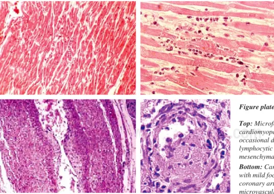
Figure plate 1.
Top: Microfocal myocarditis in HHV-6 positive cardiomyopathy and AIDS patients with occasional degenerating cardiomyocyte, mild lymphocytic infiltration and prominent mesenchymal activation.
www.medigraphic.com
RESULTSGross and microscopic findings
All 10 patients showed a prominent enlargement of their hearts, both on chest Xray and at autopsy with a reduced left ventricular ejection fraction of below 50%. Microscopy (preferentially of the left lateral ventricular wall) showed usually some inter-stitial edema with swollen and proliferated mesen-chymal cells, mild diffuse and perivascular fibro-sis, and only a scarce focal lymphocytic infiltrate (“borderline myocarditis”). Only in the two cases diagnosed as myocarditis lymphocytic infiltrates were somewhat more prominent. Cardiomyocytes in the endomyocardial biopsies showed some vac-uolization (“sarcolysis”) as known from reperfusion damage, occasional contraction bands, and in two HIV1+ autopsy cases scattered small focal necro-ses. In addition, the AIDS+ cases and cardiac transplants showed focal intimal proliferates in coronary arteries and occasional thrombotic small vessel disease in epicardial and systemic vessels (Figure plate 1).
Immunohistochemistry and in situ hybridization
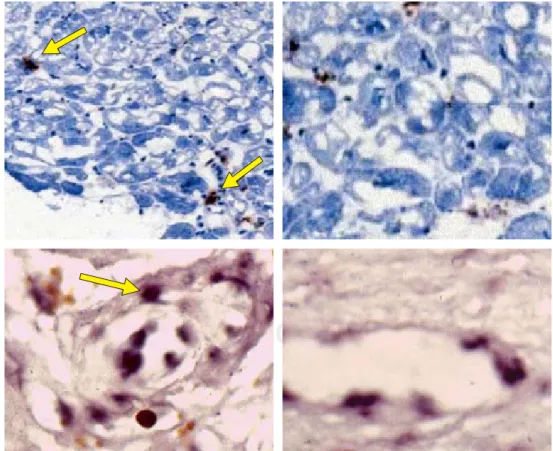
Both gp110/60 and p41 HHV-6 antigens were demon-strated in all biopsy- and autopsy cases, although usu-ally in small numbers of cells only. While HHV-6 gp 110/ 60 antigens were detected preferentially in interstitial cells and in vascular endothelial cells with some equivo-cal staining of cardiomyocytes, HHV-6 p41 was also seen in endothelial cells of small vessels (Figure plate 2). There was no unequivocal deposition of HHV-6 p41 antigens in cardiomyocytes with the techniques used. Interstitial cells that stained positive were usually larger cells probably representing macrophages rather than lymphocytes. This impression was supported by in situ
hybridization data, where HHV-6 DNA was seen in all cases only in endothelial cells of small interstitial ves-sels. The 2 transplant patients showed HHV-6 DNA fo-cally in endothelial cells of coronary vessels as previ-ously described by J.Luka using in situ PCR.12
Ultrastructural studies
Although specimens for ultrastructural studies were not optimal due to previous formalin fixation and paraf-fin embedding, careful evaluation confirmed the
micro-Figure plate 2.
Top: Myocardial biopsies of patients with HHV-6 positive cardiomyopathy and myocarditis showing prominent ballooning degeneration of cardiomyocytes and HHV-6 antigen deposition in interstitial cells and vascular endothelia (arrows).
www.medigraphic.com
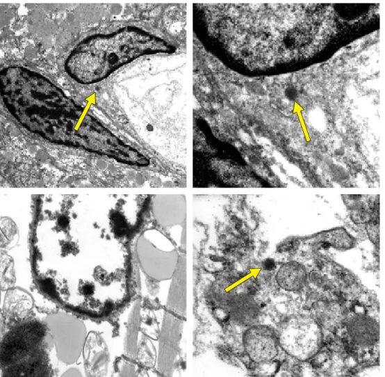
Figure plate 3.
Top: Electron microscopy showing herpesvirus particles in vascular endothelium of cardiac biopsies in HHV-6 positive cardiomyopathy and myocarditis as well as in AIDS patients (arrows).
Bottom: Electron microscopy showing a degenerating cardiomyocyte without clearly identifiable viral structures (left) while interstitial degenerating macrophages contain occasional herpesvirus particle (right, arrow).
scopic findings of mesenchymal cell activation, occa-sional mild focal inflammatory (lymphocytic) infiltration and focal cardiomyocyte degeneration. Herpes-type vi-rus particles were found in all cases preferentially in small vascular endothelial cells. Degenerating cardi-omyocytes occasionally contained dense bodies that might have been remnants of herpesvirus particles, yet proof was not possible with the techniques used. Also, monocytes contained occasional herpesvirus particle in the cases with myocarditis, cardiomyopa-thy, yet not in the transplant patients (Figure plate 3).
DISCUSSION
All cases reported in this study were selected on the basis of proven HHV-6 reactivation with circulating vi-ral DNA and p41 antigen and clinical evidence of car-diac dysfunction. Some are part of a major clinical study which is reported elsewhere.13,14 Cases with
dual viral infections such as with parvovirus B19 and
with adenovirus were excluded from this study ex-cept for the two HIV1 positive AIDS cases. Using such strict criteria for selection, all 10 cases studied showed evidence for active cardiac HHV-6 infection (without distinguishing between the subtypes HHV-6A and HHV-6B with the methods used).
As HHV-6 positive cases were selected intentionally, nothing can be said about the frequency of HHV-6 reac-tivation in the diseases discussed. This is addressed in the main clinical publication.13 The current study was
done to identify the eventual localization of HHV-6 in the heart itself of diseased patients and thus contribute to the elucidation of it’s possible pathogenesis.
unequivo-www.medigraphic.com
ESTE DOCUMENTO ES ELABORADO PORMEDIGRA-PHIC
cally a pathogenic relationship of cardiac lesions and HHV-6.15,16 HHV-6 p41, instead, is a functional
anti-gen synthesized during virus replication and thus indi-cates active infection when shown in tissues. Our finding of HHV-6 p41 antigen in scattered interstitial cells (probably macrophages) and in small vessel en-dothelial cells proves viral replication at these sites. This is further substantiated by showing HHV-6 DNA at these sites and by the ultrastructural demonstra-tion of herpesvirus particles in these cells (while there was no serologic evidence in selected patients for in-fections with other herpes viruses but HHV-6). No dif-ferentiation is possible, though, in our present study between the HHV-6 subtypes, HHV-6A or HHV-6B.
The low number of cells carrying HHV-6 DNA by in situ hybridization and HHV-6 p41 by immunohistochem-istry, and the even lower number of cells with recogniz-able viral particles in electron microscopy make a direct pathogenic effect of HHV-6 on myocardial cells less convincing. This notion is further supported by finding viral DNA and viral particles quite preferentially in small vessel endothelial cells rather than in cardiomyocytes. In addition, early microscopic lesions in the myocardi-um of HHV-6 infected hearts remind to a certain extent on what is commonly seen in reperfusion damage of the heart. In this context, the observations of Vallbracht and colleagues are most important that the endotheli-um-dependent flow-mediated vasodilatation becomes impaired in patients with myocardial virus persistence.17
Our findings are thus further supporting the previous hypothesis that cardiac pathology in persistent active HHV-6 infection is a consequence of microcirculatory dysfunction rather a of a direct toxic effect on cardi-omyocytes.7,14 This idea was further supported by the
observation of thrombotic microvasculitis in occasion-al case. The finoccasion-al proof of HHV-6 as a causative agent in certain forms of myocarditis/cardiomyopa-thies and post transplant cardiac dysfunction will rely upon the clinicial improvement following successful treatment of the viral infection.13
REFERENCES
1. Santoro F, Kennedy PE, Locatelli G, Malnati MS, Berger EA, Lusso P. CD46 is a cellular receptor for human herp-esvirus 6. Cell 1999; 99: 817-828.
2. Krueger GRF, Ablashi DV. Human herpesvirus-6: A short review of its biological behavior. Intervirology 2003; 46: 257-269.
3. Ablashi DV, Krueger GRF, Salahuddin SZ (eds). Human Herpesvirus-6. Amsterdam: Elsevier Sci Publ; 1992. p. 1-341. 4. Krueger GRF. Infections with HHV-6 and HHV-7: summa-ry of other disease associations. J Clin Virol 2006; 37 (suppl 1): S109-110.
5. HHV-6; an underestimated virus. Proc 5th Internat Conf on HHV-6 & HHV-7. Barcelona, 2006. J Clin Virol 2006; 37 (suppl 1): S1-S120.
6. Krueger GRF, Klueppelberg U, Hoffmann A, Ablashi DV. Clinical correlates of infection with human herpesvirus-6. In Vivo 1994; 8: 457-485.
7. Buja LM. HHV-6 in cardiovascular pathology. In: Krueger GRF, Ablashi DV (eds). Human Herpesvirus-6. 2nd ed. Amsterdam: Elsevier Sci Publ; 2006. p. 233-241.
8. Krueger GRF, Bertram G, Ramon A, Koch B, Ablashi DV, Brandt ME, Wang G, Buja LM. Dynamics of infection with human herpesvirus-6 in EBV-negative infectious mononu-cleosis: data acquisition for computer modeling. In Vivo 2001; 15: 373-380.
9. Wagner M, Krueger GRF, Ablashi DV, Whitman JE. Chron-ic fatigue syndrome: a critChron-ical evaluation of testing for ac-tive human herpesvirus-6 (HHV-6) infection: Review of data of 107 cases. J Chron Fatigue Syndr 1996; 2: 3-16. 10. Seyda M, Scheele T, Neumann R, Krueger GRF.
Compar-ative evaluation of nonradioactive in situ hybridization techniques for pathologic diagnosis of viral infection. Pathol Res Pract 1989; 184: 18-26.
11. Josephs SF, Salahuddin SZ, Ablashi DV, Schachter F, Wong-Staal F, Gallo RC. Genomic analysis of the human B-lymphotropic virus (HBLV). Science 1986; 234: 601-603. 12. Luka J, Aufferbach C, Carson SD, Krueger GRF. Detec-tion of human herpesvirus-6 (HHV-6) genomes by in situ
PCR and in situ hybridization in tissues from Kawasaki disease and in coronary arteries of transplanted hearts. J Clin Virol 2006; 37 (suppl 1): S111.
13. Kuehl U, Lassner D, Pauschinger M, Gross UM, Krueger GRF, Seeberg B, Noutsias M, Escher F, Poller W, Schultheiss HP. Prevalence of cardiac involvement of hu-man herpesvirus 6A and B subtypes in patients with ac-quired cardiomyopathies and heart failure symptoms. 2008 submitted for publication
14. Krueger GRF. HHV-6 in cardiac diseases. SFB Transregio 19 International Symposium on Molecular Therapies Tar-geting new Pathogenic Pathways and Networks in Human Cardiomyopathies. Harnack House of the Max-PlanckSo-ciety Berlin Oct. 25-27, 2007.
15. Krueger GRF, Ablashi DV, Salahuddin SZ (eds). Persis-tent herpesvirus infections: Current techniques for diagno-sis. J Virol Methods 1988; 21: 1-326.
16. Krueger GRF, Ablashi DV, Josephs SF, Salahuddin SZ, Lembke U, Ramon A, Bertram G. Clinical indications and diagnostic techniques of human herpesvirus-6 (HHV-6) in-fection. In Vivo 1991; 5: 287-296.
17. Vallbracht KB, Schwimmbeck PL, Kuehl U, Seeberg B, Schultheiss HP. Endothelium-dependent flow-mediated vasodilatation of systemic arteries is impaired in patients with myocardial virus persistence. Circulation 2004; 110: 2938-2945.
Correspondingauthor:
Gerhard RF Krueger MD PhD
Department of Pathology & Laboratory Medicine, UT - Houston Medical School,
6431 Fannin St., MSB 2.136, Houston, Texas 77030