P
www.permanyer.com
Rev Inves Clin. 2016;68:154-62 ORIGINAL ARTICLE
G80A Single Nucleotide Polymorphism
in Reduced Folate Carrier-1 Gene
in a Mexican Population and its Impact
on Survival in Patients with Acute
Lymphoblastic Leukemia
Myrna Candelaria
1,2*, Juan Ojeda
2, Olga Gutiérrez-Hernández
1, Lucia Taja-Chayeb
1,
Silvia Vidal-Millán
1and Alfonso Due
ñas-González
31Pharmacogenetics Laboratory; 2Hematology Department; 3Instituto de Investigaciones Biomédicas, UNAM
and Instituto Nacional de Cancerología, Mexico City, Mexico
Corresponding author:
*Myrna Candelaria
Pharmacogenetics Laboratory Instituto Nacional de Cancerología Av. San Fernando, 22
Col. Sección XVI
C.P. 14080, Ciudad de México, Mexico
E-mail: [email protected] Received for publication: 18-02-2016 Accepted for publication: 07-04-2016
ABSTRACT
Background: Hyper-CVAD is the treatment for patients with acute lymphoblastic leukemia in our institution. Objective: To
evaluate the impact of single nucleotide polymorphisms at genes associated with methotrexate metabolism on survival. Methods:
The presence of the single nucleotide polymorphisms G80A at reduced folate carrier-1 gene and C677T in the
methylenetetra-hydrofolate reductase gene was determined by denaturing high performance liquid chromatography and validated by sequencing.
Both single nucleotide polymorphisms were evaluated in 71 healthy donors and in an exploratory pilot trial with acute lympho-blastic leukemia patients to determine the influence of these single nucleotide polymorphisms on clinical outcome. Clinical characteristics, response, and outcome were registered. A Cox regression analysis was done to evaluate factors influencing response and overall survival. Results: There were no differences in the frequency of single nucleotide polymorphisms between
volunteers and acute lymphoblastic leukemia patients according to the Hardy-Weinberg test. Sensitivity and specificity were 72 and 91% for the G80A, and 64 and 75% for the C677T, respectively. The multivariate analysis showed that the T-immuno-phenotype and the presence of single nucleotide polymorphism G80A reduced folate carrier-1 were associated with a shorter relapse-free survival and overall survival. Conclusions: The presence of G80A single nucleotide polymorphism at reduced
folate carrier-1 gene in acute lymphoblastic leukemia patients was associated with a poorer prognosis. (REV INVES CLIN. 2016;68:154-62)
Key words: RFC1 gene. MTHFR4 gene. SNP. Leukemia. Methotrexate.
Sin contar con el consentimiento previo por escrito del editor
, no podrá reproducirse ni fotocopiarse ninguna parte de esta publicación.
INTRODUCTION
Acute lymphoblastic leukemia (ALL) in adults re-mains a significant treatment challenge, in contrast with pediatric ALL where considerable improve-ments in long-term survival and even cure have been achieved over the last 30 years. Long-term overall survival for this group remains relatively poor, be-tween 20 and 40%. Current research in adult ALL has mainly focused in optimizing the use of cyto-toxic drugs. In this regard, pharmacogenetics is of considerable interest, particularly for methotrexate (MTX), a folate analog drug that is essential in reg-imens for ALL1-5.
The influx of MTX into cells depends on the re-duced folate carrier protein (RFC), which efficient-ly transports folates and MTX into cells and, once inside, is converted into methotrexate polygluta-mates (MTXPG) by the enzyme folyl-polyglutamate synthetase6,7. The MTX and its polyglutamates
af-fect intracellular folate metabolism by inhibiting dihydrofolate reductase and thymidylate synthase. Therefore, the inhibition of DNA biosynthesis in-duced by MTX is multifactorial, including both par-tial depletion of reduced folates and direct inhibi-tion of folate-dependent enzymes. The effectiveness of MTX depends on its concentration and retention in cells.
The RFC1 gene is located on the long arm of chro-mosome 21 (21q22.2-22.3) and encodes a mem-brane protein called reduced folate carrier8. The
RFC1 single nucleotide polymorphism (SNP) 80G>A (rs1051266) leads to the substitution of guanine for adenine in the 80th position, which results in the
substitution of arginine for histidine at the residue 27 in the structure of the protein9. Chan, et al. showed
that this change results in decreased receptor affin-ity and variations in the transmembrane transport of folic acid antimetabolites. In ex vivo studies, the fo-late concentrations in serum were higher in the 80AA genotype than the allele G variant: 19 vs. 15 mmol/l, respectively10. Banerjee, et al. analyzed the
relation-ship between the RFC1 G80A polymorphism and the risk of relapse of ALL in children. They found that in 204 ALL patients studied, the RFC1 80AA variant was associated with higher serum concen-trations of MTX11, which has also been observed by
other authors12.
On the other hand, the enzyme 5,10-methylene-tetrahydrofolate reductase (MTHFR) catalyzes the conversion of 5,10-methylenetetrahydrofolate to 5-methyltetrahydrofolate in the folic acid cycle12.
A common genetic polymorphism in the MTHFR gene results from a C®T substitution. Individuals with the T/T genotype commonly have elevations in plasmat-ic homocysteine and differences in response to folplasmat-ic acid supplementation compared with normal (C/C) or heterozygous (C/T) genotypes. This polymorphism is highly prevalent in the Mexican population13-15,
par-ticularly among Nahua and Mixtec groups. However, in other regions of the country, such as the north, the prevalence is similar to Caucasian regions14. In
addi-tion, this SNP may influence the therapeutic response to antifolate drugs such as MTX. The frequencies of the C677T allelic variants differ according to ethnicity. In Europe, 8-20% of the Caucasian popu-lation is homozygous for the 677T allele and almost 40% is heterozygous2,16. Although some authors12,17
have described the influence of this SNP on survival and toxicity in patients with ALL whose treatment includes MTX, its role is still unclear. Thus, in this work we evaluated the feasibility of using denaturing high-performance liquid chromatography (DHPLC) to de-termine RFC1 and MTHFR gene polymorphisms as well as to correlate these SNPs with the toxicity and outcome in adults with ALL receiving MTX as part of the hyper-CVAD regimen.
MATERIAL AND METHODS
Study population
Patients
A total of 31 adult patients with ALL were included from January 2011 to December 2012 at the Na-tional Cancer Institute (INCan) in Mexico in this pro-spective, exploratory pilot trial to assess the influence of the RFC1 G80A and MTHFR C677T SNPs on re-sponse and overall survival.
Healthy individuals
Blood samples from 71 consecutive healthy, volun-teer blood donors were obtained by venous puncture from the arm; none of the donors were related to our ALL patients.
Sin contar con el consentimiento previo por escrito del editor
, no podrá reproducirse ni fotocopiarse ninguna parte de esta publicación.
Inclusion criteria
The patients were untreated, Mexican, older than 15 years of age, and with normal renal and hepatic function. After inclusion and blood sample collec-tion, patients began treatment with hyper-CVAD. Patients with a history of hypersensitivity to MTX or to any of the other drugs included in the hyper-CVAD regimen were excluded. This protocol was approved by the IRB Committee and registered at www.clinicaltrials.gov (Identifier # NCT01307241). Healthy individuals and patients signed an informed consent.
Baseline clinical and pathological characteristics were recorded. Patients were assessed for response using the International Working Group Criteria for acute leukemia18. Overall survival (OS) was defined as the
time since diagnosis until death or date of the last visit. Relapse-free survival (RFS) was defined as the time since remission was achieved until relapse was documented.
Laboratory procedures
DNA extraction
Genomic DNA was obtained using a Wizard genomic DNA purification kit (Promega, Madison, WI, USA) according to the manufacturer’s instructions. The DNA was quantified in a NanoDrop (Applied Biosys-tems) and stored at –20 °C.
Polymerase chain reaction
amplification
Polymerase chain reaction (PCR) was performed us-ing the followus-ing oligonucleotides: Forward: 5’-AGT GTCACCTTCGTCCC-3’ Reverse: 5’-TCCCGCGTGAAG TTCTTG-3’ for the RFC1 gene, and Forward: 5’- GG AGCTTTGAGGCTGACCTGAA-3’ Reverse: 5’-AGGAC GGTGCGGTGAGAGTG-3’ for the MTHFR gene. The PCR was performed in a total volume of 25 μl contain-ing 100 ng genomic DNA, 1 μmol/l oligonucleotides (forward and reverse), 200 μM dNTPs (Fermentas Life Sciences, USA), 0.25 U optimase polymerase enzyme (Applied Biosystems), and PCR 1 x buffer (15 mM MgCl2, Perkin Elmer, Foster City, CA). The PCR was performed on a GeneAmp® 9700 thermal cycler
(Applied Biosystems) using an initial denaturation
step at 94 °C for five minutes, followed by 40 cycles at 94 °C for 30 seconds, annealing (60 °C for RFC1 and 55 °C for MTHFR) for 30 seconds, and 72 °C for 30 seconds; a final extension was performed at 72 °C for seven minutes. Products were electrophoresed in 2% agarose gels.
Denaturing high-performance
liquid chromatography
analysis
The PCR products were denatured at 95 °C during 10 minutes and were cooled until 25 °C, decreasing 2 °C/minute to allow for homo- or heteroduplex for-mation (Transgenomics, Inc; San José, CA) according to conditions determined by DHPLC software. Het-erozygous chromatograms were identified by visual analysis and compared with the wild type. Homo-duplex cases, which have a single peak chromato-gram as wild type, were mixed with known wild type DNA (previously sequenced), to allow heteroduplex formation. Results were reported as wild type (wt) or polymorphic.
Sequencing
Seventy-one samples from healthy individuals were sequenced (gold standard), regardless of the results of DHPLC analysis from at least two independent am-plification products. The ALL samples were amplified and analyzed by DHPLC. The PCR amplicons were pu-rified using isopropanol precipitation, diluted and cy-cle-sequenced using a BigDye® Terminator kit v3.1
(ABI, Foster City, CA) according to manufacturer in-structions in an ABI Prism® 3100 genetic analyzer.
Electropherograms were analyzed in both sense and antisense directions.
Statistical analysis
The SNP frequencies were compared between healthy
individuals and patients using the Hardy-Weinberg test. The results of DHPLC and Sanger sequencing (gold standard) were analyzed to determine sensitiv-ity: (true positive/ [true positive + false negative]), and specificity: (true negative/ [false positive + true negative]). Survival curves were done by Kaplan-Mei-er method and compared by log rank test. Cox regres-sion analysis was done to evaluate factors influencing response and overall survival.
Sin contar con el consentimiento previo por escrito del editor
, no podrá reproducirse ni fotocopiarse ninguna parte de esta publicación.
polymorphic chromatogram, which was confirmed by sequencing in 33 (true positive); five cases were re-corded as false positive (polymorphic chromatogram by DHPLC, but the SNP discarded by sequencing). Eighteen cases had a wt chromatogram, but sequenc-ing showed the SNP (false negative), whereas 15 were true negative (wt chromatogram and no SNP by se-quencing). The calculated sensitivity and specificity were 64 and 75%, respectively, for MTHFR, and 72 and 91% for the RFC1 gene, respectively (Table 1).
Patients
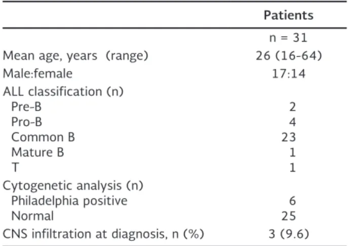
The clinical and pathological characteristics of ALL pa-tients are shown in table 2. The mean age was 26 years (range 16-64); male:female distribution was 17:14. Most (74%) had the common B subtype and six (20%) were positive for the Philadelphia chromosome.
After analyzing DHPLC chromatograms for these poly-morphisms, the frequency of the RFC1 G80A was 50.7% in healthy individuals and 40.7% among leuke-mia patients. All were heterozygous. Regarding the MTHFR C677T SNP, the frequency was 52.7 and
RESULTS
Healthy individuals
Blood samples were collected from 71 healthy donors. The mean age was 30.6 years (range 19-57) and 48% were male.
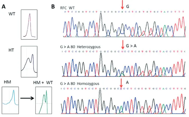
Sensitivity and specificity of DHPLC in healthy indi-viduals: PCR fragments of MTHFR and RFC1 genes were subjected to DHPLC, and the conditions for analysis (gradient and temperature) were optimized for each fragment to yield characteristic and repro-ducible profiles. As shown in figure 1, samples with wt RFC1 sequence eluted as a single peak, whereas mul-tiple peaks were indicative of heteroduplex containing 80G>A SNP. Homozygous polymorphic also showed a single peak, and an elution mixed with a wt sample was required to demonstrate this status. These re-sults were confirmed by Sanger sequencing. Likewise, MTHFR wt showed a single peak and C>T 677 SNP was documented with multiple peaks (Fig. 2). As shown in table 1, regarding the MTHFR SNP from the 71 samples from healthy individuals, 38 showed a
Figure 1. RFC gene analysis by A: DHPLC showing a single peak for the homozygous wildtype or mutant, whereas multiple peaks
were indicative of heteroduplexes containing G>A 80 SNP. When an homozygous sample mixed with wildtype DNA showed a multiple peak profile, indicated the presence of the SNP homozygously. B: Sanger sequence confirming DHPLC finding.
Sin contar con el consentimiento previo por escrito del editor
, no podrá reproducirse ni fotocopiarse ninguna parte de esta publicación.
and there were no differences in toxicity rates be-tween patients having wild type or any of the SNPs.
In accordance with the international standard crite-ria, complete response was achieved with the hyper-CVAD regimen in 25 cases (80.6%), and partial re-sponse in the remaining six cases. The three-year RFS and OS were 42 and 22%, respectively. Median RFS was 72.4%, respectively. There was only one
homozy-gous patient in the leukemia population. After using Hardy-Weinberg test, no statistically significant dif-ference was found with that expected for the ana-lyzed population.
Toxicity
As expected, all patients had grade 4 myelosuppres-sion. No grade 3-4 liver toxicities were documented,
Figure 2. MTHFR gene analysis by A: DHPLC showing a single peak for the homozygous wildtype or mutant, whereas multiple
peaks were indicative of heteroduplexes containing C > T677 SNP. When an homozygous sample mixed with wildtype DNA showed a multiple peak profile, indicated the presence of the SNP homozygously. B: Sanger sequence confirming DHPLC finding.
Table 1. Sensitivity and specificity of the denaturing high performance liquid chromatography technique for
methy-lenetetrahydrofolate reductase and reduced folate carrier-1
genes in 71 healthy blood donors
MTHFR RFC1
True positive (n) 33 34 False positive (n) 5 2 False negative (n) 18 13 True negative (n) 15 22
Total (n) 71 71
Sensitivity (%) 64 72 Specificity (%) 75 91 MTHFR: methylenetetrahydrofolate reductase; RFC: reduced folate carrier.
Table 2. Clinical characteristics of acute lymphoblastic leu-kemia patients
Patients
n = 31 Mean age, years (range) 26 (16-64)
Male:female 17:14
ALL classification (n) Pre-B
Pro-B Common B Mature B T
2 4 23 1 1 Cytogenetic analysis (n)
Philadelphia positive
Normal 625
CNS infiltration at diagnosis, n (%) 3 (9.6) ALL: acute lymphoblastic leukemia; CNS: central nervous system.
Sin contar con el consentimiento previo por escrito del editor
, no podrá reproducirse ni fotocopiarse ninguna parte de esta publicación.
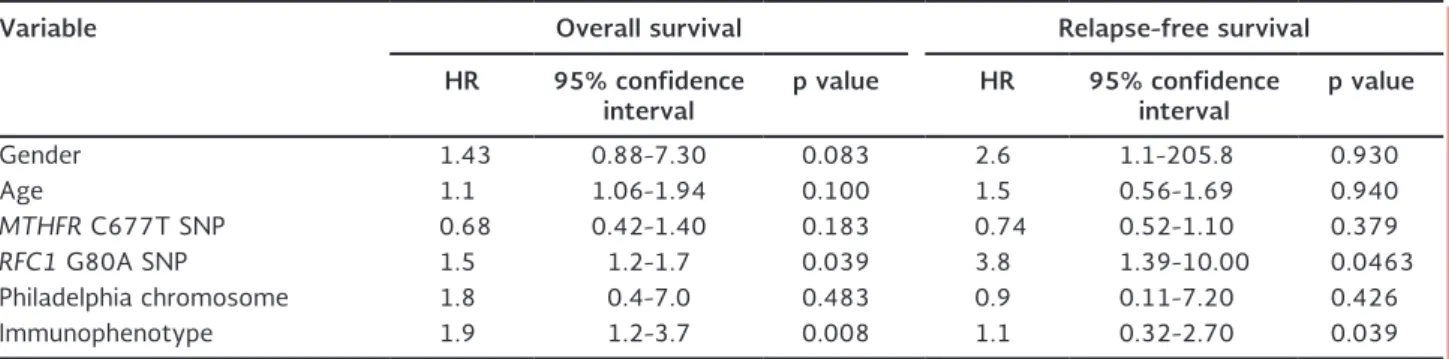
1.3 years (95% CI: 0.92-1.8). Median OS was 1.7 years (95% CI: 1.4-2.6). Cox regression analysis was done to evaluate prognostic factors, including age, cytogenetic analysis, immunophenotype of ALL, and presence or absence of any of the SNPs. The presence of 80G>A RFC SNP and T-cell immunophenotype was associat-ed with decreasassociat-ed RFS and OS (Table 3 and Fig. 3).
DISCUSSION
The pharmacogenetic approach in chemotherapy con-stitutes a research field developed to optimize drug doses and schedules for either increasing response rate or reducing toxicity. Hyper-CVAD is considered a standard of care for patients with ALL in many cen-ters worldwide, including our institution19. Within this
schema, methotrexate constitutes a key drug for ALL treatment. The results of this work add information on the field of pharmacogenetic regarding the impact of the RFC1 80G>A SNP on the efficacy of MTX when used in the context of hyper-CVAD for adult ALL pa-tients. Although many other factors related to clini-copathological characteristics of ALL patients were not taken into account, as well as other pharmacoge-netic factors that may modulate the effect of the drugs other than MTX in the hyper-CVAD regimen, Cox regression analysis clearly suggest that the RFC1 80G>A SNP may help to optimize the dose of MTX when used within this regimen, although this should be confirmed in further studies that include a larger sample of patients.
The RFC 80G>A polymorphism has been widely stud-ied in diverse populations searching for associations with increased leukemia risk and with the clinical course
(toxicity and prognosis) in leukemia patients receiving MTX. Data from the 71 healthy individuals in our study suggest that our populations have an increased frequency of this SNP (50.7%), similar to that reported among healthy French individuals (36%), whereas in a British population the frequency was 41%5. A
high-er frequency (71%) has been found in a Jordanian female population with rheumatoid arthritis20. In this
respect, some authors5 have suggested a relationship
between the RFC1 80G>A polymorphism and a 2.1-fold increased risk of ALL. Furthermore, Huang, et al.21,
after a stratified analysis by ethnicity, recently demon-strated that the association became more prominent among Caucasians (GA vs. GG: OR: 1.28; 95% CI: 1.12-1.45; p < 0.001). In contrast with these results, our findings do not support this association, since there was a trend for a higher frequency of the RFC1 80G>A polymorphism in the healthy population compared with ALL patients (50.7 vs. 40.7%, respectively).
Table 3. Cox regression analysis of factors influencing overall survival and relapse-free survival in acute lymphoblastic leukemia patients
Variable Overall survival Relapse-free survival
HR 95% confidence
interval p value HR 95% confidence interval p value
Gender 1.43 0.88-7.30 0.083 2.6 1.1-205.8 0.930
Age 1.1 1.06-1.94 0.100 1.5 0.56-1.69 0.940
MTHFR C677T SNP 0.68 0.42-1.40 0.183 0.74 0.52-1.10 0.379
RFC1 G80A SNP 1.5 1.2-1.7 0.039 3.8 1.39-10.00 0.0463
Philadelphia chromosome 1.8 0.4-7.0 0.483 0.9 0.11-7.20 0.426 Immunophenotype 1.9 1.2-3.7 0.008 1.1 0.32-2.70 0.039 MTHFR: methylenetetrahydrofolate reductase; RFC: reduced folate carrier; SNP: single nucleotide polymorphism.
Figure 3. Overall Surviva.
Sin contar con el consentimiento previo por escrito del editor
, no podrá reproducirse ni fotocopiarse ninguna parte de esta publicación.
Regarding the association between this polymorphism and shorter RFS and OS, there are experimental data supporting it, although there is also contradictory in-formation.
In contrast to our results, in a study including 70 chil-dren with ALL, an association was found between the polymorphism and a lower risk of relapse (p < 0.05): in patients with the G/A genotype it was 3.97 (95% CI: 1.12-14.06) compared with carriers of the A/A genotype (wt) who had a higher probability of relapse (7.84; 95% CI: 1.66-37.10)22. In our patients, those
with the G/A genotype had a 3.8 HR for relapse.
Other authors23,24 found that extra copies of
chromo-some 21, where the RFC1 gene is located, were associ-ated with increased expression of mRNA and reflect elevated capacities for MTX transport. Our findings are supported by a decreased functional status of this SNP, showing a lower intracellular folate concentra-tion in patients with the RFC 80G>A polymorphism, as has been confirmed by Yates and Lucock25,
al-though other manifestations of dysfunction of this 80G>A polymorphism have not been documented, such as a higher risk of neural tube defects or colorec-tal cancer25-29. In this regard, Chan, et al. showed that
this change results in decreased receptor affinity and variations in the transmembrane transport of folic acid antimetabolites. In ex vivo studies, the folate concen-trations in serum were higher in the 80AA genotype than the allele G variant: 19 vs. 15 mmol/l, respec-tively10. Banerjee, et al. analyzed the relationship
be-tween the RFC1 G80A polymorphism and the risk of relapse in 204 children with ALL and also found that the RFC1 80AA variant was associated with a higher serum concentration of MTX11, which has also been
observed by other authors12. However, Whetstine, et
al.30 reported that the change of the strongly basic
amino acid arginine to histidine, which is a weak base, in a region of the carrier documented to influence folate substrate binding and rates of uptake, might be expected to alter RFC transport properties. Neverthe-less, by directly comparing the transport properties of Arg27 and His27-hRFC in stable K562 transfec-tants, no significant differences in the uptake rates of MTX were observed, and only minor differences were calculated in the relative affinities for an as-sortment of transport substrates. Collectively, these data strongly argue for a lack of major functional differences between the Arg27- and His27-RFCs for
reduced folate cofactors and for various antifolates used in cancer chemotherapy, including MTX. On the other hand, researchers in Japan have shown increased liver toxicity in homozygous 80AA, and increased side effects in the form of severe vomiting in the 80GG group homozygotes31,32. We found no differences
re-garding toxicity dependent on this SNP, which may be explained because we had only one homozygous pa-tient (80GG).
In contrast with the RFC1 80G>A polymorphism, in our study, the SNP 677C>T at the MTHFR gene did not influence RFS or OS. Similar results have been obtained by other authors. A multicenter study from Poland published in 200632, in which MTHFR, TPMT,
GSTT1, GSTM1, GSTP1, and TS polymorphisms were determined, showed a significant relationship between genotype 677C>T and an increased death rate (OR: 4.09; 95% CI; p = 0.028). Eight of the 31 (26%) pa-tients whose death occurred during treatment had homozygous 677TT, but there was no association with the genotype 677C>T. In line with our results, Deus, et al.33 also reported that G80A polymorphism
influenced the survival of pediatric patients with ALL, but neither G677T nor A1298C in MTHFR gene had an effect on survival. In Mexico, the presence of MTHFR has been evaluated in different regions of our coun-try13-15,34 and strong differences in frequencies of
C677T polymorphism were documented, being high-er among Nahua and Mixtec groups compared with Mestizos13,14. Additionally, Ruiz-Argüelles, et al.34
eval-uated the risk of mucosal damage in 28 patients with ALL treated with MTX and, in accord with our findings, they concluded that there was no significant associa-tion with mucositis at the gene or at the genotype level. They also postulated that the risk of higher MTX toxicity in patients with decreased MTHFR activity could be neutralized by the normally folate-rich diet in Mexico.
Results of our study demonstrate by multivariate analysis that the G80A SNP is associated with shorter RFS and OS. However, because of the small sample size and unknown clinical data, it is possible that results were due to random effects. In addition, there are more pharmacogenetic variations of genes implicated not only in the metabolism of MTX, but of the other drugs in the hyper-CVAD regimen that were not studied. Therefore, it is possible that their interactions could explain the results observed.
Sin contar con el consentimiento previo por escrito del editor
, no podrá reproducirse ni fotocopiarse ninguna parte de esta publicación.
Nevertheless, our results regarding the influence of the G80A polymorphism cannot be underestimated since other studies1,5,7 have also found this association
in leukemia patients receiving MTX. Further studies are needed to establish the value of this pharmacogenetic marker in the optimization of leukemia treatment with MTX.
Finally, our study to determine the feasibility of using DHPLC as a routine method to determine the SNPs here studied and others35-37, suggest that it can be of
value, despite the fact that the sensitivity and speci-ficity we obtained were not so high. However, a num-ber of papers have documented the excellent sensitiv-ity and specificsensitiv-ity of DHPLC in detecting mutations38.
For instance, O’Donovan, et al. have reported a sensi-tivity and specificity of 100% for detecting mutations in exon H of the Factor IX and exon 16 of the neuro-fibromatosis type 1 gene39. However, under a single
hybridization condition, some probes do not have op-timal hybridization kinetics and therefore markers lo-cated near such sequence contexts cannot be de-tected. In addition, it is difficult to identify markers that are present as heterozygotes, as well as markers located close to other polymorphisms. As a result, most studies reach a sensitivity of 85-95%, with spec-ificity in some cases as low as 55%40. In this regard,
the relatively lower sensitivity and specificity here found can potentially be increased by testing differ-ent conditions. Regarding the polymorphisms studied,
these are preliminary results and require confirmation. However, we could suggest that patients with G80A may be treated with other regimens without metho-trexate, or within clinical trials.
REFERENCES
1. Mei L, Ontiveros EP, Griffiths EA, Thompson JE, Wang ES, Wetzler M. Pharmacogenetics predictive of response and tox-icity in acute lymphoblastic leukemia therapy. Blood Rev. 2015; 29:243-9.
2. Candelaria M, Taja-Chayeb L, Arce-Salinas C, Vidal-Millan S, Serrano-Olvera A, Dueñas-Gonzalez A. Genetic determinants of cancer drug efficacy and toxicity. Practical considerations and perspectives. Anticancer Drugs. 2005;16:923-33.
3. Hooijberg JH, Broxterman HJ, Kool M, et al. Antifolate resistance mediated by the multidrug resistance proteins MRP1 and MRP2. Cancer Res. 1999;59:2532-5.
4. Kool M, van der Linden M, de Haas M, et al. MRP3, an organic anion transporter able to transport anti-cancer drugs. Proc Natl Acad Sci USA. 1999;96:6914-19.
5. Hider I, Bruce N, Thomson W. The pharmacogenetics of metho-trexate. Rheumatology. 2007;46:1520-4.
6. Payne K, Newman W, Fargher E, Tricker K, Bruce IN, Ollier WE. TPMT testing in rheumatology: any better than routine moni-toring? Rheumatology. 2007;46:727-9.
7. Pirmohamed M. Pharmacogenetics and pharmacogenomics. Br J Clin Pharmacology. 2001;52:345-7.
8. Genestier L, Paillot R, Quemeneur L, Izeradjene K, Revillard JP. Mechanisms of action of methotrexate. Immunopharmacology. 2000;47:247-57.
9. Stanisławska-Sachadyn A, Mitchell LE, Woodside JV, et al. The reduced folate carrier (SLC19A1) c.80G>A polymorphism is as-sociated with red cell folate concentrations among women. Ann Hum Genet. 2009;73:484-91.
10. Chan ES, Cronstein BN. Molecular action of methotrexate in inflammatory diseases. Arthritis Res. 2002;4:266-73. 11. Banerjee D, Mayer-Kuckuk P, Capiaux G, Budak-Alpdogan T,
Gorlick R, Bertino JR. Novel aspects of resistance to drugs tar-geted to dihydrofolate reductase and thymidylate synthase. Bio-chim Biophys Act. 2002;1587:164-73.
12. de Deus DM, de Lima EL, Seabra Silva RM, Leite EP, Cartaxo Muniz MT. Influence of methylenetetrahydrofolate reductase C677T, A1298C, and G80A polymorphisms on the survival of pediatric patients with acute lymphoblastic leukemia. Leuk Res Treatment. 2012;2012:292043.
13. Antonio-Vejar A, Del Moral-Hernández O1, Alarcón-Romero LC1, et. al. Ethnic variation of the C677T and A1298C polymor-phisms in the methylenetetrahydrofolate-reductase (MTHFR) gene in southwestern Mexico. Genet Mol Res. 2014;13:7950-7. 14. Ramos MA, Mares RE, Avalos ED, et. al. Pharmacogenetic screen-ing of N-acetyltransferase 2, thiopurine s-methyltransferase, and 5,10-methylene-tetrahydrofolate reductase polymorphisms in Northwestern Mexicans. Genet Test Mol Biomarkers. 2011; 15:351-5.
15. Guéant-Rodriguez RM, Guéant JL, Debard R, et.al. Prevalence of methylenetetrahydrofolate reductase 677T and 1298C alleles and folate status: a comparative study in Mexican, West Afri-can, and European populations. Am J Clin Nutr. 2006;83:701-7. 16. Robien K, Ulrich CM. 5,10-Methylenetetrahydrofolate reductase
polymorphisms and leukemia risk: a HuGE mini-review. Am J Epidemiol. 2003;157:571-82.
17. Tas¸bas¸ O, Borman P, Gürhan Karabulut H, Tükün A, Yorgancıog˘lu R. The frequency of A1298C and C677T polymorphisms of the methylenetetrahydrofolate gene in Turkish patients with rheu-matoid arthritis: Relationship with methotrexate toxicity. Open Rheumatol J. 2011;5:30-5.
18. NCCN Guidelines Version 2.0 2015. Acute Lymphoblastic Leu-kemia. Available at: http://www.nccn.org/professionals/physi-cian_gls/pdf/all.pdf (Accessed January 5, 2016).
19. Kantarjian HM, O’Brien S, Smith TL, et al. Results of treatment with Hyper-CVAD, a dose-intensive regimen, in adult acute lym-phocytic leukemia. J Clin Oncol. 2000;18:547-61.
20. Samara SA, Irshaid Y, Mustafa KN. Association of MDR1 C3435T and RFC1 G80A polymorphisms with methotrexate toxicity and response in Jordanian rheumatoid arthritis patients. Int J Clin Pharmacol Ther. 2014;52:746-55.
21. Huang X, Gao Y, He J, et al. The association between RFC1 G80A polymorphism and cancer susceptibility: Evidence from 33 studies. J Cancer. 2016;7:144-52.
22. Leyva-Vázquez MA, Organista-Nava J, Gómez-Gómez Y, Con-treras-Quiroz A, Flores-Alfaro E, Illades-Aguiar B. Polymorphism G80A in the reduced folate carrier gene and its relationship to survival and risk of relapse in acute lymphoblastic leukemia. J Investig Med. 2012;60:1064-7.
23. Kumagai K, Hiyama K, Oyama T, Maeda H, Kohno N. Polymor-phisms in the thymidylate synthase and methylene tetrahydro-folate reductase genes and sensitivity to the low-dose metho-trexate therapy in patients with rheumatoid arthritis. Int J Mol Med. 2003;11:593-600.
24. Mandola MV, Stoehlmacher J, Zhang W, et al. A 6bp polymor-phism in the thymidylate synthase gene causes message insta-bility and is associated with decreased intratumoral TS mRNA levels. Pharmacogenetics. 2004;14:319-427.
25. Yates Z, Lucock M. G80A reduced folate carrier SNP modulates cellular uptake of folate and affords protection against throm-bosis via a non homocysteine related mechanism. Life Sci. 2005; 77:2735-42.
26. Dervieux T, Kremer J, Lein DO, et al. Contribution of common polymorphisms in reduced folate carrier and gamma-glutamyl hydrolase to methotrexate polyglutamate levels in patients with rheumatoid arthritis. Pharmacogenetics. 2004;14:733-9. 27. Dervieux T, Furst D, Lein DO, et al. Polyglutamation of
metho-trexate with common polymorphisms in reduced folate carrier, aminoimidazole carboxamide ribonucleotide transformylase, and thymidylate synthase are associated with methotrexate effects in rheumatoid arthritis. Arthritis Rheum. 2004;50:2766-74.
Sin contar con el consentimiento previo por escrito del editor
, no podrá reproducirse ni fotocopiarse ninguna parte de esta publicación.
28. Wessels JA, de Vries-Bouwstra JK, Heijmans BT, et al. Efficacy and toxicity of methotrexate in early rheumatoid arthritis are associated with single-nucleotide polymorphisms in genes coding for folate pathway enzymes. Arthritis Rheum. 2006;54: 1087-95.
29. Ranganathan P, Culverhouse R, Marsh S, et al. Single nucleotide polymorphism profiling across the methotrexate pathway in normal subjects and patients with rheumatoid arthritis. Phar-macogenomics. 2004;5:559-69.
30. Whetstine JR, Gifford AJ, Witt T, et al. Single nucleotide poly-morphisms in the human reduced folate carrier: characterization of a high-frequency G/A variant at position 80 and transport properties of the His(27) and Arg(27) carriers. Clin Cancer Res. 2001;7:3416-22.
31. Dervieux T, Furst D, Lein DO, et al. Pharmacogenetic and me-tabolite measurements are associated with clinical status in patients with rheumatoid arthritis treated with methotrexate: results of a multicenter crossectional observational study. Ann Rheum Dis. 2005;64:1180-5.
32. Dervieux T, Greenstein N, Kremer J. Pharmacogenomic and met-abolic biomarkers in the folate pathway and their association with methotrexate effects during dosage escalation in rheuma-toid arthritis. Arthritis Rheum. 2006;54:3095-103.
33. de Deus DM, de Lima EL, Seabra Silva RM, Leite EP, Cartaxo Muniz MT. Influence of methylenetetrahydrofolate reductase C677T, A1298C, and G80A polymorphisms on the survival of
pediatric patients with acute lymphoblastic leukemia. Leuk Res Treatment. 2012;2012:292043.
34. Ruiz-Argüelles GJ, Coconi-Linares LN, Garcés-Eisele J, Reyes-Nuñez V. Methotrexate-induced mucositis in acute leukemia patients is not associated with the MTHFR 677T allele in Mexico. Hematology. 2007;12:387-91.
35. Cui G, Ding H, Xu Y, Li B, Wang DW. Applications of the method of high resolution melting analysis for diagnosis of Leber’s disease and the three primary mutation spectrum of LHON in the Han Chinese population. Gene. 2013;512:108-12.
36. Frueh FW, Noyer-Weidner M. The use of denaturing high-per-formance liquid chromatography (DHPLC) for the analysis of genetic variations: impact for diagnostics and pharmacogenetics. Clin Chem Lab Med. 2003;41:452-61.
37. Taja-Chayeb L, Vidal-Millán S, Gutiérrez O, Ostrosky-Wegman P, Dueñas-González A, Candelaria M. Importance of the poly-morphisms of the Thiopurine S-methyltransferase Gene (TMPT) determination in Mexican Mestizo patients with acute lympho-blastic leukemia (ALL). Med Oncol. 2008;25:56-62.
38. Xiao W, Oefner PJ. Denaturing high-performance liquid chroma-tography: A review. Hum Mutat. 2001;17:439-74.
39. O’Donovan MC, Oefner PJ, Roberts SC, et al. Blind analysis of denaturing high-performance liquid chromatography as a tool for mutation detection. Genomics. 1998;52:44-9.
40. Steinmetz LM, Davis RW. High-density arrays and insights into genome function. Biotech Genet Eng Rev. 2000;17:109-47.
Sin contar con el consentimiento previo por escrito del editor
, no podrá reproducirse ni fotocopiarse ninguna parte de esta publicación.