P
Rev Inves Clin. 2017;69:139-45 ORIGINAL ARTICLE
Risk Factors for No-Reflow Phenomenon
after Percutaneous Coronary
Intervention in Patients with Acute
Coronary Syndrome
Tian Liang
1,2‡, Min Liu
1‡, Chengyu Wu
1*, Qing Zhang
1, Lei Lu
1and Zhongliang Wang
12Nanjing University of Chinese Medicine, Nanjing; 1Xuzhou City Hospital of TCM Affiliated to Nanjing University
of Chinese Medicine, Xuzhou; Jiangsu Province, China
Corresponding author: *Chengyu Wu
Nanjing University of Chinese Medicine Nanjing 210046, Jiangsu Province, China E-mail: [email protected]
‡Tian Liang and Min Liu contributed equally to this study.
Received for publication: 24-02-2017 Accepted for publication: 07-04-2017
ABSTRACT
Background: To explore risk factors for no-reflow phenomenon after percutaneous coronary intervention in patients with acute coronary syndrome. Methods: A total of 733 acute myocardial infarction patients with persistent ischemic chest pain within 12 or 12-24 hours after onset received emergency percutaneous coronary intervention. Patients were divided into a normal reflow group and a no-reflow group, according to TIMI grading and myocardial blush grading after percutaneous coronary inter-vention. Related risk factors were analyzed. Results: The incidence of no-reflow phenomenon after percutaneous coronary intervention was 16.1%. Univariate analysis showed that, compared with the normal reflow group, the no-reflow group was older, reperfusion time was significantly longer, preoperative systolic pressure was lower, troponin peak was higher, and creatine kinase enzyme peak was higher (p < 0.05). The proportions of preoperative cardiac function Killip grade ≥ 2 and number of patients using preoperative intra-aortic balloon pump were significantly different (p < 0.05). Multivariate logistic regression analysis showed that age > 65 years (OR: 1.471; 95% CI: 1.462-1.492; p = 0.007), reperfusion time > 6 hours (OR: 1.274; 95% CI: 1.164-1.405; p = 0.001), low systolic pressure at admission (< 100 mmHg) (OR: 1.918; 95% CI: 1.017-3.897; p = 0.004), intra-aortic balloon pump use before percutaneous coronary intervention (OR: 1.949; 95% CI: 1.168-3.253; p = 0.011), low TIMI grade (≤ 1) before percutaneous coronary intervention (OR: 1.100; 95% CI: 1.086-1.257; p < 0.01), high thrombus load (OR: 1.274; 95% CI: 1.423-2.761; p = 0.030), and long target lesion (OR: 1.948; 95% CI: 1.908-1.990; p = 0.019) were independent risk factors. Conclusions: No-reflow phenomenon after percutaneous coronary intervention in patients with acute coronary syndrome was affected by complicated pathological factors. (REV INVES CLIN. 2017;69:139-45)
Key words: Myocardial infarction. No-reflow phenomenon. Percutaneous coronary intervention.
INTRODUCTION
Rapid opening of infarct-related artery (IRA) in emer-gency percutaneous coronary intervention (PCI), which can effectively recover the blood flow in infarcted
of IRA distal thrombus fragments. Delayed angiogra-phy was conducted after intracoronary injection of 50-100 μg nitroglycerin. With respect to the vascular diameter, when the residual stenosis was < 70% or the stent could successfully pass, stent implantation was initiated. Alternatively, at a proper pressure using a balloon, pre-dilation was performed before implan-tation (5-14 atm). Before stent implanimplan-tation or bal-loon pre-dilation, the amount of contrast agent was controlled, the number of angiographic procedures was reduced, and the interval between two angio-graphic examinations was prolonged during surgery.
Angiographic characteristics
of target lesions
The following angiographic characteristics were record-ed: (i) thrombus load (low, moderate or high); (ii) if the blood vessel with lesion was completely occlud-ed, the characteristics of vascular morphology should be recorded; (iii) centrifugal or centripetal lesions should be recorded in case of a sub-occluded lesion; (iv) length of the target lesion; (v) position of the lesion (proximal, middle or distal). Thrombosis was scored according to the Gibson standard7 and classi-fied into TIMI grades 0-5. Grades 0-1, low thrombus load; grades 2-3, moderate thrombus load; ≥ 4, high thrombus load. During surgery, it was determined whether balloon dilation or stent implantation was conducted for the blood vessel with lesion. Unless the stent failed to pass due to serious lesion calcification or because the diameter of the normal control seg-ment near the lesion was < 2.25 mm, stent implanta-tion should be performed for all available ischemia-related blood vessels. Only drug-eluting stents were used. The patients took Plavix routinely after surgery. The range of infarction was evaluated according to the peak level of myocardial enzyme; the level was mea-sured 5-6 times at regular intervals 96 hours after onset. The ST-segment depression was observed be-fore and one hour after emergency PCI.
Grouping criteria
According to TIMI grading after PCI, the patients were divided into a no-reflow group (TIMI grade ≤ 2) and a normal reflow group (TIMI grade 3). No-reflow phe-nomenon was diagnosed if there was successful lesion dilation without mechanical complications such as dis-section, spasm or obvious distal embolization, and TIMI epidemiological study showed that the annual incidence
of no-reflow phenomenon after PCI was about 5-25%. The phenomenon is closely related to the clinical out-come of patients, and is also one of the leading causes of death after PCI4. It is of both theoretical and clini-cal value to analyze the risk factors associated with no-reflow phenomenon to improve the safety and ther-apeutic effects of PCI. Adequate measures can then be taken to improve the clinical outcome of patients5,6. In this study, we explored the risk factors for no-re-flow phenomenon after PCI through univariate and multivariate analyses of clinical data.
MATERIALS AND METHODS
Baseline clinical data
Acute myocardial infarction patients with acute ST-segment elevation, who underwent emergency PCI in our hospital between January 2011 and December 2012, were selected. The study was approved by the ethics committee of our hospital, and written consent was obtained from all patients. Inclusion criteria were: severe chest pain lasting for over 30 min; ≥ 2 con-secutive ST-segment elevations for > 1 minute in ECG leads or new left bundle branch block; myocardial en-zyme level increased more than twice the normal val-ue; presenting signs and symptoms of persistent isch-emia within 12 or 12-24 hours after onset; successful PCI; with complete clinical data. A total of 733 patients were enrolled. Exclusion criteria were: coronary artery spasm or stenosis degree of culprit lesion ≤ 50% con-firmed by coronary angiography; with normal coronary artery blood flow after receiving conservative drug treatment; severe lesion in the left main coronary ar-tery or triple-vessel lesion that required emergency coronary artery bypass grafting; culprit lesion in the graft blood vessel or the left internal mammary artery; and failure to open the coronary artery in surgery.
Percutaneous coronary intervention
method
grade of the target blood vessel was ≤ 22. Depending on the blood pressure, the no-reflow group was treat-ed with intracoronary administration of 200-400 μg nitroglycerin and/or 1 mg verapamil; the patients with systolic pressure < 90 mmHg were intravenously giv-en dopamine to increase the pressure and coronary perfusion. As a result, the patients were relieved from symptoms.
Statistical analysis
Numerical data were expressed as percentage, and categorical data were expressed as mean ± standard deviation. The continuous variables were analyzed us-ing the Wilcoxon signed-rank test for comparison of two independent samples. The binary logistic regres-sion analysis was used to analyze the correlations between the basic clinical data of AMI patients (gen-der, age, body mass index, smoking, diabetes, hyper-tension, hyperlipidemia history, myocardial infarction history, preoperative blood pressure, heart rate, time
from onset to PCI, Killip grade, preoperative intra-aor-tic balloon pump [IABP] use, etc.), angiographic results, surgery-related data (number of blood vessels with lesions, IRA, preoperative TIMI grade, degree of throm-bus compliance, length, diameter and position of le-sions, method of reperfusion, number of implanted stents) and no-reflow phenomenon after emergency PCI. All data were analyzed by the SPSS 21.0 soft-ware. P < 0.05 was considered statistically significant.
RESULTS
Baseline clinical data
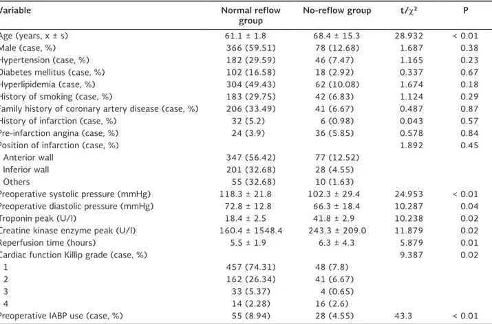
Of the 733 enrolled patients, 54 presented with the no-reflow phenomenon after PCI, with an incidence of 16.1%. The baseline clinical data are summarized in table 1. The normal reflow and no-reflow groups were similar with respect to gender, main risk factors for coronary artery disease (e.g. diabetes mellitus,
Table 1. Baseline clinical data of acute coronary syndrome patients with normal reflow and no-reflow after percutaneous coro-nary intervention
Variable Normal reflow
group No-reflow group t/χ
2 P
Age (years, x ± s) 61.1 ± 1.8 68.4 ± 15.3 28.932 < 0.01
Male (case, %) 366 (59.51) 78 (12.68) 1.687 0.38
Hypertension (case, %) 182 (29.59) 46 (7.47) 1.165 0.23
Diabetes mellitus (case, %) 102 (16.58) 18 (2.92) 0.337 0.67
Hyperlipidemia (case, %) 304 (49.43) 62 (10.08) 1.674 0.18
History of smoking (case, %) 183 (29.75) 42 (6.83) 1.124 0.29
Family history of coronary artery disease (case, %) 206 (33.49) 41 (6.67) 0.487 0.87
History of infarction (case, %) 32 (5.2) 6 (0.98) 0.043 0.57
Pre-infarction angina (case, %) 24 (3.9) 36 (5.85) 0.578 0.84
Position of infarction (case, %) 1.892 0.45
Anterior wall 347 (56.42) 77 (12.52)
Inferior wall 201 (32.68) 28 (4.55)
Others 55 (32.68) 10 (1.63)
Preoperative systolic pressure (mmHg) 118.3 ± 21.8 102.3 ± 29.4 24.953 < 0.01
Preoperative diastolic pressure (mmHg) 72.8 ± 12.8 66.3 ± 18.4 10.287 0.04
Troponin peak (U/l) 18.4 ± 2.5 41.8 ± 2.9 10.238 0.02
Creatine kinase enzyme peak (U/l) 160.4 ± 1548.4 243.3 ± 209.0 11.879 0.02
Reperfusion time (hours) 5.5 ± 1.9 6.3 ± 4.3 5.879 0.01
Cardiac function Killip grade (case, %) 9.387 0.02
1 457 (74.31) 48 (7.8)
2 162 (26.34) 41 (6.67)
3 33 (5.37) 4 (0.65)
4 14 (2.28) 16 (2.6)
Preoperative IABP use (case, %) 55 (8.94) 28 (4.55) 43.3 < 0.01
hypertension, hyperlipidemia, family history of coro-nary artery disease), history of infarction, location of infarction, and pre-infarction angina. Compared with the normal reflow group, the no-reflow group was older (68.4 ± 15.3 vs. 61.1 ± 1.8 years); the reperfu-sion time was significantly longer (6.3 ± 4.3 vs. 5.5 ± 1.9 hours); the preoperative systolic pressure was lower (102.3 ± 29.4 vs. 118.3 ± 21.8 mmHg); the troponin peak was significantly higher (18.4 ± 2.5 vs. 41.8 ± 2.9 U/l) (p < 0.05), and the CK enzyme peak was significantly higher (243.3 ± 209 vs. 160.4 ± 1,548.4 U/l) (p < 0.05). There were significant differ-ences (p < 0.05) in the proportion of preoperative cardiac function Killip grade ≥ 2 and the number of patients using preoperative IABP between the two groups.
Angiography and emergency
percutaneous coronary intervention
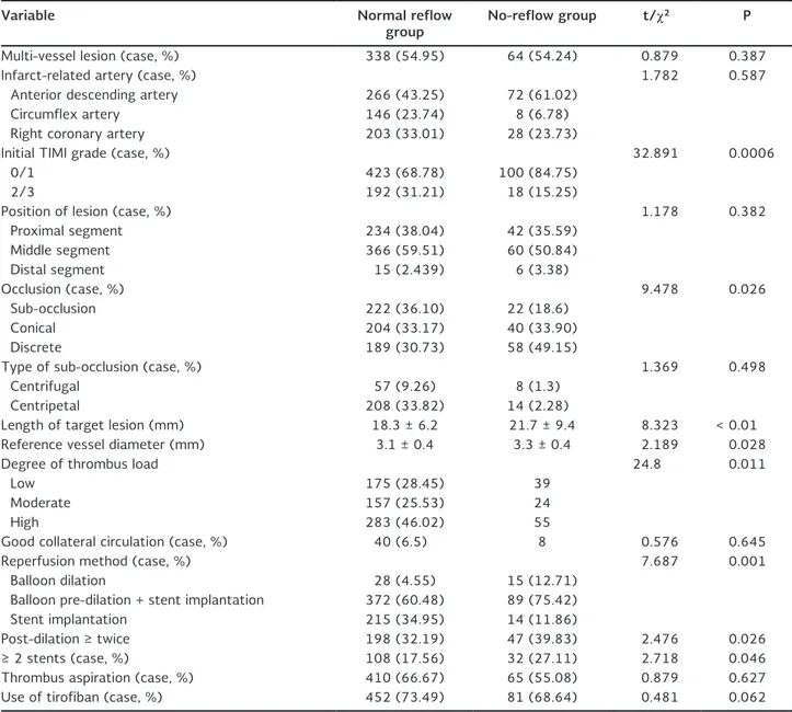
During angiography and emergency PCI, the normal reflow and no-reflow groups had significantly differ-ent patidiffer-ents of preoperative TIMI grade ≤ 1, discrete complete occlusion, long target lesion and reference vessel diameter (p < 0.05) (Table 2). The patients with reperfusion time > 6 hours and high thrombus load were more prone to no-reflow phenomenon, also with significant inter-group differences (p < 0.05). The two groups had similar IRA, number of blood vessels with lesion, position of target lesion, type of sub-oc-clusion and collateral blood flow (p > 0.05). The meth-od of reperfusion also determined whether no-reflow phenomenon occurred in the two groups (p < 0.05). In contrast, the number of implanted stents, maxi-mum distending pressure, or repeated balloon dilation did not affect such phenomenon (p > 0.05) (Table 2).
Univariate and multivariate logistic
regression analysis results for risk
factors related to no-reflow
phenomenon after emergency
percutaneous coronary intervention
Currently, the factors related to no-reflow phenom-enon have not been widely studied. Here we performed univariate and multivariate logistic regression analyses for the factors with significant differences in the base-line clinical data of two groups as well as those with significant differences in angiographic and emergency PCI results. Age, time from appearance of symptoms
to PCT (reperfusion time), systolic pressure upon hos-pitalization, cardiac function Killip grade, preoperative use of IABP, type of occlusion, length of target lesion, degree of thrombus load, preoperative TIMI grade, ref-erence vessel diameter and reperfusion method were selected as the covariates. Finally, age > 65 years (OR: 1.471; 95% CI: 1.462-1.492; p = 0.007), reper-fusion time > 6 hours (OR: 1.274; 95% CI: 1.164-1.405; p = 0.001), low systolic pressure at admission (< 100 mmHg, 1 mmHg = 0.133 kPa) (OR: 1.918; 95% CI: 1.017-3.897; p = 0.004), IABP use before PCI (OR: 1.949; 95% CI: 1.168-3.253; p = 0.011), low TIMI grade (≤ 1) before PCI (OR: 1.100; 95% CI: 1.086-1.257; p < 0.01), high thrombus load (OR: 1.274; 95% CI: 1.423-2.761; p = 0.030) and long target lesion (OR: 1.948; 95% CI: 1.908-1.990; p = 0.019) were identified as independent risk factors for no-reflow phenomenon after PCI (Table 3).
DISCUSSION
No-reflow phenomenon is one of the common com-plications after emergency PCI, with incidence rates of 5-25%8. Its pathophysiological mechanisms mainly include endothelial dysfunction, microvascular dys-function, microvascular spasm, reperfusion injury, and microvascular embolism, among others. Old age, de-layed reperfusion (a relatively long time from the onset of symptoms to PCI), low TIMI grade before PCI, hypo-tension at admission (systolic pressure < 100 mmHg), preoperative IABP use, long target lesion, and high thrombus load are all independent predictive factors for no-reflow phenomenon. In this study, the incidence of no-reflow phenomenon after emergency PCI was approximately 16.1%, being consistent with that in previous literature9-11.
Table 2. Angiographic findings and emergency percutaneous coronary intervention
Variable Normal reflow
group No-reflow group t/χ
2 P
Multi-vessel lesion (case, %) 338 (54.95) 64 (54.24) 0.879 0.387
Infarct-related artery (case, %) 1.782 0.587
Anterior descending artery 266 (43.25) 72 (61.02)
Circumflex artery 146 (23.74) 8 (6.78)
Right coronary artery 203 (33.01) 28 (23.73)
Initial TIMI grade (case, %) 32.891 0.0006
0/1 423 (68.78) 100 (84.75)
2/3 192 (31.21) 18 (15.25)
Position of lesion (case, %) 1.178 0.382
Proximal segment 234 (38.04) 42 (35.59)
Middle segment 366 (59.51) 60 (50.84)
Distal segment 15 (2.439) 6 (3.38)
Occlusion (case, %) 9.478 0.026
Sub-occlusion 222 (36.10) 22 (18.6)
Conical 204 (33.17) 40 (33.90)
Discrete 189 (30.73) 58 (49.15)
Type of sub-occlusion (case, %) 1.369 0.498
Centrifugal 57 (9.26) 8 (1.3)
Centripetal 208 (33.82) 14 (2.28)
Length of target lesion (mm) 18.3 ± 6.2 21.7 ± 9.4 8.323 < 0.01
Reference vessel diameter (mm) 3.1 ± 0.4 3.3 ± 0.4 2.189 0.028
Degree of thrombus load 24.8 0.011
Low 175 (28.45) 39
Moderate 157 (25.53) 24
High 283 (46.02) 55
Good collateral circulation (case, %) 40 (6.5) 8 0.576 0.645
Reperfusion method (case, %) 7.687 0.001
Balloon dilation 28 (4.55) 15 (12.71)
Balloon pre-dilation + stent implantation 372 (60.48) 89 (75.42)
Stent implantation 215 (34.95) 14 (11.86)
Post-dilation ≥ twice 198 (32.19) 47 (39.83) 2.476 0.026
≥ 2 stents (case, %) 108 (17.56) 32 (27.11) 2.718 0.046
Thrombus aspiration (case, %) 410 (66.67) 65 (55.08) 0.879 0.627
Use of tirofiban (case, %) 452 (73.49) 81 (68.64) 0.481 0.062
Table 3. Univariate and multivariate logistic regression analysis results for risk factors related to no-reflow phenomenon after emergency percutaneous coronary intervention
Variable Univariate logistic regression Multivariate logistic regression
P OR (95% CI) P OR (95% CI)
Age > 65 years < 0.010 1.884 (1.830-1.980) 0.007 1.471 (1.462-1.492)
Low systolic pressure at admission (< 100 mmHg) 0.005 1.570 (1.390-1.850) 0.004 1.918 (1.017-3.897)
Initial TIMI grade (0-1) 0.020 1.423 (1.062-1.854) 0.030 1.600 (1.473-2.764)
IABP use before PCI 0.003 1.879 (1.152-3.172) < 0.01 1.100 (1.086-1.257)
High thrombus load 0.020 1.423 (1.062-1.854) 0.030 1.948 (1.908-1.990)
Reperfusion event > 6 hours 0.005 1.564 (1.362-1.872) 0.001 1.274 (1.423-2.761)
Long target lesion 0.027 1.268 (1.156-1.405) 0.019 1.948 (1.908-1.990)
embolism, and microcirculation disorders. These path-ological changes are associated with age and the lack of ischemic preconditioning, collateral circulation, and neurohumoral changes, which easily lead to distal em-bolization of coronary artery during emergency PCI and then to no-reflow phenomenon14-16. In our study, univariate analysis showed that the no-reflow group was older (68.4 ± 15.3 vs. 61.1 ± 1.8 years old) than the normal reflow group, like in previous studies. Additionally, the reperfusion time was significantly prolonged in the patients with no-reflow after PCI (6.3 ± 4.3 vs. 5.5 ± 1.9 hours). The thrombus load was significantly elevated in the patients with reper-fusion time > 6 hours, and the no-reflow incidence was increased about 1.3-fold compared with that of the patients with shorter reperfusion time. Pathophys-iologically speaking, myocardial cells in the infarcted region become completely necrotic owing to the loss of effective blood supply six hours after the onset of myocardial infarction, so recanalization within six hours can exert evident preventive effects. In the case of no-reflow phenomenon after PCI, the ischemic time of myocardial cells in the infarcted region is prolonged, which can also lead to edema of the distal capillary bed, swelling of myocardial cells, neutrophil chemo-taxis, alteration of the integrity of capillaries, and microvascular bed damage. Microvascular bed dam-age can further promote the occurrence of no-reflow phenomenon after PCI. In the early stages of AMI, us-ing thrombolytic drugs to reduce small thrombus in the infarcted region has an obvious effect, and thromboly-sis is also relatively easy. However, the no-reflow phe-nomenon prolongs the duration of infarction in the infarcted region, and many red blood cells are recruited in micro-thrombi of small arteries, so the thrombi be-come more compact and firmer from being dissolved. During balloon dilation, the thrombi easily fracture and flow to downstream smaller arteries with the recana-lization of blood flow, resulting in adverse events such as distal coronary embolization and secondary embo-lism after recanalization. Moreover, prolonged reper-fusion time can also lead to organization of a large number of intracoronary thrombi and form fibrous tissues, further increasing the difficulty of recanaliza-tion as well as the risk of PCI distal embolizarecanaliza-tion17-20. Yip, et al.21 reported that the subgroup of reperfusion time < 4 hours had a lower incidence of no-reflow phenomenon among AMI patients with high thrombus
load, suggesting that reperfusion time was of great clinical significance to the incidence of no-reflow and correlated with thrombus load. In myocardial infarction patients with long reperfusion time and high throm-bus load, using a distal protection device during PCI can effectively relieve thrombosis and other adverse reactions as well as obviously improve the perfusion of myocardial tissues in the infarcted region. However, the no-reflow phenomenon also endangers patients with long reperfusion time and moderate thrombus load, possibly because small particles such as throm-bus fragments destroy the integrity of the microvas-cular bed, inducing the no-reflow phenomenon. Before PCI, the incidence of no-reflow phenomenon in the patients with TIMI grade of IRA ≤ 1 is 1.1 times that of the patients with TIMI grade ≥ 2. De Luea, et al.22 found that before emergency PCI, high TIMI grade was closely related to whether postoperative grade could reach 3 and myocardial perfusion grade could reach 2-3, small infarct area, etc. It is generally believed that TIMI blood flow before PCI indicates good IRA patency, low thrombus load, spontaneous thrombus dissolution, and apparent release of vascular spasm, so the myocar-dial infarction area decreases. The main purpose of AMI treatment is to open IRAs as much as possible, thereby promoting the recovery of normal forward blood flow.
no-reflow phenomenon. In terms of blood hydrody-namics, a larger diameter means a slower blood flow. Hence, a longer infarcted target lesion indicates a larger plaque volume in the lesion, more thrombi in blood vessels, a higher thrombus load, and slow blood flow or no-reflow phenomenon after PCI.
In summary, complex pathological factors affect the occurrence of no-reflow phenomenon after PCI, and these factors, with their combined action, can in-crease the incidence. However, this study has some limitations. Since some patients with incomplete clin-ical data upon hospitalization were excluded, there may be biases that led to a small sample size, thus giving non-significant results. Furthermore, we could not include all possible indices, so there may be other independent risk factors for no-reflow phenomenon.
REFERENCES
1. Tobis J. Is no-no-reflow following PCI in AMI due to distal em-bolization of plaque and thrombus? Catheter Cardiovasc Interv. 2013;82:210-1.
2. Arslan U, Yaman M, Kocaog˘lu I˙, et al. Risk of no-reflow in culprit lesion versus culprit vessel PCI in acute STEMI. Coron Artery Dis. 2015;26:510-5.
3. Gupta S, Gupta MM. No reflow phenomenon in percutaneous coronary interventions in ST-segment elevation myocardial in-farction. Indian Heart J. 2016;68:539-51.
4. Kim MC, Cho JY, Jeong HC, et al. Long-term clinical outcomes of transient and persistent no reflow phenomena following per-cutaneous coronary intervention in patients with acute myocar-dial infarction. Korean Circ J. 2016;46:490-8.
5. Wang L, Liu G, Liu J, Zheng M, Li L. Effects of no-reflow phenom-enon on ventricular systolic synchrony in patients with acute anterior myocardial infarction after percutaneous coronary in-tervention. Ther Clin Risk Manag. 2016;12:1017-22. 6. Yang L, Mu L, Sun L, Qi F, Guo R. Effect of intracoronary
nitroprus-side injection time point on flow recovery during primary percu-taneous coronary intervention in patients with ST elevation acute myocardial infarction. Minerva Cardioangiol. 2017;65:111-18. 7. Leonardi S, Lopes RD, Steg PG, et al. Implications of different
criteria for percutaneous coronary intervention-related myocar-dial infarction on study results of three large phase III clinical trials: The CHAMPION experience. Eur Heart J Acute Cardiovasc Care. 2016. (Epub ahead of print).
8. Amano H, Ikeda T, Toda M, et al. Plaque composition and no-reflow phenomenon during percutaneous coronary intervention
of low-echoic structures in Grayscale intravascular ultrasound. Int Heart J. 2016;57:285-91.
9. Reffelmann T, Kloner RA. The no-reflow phenomenon: basic science and clinical correlates. Heart. 2002;87:162-8.
10. Ipek G, Gungor B, Karatas MB, et al. Risk factors and outcomes in patients with ectatic infarct-related artery who underwent primary percutaneous coronary intervention after ST elevated myocardial infarction. Catheter Cardiovasc Interv. 2016;88: 748-53.
11. Sensoy B, Uzunget SB, Acikgoz S, et al. Renal dysfunction on admission predicts no-reflow phenomenon in patients undergo-ing manual thrombus aspiration durundergo-ing primary percutaneous coronary intervention. Acta Cardiol Sin. 2016;32:185-93. 12. Suda A, Namiuchi S, Kawaguchi T, et al. A simple and rapid
method for identification of lesions at high risk for the no-reflow phenomenon immediately before elective coronary stent im-plantation. Heart Vessels. 2016;31:1904-14.
13. Levi Y, Sultan A, Alemayehu M, Wall S, Lavi S. Association of endothelial dysfunction and no-reflow during primary percuta-neous coronary intervention for ST-elevation myocardial infarc-tion. Cardiovasc Revasc Med. 2016;17:552-5.
14. Balta S, Celik T, Ozturk C, et al. The relation between monocyte to HDL ratio and no-reflow phenomenon in the patients with acute ST-segment elevation myocardial infarction. Am J Emerg Med. 2016;34:1542-7.
15. Wagdy S, Sobhy M, Loutfi M. Neutrophil/lymphocyte ratio as a predictor of in-hospital major adverse cardiac events, new-onset atrial fibrillation, and no-reflow phenomenon in patients with ST elevation myocardial infarction. Clin Med Insights Cardiol. 2016; 10:19-22.
16. Murat SN, Kurtul A, Celik IE, et al. The association of serum procalcitonin level with the no-reflow phenomenon after a primary percutaneous coronary intervention in patients with ST-elevation myocardial infarction. Coron Artery Dis. 2016;27: 116-21.
17. Falk E, Thuesen L. Pathology of coronary microembolisation and no reflow. Heart. 2003;89:983-5.
18. Nukada H, Anderson GM, McMorran PD. Reperfusion nerve in-jury: pathology due to reflow and prolonged ischaemia. J Pe-ripher Nerv Syst. 1997;2:60-9.
19. Dong M, Mu N, Ren F, Li F, Zhang C, Yang J. Matrix metallopro-teinase-9 in the culprit coronary artery and myocardial no-re-flow. Am J Med Sci. 2015;350:352-6.
20. Li J, Wu L, Tian X, Zhang J, Shi Y. Intravascular ultrasound ob-servation of the mechanism of no-reflow phenomenon in acute myocardial infarction. PLoS One. 2015;10:e0119223. 21. Yip HK, Chen MC, Chang HW. Angiographic morphologic
fea-tures of infarct-related arteries and timely reperfusion in acute myocardial infarction: predictors of slow-flow and no-flow. Chest. 2002;122:1322-32.
22. De Luca G, Suryapranata H, Zijlstra F. Symptom-onset-to-bal-loon time and mortality in patients with acute myocardial infarc-tion treated by primary angioplasty. J Am Coll Cardiol. 2003; 42:991-7.
23. Ishikura F, Miki A, Iwata A. Effect of systemic blood pressure in microcollateral circulation evaluated by real-time contrast echo-cardiography. J Am Soc Echocardiogr. 2008;21:765-9. 24. Lee KL, Woodlief LH, Topoi EJ. Predictors of 30-day mortality in