dysfunction in chronic respiratory conditions:
influence of disease severity and body composition
Ester Puig Vilanova
Director:
Dr. Esther Barreiro Portela
UPF Doctoral Thesis / 2014
A vosaltres:
AGRAÏMENTS
Aquesta tesi no hauria estat possible sense la col·laboració de molta gent. Sens dubte, no puc agrair tota l’ajuda rebuda al llarg d’aquests 4 anys amb una simple pàgina! Per tant, aquest apartat només té la finalitat de fer un breu recull de totes les persones que, directament o indirectament, han fet possible aquesta tesi, i de deixar constància de com us estic d’agraïda.
Primer de tot, voldria donar les gràcies a tots aquells que hi han contribuït directament:
A la Dra. Esther Barreiro, per deixar-me formar part del seu grup de recerca i confiar en mi per tirar endavant un projecte i, finalment, tota una tesi! Donar-li gràcies també per tota la tutela rebuda, tant científica com personal, durant aquesta estada al grup.
Gràcies també a tots els membres de l’Hospital del Mar; entre ells el Dr. Lluis Molina i la Mireia així com d’altres companys que també han col·laborat amb part dels experiments de la tesi. Sense la seva contribució, de ben segur que aquest projecte no s’hauria pogut dur a terme.
Haig d’agrair immensament l’ajuda rebuda durant aquests 4 anys a les meves actuals “companyes“ de treball: la Mònica, l’Alba i la Mercè; així com a les “excompanyes”: la Lluïsa, la Marina, la Noèlia, l’Alba i la Cristina. Totes em vau ajudar en el seu moment i encara ara m’ajudeu en tot el que podeu! Gràcies a totes vosaltres, “companyes”, tant per l’ajuda professional com per l’emocional, perquè no només m’heu facilitat el dia a dia, sinó que també m’heu donat una gran amistat que va començar en el seu dia com a “companyes” tot continuant, i de ben segur continuarà, com a grans amigues.
Per altra banda, vull donar les gràcies també a tots aquells que han contribuït d’una manera indirecta en aquesta tesi. La vostra contribució ha estat essencial:
Als companys del gimnàs, i especialment a l’Asún, per ser allà cada matí disposats a escoltar, i amb un somriure per ajudar-me a començar el dia amb plena energia.
Gràcies Ana, Xavier, Kathi, Marina, Ana, Laura, Lorena, Eva, Federico i Joana, pels retrobaments i pels bons moments compartits. Tots junts vam iniciar aquesta etapa amb un màster a Barcelona i de ben segur que continuarem compartint junts molts més bons moments.
Gràcies Rosa per les teves paraules i visió de la vida, sempre a punt fossis on fossis.
Vull donar les gràcies a l’Iris per tota l’ajuda rebuda de forma totalment desinteressada, i per estar sempre disposada a col·laborar en el que fes falta.
També vull agrair a tots els meus amics, en Sergi i la Vane, la Sílvia, tots els de la Conga Catalana, però sobretot a les Nenes, la confiança dipositada en mi. Per creure fermament en el que estava fent tot i no saber massa de què anava, però tot i així, sempre m’heu animat a continuar endavant!
I, finalment, vull donar gràcies a tota la meva família, però especialment a en Costa, i als meus pares, per la immensa paciència que heu tingut durant aquests 4 anys i per estar sempre disposats a fer i entendre el que fes falta!
V
TABLE OF CONTENTS
ABSTRACT
XI
RESUM
XIII
PREFACE
XV
Scientific Collaborations XV
Publications XV
Communications XVII
Funding XVIII
ABBREVIATIONS
XIX
INTRODUCTION
1
1- Skeletal muscles 1
1.1- Muscle formation, structure and organization 1
1.2- Muscle function (Diaphragm and Vastus lateralis) 4
1.3- Skeletal muscle fiber types 4
2- Muscle dysfunction and muscle mass loss 6
2.1- Definition of muscle dysfunction 6
2.2- Cachexia 6
2.3- Conditions associated with muscle dysfunction 7
2.3.1- Chronic obstructive pulmonary disease (COPD) 7
2.3.2- Cancer 9
2.4- Biological mechanisms involved in the process of 9
muscle dysfunction and mass loss 2.4.1- Oxidative and nitrosative stress 9
2.4.2- Inflammation 10
2.4.3- Signaling pathways 11
2.4.3.1- Mitogen-activated protein kinase (MAPK) pathway 11
2.4.3.2- Nuclear Factor-KappaB (NF-κB) pathway 12
2.4.3.3- Forkhead box O class (FoxO) pathway 13
2.4.3.4- Myostatin/Activin pathway 13
VI
2.4.4- Proteolysis 15
2.4.4.1- ATP ubiquitin dependent proteolytic pathway 16
Ubiquitination 16
Sumoylation 17
2.4.4.2- Autophagy-lysosomal pathway 18
2.4.4.3- Calcium activated system 19
2.4.5- Epigenetic mechanisms 19
2.4.5.1- Histone modifications 20
Histone methylation 20
Histone acetylation 21
2.4.5.2- MicroRNAs 23
HYPOTHESIS
27
OBJECTIVES
29
1- Specific objectives of study #1 30
2- Specific objectives of study #2 31
3- Specific objectives of study #3 32
4- Specific objectives of study #4 33
METHODS
35
1- Methods Study #1 41
1.1- Study subjects 41
1.2- Antropometrical and funcional assessment 42
1.3- Muscle biopsies and blood samples 42
1.4- Molecular biology analyses 42
1.4.1- Immunoblotting of 1D electrophoresis 42
1.4.2- Superoxide dismutase and catalase activity assays 45
1.4.3- Protein carbonylation using enzyme-linked immunosorbent assay 45
1.4.4- Cytokine enzyme-linked immunosorbent assay 46
1.4.5- Measurement of superoxide anion radicals 47
by lucigenin-derived hemiluminescence 1.4.6- Muscle fiber counts and morphometry 47
1.4.7- Muscle structure abnormalities 47
1.4.8- Ultrastructural evaluation 48
1.4.8.1- Sarcomere length 49
1.4.8.2- Mitochondrial diameter 49
VII
1.5- Statistical analyses 50
2- Methods Study #2 51
2.1- Study subjects 51
2.2- Antrophometrical and funcional assessment 51
2.3- Muscle biopsies and blood samples 52
2.4- Molecular biology analyses 53
2.4.1- RNA isolation 53
2.4.2- MicroRNA and mRNA reverse transcription (RT) 53
2.4.3- Real time-PCR amplification (qRT-PCR) 53
2.4.4- Immunoblotting of 1D electrophoresis 54
2.4.5- Muscle fiber counts and morphometry 56
2.5- Statistical analyses 57
3- Methods Study #3 59
3.1- Study subjects 59
3.2- Antrophometrical and funcional assessment 60
3.3- Muscle biopsies and blood samples 60
3.4- Molecular biology analyses 60
3.4.1- RNA isolation 60
3.4.2- MicroRNA and mRNA RT 61
3.4.3- qRT-PCR 61
3.4.4- Immunoblotting of 1D electrophoresis 61
3.4.5- Muscle fiber counts and morphometry 63
3.5- Statistical analyses 64
4- Methods Study #4 65
4.1- Study subjects 65
4.2- Antrophometrical and funcional assessment 65
4.3- Muscle biopsies and blood samples 66
4.4- Molecular biology analyses 66
4.4.1- RNA isolation 66
4.4.2- MicroRNA and mRNA RT 66
4.4.3- qRT-PCR 67
4.4.4- Immunoblotting of 1D electrophoresis 67
4.4.5- Muscle fiber counts and morphometry 69
VIII
RESULTS
71
1- Results Study #1 71
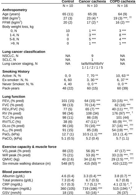
1.1- Clinical characteristics 71
1.2- Muscle and systemic redox balance 73
1.2.1- Protein oxidation 73
1.2.2- Antioxidants 74
1.3- Muscle and systemic inflammation 76
1.4- Redox-signaling markers in muscles 77
1.4.1- MAPK signaling pathway 77
1.4.2- NF-kB signaling pathway 78
1.4.3- FoxO signaling pathway 80
1.4.4- AMPK signaling pathway 81
1.5- Muscle proteolysis markers 82
1.6- Muscle growth and differentiation 83
1.7- Contractile and funcional muscle proteins 83
1.8- Muscle structure 84
1.8.1- Fiber type composition 84
1.8.2- Muscle abnormalities 85
1.9- Electron microscopy features 85
2- Results Study #2 91
2.1- Clinical characteristics 91
2.2- Muscle biological markers 93
2.2.1- MicroRNAs expression 93
2.2.2- Histone modifications 95
2.2.3- Myogenic transcription factors 96
2.2.4- Expression of SUMO 97
2.3- Muscle structure 97
3- Results Study #3 99
3.1- Clinical characteristics 99
3.2- Muscle biological markers 101
3.2.1- MicroRNAs expression 101
3.2.2- Histone modifications 105
3.2.3- Myogenic transcription factors 108
3.2.4- Expression of SUMO 109
IX
4- Results Study #4 111
4.1- Clinical characteristics 111
4.2- Muscle biological markers 113
4.2.1- MicroRNAs expression 113
4.2.2- Histone modifications 115
4.2.3- Myogenic transcription factors 117
4.2.4- Expression of SUMO 118
4.3- Muscle structure 118
5- Summary of main findings 121
5.1- Study #1 121
5.2- Study #2 122
5.3- Study #3 123
5.4- Study #4 125
DISCUSSION AND CONCLUSIONS
127
1- Discussion 127
2- Study limitations 137
3- Concluding remarks 141
XI
ABSTRACT
ABSTRACT
XII
XIII
RESUM
La disfunció muscular esquelètica i la pèrdua de massa muscular són dues manifestacions sistèmiques freqüents en malalties com la Malaltia Pulmonar Obstructiva Crònica (MPOC) i el Càncer de Pulmó (CP). Malgrat que en els malalts amb MPOC, els músculs perifèrics sovint es veuen més greument afectats, els músculs respiratoris també mostren alteracions estructurals i funcionals. La disfunció muscular que pateixen aquests pacients afecta negativament la seva qualitat de vida, no només reduint la seva tolerància a l’exercici físic sinó també a les seves activitats quotidianes. Diferents mecanismes moleculars estan implicats en la etiologia de la disfunció muscular en la MPOC. Hipòtesi: La nostra hipòtesi és que l’estrès oxidatiu
podria ser un desencadenant de l’augment de proteòlisi i disfunció muscular en els músculs perifèrics de pacients amb caquèxia associada a processos respiratoris com ara la MPOC i el CP. Els mecanismes epigenètics podrien estar també implicats en la fisiopatologia de la disfunció muscular en la MPOC. Objectius: 1) Avaluar els mecanismes moleculars que contribueixen a la disfunció muscular i pèrdua de massa muscular en els músculs perifèrics de pacients amb MPOC i CP, 2) Explorar l’expressió de varis fenòmens epigenètics en el múscul diafragma de pacients amb MPOC d’un ampli rang d’obstrucció aèria, 3) Analitzar els marcadors epigenètics en el múscul vast extern (VE) del quàdriceps en pacients d’un ampli rang d’obstrucció aèria i composició corporal, i 4) Avaluar els mecanismes epigenètics en el VE de pacients amb MPOC lleu o molt lleu. Mètodes: Estudi #1: L’equilibri redox, la inflamació, les
RESUM
XIV
oxidació proteica, tant sistèmica com muscular, major contingut i activitat muscular d’enzims antioxidants, major nivells de citoquines i factors de creixement musculars, nivells augmentats de les vies de senyalització redox (NF-kB, FoxO-3), i un augment dels nivells d’enzims lligases E3 i de la ubiquitinació proteica total. Estudi #2: en el diafragma de pacients amb MPOC moderada, l’expressió de microARNs específics de múscul (miR-1, -133, i -206) estava disminuïda, mentre que els nivells proteics de HDAC4 i MEF2C estaven augmentats. Estudi #3: en els músculs perifèrics de pacients amb MPOC greu sense pèrdua de massa muscular, l’expressió de microARNs enriquits en el múscul estava augmentada, mentre que l’expressió dels mateixos microARNs estava disminuïda en els pacients amb MPOC greu i pèrdua de massa muscular en comparació amb aquells sense pèrdua muscular. A més, en els músculs perifèrics dels pacients amb pèrdua de massa muscular els nivells d’acetilació estaven augmentats, mentre que els nivells de la HDAC3 i el SIRT-1 estaven disminuïts. Estudi #4: Els músculs perifèrics de pacients amb MPOC lleu presentaren un augment en l’expressió de miR-1 i en els nivells proteics de la HDAC4. Conclusions: 1) L’estrès
XV
PREFACE
Scientific collaborations
In the current thesis, the investigations have been conducted in the Muscle and Respiratory System Research Unit (URMAR), IMIM-Hospital del Mar, in collaboration with researchers from other departments and centers as described below.
Study #1 was in collaboration with the Pulmonology Department at Hospital del Mar, Barcelona, Spain, the Pathology Department at Hospital del Mar, Barcelona, Spain, and with the Pulmonology Department (ICT) at Hospital Clinic-IDIBAPS, Barcelona, Spain.
Study #2 was in collaboration with the Pulmonology Department at Hospital del Mar, Barcelona, Spain, and the Thoracic Surgery Department at Hospital del Mar, Barcelona, Spain.
Study #3 was in collaboration with the Pulmonology Department at Hospital del Mar, Barcelona, Spain, and with the Pulmonology Department (ICT) at Hospital Clinic-IDIBAPS, Barcelona, Spain.
Study #4 was in collaboration with the Pulmonology Department at Hospital del Mar, Barcelona, Spain.
Publications
The results of the investigation included in the current PhD thesis have been recently submitted in international journals. Additionally, during the four years in the Molecular Mechanisms of Lung Cancer predisposition group, under the supervision of Dr. Esther Barreiro, I had also the opportunity to participate in other studies apart from those that compose the current PhD thesis. Some of these studies have been already published in several international journals.
Study #1- Puig-Vilanova E, Rodriguez DA, Lloreta J, Ausín P, Pascual-Guardia S, Broquetas J, Roca J, Gea J, Barreiro E.
Oxidative stress and redox signaling pathways in cachectic muscles of patients with advanced COPD and lung cancer.
PREFACE
XVI
Study #2- Puig-Vilanova E, Aguilo R, Rodriguez-Fuster A, Martínez-Llorens J, Gea J, Barreiro E.
Epigenetic mechanisms in respiratory muscle dysfunction of patients with chronic obstructive pulmonary disease.
Submitted.
Study #3 - Puig-Vilanova E, Martínez-Llorens J, Ausín P, Roca J, Gea J, Barreiro E. Epigenetic events in the vastus lateralis of patients with severe COPD and muscle atrophy.
Submitted.
Study #4 - Puig-Vilanova E, Ausín P, Martínez-Llorens J, Gea J, Barreiro E.
Do epigenetic events take place in the vastus lateralis of patients with mild chronic obstructive pulmonary disease?
Revised version submitted.
1- Fermoselle C, Garcia-Arumi E, Puig-Vilanova E, Andreu AL, Urtreger AJ, Bal de Kier Joffé ED, Tejedor A, Puente-Maestu L, Barreiro E.
Mitochondrial dysfunction and therapeutic approaches in respiratory and limb muscles of cancer cachectic mice.
Exp Physiol. 2013 Sep;98(9):1349-65. Epub 2013 Apr 26
2- Fermoselle C, Rabinovich R, Ausín P, Puig-Vilanova E, Coronell C, Sanchez F, Roca J, Gea J, Barreiro E.
Does oxidative stress modulate limb muscle atrophy in severe COPD patients? Eur Respir J. 2012 Oct;40(4):851-62. Epub 2012 Mar 9.
3- Barreiro E, del Puerto-Nevado L, Puig-Vilanova E, Pérez-Rial S, Sánchez F, Martínez-Galán L, Rivera S, Gea J, González-Mangado N, Peces-Barba G.
Cigarette smoke-induced oxidative stress in skeletal muscles of mice. Respir Physiol Neurobiol. 2012 Jun 15;182(1):9-17.
4- Rodriguez DA, Kalko S, Puig-Vilanova E, Perez-Olabarría M, Falciani F, Gea J, Cascante M, Barreiro E, Roca J.
XVII
Communications
The results of the investigation included in the current PhD thesis as well as those from other studies, which are not included in the thesis, have been previously presented in the form of an abstract at several national and international conferences.
1- Barreiro E, Puig-Vilanova E, Martínez-Llorens M, Pascual-Guardia S, Casadevall C, Gea J.
Differential profile of epigenetic events in diaphragm and limb muscles of COPD patients.
American Thoracic Society (ATS) International Conference, San Diego, May 2014
2- Puig-Vilanova E, Busquets S, Toledo M, Sanchez F, Argiles JM, Gea J, Lopez-Soriano F, Barreiro E.
Redox balance and mitochondrial dysfunction in respiratory and limb muscles of cancer cachectic rats.
The 4th EMBO meeting (Abstract book, page 200), Nice, France, September 2012
3- Barreiro E, Puig-Vilanova E, Ausín P, Sánchez F, Martínez-Llorens J, Curull V, Broquetas J, Gea J.
Patrón diferencial de estrés oxidativo e inflamación muscular en la caquexia asociada al cáncer y a la EPOC.
XIII International Symposium on COPD, Barcelona, Spain, April 2012.
4- Barreiro E, del Puerto L, Puig-Vilanova E, Pérez-Rial S, Sánchez F, Martínez-Galán L, Gea J, González-Mangado N, Peces-Barba G.
Estrés oxidativo en los músculos respiratorios y periféricos en un modelo experimental de enfisema inducido por el humo del cigarrillo.
XIII International Symposium on COPD, Barcelona, Spain, April 2012.
5- Puig-Vilanova E, Marín-Corral J, Sabaté M, Sánchez F, Gea J, Molina L, Barreiro E. Papel de los antioxidantes y de la inhibición de la proteólisis en la caquexia asociada al cor pulmonale en un modelo experimental.
PREFACE
XVIII
6- Puig-Vilanova E, Marín-Corral J, Sánchez F, Sabaté M, Gea J, Molina L, Barreiro E. Efecto de los antioxidantes y el bortezomib sobre la proteólisis muscular en un modelo experimental de insuficiencia cardiaca derecha.
Cuartas Jornadas de Formación del CIBERES (Abstract book, page 22), Bunyola, Mallorca, Illes Balears, Spain, October 2011.
7- Barreiro E, Ausín P, Sánchez F, Puig-Vilanova E, Martinez-Llorens J, Curull V, Broquetas J, Gea J.
Role of systemic and muscle oxidative stress and cytokines in patients with lung cancer cachexia.
European Respiratory Society (ERS) Annual Congress. Eur Respir J 2011; 38 (suppl 55). Amsterdam, September 2011
8- Marín-Corral J, Fornaguera C, Sánchez F, Sabaté M, Puig-Vilanova E, Gea J, Molina L, Barreiro E.
Efecto de los antioxidantes y el bortezomib sobre la proteólisis muscular en un modelo experimental de insuficiencia cardiaca derecha.
Arch Bronconeumol 2011; 47 (especial congreso): 96.
9- Puig-Vilanova E, Busquets S, Toledo M, Sánchez F, Argilès JM, Gea J, López-Soriano F, Barreiro E.
Efectos del formoterol sobre el estrés oxidativo, la inflamación, la estructura y el daño muscular en los músculos respiratorios y periféricos de ratas con caquexia.
Terceras Jornadas de Formación del CIBERES (Abstract book, page 26), Bunyola, Mallorca, Illes Balears, Spain, October 2010.
Funding
XIX
ABBREVIATIONS
ActRIIB: activin receptor type-IIB ADP: adenosine diphosphate
AIDS: acquired immunodeficiency syndrome Akt: RAC-alpha serine/threonine-protein kinase AMP: adenosine monophosphate
AMPK: adenosine monophosphate-activated protein kinase APR: acute phase response
Atg: autophagy-related gene ATP: adenosine triphosphate bHLH: basic-helix-loop-helix BMI: body mass index BSA: bovine serum albumin
C8-20S: C8 alpha-subunit of the 20S proteasome CAT: catalase
CBP: cAMP response element-binding protein cDNA: complementary deoxyribonucleic acid CHF: chronic heart failure
CKD: chronic kidney disease CO2: carbon dioxide
CoA: coenzyme A
COPD: chronic obstructive pulmonary diseases CRP: C-reactive protein
CSA: cross-sectional area
CuZn-SOD: copper zinc superoxide dismutase DLCo: diffusing capacity
DNA: deoxyribonucleic acid DNP: dinitrophenol
E1: ubiqiutin-activating enzyme E2: ubiquitin-conjugating enzyme E3: ubiquitin-ligase enzyme
EDTA: ethylenediaminetetraacetic acid
ABBREVIATIONS
XX FEV1: forced expiratory volume in one second
FFMI: fat free mass index FoxO: forkhead box O class FVC: forced vital capacity
GAPDH: glyceraldehyde-3-phosphate dehydrogenase GDF-8: growth factor differentiation factor-8
GNAT: Gcn5-related N-acetyltransferase
GOLD: global initiative for chronic obstructive lung disease GSV: globular sedimentation velocity
HAT: histone acetyltransferase HDAC: histone deacetylase
HKMT: histone lysine methyltransferases HNF4: hepatocyte nuclear factor 4
HNE: hydroxynonenal H2O: water
HRMT: histone arginine methyltransferases HRP: horseradish peroxidase
IGF-1: insulin-like growth factor 1
IkB-α:nuclear factor kappa-light-chain-enhancer of activated B cells inhibitor-alpha IKK: I kappaB kinase
IL-1β: interleukin-1-beta IL-6: interleukin-6 IL-8: interleukin-8
INF-γ: interferon-gamma
JNK: c-Jun NH2-terminal kinase
KAT: lysine acetyltransferase KCO: Krough transfer factor
LC: lung cancer
LDCL: lucigenin-derived chemiluminescence MAFbx: muscle atrophy F box
MAPK: mitogen-activated protein kinase MDA: malondialdehyde
MEF2: myocyte enhancer factor 2 miR: microRNA
XXI Mn-SOD: manganese superoxide dismutase MRF: myogenic regulatory factor
Mrf4: muscle regulatory factor 4 mRNA: messenger ribonucleic acid mTOR: mammalian target of rapamycin MuRF1: muscle ring finger protein 1 MyHC: myosin heavy chain
MyoD: myogenic differentiation Myf5: myogenic factor 5
Myf6: myogenic factor 6
MIP: maximal inspiratory pressure NAD: nicotinamide adenine dinucleotide
NF-kB: nuclear factor kappa-light-chain-enhancer of activated B cells NT: nitrotyrosine
NSCL: non-small cell lung cancer O2·-: superoxide anion
p300: E1A binding protein p300
PaCO2: arterial carbon dioxide partial pressure
PaO2:arterial partial pressure of oxygen
PGC1-α: peroxisome proliferator-activated receptor-γ coactivator 1-alpha PI3K: phosphoinositide 3-kinase
PRMT: protein arginine methylatransferases Pdi max: maximal transdiaphragmatic pressure PVDF: polyvinylidene difluoride
QMVC: quadriceps isometric maximum voluntary contraction
qRT-PCR: quantitative reverse transcription - polymerase chain reaction RISC: ribonucleic acid-induced silencing complex
RNA: ribonucleic acid
RNS: reactive nitrogen species ROS: reactive oxygen species RV: residual volume
SDS-PAGE: sodium dodecyl sulfate – polyacrylamide gel electrophoresis Sirt1: silent information regulator 1
ABBREVIATIONS
XXII SR: sarcoplasmic reticulum
SUMO: small ubiquitin-related modifier SCLC: small cell lung cancer
TGF-β: transforming growth factor-beta TLC: total lung capacity
TNF-α: tumour necrosis factor-alpha
TNM: tumor, nodes, metastasis classification of malignant tumors TV: tidal volume
UBL: ubiquitin-like protein UTR: untranslated region
VEGF: vascular endothelial growth factor VO2 peak: peak exercise oxygen uptake
WR peak: peak work rate XPO5: exportin-5
1
INTRODUCTION
1- Skeletal muscles
The skeletal muscle accounts for approximately 40-50% of human body weight. It is a specialized tissue with the ability to contract and relax due to its internal organized arrangement that confers the characteristic striated appearance. This property of the muscle to contract and relax can be controlled either in a voluntary or involuntary manner depending on the muscle type, with the primarily function to assist to the maintenance of homeostasis and adapt to our needs (1-3).
1.1- Muscle formation, structure and organization
Each muscle of our body is composed for several muscle fascicles which in turn are formed by numerous muscle cells that lie parallel to each other (3). These muscle cells are named muscle fibers or also myofibers due to their elongated and cylindrical shape fiber like, which is usually long as the muscle fiber in which they are contained (4). Muscle fibers are formed by the process of myogenesis, which is divided into two main distinct phases: muscle growth and muscle differentiation. Initially, during the skeletal muscle development, muscle progenitor cells called myoblasts exclusively proliferate, but later on, in the absence of growth factors these myoblasts quits the cell cycle and starts to differentiate and fuse into multi-nucleated fibers called myotubes (3, 5). These two processes of the skeletal muscle development are strictly regulated by a family of transcription factors known as myogenic regulatory factors (MRFs), which are responsible for the regulation of downstream targets required for the terminal differentiation of a myoblast into a functional myofiber (6, 7). Myf5, MyoD, Myogenin and Mrf4, also known as Myf6, are members of the MRFs, which belong to the basic-helix-loop-helix (bHLH) class of transcription factors specifically expressed in the skeletal muscles (5, 6, 8). In order to accomplish their function, these proteins bind to the regulatory region of muscle specific genes and provide a cascade of gene expression (8) that results in a functional myotube (5, 6).
INTRODUCTION
2
fibers. In turn, each myofibril is formed by two types of filaments; the thick and the thin filaments that correspond to clusters of the contractile proteins myosin and actin respectively (2, 4). The organized arrangement of these two contractile proteins along with the other cytoskeletal proteins confers to the skeletal muscle the characteristic repetitive pattern of light and dark striations appearance. This striated appearance of the myofibril is divided into sarcomeres of 2 um length which are the functional unit of skeletal muscles (3). Within the sarcomere, the pattern of overlap between the myosin and actin proteins creates a variety of zones and bands that can be identified under a microscope by the following characteristics (3, 4) (shown in figure 1):
The Z-line: which is a dense region of proteins that delimitates one sarcomere form the next.
The A-band: which corresponds to the dark stripe and is found in the middle part of the sarcomere thus extending the entire length of thick filament (myosin).
The I-band: which is a less dense area corresponding to the light stripe due to the presence of thin filament (actin) and non-thick filament.
The H-zone: which corresponds to the light area at the centre of a sarcomere, concretely in the middle of A-band. In this region there is no overlapping of the cytoskeletal proteins actin and myosin and only the thick filament is found. Particularly, the width of this zone is diminished during contraction.
The M-region: which is the dark line in the middle of H-zone, is located exactly in the centre of sarcomere and contains the myosin and other supporting proteins (myomesin and creatine kinase) that are critical for organization and alignment of the thick filament.
3
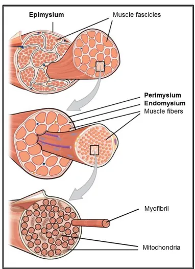
[image:27.595.202.393.424.687.2]Apart from the presence of numerous myofibrils the muscle fiber is abundant in mitochondria. The mitochondria length is up to 1 to 2 um of diameters and adopts an oval shape within the muscle fibre. Mitochondria are found mainly between myofibrils and are the organelles responsible to form the ATP, the main energy of the living cells. Therefore, mitochondria are required for muscle contraction. Mitochondria produced the ATP from the krebs tricarboxylic acids cycle trough the cytochrom chain and the ATP-synthase by degrading two components that can be found in the cytoplasm of the cell; the glyocogen granules and the lipid droplets. However, mitochondria are also a major source of reactive oxygen species production inside skeletal muscle fibers (3, 9). Each subunit of muscle is surrounded by connective tissue that adopts different names according to their localization. As shown in figure 2, there are three layers of muscle connective tissue: the epimysium, the perimysium and the endomyisium. While epimysium is the most outer layer surrounding the whole muscle, the perimysium is the sheat of connective tissue that surrounds the muscle fascicle being the endomysium the most indoor sheat of connective tissue surrounding the muscle cell. The role of the connective tissue is to transmit to the tendon and skeleton the action of the contractile proteins in order to effect the movement (2).
INTRODUCTION
4
1.2- Muscle function (Diaphragm and Vastus lateralis)
The interactions between myosin and actin filaments lead to the cross-bridge formation and the subsequently cross-bridge cycle or sliding filament theory, the mechanisms by which the myosin produces force causing the filaments to slide and shortens the sarcomere. This ability to contract and relax allows the skeletal muscles to perform three main functions: 1) support the skeleton, 2) allow movements such as locomotion and manipulation of objects, and 3) the vital function of ventilation through the movement of the respiratory muscles (4, 11, 12).
The diaphragm is the main respiratory muscle and a vital organ that trough the continuous involuntary contractions and relaxations assist to the function of adequate gas exchange through the movement of air into and out of the lungs (11, 13, 14). In contrast, the vastus lateralis is a peripheral muscle exposed to voluntary manner that through contraction make possible movements of daily life such as rise from a position, walking, etc.Most studies focus on the analysis of the vastus lateralis, which is the largest and the most accessible muscle of the quadriceps femoris group of muscles composed by the rectus femoris, vastus medialis, vastus intermedius, and the vastus lateralis, located in the lateral side of the thigh (1, 11, 14).
1.3- Skeletal muscle fiber types
Each muscle can perform different tasks in order to adapt to our needs due to the different types of skeletal muscle fibers that coexist within the same muscle and differ one from another by their structural and functional properties (15, 16). Proportions of each muscle fiber type varies depending on the action of the muscle although in some muscles one or another type of muscle fiber predominates depending upon its requirements (17). In addition, due to its higher plasticity, muscle fiber can convert from one type to another and change its size in response to different stimuli such as exercise, training and environmental factors, which regulate muscle phenotype, both, fiber type composition and fiber size, in order to adapt to the different functional demands (15, 16).
5
fibers are classified into three main types according to their histochemical and biochemical characteristics as illustrated in the following table (Table 1):
Properties Human skeletal muscle fiber types
MyHC isoform Type I Type IIa Type IIx Speed of contraction Slow Fast Very fast Metabolism Oxidative Oxidative glycolytic Glycolytic Myofibrillar ATPase
activity (Force production)
Low High Very High
Resistance to fatigue High Intermediate Low
Activity used for Aerobic Long term anaerobic Short term anaerobic Muscle colour
Myoglobin content
Red High
Intermediate Medium
White Low Number of mitochondria High Medium Low Capillary density High Intermediate Low
Table1: Main characteristics of the three types of human skeletal muscle fibers (16, 18).
Type I muscle fibers are the slow-twitch oxidative fibers, characterized by being the smallest in diameter and red due to their large amounts of myoglobin content that confers them the red color. These fibers are supplied with many blood capillaries and mitochondria and generate ATP mainly by aerobic cellular respiration. That is the reason why they are called oxidative fibers. These fibers are also referred to as slow, since the ATP is hydrolysed by the ATPase relatively slowly and consequently the speed of contraction is slow. They are the least powerful type of muscle fibers but they are resistant to fatigue, thus being suitable to low intensity but sustained activities (16, 18).
INTRODUCTION
6
relatively few blood capillaries and mitochondria. In contrast to other types, they contain large amounts of glycogen and generate the ATP mainly by glycolysis. These fibers have the ability to hydrolyze the ATP rapidly so they contract strongly and fast but they come quickly fatigue. This type of fibers is appropriate for intense exercise of short duration (16, 18).
2 – Muscle dysfunction and muscle mass loss
2.1- Definition of muscle dysfunction
Muscle dysfunction is defined by either the loss of at least one of the two main functional properties of the muscle: strength and endurance (11, 14). While strength is the capacity of the muscle to generate force through muscle contraction, endurance is the capacity of the muscle to maintain that force over time. Both properties depend on muscle characteristics: while strength depends on muscle mass, endurance depends on muscle composition, concretely on the proportion of type I fibers, those more resistant to fatigue. Therefore, the loss of these two properties leads to muscle weakness and fatigue respectively (11, 14, 19, 20).
Several respiratory diseases and malignant conditions including chronic obstructive pulmonary disease (COPD) and lung cancer (LC) are associated with muscle dysfunction and weakness of both respiratory and limb muscles, thus affecting the quality of life of these patients and aggravating the disease itself. Whereas peripheral muscle weakness limits the activities of daily life, leading to inactivity and loss of independence, the loss of respiratory muscle function is critical since implies respiratory muscle weakness and consequently a ventilator insufficiency that may lead to respiratory failure (13, 21-23).
2.2- Cachexia
7
mass it is known as cachexia whereas when it is due to the process of normal aging it is known as sarcopenia (21, 22).
Cachexia is a highly prevalent condition ranging from 5 to 15% in COPD patients and 60 to 80% in patients with advanced cancer. These cachectic patients can reach a progressive weight loss, up to 80% along with alterations in body composition and disturbed homeostasis of fat and muscle tissues (23, 24).
In order to diagnose cachexia, the international consensus statement (25) defined that cachexia is present when at least one of the following clinical conditions are found in patients:
• Weight loss >5% over the past 6 months (in absence of simple starvation); • Body mass index (BMI) <20 kg/m and any degree of weight loss >2%;
• Appendicular skeletal muscle index consistent sarcopenia (males <7.26 kg/m; females <5.45 kg/m; determined by dual energy x-ray absorptiometry and sex-specific defined reference values) and any degree of weight loss >2%
2.3- Conditions associated with muscle dysfunction
2.3.1- Chronic obstructive pulmonary disease (COPD)
Chronic obstructive pulmonary disease (COPD) is described by an airflow limitation that is usually progressive and is not fully reversible (11, 26). Cigarette smoke is the main risk factor for the development of the pathogenesis of COPD that causes a parenchymal destruction of the lungs along with extrapulmonary (systemic) effects that lead to other comorbid conditions, and contribute to the severity of the disease (26, 27). Different symptoms are present during the course of COPD. While chronic cough and sputum are preceded symptoms that can be present many years before of the development of the airflow limitation, progressive dyspnea appears once COPD is present. Throughout the advanced severity of COPD other symptoms such as weight loss, anorexia and other psychiatric morbidities such as depression or anxiety become evident (26).
COPD is a highly prevalent and costly disease (26) that ranks one of the most important causes of death worldwide either consequence of the disease itself of its complications, and is expected to become the third leading cause of death worldwide by 2020 (26, 28).
Several lung function parameters are used to evaluate and monitor disease progression in COPD patients: forced expiratory volume in 1 second (FEV1), forced
INTRODUCTION
8
(DLCO) (26, 29). Briefly, FEV1 is the volume of air forcibly exhaled during the first
second; FVC is the volume of air that can be forcibly exhaled in the point of maximal inspiration; TV is the volume of air inhaled and exhaled at rest; TLC is the maximum volume of air that can be present in the lungs; and DLCO is the carbon monoxide uptake from a single inspiration usually in 10 sec.
According to the Global Initiative for Chronic Obstructive Lung Disease (GOLD) guidelines (26), these parameters are used to classify the severity of COPD into four stages as follow (Table 2):
Stage I: mild FEV1/FVC < 0.70 FEV1 > 80% predicted
Stage II: moderate FEV1/FVC < 0.70
50% < FEV1 , 80% predicted
Stage III: severe FEV1/FVC < 0.70
30% < FEV1 , 50% predicted
Stage IV: very severe
FEV1/FVC < 0.70
FEV1 < 30% predicted or FEV1 < 50% predicted
plus chronic respiratory failure*
Table 2: Spirometric classification of COPD based on post-bronchodilator FEV1 (26). *
Respiratory failure: arterial partial pressure of oxygen (PaO2) < 8.0 kPa (60 mm Hg) with or without arterial
partial pressure of CO2 (PaCO2) > 6.7 kPa (50 mm Hg) while breathing air at sea level.
9
2.3.2- Cancer
Cancer is a major condition that among other things is also associated with muscle dysfunction and wasting. Unlike COPD, muscle mass loss in cancer is more strikingly evident and called cachexia, whereas in COPD is more frequently used the term wasting to describe the muscle mass loss. Patients with several types of cancer often develop cachexia with or without anorexia (33). However, in lung cancer, the cachexia syndrome is more evident than in other types of cancer (34, 35). Lung cancer is the leading cause of cancer-related deaths worldwide accounting for more than one million people deaths each year (27, 36). In addition, at the time of their diagnosis, approximately the 60% of patients have already experienced considerable body weight loss (33, 37).
Cachexia has been associated with reduce survival and estimated to be the responsible cause of death in 20 to 40% of cancer patients. However, it has also other negative implications; it also increases surgical risk and decreases the response to chemotherapy. Together with a decreased physical function, cachexia really impairs the quality of life of cancer patients (25, 33, 35).
2.4- Biological mechanisms involved in the process of muscle dysfunction
and mass loss
2.4.1- Oxidative and nitrosative stress
INTRODUCTION
10
Proteins and consequently the structural myofilament proteins of skeletal muscle are the major targets of oxidative stress. The posttranslational modification that they undergo makes them more susceptible to further degradation (40) suggesting a role of oxidative stress in muscle dysfunction and wasting (9, 40). Protein carbonylation along with the final products of lipidic peroxidation malondialdehyde (MDA), 4-hydroxy-2-nonenal (HNE), and 3-nitrotyrosine formation (NT) are important markers of oxidative stress (9, 39-42). The assessment of these markers has been widely studied in our group demonstrating a link between oxidative stress and skeletal muscle dysfunction and wasting of COPD patients (31, 43) in both respiratory (31) and limb muscles (9, 11, 14, 31, 40) as well as in cancer animal models (9, 38, 39).
2.4.2- Inflammation
Inflammation is a physiologic response mediated by different cells and molecules of the organism against aggressions of external agents. Inflammatory response have the objective to defend, aisle and destroy the damaging agents as well as repair the tissue or organ damaged. However, when this response is persistent or excessive, inflammation can become detrimental (44, 45).
During the inflammatory response, different cells become activated, attracted to the injured tissue and deliver different substances such as cytokines and growth factors that are essential for elimination of the detritus and repairing the damaged tissue (44, 45). Inflammation of skeletal muscle can be both a systemic and a local factor. Presence of inflammatory mediators can affect systemic circulation, reaching various organs and tissues, including muscles, and directly damage the muscle structure through affecting the contractile performance of muscle fibers and thus contributing to their dysfunction (11, 46).
11
can up-regulate the production of other proinflammatory substances (45). Most of these cytokines exert their function by binding to specific extracellular receptors that execute intracellular signaling pathways that results in muscle atrophy and dysfunction (14) through inducing increase protein degradation by the activation of catabolic pathways (11, 14, 45). In addition, evidence shown that oxidative/nitrosative stress and inflammation are related molecular mechanisms that could act cooperatively in the triggering of muscle dysfunction and muscle mass loss since oxidative stress can activate the expression of inflammatory mediators and in turn, inflammation can modulate oxidative stress through the regulation of ROS production levels (11, 14, 39, 48, 52).
However, contradictory results have also been reported regarding the role of proinflamamtory cytokines in muscle (45). Furthermore and probably due to the differences of activity and function of each muscle, the expression of proinflammatory cytokines seems to be differentially expressed between respiratory and limb muscles (32, 50).
2.4.3- Signaling pathways
Cell signaling pathways are responsible for the transmission of information within the cell and coordinate cellular activities. Cell signaling pathways are able to respond either to an external or internal stimulus and generate a cellular response. This signal transduction process usually involves several steps, which in turn; each step amplifies the signal response. Thus one signaling molecule can cause many cellular responses. Usually, the names of these signaling pathways receive the name of the major component of the pathway (53).
Oxidative stress and inflammation are among others, triggers to multiple intracellular signaling pathways that contribute to skeletal muscle atrophy by tipping the homeostatic balance of protein metabolism towards proteolysis (54, 55). The signaling pathways, p38 mitogen-activated protein kinase (MAPK), nuclear factor-KappaB (NF-kB), and forkhead box O class (FoxO) have been shown to be involved in several conditions of muscle wasting (54, 55).
2.4.3.1- Mitogen-activated protein kinase (MAPK) pathway
INTRODUCTION
12
or big MAPK (54, 55). All of them are stimulated by cytokines, growth factors and cellular stress (54) and through a phosphorylation mechanism, they signal to either substrates or transcription factors which are involved in the processes of carbohydrate and fat metabolism, cell proliferation, hypertrophy, apoptosis, and inflammation (54). Specifically, p38 MAPK subclass is formed by four isoforms (p38-α, p38-β, p38-γ, and p38-δ) of which only the p38-γ isoform, almost exclusively, is expressed in skeletal muscle (54, 55). p38 is mainly involved in inflammation, cell growth and differentiation, cell cycle regulation, cell death, glucose metabolism and energy expenditure (55). The activation of p38 MAPK pathway through oxidative stress stimuli has been shown to upregulate the downstream genes involved in protein degradation (E3 ligase MAFbx /Atrogin-1 and the autophagy-related gene Atg7) participating by this way in muscle wasting through both, the ubiquitin-proteasome and autophagy-lysosome dependent proteolytic mechanisms (56). However, MAPK has also shown to signals upregulation of antioxidant enzymes in order to neutralize free radicals and thus regulating the redox status of skeletal muscle (54). Together, these evidences suggest that there is a link between oxidative stress and proteolysis through the p38 MAPK pathway (55).
2.4.3.2- Nuclear Factor-KappaB (NF-κB) pathway
The Nuclear Factor kappaB (NF-kB) or Rel signaling pathway is composed for 5 proteins named: p50, p52, p65 (Rel A), Rel B and c-Rel. The dimerization of two of them either as homodimers or heterodimers makes them to be able to act as transcription factors by binding to DNA and regulate gene expression. However, each dimer regulate and function differentially depending on the stimulus received (54). In normal conditions, NF-kB is bound to the inhibitory proteins IkappaB-alpha (IkB-α). Various stimulus leads to the activation of Ikappa kinase (IKK), which in turn phosphorylates IkB-α leading them to degradation through the ubiquitin-proteasome system. Only then, the free NF-kB complexes can translocate into the nucleus and influence gene expression to consequently regulate multiple signaling pathways including apoptosis, inflammation and differentiation programs (54).
13
2.4.3.3- Forkhead box O class (FoxO) pathway
Forkhead box transcription factors (Fox) are a family of proteins, which are characterized by a DNA binding domain called Forkhead box (57). In mammals, there are four Fox O class subfamily of proteins: FoxO-1, FoxO-3, FoxO-4 and FoxO-6 (58). FoxO transcription factors regulate various cellular processes such as cell cycle progression, cell size, cell death, cell differentiation, cellular resistance and metabolism (57, 58).
As proteins, FoxO transcription factors can be post-translationally modified by phosphorylation or acetylation resulting in different signaling responses (58). These covalent modifications, as well as protein-protein interactions can modulate the transcription of specific target genes (58).
As a transcription factors, FoxO proteins also shuttle between nucleus and cytoplasm according to the changes in their phosphorylation status (58). Depending to the stimuli that trigger FoxO phosphorylation, it causes opposing effects on FoxO localization; In response to growth factors, the phosphorylation of FoxO proteins causes the exclusion from the nucleus, whereas in response to oxidative stress, FoxO phosphorylation results in a shuttle into the nucleus (58). However, when both stimuli are present, the effect of oxidative stress seems to prevail on the effect of growth factors resulting in a FoxO nuclear localization (58).
The activation of FoxO transcription factors, either FoxO-1 or FoxO-3, lead to the upregulation of the two major proteolytic systems, the ubiquitin-proteasome and the autophagy-lysosomal proteolytic systems through the increased expression of the muscle-specific ubiqutin ligases and the autophagy related genes respectively (55, 56).
2.4.3.4- Myostatin/Activin pathway
INTRODUCTION
14
downstream target genes including MyoD, Myf5 and myogenin among others (61). Myostatin also signals through different pathways such as the p38 MAPK and the ERK1/2, which in turn also down-regulate the myogenesis-related genes (59, 61). A part from the SMAD family of transcription factors, the activation of others transcription factors such as the FoxO signaling pathway has also been found to be increased upon myostatin-activin binding leading to muscle mass loss through an upregulation of the components of the ubiquitin proteasome system (Murf-1 and atrogin-1) and autophagy-related genes (60, 61).
On the other hand, myostatin overexpression has also showed to inhibit the IGF-1-PI3k-Akt pathway in muscle wasting conditions, through the inhibition of the Insulin-growth factor 1 (IGF-1), the main positive regulator of muscle Insulin-growth responsible for protein synthesis through the downstream kinase mammalian target of rapamycin (mTOR). Together, myostatin signaling involves activation and inhibition of several cellular signaling pathways that result in downregulation of myogenic factors, activation of the ubiquitin proteasome system and a decrease in protein synthesis (59-61).
2.4.3.5- Adenosine monophosphate (AMP)-activated protein kinase (AMPK) pathway
Muscle 5’ adenosine monophosphate (AMP)-activated protein kinase (AMPK) is a heterotrimeric complex composed by a catalytic α-subunit and two regulatory β- and γ- subunits (62, 63). This latter has three sites of binding; two of them can bind either AMP or adenosine triphosphate (ATP) whereas the third site already contains a tightly bound AMP molecule (62, 63). Adenosine monophosphate-activated protein kinase is a protein kinase enzyme regulator of cellular energy homeostasis, which in turn also regulates protein synthesis (62, 63). Although increase protein degradation has been widely studied and demonstrated in several muscle wasting conditions, a reduction of protein synthesis has also been shown, thus accounting for the misbalance between protein synthesis and degradation that occurs during the process of muscle mass loss (64).
15
AMPK increases ATP generation (fatty acid oxidation and glucose transport) while decreases others that consume ATP (lipid and protein synthesis, cell growth and proliferation) (63, 64). Therefore, under normal conditions of energy availability, the decreased levels of AMP inhibit AMPK activation, whereas during conditions of energy restriction, the ATP is hydrolyzed into adenosine diphosphate (ADP) being rapidly converted to AMP through the adenylate kinase reaction. The increased AMP levels lead to AMPK activation (58, 63) resulting in PI3K/Akt/mTOR signaling pathway inhibition and consequently protein synthesis suppression (64). However, if ATP remains depleted, AMPK can also phophsphorylate the transcription factors and co-activators FoxO-3, peroxisome proliferator-activated receptor-γ coactivator (PGC)1-α, E1A binding protein p300 (p300), and hepatocyte nuclear factor 4 (HNF4), that further regulate gene expression (62).
2.4.4- Proteolysis
The process of protein degradation or proteolysis is necessary to maintain the homeostasis through the continually hydrolyses of intracellular and many extracellular proteins, to their forming amino acids, in order to be used for a new synthesis (65). In addition, protein degradation is necessary for the removal of regulatory proteins. This is essential for the control of cell growth and metabolism (65, 66), and also as act of quality control, by selectively eliminating the abnormally folded or damaged proteins for example as consequence of oxygen radicals (65).
Mammalian cells contain multiple proteolytic systems in order to achieve the functions of proteolysis, and these systems need to be highly regulated and precisely balanced in order to prevent excessive breakdown of key proteins (65).
INTRODUCTION
16
2.4.4.1- ATP ubiquitin dependent proteolytic pathway
The ATP-ubiquitin dependent proteolytic pathway, also shorten as ubiquitin-proteasome pathway, is the responsible to degradate the bulk of intracellular proteins found in the cytosol (65). An upregulation of this pathway has been widely associated with muscle wasting and cachexia (35, 65, 68). The initiation of the proteolytic pathway is the ubiquitination process in which proteins are marked for degradation by ubiquitin moieties and are subsequently recognized and processed in the catalytic core of the ubiquitin-proteasome pathway (48), where proteins are degraded to small peptides (65, 66, 68, 72).
Ubiquitination of proteins is summarized in a cascade of reactions involving at least three different enzymes: the ubiquitin-activating enzyme E1, the ubiquitin conjugating enzyme E2 and the ubiquitin ligase E3 (65, 68). This process detailed below and illustrated in figure 2, is finalized when the proteins marked by a polyubiquitin chain are recognized by the 26S proteasome proteolytic system and degraded in the catalytic core 20S which is a complex composed of four stacked rings surrounding a central cavity that forms a barrel shaped structure (65, 66, 68, 72).
Ubiquitination
The first step in the ubiqutination process of proteins requires the ubiquitin-activating enzyme E1 that utilizes ATP to generate a highly reactive form of ubiquitin, and consequently activate the ubiquitin molecule. The E1-activating enzyme, transfer this ubiquitin activated molecule to the ubiquitin conjugating enzyme E2. E1 is essential for the attachment of the protein substrate to the ubiquitin moieties during next reactions. E2, which is the ubiquitin carrier enzyme, accept the activated ubiquitin molecule and transfer it to the enzyme E3 that is linked to the protein substrate. The E3 enzymes are the determinants of substrate specificity and catalyse the formation of long ubiquitin chains (65) (Figure 3).
17
Once the protein is completely degraded, deubiquitination process takes place resulting in the release of the ubiquitin molecule to further reuse in subsequent proteolytic cycles (23, 65, 68).
Sumoylation
Sumoylation is a highly dynamic and reversible modification similar to ubiquitination that involves a ubiquitin-like protein (UBLs) called small ubiquitin-related modifier (SUMO) protein (73). These proteins are structurally related to ubiquitin and are mainly involved in the regulation of transcription factors (74). There are four SUMO isoforms in mammals able to mediate the process of sumoylation: SUMO-1, SUMO-2, SUMO-3 and SUMO-4 (74). The small ubiquitin-related modifier 2 and 3 are highly related proteins, which differ from one another by only three N-terminal residues, and appear to be functionally redundant. In addition, SUMO2/3 are 50% identical in sequence to SUMO1 (73, 75, 76).
As ubiquitin moieties, SUMO is attached to lysine residues in target proteins (74) and the conjugation cycles can be repeated to produce polymeric chains (73). In an analogous way to ubiquitination, SUMO is activated by the E1-SUMO activation enzyme and transferred to the E2-SUMO conjugation enzyme, which mediates the further conjugation of SUMO to the substrate by a specific E3-SUMO ligase (74-77) (Figure 3).
Figure 3: Steps of protein ubiquitination (top) and protein sumoylation (bottom).
INTRODUCTION
18
interactions, protein stability, cellular localization, DNA-binding, and control of gene expression (74-77). Target proteins of sumoylation are mainly transcription factors and it seems to have a negative effect on their expression thus promoting gene repression (74, 76). Members of the myocyte enhancer factor 2 (MEF2) family of transcription factors, MEF2C and MEF2D, are examples of target proteins which are repressed through sumoylation by SUMO-2 and SUMO-3. Consequently, the expression of muscle-specific genes required for myogenesis is also being repressed (74).
However, recent findings suggest that SUMO, like ubiquitination can target proteins to degradation through signaling for the recruitment of E3 ubiquitin ligases, which leads to the ubiquitination and degradation of the modified protein (75). It remains unclear, what is the signal that triggers ubiquitination of the SUMO modified protein. It was suggested that an important determinant could be the length of SUMO chains (75), which is regulated by SUMO-specific isopeptidases that are the responsible of deconjugating and remove the SUMO moieties (74, 77).
2.4.4.2- Autophagy-lysosomal pathway
The Autophagy-Lysosomal pathway is mainly responsible for the degradation of extracellular proteins and cell surface receptors that are taken up by endocytosis and completely degraded within lysosomes (78-80). These organelles contain several acid proteases including the cysteine proteases cathepsins (B,H,L and D) and many other hydrolases (carbohydrases, lipases, and nucleases) required for the digestion of macromolecules (56).
19
2.4.4.3- Calcium activated system
The calcium-activated system is an ATP independent proteolytic process that is regulated by the cysteine proteases called calpains. There are two forms of calpains, I and II, which differ in their affinity for Ca2+ (65). Calpains are involved in tissue injury, necrosis and autolysis (23, 35, 48) and have been implicated in the initial degradation of myofibrillar proteins during muscle wasting together with the ATP-ubiquitin dependent proteolytic pathway (71).
Apart from these three main proteolytic systems detailed above, there are also other systems of protein degradation within the cell such as the cytosolic proteases called Caspases and mitochondrial proteases (65). On one hand, caspases are activated in response to a variety of toxic stimuli, and are involved in the apoptotic pathway leading to a programmed cell death (65). On the other hand, mitochondrial proteases are located within the mitochondrial matrix where, the protein turnover of these organelles is regulated by the ATP-dependent pathway (65). However, the calcium activated system does not involve the intermediate step process of ubiquitination (65).
2.4.5- Epigenetic mechanisms
Epigenetics are the heritable changes that regulate gene expression without affecting the DNA sequence (82). These changes may be induced spontaneously or in response to environmental factors and are maintained through meiosis and transmitted from one generation to the next (83-85). Our genome is packaged within the cell as chromatin. Chromatin is the complex of DNA and histones that forms the functional unit nucleosome which in turn, is composed by 147 base-pairs of DNA that are wrapped around an octamer of the two of each four core histones H2A, H2B, H3 and H4 (85, 86). Chromatin can be modified depending on the environment, thus regulating the tightness of the packing DNA to histones (83). Depending on this tightness, the chromatin can be divided into heterochromatin, which is a highly packed and condensed region, and euchromatin, which is a more relaxed genomic region (83, 85-87). While heterochromatin is usually associated with gene repression, due to their higher level of compactness that difficult the accessibility to the transcription factors, euchromatin contains principally the active genes since transcription factors are able to access it more easily and therefore activate transcription (87).
INTRODUCTION
20
modification of chromatin structure, associated with gene silencing (82, 83, 86); 2) modifications to histones, which comprehend: methylation, acetylation, phosphorylation, ubiquitylation, sumoylation, deamination, ADP ribosylation and proline isomerization; despite, the highly abundance of histone tail modifications that make easier the crosstalk between them, histone methylation and acetylation are the best known post translational chromatin modifications and the central mechanisms in the control of skeletal muscle development and gene expression that seems to act synergistically to either activate or repress transcription of muscle genes (83, 85, 88); 3) noncoding RNAs such as microRNAs; and 4) histone variants (82). These modifications are very dynamically controlled by chromatin-modyifing enzymes that act through two main mechanisms to accomplish the modification (85, 86): i) through the alteration of chromatin structure and thereby chromatin accessibility to transcription factors, or ii) by recruiting additional DNA interacting proteins with enzymatic activities (chromatin modifiers and remodelling enzymes) which further modify the chromatin (83, 85, 86, 89, 90).
Recent evidence suggests that these epigenetic mechanisms could play a relevant role in the process of muscle wasting and dysfunction (82).
2.4.5.1- Histone modifications
Histone methylation
21
In contrast, demethylases are the opposing enzymes whose function is to remove the methyl groups having important roles in various cellular processes in which a switch in gene expression is required (85, 87).
Histone acetylation
Histone acetylation is the most frequently found post translational modification of histones, in which an acetyl group from the acetyl-CoA is transferred to an epsilon-amino group of a lysine residue within a histone tail (82, 87, 93). The addition of this acetyl group, can act through two different mechanisms that lead to the same final scenario: an increase in gene expression. The first mechanism of action is through the weakening of interactions between histones and DNA with nucleasomes that result from the neutralization of the positive charge of the lysine residue as a consequence of the acetyl group addition. Such alteration results in a conformational change in the structure of the histone towards a more open chromatin (euchromatin) as well as in the recruitment of remodelling enzymes to specific regions of the chromatin (82, 83, 85, 94, 95). In contrast, deacetylation reverses this process, through the restoration of the positive charge of lyisine, hence resulting in a close chromatin structure (heterochromatin) that is transcriptionally blocked (82, 83, 85, 92).
Two families of enzymes; the histone acetyltransferases (HATs) and histone deactylases (HDACs) control the modulation of these opposing processes (82, 87, 88).The interplay between these two families of enzymes, ensure that acetylation is a transient and a dynamic alteration (83, 88).
INTRODUCTION
22
Histone deacetylases (HDACs) are the enzymes that carry out the opposing effect, thus blocking transcription and promoting gene repression by removing the acetyl group of the lysine residues. To date, there are 18 HDACs classified into four classes (82, 85, 87, 92): Class I comprises the HDAC members 1,2,3 and 8 which are located primarily in the nucleus and are ubiquitously expressed (88). Class IIa is formed by the HDAC members 4,5,7 and 9, which are able to shuttle in and out of the nucleus and are found to be expressed in high levels in heart, skeletal muscle and brain tissues (88). Class IIb includes the members HDAC6 and 10 which are located in the cytoplasm and distributed primarily in liver and kidney tissues (88). Class III or also called Sirtuins is constitued by the members silent information regulator (Sirt)1,2,3,4,5,6 and 7 which are nicotinamide adenine dinucleotide (NAD)-dependent enzymes that requires NAD+ as specific cofactor for their activity (88, 96). Lastly, class IV, only formed by HDAC11, is able to shuttle in and out of the nucleus and it is also found in various tissues including skeletal muscle (88, 96).
These enzymes have low substrate specificity, being capable of deacetylating multiple sites within histones (87). Therefore, they are involved in multiple signaling pathways by repressing chromatin complexes (82, 85). One member of the Class III, silent information regulator 1 (Sirt1), is an important master enzyme involved in controlling gene expression of active metabolic tissues and regulates multiple cellular functions and biological processes such as differentiation of cultured skeletal muscle cells, satellite cell proliferation, and senescence among others (62). Sirt1 acts through the deacetylation of transcription factors that can either increase or decrease their transcriptional activity depending on the target gene. Therefore it has been shown to inhibit muscle atrophy and promoting muscle growth through blocking the activity of the transcription factors FoxO-1 and 3 (97, 98). In this regard, the HDAC activity of Sirt1 along with the other HDAC members, 3 and 6, have been found downregulated in muscle wasting conditions (99).
23
2.4.5.2- MicroRNAs
MicroRNAs (miRNAs) are noncoding single-stranded ribonucleic acids of approximately 22 nucleotides long, that play a role in gene expression through transcriptional and translational regulation through targeting the transcripts in the cytoplasm (82, 103). miRNAs inhibit protein translation or enhance mRNA degradation through base-pairing with complementary sequences of targets mRNAs (82, 103, 104). The efficacy of miRNA on mRNA degradation depends on its binding capacity which is usually located in the 3 untranslated region (UTR) of the target mRNA. The binding between miRNA-mRNA target molecules can be perfectly paired leading to degradation or imperfectly paired promoting gene repression of the target transcript (103, 105). The process of gene regulation through miRNA is complex since miRNAs have multiple gene targets (mRNA) which in turn can be regulated by multiple miRNAs (103). Thereby miRNAs are implicated in numerous processes including the control of muscle mass and in the regulation of muscle phenotype (82, 103, 104).
INTRODUCTION
24
Figure 4: Formation of miRNAs.
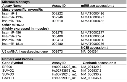
Some miRNAs are tissue specific and those abundantly expressed in skeletal muscles are known as muscle-specific miRNAs or also named myomiRs (82, 103, 105). miR-1, miR-133a, miR-133b, miR-206, miR-208, miR-486 and miR-499 are these muscle-specific miRNAs and among them, miR-1, miR-133 and miR-206 are the most widely studied myomiRs which in turn are arranged from biscistronic clusters and transcribed together (103, 105, 107). Other miRNAs, such as miR-27, miR-29 and miR-181 are considered non-specific muscle miRNAs besides are also found to be expressed in skeletal muscle tissue (103, 105).
25
proliferation (103). In addition, all except miR-29 also participate in muscle regeneration (103). Other aspects of muscle phenotype are also controlled by these myomiRs; miR-1, 133 and 206 contribute to regulation of muscle hypertrophy and atrophy while the miR-208 and 499 controlled the muscle fiber type and muscle performance (103).