Basis set superposition error effects, excited-state potential energy surface and photodynamics of thymine
232
0
0
Texto completo
(2)
(3) TESI DOCTORAL. Basis Set Superposition Error effects, excited-state Potential Energy Surface and photodynamics of thymine. David Asturiol Bofill. 2009. Doctorado Interuniversitario en Química Teórica y Computacional. Dirigida per: Lluís Blancafort San José, Pedro Salvador Sedano i Miquel Duran Portas. Memòria presentada per a optar al títol de Doctor per la Universitat de Girona. i.
(4) Lluís Blancafort San José, Pedro Salvador Sedano i Miquel Duran Portas professors titulats del Departament de Química de la Universitat de Girona,. CERTIFIQUEM: Que aquest treball titulat “Basis Set Superposition Error effects, excited-state Potential Energy Surface and photodynamics of thymine”, que presenta en David Asturiol Bofill per a l’obtenció del títol de Doctor, ha estat realitzat sota la nostra direcció i que compleix els requeriments per poder optar a Menció Europea.. Signatura. Lluís Blancafort. Pedro Salvador. Miquel Duran. Girona, 9 de Novembre de 2009. iii.
(5) Summary of the thesis The study of the photophysics of thymine is the main objective of this thesis. This work has been divided in 4 parts; the first two parts are devoted to find a proper level of theory for the study of thymine, whereas in the third and fourth parts the photohpysics of thymine are studied. Moran et al.140 found that correlated methods such as the Configuration Interaction with Single and Double excitations (CISD) and Møller-Plesset up to second order (MP2) when used with some of Pople’s basis sets, can not describe the planar structure of benzene. In addition, if a planar stationary point is optimized, the frequency analysis shows one or more imaginary frequencies. Given that thymine is a planar aromatic molecule as benzene, a benchmark study has been performed to determine if thymine can also suffer from such pitfalls. For completeness, the study is extended to the rest of the nucleobases, namely uracil, cytosine, adenine, and guanine. Our results show that, when Pople’s basis sets are used in conjunction with the MP2 method, minima structures of nucleobases with planar rings present imaginary frequencies. However, the same basis sets studied by Moran et al. have been analyzed at the Complete Active Space Self Consistent Field (CASSCF) level for thymine, and no imaginary frequencies have been found in any case. Thus according to our results, it can be concluded that the pitfalls reported for benzene seem to be common to correlated methods describing planar aromatic rings with Pople’s basis sets. In contrast, we have shown that the 6-31G* and 6-311G* basis sets, which are of general use in computational studies, can properly describe the minima structures of nucleobases. In addition, we have determined that the CASSCF/6-31G* and CASSCF/6-311G* levels of theory will be used for the study of the photophysics of thymine. In the first part of the thesis, we have also analyzed the origin of the pitfalls described above. We have shown that they can be explained in terms of intra-molecular Basis Set Superposition Error (BSSE), and that they can be fixed by using a typical BSSE correction technique such as the Counterpoise method (CP), which is implemented in general electronic structure modeling softwares. This method divides the molecule into fragments and this can be a problem as the multiplicity has to be assigned to each fragment. We have shown that independently of the fragments’ definition and fragment’s multiplicity assignment, the Counterpoise method fixes the imaginary frequencies where present and has no meaningful effects on the descriptions that were already correct. Nevertheless, we stress that one has to take into account that the isolated fragment and the associated ghost orbital calculations must correspond to the same state with the same orientation of singly-occupied v.
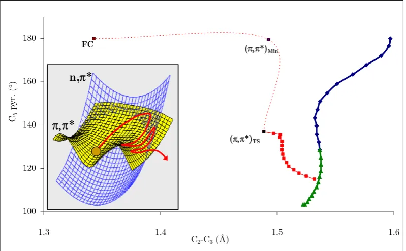
(6) degenerate orbitals, otherwise artifacts might arise during the BSSE removal which can result in a bad description of the molecule. By using our own code, which allows for a flexible definition of the Counterpoise function, we have been able to fix pitfalls in complicated systems such as the cyclopentadienyl and indenyl anions and naphthalene, in which a negative charge has to be considered and up to five imaginary frequencies were found, respectively. In addition, we have observed that the BSSE has a delocalized nature given that although the imaginary frequencies can be removed by just correcting BSSE for a single fragment, all fragments need to be included in the CP function to recover the frequency values of the correct descriptions. Experimental studies23,24,85-88 show that the relaxation of thymine after photon absorption can be described with a biexponential decay. That is, there exist two decay mechanisms, one in the subpicosecond and another one on the picosecond time scale, that lead photoexcited thymine to its initial structure. The existence of a longer component of hundreds of ns has also been reported.24,85 In the second part of this thesis, the photophysics of thymine have been studied. First, the PES of thymine has been optimized with a high level of theory to determine the decay paths of thymine. This has been carried out with the MS-CASPT2(12,9)//CASSCF(12,9)/6-311G* approach, in which Multi State Complete Active Space Møller-Plesset (MS-CASPT2) single point calculations, which include dynamic correlation, are carried out along minimum energy paths optimized at the Complete Active Space Self Consistent Field (CASSCF) level. Our results show that there exist two paths that after photon absorption can lead to the regeneration of the initial structure (for a better description of the PES we refer to Figure 28 and Figure 31). The first path, Path 1 in Figure 28, leads directly from the Franck-Condon (FC) point to a conical intersection (CI) with the ground state (GS), namely (Eth)X. Due to its barrierless character, this path has been assigned to the subpicosecond decay component determined experimentally. The second path, Path 2 in Figure 28, is indirect and is separated from (Eth)X by a minimum, (π,π*)Min, a barrier, (π,π*)TS, and a CI between the π,π* and n,π* states, (π,π*/n,π*)X. Two paths connect this CI with the GS. The first path leads directly to (Eth)X with no further barriers, and the second one leads to a minimum of the n,π* state and further to a CI with the GS, namely (n,π*/GS)X. Given that the barrier that separates (Eth)X from the FC structure is only of 0.05 eV, we assign the deactivation through this path to the subpicosecond decay. On the other hand, since the n,π* state can be accessed via (π,π*/n,π*)X, and the CI that leads the population of this state to the GS is not accessible (it lies 0.2 eV above the FC point), we assign the deactivation from this state to the picosecond decay component determined experimentally. vi.
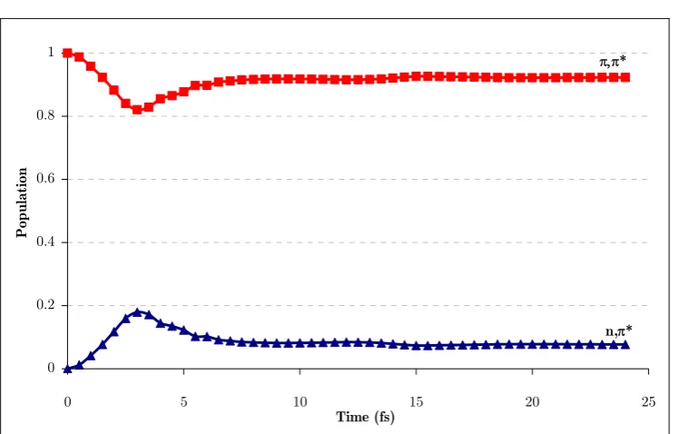
(7) We have carried out quasi-classical dynamics, i.e. classical dynamics simulations with a surface hopping algorithm which allows the propagation of nuclei on different surfaces, on the indirect path of thymine. The trajectories have been run at the CASSCF(8,6)/6-31G* level of theory because of their high computational cost. Unfortunately, at this level of theory the FC region is not well described and the barrier is overestimated by 0.2 eV. Thus, the direct path cannot be described because of the lack of dynamical correlation. A previous study138 showed that trajectories starting on the FC point got trapped at (π,π*)Min for more than 500fs, a time range that exceeds our simulations. Thus, we have run quasi-classical dynamics on the indirect path of thymine starting at (π,π*)TS, from which the behavior at the S2/S1 CI and further regeneration of the GS have been sampled. The results confirm the role proposed for this path at the MS-CASPT2 level. That is, part of the population of the indirect path is responsible for the picosecond component since it is funneled to the n,π* state at (n,π*/π,π*)X, whereas the rest of the population decays in the subpicosecond range. Because of the importance of (n,π*/π,π*)X in the photophysics of thymine, a topological analysis of this seam has been carried out. For consistency with the level of theory used in the dynamics simulations, such analysis has also been carried out at the CASSCF(8,6)/6-31G* level of theory. A structure of Cs symmetry has been optimized on the seam of intersection from which a constrained IRC has been performed. This shows that this CI seam presents a sloped-to-peaked topology and that all its parts are energetically accessible from the FC point. This slope-to-peaked topology has also been described in other works,284-286 where it is observed that the different parts of the seam can determine the photophysics of the molecule. In order to study this possibility, we have carried out quantum dynamics simulations on the indirect path of thymine with a novel method, namely the Direct Dynamics vibrational Multi Configurational Gaussian (DD-vMCG). This method uses diabatic states to propagate a wavepacket which is approximated by a set of Gaussian functions, whose centers move classically. This approximation implies that only local evaluations of the PES calculated on-the-fly are needed to propagate the wavepacket, rather than a precalculated grid of points which represents the PES. As in the semi-classical case, the simulations were started at (π,π*)TS. The different parts of the seam were analyzed by adding momentum on given coordinates which lead the wavepacket toward the regions of interest. Our results show that the segment of the seam that is reached during the decay has a large influence on the photophysics. In general it is observed that the peaked region of the seam favors the regeneration of the ground state, whereas the sloped one delays the deactivation as that region is responsible for trappings at (π.π*)Min and the n,π* state. vii.
(8) The DD-vMCG method has been applied to the present study of thymine, and all degrees of freedom of the molecule (39) have been taken into account. Such a large amount of degrees of freedom has been taken into account for the very first time with the DD-vMCG method. Problems associated with the use of a small active space, which limits the number of Gaussian centered wavefunctions that form the wavepacket, and the appearance of intruder states that invalidate the diabatic transformation at some points, have been encountered. However, we consider the performance of the method is encouraging as in its initial version it can be used to get a qualitative, mechanistic insight into the photophysics of thymine.. viii.
(9) Resum de la tesi L’estudi de la fotofísica de la timina és el principal objectiu d’aquesta tesi doctoral. Aquest treball ha estat dividit en 4 parts. En les dues primeres, s’ha realitzat un estudi de metodologies per tal de trobar la més adient per dur a terme l’objectiu principal de la tesi. En les altres dues parts de la tesi s’ha estudiat la fotofísica de la timina en detall. Moran i col·laboradors140 van publicar un treball en el qual es descrivia que alguns mètodes correlacionats com són el Configuració d’Interaccions amb excitacions Simples i Dobles (CISD) i Møller-Plesset de segon ordre (MP2), al utilitzar-los conjuntament amb bases de Pople per optimitzar l’estructura de mínima energia del benzè, s’obtenien geometries no planes. A més a més, si la optimització es duia a terme forçant la simetria Cs, els anàlisis posteriors de freqüències mostraven una o més freqüències imaginàries, tot indicant que no es tractava d’estructures de mínima energia. Degut al fet que la timina, igual que el benzè, és una molècula plana, formada per un anell de sis membres i aromàtica, vam decidir dur a terme una calibratge de mètodes per tal de comprovar si els errors descrits pel benzè es reproduïen en la timina. Per dur a terme un treball més complert, aquest estudi es va ampliar a la resta de bases de l’ADN: uracil, citosina, adenina i guanina. El nostre estudi mostra que, quan les bases de Pople es fan servir amb el mètode MP2, les estructures de mínima energia de les nucleobases, optimitzades forçant la planaritat de l’anell, presenten freqüències imaginàries. No obstant, si el mateix estudi fet per Moran i col·laboradors es fa amb el mètode d’Espai Actiu Complert de Camp Auto-Consistent (CASSCF) per al cas de la timina, en cap cas apareixen freqüències negatives. Per tant, en base als nostres resultats, podem concloure que els problemes que presenten els mètodes correlacionats amb les bases de Pople descrits per Moran i col·laboradors pel benzè, semblen ser comuns a les molècules aromàtiques amb anells plans. Sorprenentment, hem observat que les bases 631G* i 6-311G*, que són d’ús generalitzat en estudis computacionals, poden descriure perfectament l’aplanament de les estructures de mínima energia de les nucleobases. A més a més, aquest estudi ens ha ajudat a determinar que els nivells de càlcul CASSCF/6-31G* i CASSCF/6-311G* seran els que farem servir per estudiar la fotofísica de la timina. En la primera part de la tesi, també hem analitzat l’origen d’aquest problemes descrits anteriorment. Hem observat que es poden explicar en termes d’Error de Superposició de Base (BSSE) intra-molecular que, com a tals, es ix.
(10) poden arreglar fent servir tècniques de correcció del BSSE típiques; com per exemple el mètode Counterpoise. Una de les avantatges d’aquest mètode és que està implementat en la majoria de paquets de programari i, per tant, és de fàcil accés. Aquest mètode es basa en la separació de la molècula en fragments, fet que pot suposar un problema ja que, amb la versió actual, s’ha d’especificar la multiplicitat de cada un. La complicació pot ser major en el cas que es treballi amb una sola molècula. En aquesta tesi es demostra que, independentment de la definició dels fragments i de la seva multiplicitat, el mètode Counterpoise arregla les freqüències negatives en els casos on apareixen i que no té efectes apreciables en els que no se n’observen. Cal tenir en compte però, que per tal que el mètode arregli correctament els errors, els càlcul dels fragments aïllats i els dels càlcul corresponent que inclou totes les funcions de base del sistema (ghost orbital), s’han de dur a terme en el mateix estat i amb els orbitals degenerats mono-ocupats igualment orientats. La utilització d’un programari propi que permet la lliure definició de la funció de Counterpoise, ens ha permès arreglar els errors de sistemes complicats com els anions de ciclopentadiè i indenil i el naftalè. En el cas dels anions, la complicació ve donada pel fet d’haver de tractar amb una càrrega negativa, mentre que pel benzè ve donada pel fet d’haver de corregir 5 freqüències imaginaries. A part d’això, amb l’ajuda d’aquest codi hem observat que el BSSE té un caràcter deslocalitzat, ja que tot i que les freqüències negatives es poden eliminar corregint el BSSE d’un sol fragment, els valors “correctes” no es poden obtenir si no s’inclouen tots els fragments en la funció de Counterpoise. Un cop trobada la metodologia que es farà servir per estudiar la timina, ens centrarem en el seu estudi. Els estudis experimentals23,24.85-88 de la timina mostren que el relaxament posterior a l’absorbància d’un electró es pot descriure amb una funció biexponencial. És a dir, que existeixen dos mecanismes de desactivació, un en l’escala de fs i l’altre en la de ps. També s’ha detectat24,85 la presència d’un altre mecanisme que ajuda a la desactivació de la timina però més lentament (centenars de ns). Per tal de dur a terme l’estudi, primer hem optimitzat la Superfície d’Energia Potencial (PES) de la timina amb un alt nivell de càlcul. Concretament, hem fet servir l’aproximació MSCASPT2(12,9)//CASSCF(12,9)/6-311G* en la qual càlculs puntuals a nivell de Teoria de Pertorbacions de segon ordre amb una referència d’Espai Actiu Complert (CASPT2) es duen a terme al llarg dels perfils optimitzats a nivell CASSCF. Els nostres resultats mostren que existeixen dos camins de reacció que porten la molècula fotoexcitada cap a la seva estructura inicial (per una millor comprensió del perfil d’aquest camins, es recomana seguir les imatges 26 i 29). El primer camí, Path 1 en la imatge 26, porta directament des del punt d’excitació (FC) a una intersecció cònica amb l’estant fonamental (GS), que l’anomenarem (Eth)X. Degut a la manca de barreres al llarg d’aquest camí, x.
(11) l’hem assignat a la component de relaxament ultraràpid (fs) determinat experimentalment. El segon camí, Path 2 de la imatge 26, també porta a (Eth)X, però és indirecte. Al llarg del camí hi ha un mínim, (π,π*)Min, una barrera, (π,π*)TS, i una intersecció cònica entre els estats π,π* i n,π*, (π,π*/n,π*)X. Dos camins connecten aquesta intersecció amb l’estat fonamental. El primer camí no té barreres i porta directament al GS a través de (Eth)X. Per altra banda, el segon camí transcorre sobre l’estat n,π* i porta primer al mínim d’aquest estat, (n,π*)Min, i posteriorment a una intersecció amb l’estat fonamental, (n,π*/GS)X. Com que la barrera que separa (Eth)X del punt FC és de només 0.05 eV, hem assignat el relaxament a través d’aquest camí indirecte i (Eth)X, a la mateixa component ultraràpida d’abans: la de fs. Per altra banda, com que es pot accedir a l’estat n,π* a través de (π,π*/n,π*)X, i la intersecció que permet la desactivació d’aquest estat no és accessible ja que està 0.2 eV per sobre de l’energia del punt FC, hem assignat el relaxament des d’aquest estat a la component de ps. Un cop explicat l’estudi estàtic de la PES de la timina, procedirem a descriure l’estudi dinàmic. Hem fet simulacions de dinàmiques semi-clàssiques del camí indirecte de relaxament de la timina. És a dir, hem fet servir dinàmiques clàssiques amb un algoritme de salt de superfícies que permet propagar els nuclis en diferents superfícies. Les trajectòries s’han dut a terme al nivell de càlcul CASSCF(8,6)/6-31G* ja que tenen un alt cost computacional. Desgraciadament, a aquest nivell de càlcul, la zona FC no està ben descrita, el que impossibilita la optimització del camí indirecte. A més, la barrera del camí indirecte se sobreestima en 0.2 eV. Un estudi previ semblant al que es vol realitzar, en el qual les trajectòries es van iniciar al punt FC, mostra que totes queden atrapades al (n,π*)Min durant més de 500fs, un temps superior al de les nostres simulacions. Degut a això, i que la zona FC no es pot descriure correctament, hem decidit començar les trajectòries al (π,π*)TS, des del qual es pot estudiar el comportament de la CI S2/S1 i el posterior relaxament cap al GS. Els nostres resultats mostren que la població del camí indirecte és la responsable del component de ps ja que part de la població es pot transferir a l’estat n,π*, la desactivació del qual es dur a terme en ps. Degut a la importància d’aquesta CI en la fotofísica de la timina, se li ha realitzat un estudi topològic. Per consistència amb metodologia de les dinàmiques, aquest estudi s’ha dut a terme al mateix nivell de càlcul. Hem optimitzat una estructura amb simetria Cs en l’espai d’intersecció de la CI des de la qual s’ha optimitzat un camí de mínima energia restringit a aquest espai. Aquest camí porta directament al punt de mínima energia de la CI i l’estudi dels gradients dels estats al llarg d’aquest camí ens mostra que l’espai d’intersecció té una topologia “sloped-to-peaked”. Aquest tipus d’espai d’intersecció ha estat descrit en algun altre treball,284-286 en els quals s’ha xi.
(12) observat que les diferents parts de l’espai d’intersecció poden determinar la fotofísica de la molècula. Per tal d’estudiar aquesta possibilitat, hem dut a terme simulacions dinàmiques quàntiques al llarg del camí indirecte amb un nou mètode, DD-vMCG. Aquest mètode propaga paquets d’ona sobre, formats per una sèrie de funcions Gaussianes, en estats diabàtics. Això implica que la PES només s’ha d’avaluar localment en el centre de les Gaussianes en comptes d’haver de generar una xarxa de punts que descriguin tota la PES. El punt inicial d’aquestes simulacions és el mateix que per les dinàmiques clàssiques, (π,π*)TS. Les diferents part de l’espai d’intersecció s’han analitzat dirigint el paquet d’ones cap aquell direcció en concret. Això es pot fer afegint un moment d’inèrcia en la coordenada o coordenades que porten cap a la zona d’interès. Els nostres resultats mostren que la topologia de l’espai d’interacció té una gran influència en el mecanisme de relaxament. En general, s’observa que la regió “peaked” de l’espai d’interacció afavoreix el camí de relaxament que porta directament a l’estat fonamental, mentre que la regió “sloped” retarda la desactivació ja que afavoreix la confinament tant en (π,π*)Min com en l’estat n,π*. Tal com s’ha dit abans, hem utilitzat el mètode DD-vMCG per dur a terme les dinàmiques quàntiques. Per primer cop amb aquest mètode s’han fet servir 39 graus de llibertat (tots els de la timina). Hem observat problemes associats a l’ús d’un espai actiu reduït, el qual ha limitat el nombre de Gaussianes que formaven el paquet d’ona i també l’aparició d’estats intrusos que invalidaven la transformació diabàtica. No obstant, considerem que el comportament del mètode és satisfactori ja que en la seva primera versió hem obtingut uns resultats qualitatius d’alguns aspectes mecanístics del relaxament de la timina. Tot i això, cal tenir en compte que un mètode com aquest ha de poder oferir dades quantitatives.. xii.
(13) Agraiments No ha sigut fàcil arribar a poder escriure aquestes línies i ara que ho estic fent m’adono que m’entristeix una mica perquè significa que s’acaba l’etapa més meravallosa de la meva vida. Espero ser capaç d’incloure a tothom qui l’ha fet possible en aquest parell de fulls que venen a continuació. En aquest text no hi ha un ordre establert i hi ha faltes d’ortografia, això és degut a què és la part més personal de la tesi i he volgut que sigui així, imperfecte, com jo. La major part d’aquesta tesi no és mèrit meu, és mèrit de les persones que sempre han estat al meu costat donant-me suport fins i tot quan el rebutjava perquè pensava que no el necessitava. És mèrit de les persones que han patit amb i per mi des de sempre, de les dues persones que fan que em senti afortunat cada cop que les veig. Gràcies i us estimo no són suficients per expressar els meus sentiments, però no se m’acut cap altra manera de fe-rho en un paper. Gràcies papa i mami. Algú va dir que del que realment ens hem de penedir és de no haver fet alguna cosa, i no pas d’haver-la fet. Si d’algo em penedeixo d’aquest darrers anys, és de no haver passat tant temps com m’hagués agradat amb els meus avis. Tot i que potser no entenen ben bé què he fet durant tot aquest temps, segurament seran ells qui més s’alegrin quan sigui doctor. Només per aquest fet ja em sento orgullós i afortunat, i no els hi puc dir res més que me’ls estimo i que: “avi, àvia, avi Joan; gràcies”. Amb els que sí que he passat moltes estones aquest temps ha estat amb els meus companys de doctorat, i també amics. Vam començar al llegendari 166, edu, albert, david, quim, juanma i mireia. Alguns d’ells van marxar, i la veritat és que s’han trobat a faltar els acudits d’en quim, les vajanades de l’albert, el coneixement i capacitat organitzativa de l’edu i els cotilleos d’en torrente. Lo maco de tot això és que s’ha mantingut el contacte i que en dates senyalades com fires, ens tornem a reunir tots i t’adones que l’amistat segueix sent la que era. Amb el desterrament al parc ens vam ajuntar els dos despatxos, el 166 i 177, tot i que alguns com en Dani, que sempre està disposat a ajudar i comprar gadgets al dealxtreme, es van quedar a la universitat. El trasllat al “parque” va fer que deixés de compartir el despatx amb els meus companys, per fer-ho amb els meus amics. Cada un amb el seu toc freak característic que els fa únics. Gràcies a en juanma, la mireia, la sílvia i en ferran he viscut moments inoblidables amb les activitats “extra-escolars” (partits del barça, pàdels, futbols, bàsquets, voleis, esquí, play, fórmula de, sopars, can mià, lujuria, sessions de photoshop, cremats, karts, etc.), però sobretot amb el dia a dia. Un capítol apart mereixen els congressos viscuts amb tots ells: manchester-liverpool xiii.
(14) (la pocha, antrus i pudors), brussel·les (dnis, trepitjades, discoteques, ...), amsterdam (bicicletes, bolets i pastissos), colònia (cuba bars i kebabs), goteborg (pizzes i sueques) i helsinki (sol, insomni i resets). Apart d’ells, també he tingut companys meravallosos com la cristina, en samat, l’oscar, en quantum li, les annes, i més recentment en sergi, en rambo, en ievgeny i l’eloy. Els post-docs (marcel, annapaola i jordi) també mereixen un agraiment, ja que han sigut com els nostres germans grans allà al parc, que sempre que hem necessitat ajuda ens l’han ofert. A tots gràcies. Bona part del doctorat me l’he passat voltant per aquest món. Tot i que marxar a fora és difícil, he trobat persones que m’han fet sentir com a casa allà on fós. L’istvan i els erasmus a budapest, però sobretot la giulia, en ben, en jacob i en fabri a londres. Sense ells, tot hagués sigut molt més difícil. Gràcies. Lo bo que tenia estar a la universitat era que et podies relacionar amb tot tipus de gent, fins i tot aquells que van tot el dia amb bata i ulleres i estan envoltats de productes tòxics i al·lucinògens, vaia, els “expis”. Sense ells els cafès, sopars, partidillus i sobretot les “jodete” no haguéssin sigut el mateix. Gràcies per les bones estones que em passat. Lo més important de treballar/estudiar (algú sap com es defineix fer el doctorat?) és estar agust en el lloc on ho fas. El fet que l’iqc sigui un dels millors llocs on fer-ho és degut a les persones que el formen, però sobretot als miquels que són qui han marcat les directrius perquè sigui així. Per això, també vull aprofitar l’ocasió per agrair-los haver-me donat la oportunitat de fer el doctorat aquí i per les facilitats i llibertat que ens han donat per fer tot tipus de coses. No em voldria deixar la resta de l’iqc, josep mª, sergei, sílvia, emili i molt especialment la carme, que en el fons és la mama de tots nosaltres. No hauria pogut escriure aquesta tesi si no hagués tingut dos jefes com en pedro i en lluís. Desgraciadament no en sabré mai tant com ells però almenys he tingut la oportunitat d’aprendre’n. M’han ensenyat moltes coses, però sobretot els he d’agrair la paciència i disponibilitat que han tingut amb mi per explicarme les coses que no entenia i també per motivar-me quan no veia les coses gaire clares. Haver voltat pel món també m’ha ensenyat que de jefes com ells, no n’hi ha. Gràcies. Per sort aquests anys també he tingut vida fora de la universitat. Aquesta “altra” vida ha estat marcada per les meves companyes de pis, martona, txell, anna, i també l’alba. Elles han sigut durant molt de temps la meva família aqui a girona, i m’han ajudat a superar els mals trànguls i a tirar endavant. Juntament amb en pedro, hem passat molt bones estones i en tinc molt bons xiv.
(15) records, però de tots ells em quedo amb els sopars i festes a “ca la pacheca”. Gràcies. Tothom necessita una via d’escape, una forma de desestressar-se, i jo l’he trobat amb l’scalextric. Gràcies a en jordi, l’anna, en lluís, en jokin i en sergi he pogut desconnectar dels problemes i passar-m’ho molt bé, no per l’scalextric en si que a vegades pot ser avorrit, sinó per tot lo que l’envolta: viatges a salou, oviedo, león, madrid, igualada, les històries dels dilluns, els sopars al taco-taco, les festes, els carnavals, ..., però sobretot pel dia a dia. La bisbal també existeix, potser no per tothom però per mi sí. Allà hi ha els meus amics, sempre hi han sigut i sempre hi seran. Tot ells m’han ajudat sempre que han pogut, però lo que més valoro de tot plegat és que sempre hi són. La llista no està completa sense en runaldu i en mau que, tot i que poc, quan ens veiem sempre aconsegueixen que passem una bona estona. Gràcies. No em voldria deixar a la ceaccm, (jo tampoc sé que vol dir), que m’han acollit com un d’ells i també m’han fet costat tot aquest temps. A vosaltres també, gràcies. Ara que ja estic acabant d’escriure aquestes línies d’agraiment, miro enrera i se’m dibuixa un somriure a la boca perquè veig tot el que m’emporto d’aquest període, tot es bo, però si alguna cosa em fa realment feliç de tot això, ets tu. Tu que has estat amb mi a les bones i dolentes, que m’has fet costat sempre i m’has ajudat en tot. Tu que fas que tot sigui més fàcil i que cada dia sigui especial. Tu que has aconseguit que perdi la por, i m’has fet veure que lo millor que un pot tenir és il·lusió. Tu que dones sentit a frases com “jo més perquè sóc més gran” i “no existeix pq és infinit”. Gràcies. I per acabar, només em queda donar les gràcies al Ministerio de Educación y Ciéncia per haver finançat els meus estudis durant aquests anys, moltes gràcies.. xv.
(16) List of publications of this thesis (1). Asturiol, D.; Duran, M.; Salvador, P.; “Intramolecular Basis Set. Superposition Error Effects on the Planarity of Benzene and other aromatic molecules: A solution to the problem“; J. Chem. Phys. 2008, 128. (2). Asturiol, D.; Duran, M.; Salvador, P.; “Intramolecular Basis Set. Superposition Error Effects on the Planarity of DNA and RNA Nucleobases“; J. Chem. Theor. Comput. 2009, 5, 2574-2581. (3). Asturiol, D.; Lasorne, B.; Robb, M. A.; Blancafort, L.; “Photophysics of the π,π* and n,π* States of Thymine: MS-CASPT2 Minimum-Energy Paths and CASSCF on-the-Fly Dynamics”; J. Phys. Chem. A 2009, 113, 10211-10218.. (4). Asturiol, D.; Lasorne, B.; Robb, M. A.; Blancafort, L.; “Thymine S2/S1. Conical Intersection analysis and quantum dynamics”; J. Phys. Chem. A. (Submitted).. xvii.
(17) Publications not included in this thesis (1). Asturiol, D.; Duran, M.; Salvador, P.; Torrent-Sucarrat, M. BSSE-free. hardness profiles of hydrogen bond exchange in the hydrogen fluoride dimer; Int. J. Quant. Chem. 2006, 106, 2910-2919. (2). Salvador, P.; Asturiol, D.; Mayer, I.; “A general efficient implementation. of the BSSE-free SCF and MP2 methods based on the Chemical Hamiltonian Approach“; J. Comput. Chem. 2006, 27, 1505-1516. (3). Asturiol, D.; Salvador, P.; Mayer, I.; “Dissecting the hindered rotation of ethane”; Chem. Phys. Chem 2009, 10, 1987-1992.. (4). Kobylecka, M.; Migani, A.; Asturiol, D.; Rak, J.; Blancafort, L.; “Benign. Decay vs. Photolysis in the Photophysics and Photochemistry of 5Bromouracil. A Computational Study”; J. Phys. Chem. A 2009, 113, 5489-5495.. xviii.
(18) List of figures Figure 1. Scheme6 of the double strand of DNA. Adapted from Access Excellence @ the National Health Museum. ........................................................................................................1 Figure 2. Thymine UV induced photoproducts (adapted from Medical Ecology online resources16). .............................................................................................................................3 Figure 3. Absorption and emission spectra (adapted from Whitman College’s webpage17). .............................................................................................................................5 Figure 4. Franck-Condon principle energy diagram. The blue arrow corresponds to the vertical excitation from the ground state (E0) to the vibrational level of the first excited state (E1) with highest overlap with the initial state. Similarly, the green arrow denotes the vertical deexcitation (adapted from IUPAC Compendium of Chemical Terminology, 2nd Edition, 1997). ..................................................................................................................6 Figure 5. Jablonsky energy diagrams of (a) fluorescence (b) phosphorescence and (c) delayed fluorescence deactivation mechanisms (adapted from Molecular Expressions website21). .............................................................................................................................8 Figure 6. Possible photoprocesses for a molecule: a) Emissive deactivation to the ground state (adiabatic). b) Emissive photoreaction (adiabatic). c) Radiationless deactivation to the initial position (non-adiabatic). d) Internal conversion to a photoproduct (non-adiabatic) (Adapted from Encyclopedia of Computational Chemistry (1998)).................................................................................................................. 12 Figure 7. Plot of the potential energy surface as a function of the branching space (x1,x2) (IUPAC Compendium of Chemical Terminology, 2nd Edition, 1997) ........................ 14 Figure 8. Sloped and peaked crossings as defined by Ruedenberg. ........................................ 15 Figure 9. Second order conical intersection picture. a) 3-coordinate model potential energy surface along x3. b) 3-coordinate model along x1 and x3. Adapted from Ref. 35. ....... 17 Figure 10. Projection of a seam of intersection on the x1,x3 plane including second order effects. Adapted from Ref. 39. ............................................................................................... 18 Figure 11. González-Schlegel IRC algorithm ......................................................................... 24 Figure 12. IRDs calculated from a circular cross-section and corresponding MEPs from the FC structure. ................................................................................................................... 25 Figure 13. Characterization of a PES with successive IRD calculations of increasing radius (d). ........................................................................................................................... 26 Figure 14. Deactivation paths from a CI and energy profile along a circular crosssection centered on the CI point of radius d. Note: the general IRD procedure extends. xix.
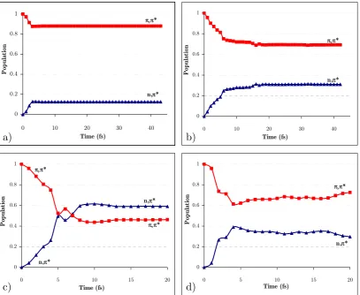
(19) the energy minima search to an (n-1)-dimensional spherical cross-section (hypersphere) rather than a mono-dimensional cross-section as depicted above. Adapted from Ref. 49...... 27 Figure 15. Representative structures of base multimers from Ref. 91: (a) Watson and Crick base pair, A-T (top) and G-C (bottom); (b) base-stacked form of the dinucleoside monophosphate ApA; (c) B-form double-stranded DNA, views down the helical axis (left) and from the side (right); (d) A-form double-stranded DNA, views down the helical axis (left) and from the side (right). ........................................................................... 34 Figure 16. Summary of thymine’s relaxation models found in the literature. a) 3 components corresponding to deactivations from the π,π* state, (π,π*)Min, and 3π,π* state were reported131,137 to lie in the fs, ps and ns time ranges, respectively. b) 2 components corresponding to relaxation from FC to (π,π*)Min, and further deactivation from that minimum were assigned138 to the fs and ps components, respectively. c) 2 fs components, fs’ (<50 fs) and fs’’ (490 fs), were reported139 for two different two-step mechanisms corresponding to π,π*-GS, and π,π*-n,π*-GS, respectively. .............................. 40 Figure 17. Effects of geometrical optimization anomalies in the description of deactivation paths.................................................................................................................. 47 Figure 18. BSSE corrected and uncorrected PES for a given system. ................................... 61 Figure 19. Potential energy surface for the two lowest 1Σ+ states of LiF. Dashed lines represent CASPT2 energies. Solid lines correspond to MS-CASPT2 calculations and the dots correspond to FCI values (Adapted from Ref. 232). ...................................................... 82 Figure 20. Typical microcanonical sampling procedure. Adapted from Ref. 49..................... 87 Figure 21. Arenes considered in this study. ......................................................................... 108 Figure 22. CP-corrected energies along the b2g vibrational mode in benzene....................... 110 Figure 23. Orbitals used in the CASSCF calculations for thymine. .................................... 115 Figure 24. CCSD and MPn energies along the vibrational mode associated to the imaginary frequency for thymine at the MP2 level with the 6-31+G* (left) and 6311+G* (right) basis sets. ................................................................................................... 118 Figure 25. Nucleobases considered in this study .................................................................. 119 Figure 26. Density difference plot between ghost-orbital and isolated calculations for an N-H fragment in thymine for a) triplet and b) singlet electronic states. The position of the ghost-atoms is shown with semitransparent blue spheres. ............................................. 121 Figure 27. Intramolecular fragments used for the CP-correction in thymine....................... 123 Figure 28. Two-dimensional sketch of the two lowest excited state potential energy surfaces (S1 and S2) of thymine in the vicinity of the Franck-Condon region. Insets: FC. xx.
(20) structure with atom numbering and energy profiles for the paths contained in the twodimensional sketch. .............................................................................................................. 128 Figure 29. LIIC CASSCF(8,6)/6-31G* of the indirect path (FC-(π,π*)Min-(π,π*)TS(n,π*/π,π*)X-(Eth)X)............................................................................................................ 130 Figure 30. MS-CASPT2(12,9) energy profiles along the CASSCF/6-311G* minimum energy paths from the FC structure: a) Direct path (path 1 on Figure 28); b) Indirect path (path 2 on Figure 28). ................................................................................................. 132 Figure 31. Energies of relevant critical points on the excited state surface of thymine at the MS-CASPT2(12,9)/6-311G* level of theory (CASSCF(8,6)/6-31G* optimized energies in brackets). ........................................................................................................... 133 Figure 32. Transition vector at (π,π*)TS and branching space vectors (interstate coupling, IC, and gradient difference, GD) at (n,π/π,π*)X, calculated at the CASSCF(8,6)/6-31G* level.................................................................................................. 134 Figure 33. Time evolution of the CASSCF(8,6) energy of the S0-S2 states of thymine and the C4-O8 distance for a representative trajectory on S2 from (π,π*)TS. The labels of the states refer to the order at the beginning of the trajectory. ...................................... 135 Figure 34. a) Time evolution of the CASSCF(8,6) energy of the S0-S1 states of thymine for a representative trajectory on S1 (π,π* state); (b) the same for the C4-O8 and C5-C6 distance and the C5 pyramidalization (C9-C5-C4-N3 dihedral angle). The label of the states in (a) refers to the order at the beginning of the trajectory. ..................................... 136 Figure 35. Branching space vectors (interstate coupling, IC, and gradient difference, GD) at (Eth)X, calculated at the CASSCF(8,6)/6-31G* level. ............................................ 137 Figure 36. Time evolution of the CASSCF(8,6) energy of the S0-S1 states of thymine and the C4-O8 distance for a representative trajectory on S1 (n,π* state). The label of the states refers to the order at the beginning of the trajectory. ......................................... 138 Figure 37. Sketch of a sloped-to-peaked CI intersection seam. ............................................ 141 Figure 38. The two lowest vibrational modes and branching space vectors of the Cs structure of the S2/S1 CI. ..................................................................................................... 144 Figure 39. a) Energy profile of the three lowest states along the interstate vector. b) C5 pyramidalization angle vs C4-O8 distance along the intersection space of the S2/S1 seam. .. 144 Figure 40. Norm of the two state gradient vectors along the constrained IRC on the seam ………. ........................................................................................................................ 145 Figure 41. Vibrations of the 3 lowest normal modes at the dynamics starting point. Frequencies expressed in cm-1 (imaginary values in italics). ................................................ 146. xxi.
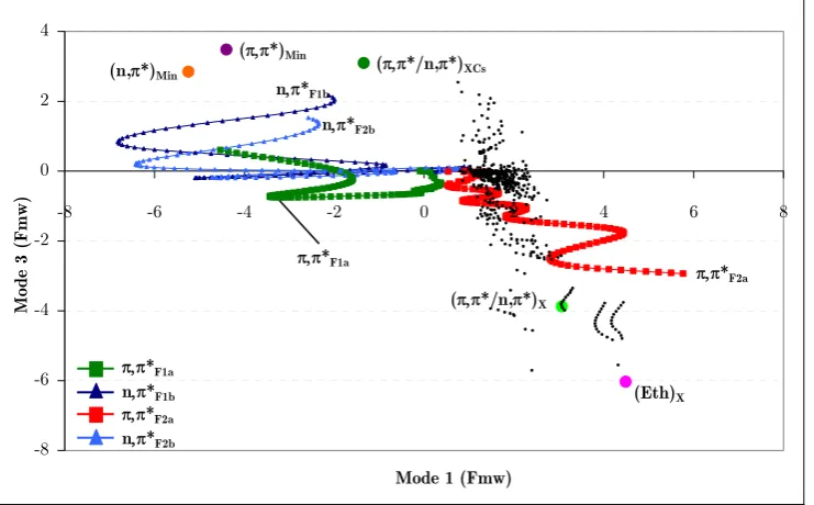
(21) Figure 42a. Position of the center of the Gaussian functions of run 1a with respect to mode 1 and mode 3 (position relative to (π,π*)TS). ............................................................. 149 Figure 43a. Diabatic and adiabatic energies of the Gaussian function of the π,π* state (F1a) of run 1b. ................................................................................................................... 151 Figure 44. Diabatic populations of propagations with 0.1 eV of extra momentum on mode 1 with 1, 2, 4, and 8 functions per state, respectively (runs 1a, 1c, 1d, and 1e). ....... 154 Figure 45. Position of the center of the Gaussian functions with respect to mode 1 and mode 3 along run 2b (position relative to (π,π*)TS)............................................................. 155 Figure 46a. Position of the center of the Gaussian functions of the π,π* state of run 3a (position relative to (π,π*)TS). ............................................................................................. 156 Figure 47a. Position of the center of the Gaussian functions of the π,π* state of run 3b (position relative to (π,π*)TS)............................................................................................... 157 Figure 48. Representation of the S2/S1 CI seam and MEP from the dynamics starting point. Inset represents the typical behavior of a wavepacket accessing the peaked region of the seam. ......................................................................................................................... 159. xxii.
(22) List of tables Table 1. Lowest uncorrected and CP-corrected vibrational frequencies (cm-1) of benzene for different levels of theory and basis sets (spurious frequency values in italics). .............. 111 Table 2. Average error (in %) for the computed harmonic frequencies of benzene with respect to experiment.264 In parenthesis the error computed without the lowest five frequencies. ......................................................................................................................... 111 Table 3. Lowest MP2 uncorrected and CP-corrected vibrational frequencies of naphthalene (cm-1) for different basis sets (spurious frequency values in italics)................. 112 Table 4. Lowest MP2 uncorrected and CP-corrected vibrational frequencies (cm-1) of indenyl and cyclopentadienyl anions (spurious frequency values in italics). ........................ 113 Table 5. Lowest harmonic vibrational frequencies of thymine (cm-1) at the CASSCF, MP2 and Counterpoise-corrected MP2 levels of theory. Basis sets in black indicate benzene is not planar at the corresponding MP2 level. Imaginary frequencies are displayed in italics. .............................................................................................................. 116 Table 6. CP-corrected and uncorrected frequencies in cm-1 of optimized planar structures of pyrimidine nucleobases. Imaginary frequencies are displayed in italics. ......... 120 Table 7. Lowest vibrational frequency value in cm-1 (Freq.) of various partial CPcorrected calculations. The numbers of the fragments included in the CP-function are defined in Figure 27. The first value corresponds to the uncorrected calculation. Imaginary frequencies are displayed in italics...................................................................... 122 Table 8. CP-corrected and uncorrected frequencies of optimized planar structures of purine nucleobases. Imaginary frequencies are displayed in italics. ..................................... 126 Table 9. Critical points position with respect to (π,π*)TS in Fmw coordinates. * The total distances of these points with respect to the (π,π*)TS in Fmw coordinates are overestimated because of a rotation of the methyl group. ................................................... 147 Table 10. Simulations described in this chapter with their corresponding characteristics (modes to which momentum has been added, momentum (eV), functions per state, and figures that describe each run)............................................................................................. 148. xxiii.
(23) List of Acronyms ANO BSIE BSSE CAS CASPT2 CASSCF CC CCSD CI CIS CISD CP CP-CISD CP-HF CP-MP2 DFT DIIS DNA E0 E1 E2 EA FC FCI GD GS HF HOMO IC IRC IRD ISC LCAO LHA LIIC. Atomic Natural Orbital Basis Set Incompleteness Error Basis Set Superposition Error Complete Active Space Complete Active Space Second Order Perturbation Theory Complete Active Space Self Consistent Field Coupled Cluster Coupled Cluster Singles-Doubles Conical Intersection Configuration Interaction Singles Configuration Interaction Singles-Doubles Counterpoise Counterpoise corrected CISD Counterpoise corrected HF Counterpoise corrected MP2 Density Functional Theory Direct Inversion in the Iterative Subspace Deoxyribonucleic Acid Energy of the Ground State Energy of the first excited state Energy of the second excited state Electron Affinity Franck-Condon Full Configuration Interaction Gradient Difference Ground State Hartree-Fock Highest Ocuppied Molecular Orbital Interstate Coupling Intrinsic Reaction Coordinate Initial Relaxation Direction Intersystem Crossing Linear Combination of Atomic Orbitals Local Harmonic Approximation Linear Interpolation of Internal Coordinates xxv.
(24) MC MCSCF MCTDH MEP MMVB MO MP MP2 MP3 MP4 MRCI OM2 PES QM/MM RASSCF REMPI RHF RNA S0 S1 S1/S0 S2 S2/S1 S3 SCF SOMO T1 T2 TD-DFT TDH TDM TRPES TS T-T UV ZPE. xxvi. Multi-Configurational Multi-Configurational Self Consistent Field Multi-Configurational Time-Dependent Hartree Minimum Energy Path Molecular Mechanics of Valence Bond theory Molecular Orbital Møller-Plesset Second order Møller-Plesset perturbation theory Third order Møller-Plesset perturbation theory Fourth order Møller-Plesset perturbation theory Multi-Reference Configuration Interaction Orthogonalization method 2 Potential Energy Surface Quantum Mechanics/Molecular Mechanics Restricted Active Space Self Consistent Field Resonance-enhanced Multiphoton Ionization Restricted Hartree-Fock Ribonucleic Acid Ground state First Excited State Conical intersection between S1 and S0 Second Excited State Conical intersection between S2 and S1 Third Excited State Self Consistent Field Single Occupied Molecular Orbital First Triplet State Second Triplet State Time Dependent Density Functional Theory Time Dependent Hartree Transition Dipole Moment Time-Resolved Photoelectron Spectra Transition State Thymine-Thymine Ultra-Violet Zero Point Energy.
(25) CONTENTS Summary of the thesis ..................................................................................................... v Resum de la tesi ............................................................................................................. ix Agraiments ................................................................................................................... xiii List of publications of this thesis ................................................................................. xvii Publications not included in this thesis ...................................................................... xviii List of figures ................................................................................................................ xix List of tables ............................................................................................................... xxiii List of Acronyms.......................................................................................................... xxv 1. INTRODUCTION ................................................................................................... 1 1.1. PHOTOCHEMICAL CONCEPTS ............................................................................ 4. 1.1.1. Absorption and emission spectra.................................................................. 4. 1.1.2. Relaxation mechanisms ................................................................................ 7. 1.1.3. Potential Energy Surface(s) ......................................................................... 9. 1.1.3.1. PES characterization.......................................................................................11. 1.1.3.2. Touching surfaces regions ...............................................................................11. 1.1.3.2.a 1.1.3.3. Conical Intersections...................................................................................13. Second order effects at CIs..............................................................................16. 1.1.3.3.a. Intersection space Hessian ..........................................................................19. 1.1.3.4. Optimizing conical intersections .....................................................................21. 1.1.3.5. Interconnecting stationary points ...................................................................23. 1.1.4 1.2. 1.1.3.5.a. González-Schlegel IRC algorithm ...............................................................24. 1.1.3.5.b. IRD .............................................................................................................25. Dynamics simulations................................................................................. 27 EXPERIMENTAL AND COMPUTATIONAL BACKGROUND .................................. 31. 1.2.1. Thymine experimental studies.................................................................... 36. 1.2.2. Computational studies................................................................................ 38. 1.3. COMPUTATIONAL METHODOLOGY .................................................................. 41. 1.3.1. Failures of general computational methods in ring planarity description .. 41. 1.3.1.1. Intramolecular BSSE.......................................................................................42. xxvii.
(26) 1.3.2 2. Pitfalls on DNA and RNA nucleobases ...................................................... 43. THEORETICAL METHODS ................................................................................ 47 2.1. AB INITIO METHODS ....................................................................................... 47. 2.1.1. Schrödinger equation.................................................................................. 48. 2.1.2. The Born-Oppenheimer approximation...................................................... 49. 2.1.2.1. 2.1.3. Born-Oppenheimer approximation breakdown ...............................................50. Molecular orbital theory............................................................................. 52. 2.1.3.1. Basis Sets ........................................................................................................54. 2.1.3.1.a. Minimal Basis Set .......................................................................................55. 2.1.3.1.b. Double Zeta Basis Sets ...............................................................................55. 2.1.3.1.c. Polarization functions .................................................................................56. 2.1.3.1.d. Diffuse functions .........................................................................................57. 2.1.3.1.e. ANO-type basis sets....................................................................................57. 2.1.3.2. Basis Set Superposition Error .........................................................................58. 2.1.3.2.a. 2.1.4. The Hartree-Fock method .......................................................................... 62. 2.1.4.1. Hartree-Fock equations ...................................................................................62. 2.1.4.1.a. 2.1.5. Counterpoise method ..................................................................................59. HF limitations.............................................................................................64. Multi-Configurational Methods .................................................................. 66. 2.1.5.1. The Configuration-Interaction method ...........................................................67. 2.1.5.2. CASSCF ..........................................................................................................68. 2.1.6. 2.1.5.2.a. CASSCF wavefunction optimization ..........................................................69. 2.1.5.2.b. CASSCF limitations ...................................................................................73. Including Dynamic correlation ................................................................... 74. 2.1.6.1. Møller-Plesset perturbation theory .................................................................74. 2.1.6.1.a 2.1.6.2. CASPT2 ..........................................................................................................77. 2.1.6.2.a 2.1.6.3. 2.2. MP2 ............................................................................................................75 Intruder states and Level Shift ...................................................................80. MS-CASPT2 ...................................................................................................82. MOLECULAR DYNAMICS ................................................................................. 85. 2.2.1. Quasi-classical dynamics ............................................................................ 85. 2.2.2. Quasi-classical dynamics propagation ........................................................ 87. 2.2.2.1. 2.2.3. Surface hopping...............................................................................................89. Quantum dynamics .................................................................................... 90. 2.2.3.1. TDH ................................................................................................................91. 2.2.3.2. MCTDH ..........................................................................................................93. 2.2.3.2.a. xxviii. DD-vMCG ..................................................................................................97.
(27) 2.2.4. Non-adiabatic events with quantum dynamics........................................... 99. 2.2.4.1. Diabatic representation .................................................................................100. 2.2.4.1.a. Regularized diabatic states .......................................................................101. 3. OBJECTIVES.......................................................................................................105. 4. RESULTS .............................................................................................................107 4.1. BSSE EFFECTS ON THE PLANARITY OF BENZENE AND OTHER PLANAR ARENES:. A SOLUTION TO THE PROBLEM ..................................................................................108. 4.1.1. Computational details ............................................................................... 109. 4.1.2. Fragments’ definition ................................................................................ 109. 4.1.3. Vibrational frequencies.............................................................................. 110. 4.2. GLOBAL AND LOCAL BSSE EFFECTS ON NUCLEOBASES .................................114. 4.2.1. Computational details ............................................................................... 114. 4.2.2. Thymine benchmark..................................................................................115. 4.2.3. BSSE removal on nucleobases ................................................................... 118. 4.2.3.1. 4.2.4. Local BSSE ...................................................................................................122. BSSE effects on nucleobases...................................................................... 124 PHOTOPHYSICS OF THE π,π* AND n,π* STATES OF THYMINE.........................127. 4.3. 4.3.1. Starting scenario........................................................................................127. 4.3.2. Computational Details ..............................................................................129. 4.3.3. High level potential energy surface............................................................ 132. 4.3.4. Dynamics simulations................................................................................134. 4.3.5. Discussion.................................................................................................. 138. 4.4. THYMINE S2/S1 CI SEAM ANALYSIS AND QUANTUM DYNAMICS ......................141. 4.4.1. Computational details ............................................................................... 142. 4.4.2. Topological analysis of the S2/S1 CI seam .................................................143. 4.4.2.1. Intersection space characterization ...............................................................143. 4.4.2.2. Analysis of the normal modes at the dynamics starting point (π,π*)TS ........146. 4.4.3. Quantum dynamics at the S2/S1 CI seam ................................................. 147. 4.4.3.1. Propagation with additional momentum on mode 1.....................................148. 4.4.3.2. Propagation with additional momentum on mode 2.....................................154. 4.4.3.3. Propagation with additional momentum on mode 3.....................................155. 4.4.4. Discussion.................................................................................................. 159. xxix.
(28) 5. CONCLUSIONS ...................................................................................................163. 6. BIBLIOGRAPHY.................................................................................................165. APPENDIX I ...............................................................................................................177 APPENDIX II ..............................................................................................................184 APPENDIX III.............................................................................................................190. xxx.
(29) INTRODUCTION. 1. 1. INTRODUCTION. Probably there is not a more fascinating molecule than the Deoxyribonucleic acid, commonly known as DNA. Maybe its charm lies in its contradictory and uncertain nature, or perhaps, in the fact it might be unique and special. It has been “out there”, almost untouched, for thousands of years and amazingly, it was not until 1871 when we first heard1 of it. Many resources and money have been spent on its study since then. Unfortunately, although many advances have been performed, we still know very little about it. We do not even know for sure who discovered the famous double helix structure of DNA,2 as its discovery was first credited to James Watson and Francis Crick but it has lately been suggested3-5 that Maurice Wilkins and Rosalind Franklin should also be recognized for their essential contribution to the discovery. DNA is an anti-parallel double sequence of nucleobases, namely Adenine, Thymine, Cytosine, and Guanine, as shown in Figure 1. They are coded in genes that contain all the necessary information for the development and functioning of every living being.. Figure 1. Scheme6 of the double strand of DNA. Adapted from Access Excellence @ the National Health Museum. Any variation in the sequence of the bases would translate into gene mutation which would have an unpredictable repercussion in the cell functionality. Taking into account that the importance of the information coded in the DNA, it is not strange that Mother Nature has provided a set of.
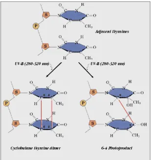
(30) 2. INTRODUCTION. protection mechanisms to preserve it from external agents. Some of the protection tactics featured by DNA are outlined next. Among the various protection mechanisms present in nature, one of the most efficient and widespread is isolation. The isolation of nucleobases starts by being kept in the cell’s nucleus, which is the most inaccessible part of the cell as two membranes protect it from external agents. In addition, a highly packed conformation of the DNA strand inside the nucleus makes nucleobases even more unreachable. Again, contradiction is present in DNA, since it must be protected to avoid mutations, while at the same time it needs to be easily accessible to perform vital processes for the cell such as replication, transcription, and translation. Physical barriers can keep (physical) agents away from the coded information, but they cannot protect them from radiation. Nucleobases are the chromophores of DNA, i.e. the parts of the DNA that absorb light. If they are exposed to UV light, photons are absorbed bringing the molecule to an excited electronic state where it is prone to react because it has extra energy. Thus, UV light is a potential DNA damaging agent as it can promote photoreactions which, within the DNA strand, can cause gene mutation. It is obvious that nucleic acid bases must feature several protection tactics against UV radiation, because otherwise the evolution could not have taken place as individuals can not survive to major changes in their DNA. One of the protection tactics of DNA against radiation is external, and corresponds to the ozone layer. Nucleobases have the lowest energy transitions located at the same spectral region as ozone, thus, the most dangerous UV radiation cannot reach the Earth’s surface, as it is absorbed by the ozone layer. A more particular shield of DNA is the complex packed conformation it adopts inside the nucleus. It reduces its exposure to light, which hinders photon absorption. However, in spite of these protection tactics, nucleobases are still reached by the UV radiation. Fortunately, DNA also has some tools to minimize the effect of the photoreactions. For instance, the excited states of nucleobases are characterized by an ultra-short lifetime. They get rid of the UV induced extra energy in the sub-picosecond or picosecond time scale, which reduces the probability of photoreaction. In addition, the energy gained in photon absorptions can be redistributed along the DNA structure, which also helps in minimizing the probability of photoreactions. As seen above, DNA has a large number of protection mechanisms, however, they do not provide 100% of security. In the cases where mutations take place, there are enzymes that can repair7 DNA mutagenic8-11 photoproducts.
(31) INTRODUCTION. 3. such as cyclobutane pyrimidine dimers12,13 and 6-4 pyrimidine adducts14,15 (see Figure 2).. Figure 2. Thymine UV induced photoproducts (adapted from Medical Ecology online resources16). It is obvious that the response of DNA to light is a complex and multivariable process and that its study cannot be faced globally. Here, we will study the photophysics of thymine as a first step towards the full understanding of the DNA protection mechanisms. A broad overview of the most important experimental and theoretical works on this field is presented in section 1.2. However, before reviewing the results present on the literature and explaining the results of this thesis, a brief overview of some useful photochemical concepts will be given..
(32) 4. Photochemical concepts. 1.1. Photochemical concepts. Light can interact with molecules changing their properties such as color, structure, stability, reactivity, etc. In this thesis we will study the way nucleobases interact with UV light. As mentioned above, nucleobases are the parts of DNA that absorb and/or emit light. In general, the light a molecule or an object absorbs is indicated by its color. That is, if an object is irradiated with “white” light (light compound of different wavelengths, as that of the sun) depending on what “part” of the light is absorbed, it will adopt one color or another. Actually, the colors of objects do not correspond to the light that they absorb but the one that is reflected. This is because only the light that is reflected reaches our eyes, and therefore, is the one that defines the colors of objects. For instance, it is known that plant leaves absorb “yellow” light because most of the sunlight that reaches the earth is made of “yellow” light and they use it for the photosynthesis. However, most plant leaves are green. This is explained because we only see the light that has been reflected by the plant (the green one), not the absorbed one (the yellow one). Spectrophotometers can determine the wavelength (color) of the light that is absorbed/emitted by a given molecule. The importance of absorption and emission spectra is explained next together with the functioning of spectrophotometers.. 1.1.1. Absorption and emission spectra. A spectrophotometer is an apparatus that irradiates samples with white light and records the light that has been absorbed and/or emitted by them, thus, it records absorption and/or emission spectra. An absorption spectrum (see Figure 3) consists of a continuous spectrum (Inset a of Figure 3) with some “lines” which denote the energy of the light that has been absorbed by the sample. These lines, called bands in molecules, appear because the light of that part of the spectrum was used to promote an electron of the sample from a given molecular orbital to another orbital of higher energy. Thus, all irradiated light reaches the detector except that which has been absorbed by the sample (Inset b of Figure 3). On the other hand, the emission spectrum of a given molecule (Inset c of Figure 3) is the light emitted by a molecule which has been previously irradiated. In principle, an emission spectrum should be complementary to the absorption one, nevertheless, usually part of the absorbed energy is transformed, and then the remaining energy is emitted as light, which composes the emission spectrum..
(33) Absorption and emission spectra. 5. Figure 3. Absorption and emission spectra (adapted from Whitman College’s webpage17). Kasha’s rule18 states that photon emissions occur only from the lowestenergy excited state of a molecule. That is, if an electron is promoted to an excited state (blue arrow of Figure 4) and has extra energy to populate higher vibronic levels (ν’ > 0 in Figure 4), it will relax to the lowest vibronic level (ν’ = 0) from which it will deactivate emitting light (green arrow of Figure 4). Kasha’s rule is complementary to the Franck-Condon principle19,20, which states that an electronic transition is most likely to occur without changes in the positions of the nuclei in the molecular entity and its environment. The resulting state is called a Franck–Condon state and the transition involved a vertical transition. The quantum mechanical formulation of this principle is that the intensity of a vibronic transition is proportional to the square of the overlap integral between the vibrational wavefunctions of the two states that are involved in the transition (see Figure 4)..
(34) 6. Absorption and emission spectra. Figure 4. Franck-Condon principle energy diagram. The blue arrow corresponds to the vertical excitation from the ground state (E0) to the vibrational level of the first excited state (E1) with highest overlap with the initial state. Similarly, the green arrow denotes the vertical deexcitation (adapted from IUPAC Compendium of Chemical Terminology, 2nd Edition, 1997). Electronic absorptions and emissions are transitions within different electronic states of a molecule. However, electrons cannot be promoted to whatever orbital since selection rules apply. In short, only transitions between states of the same multiplicity can take place. The fact that two states have the same multiplicity does not necessarily imply that such a transition will appear in the spectrum as it might correspond to a low intensity transition. The intensity of transitions is governed by the oscillator strength ( f ij ), which is a dimensionless quantity that ranges from 0 to 1 and indicates the intensity of transitions, and reads as. f ij =. 2 ⋅ λij ⋅ (TDMij )2 3. (1.1). where λij corresponds to the energy of the transition between the states i and j, and TDM stands for Transition Dipole Moment. The electronic structure of a molecule gets modified when a photon is absorbed. Due to the extra energy gained in the absorption, the occupation of the molecular orbitals varies inducing a polarization of the molecule which generates a transition dipole moment. It can be calculated from an integral taken over the product of the.
Figure

+7

Documento similar
The quantum chemical methods employed make use of explicit wave functions expanded in terms of flexible basis sets, multiconfigurational self-consistent-field and
The rst conical intersection labelled a) is a peaked CI. In that case, the minimum of the conical intersection is a minimum on excited state surface and a maximum on the ground
"An Overview of Design Strategies in
•Power law distribution of recurrence times, and recurrence times dependent on energy deposition (consistent with DEM observations, Cargill 2014) with an average recurrence time
Suspect screening and target quantification of multi-class pharmaceuticals in surface water based on large-volume injection liquid chromatography and time-of-flight
The decay into bound final states (inset) takes place while the vibrational wavepacket on the Q 1 doubly excited state potential energy surface is still in the vicinity of the
T F is folding temperature and it depends on ∆E the energy gap between funnel minima and random states, and configurational entropy Sc.. T G is glass transition temperature
As we have seen, even though the addition of a cosmological constant to Einstein’s eld equations may be the simplest way to obtain acceleration, it has its caveats. For this rea-
