for tissue plasminogen activator (tPA)
A thesis presented by RAM MOHAN.P.R
in fulfillment of the requirements for the degree of Doctor of Philosophy in Biomedicine at the Department of Experimental and Health Sciences,
Faculty of Health and Life Sciences
Universitat Pompeu Fabra
This thesis was performed under the supervision of
Dr. Navarro Medrano Pilar in the Cancer Research Programme, IMIM (Institut de Recerca Hospital del Mar)
Dedicated to my everloving Dedicated to my everloving Dedicated to my everloving
Dedicated to my everloving Grandma Grandma Grandma Grandma Late Late Late Late
Smt. Smt. Smt.
shown to me over these years by a large number of people.
I would firstly like to thank my supervisor Dr Navarro Medrano Pilar, for giving me the chance to be her PhD student, for all her support, guidance and kind advice throughout the years. I am grateful for the patience she showed and for her constructive criticism during the preparation of this manuscript. Her help was valued and much appreciated. I am very grateful for her patience, motivation, enthusiasm, and that, taken together, make her a great mentor.
I thank Dr. David Pineda Tomas for his valuable moral support and helping with the experiments. He was very friendly and always had time to give me valuable tips in scientific and non-scientific side !
Special thanks to Neus Martinez Bosch for her moral support and for the good times we had.
Thank you to Professors, Inmaculada Hernández Muñoz,Anouchka Skoudy, Xavier, Antonio for their assistance.
Last but not least, very special thanks to Mohammad Massumi for his friendship, for all his support during my PhD years.. Thank you to Fabien Delaspre, Carles Romero for the fun we had during my early times.
You people have all been a great and generous family to me!
I.1 The Pancreas and Pancreatic Cancer... 18
I.1.1 PDAC origin, characteristics, and molecular alterations. 19 I.1.1 PDAC Treatments... 24
I.2 Angiogenesis and its molecular mechanisms………..
26I.2.1 Soluble Factors in regulating Angiogenesis………. 29
I.2.1.1 Vascular Endothelial growth factor……….. 29
I.2.1.2 Angiopoietins and Tie receptors……… 30
I.2.1.3 Fibroblast growth factor………. 31
I.2.1.4 Transforming Growth Factor-Beta……….. 32
I.2.1.5 Interleukins……… 32
I.2.1.6 Matix Metalloproteinase’s (MMPs)………. 32
I.3 Angiogenesis in Pancreatic Cancer………
34I.3.1 Hypoxia in Pancreatic Cancer………. 40
I.3.2 Angiogenesis Inhibitors in PDAC……… 42
I.4 Plasminogen System………
45I.4.1 Components of Plasminogen system... 46
I.4.1.1 Plasminogen and Plasmin………. 46
I.4.4 Tissue Plasminogen Activator in Cancer and PDAC…. 58
I.5 Galectins………
62I.5.1 Galectin Expression………... 63
I.5.2 Galectin-1……… 64
I.5.3 Galectin-1 in Cancer………... 65
I.5.4 Galectin-1 in Pancreatic Cancer……… 66
I.5.5 Galectin-1 in Angiogenesis……….. 67
OBJECTIVES...
70RESULTS...
72R.1 Effect of tPA on Angiogenic factors... 72
R.2 tPA induces endothelial cell Proliferation, Migration and Tubulogenesis ... 75
R.3 tPA induces catalytic-independent activation of ERK1/2, AKT and JNK signalling pathways in Ecs... 78
R.4 ERK1/2, AKT and JNK signalling pathways are responsible for tPA-mediated EC Proliferation, Migration and Tubulogenesis... 82
R.5 AnnexinA2, EGFR and Galectin-1 are involved in tPA-mediated signalling activation in endothelial cells... 87
R.6. TGF-β and IL-1 induce tPA in HUVEC and PDAC cells.. 93
R.7.Hypoxia induces tPA in PDAC cells…………... 95
endothelial cells ………..………... 98
D.2 tPA induces endothelial cell proliferation, migration and tubulogenesis by activation of ERK1/2, AKT and JNK signaling pathways and independently of tPA proteolytic activity ………... 101
D.3 Annexin A2, EGFR and Galectin-1 are involved in binding of tPA to initiate the activation of signaling pathways in endothelial cells ………. 105
D.4 Cytokines (IL-1α and TGFβ) induce tPA expression in Pancreatic cancer and Endothelial cell lines ………... 107
D.5 Hypoxia induces tPA expression in Pancreatic cancer cell lines ………... 109
CONCLUSIONS……… 112
EXPERIMENTAL PROCEDURES... 113
E.P.1 Cell Line Culture... 113
E.P.2 Cell treatments... 114
E.P.3 siRNA oligonucleotde transfection... 114
E.P.4 shRNA oligonucleotde transfection... 116
E.P.5 Total RNA isolation... 117
E.P.6 Semi Quantitative RT-PCR... 118
AnxA2: Annexin A2 bp: Base Pair
BSA: Bovine Serum Albumin cDNA: Complementary DNA CSC: Cancer Stem cells
DMEM: Dulbescco’s Modified Eagle Medium DMSO: Dimethyl Sulfoxide
DNA: Deoxyribonucleic acid EC: Endothelial Cells
ECM: Extracellular Matrix EGF: Epidermal Growth Factor
EGFR: Epidermal Growth Factor Receptor ERK: Extracellular-Regulated Kinase FBS: Fetal Bovine Serum
FGF: Fibroblast Growth Factor Gal-1: Galectin 1
HGH: Hepatocyte Growth Factor
HPRT: Hypoxanthine Guanine Phosphoribosyl Transferase IF: Immunoflouresence
IGF: Insulin Growth Factor IL: Interleukin
IP: Immunoprecipitation JIP: JNK peptide inhibitor JNK: Jun N-terminal kinase kDa: Kilo Dalton
KO: Knock out
LPS: Lipopolysacchride mAb: Monoclonal Antibody
MAPK: Mitogen-Activated Protein Kinase MMP: Matrix metalloproteinase
mRNA: Messenger RNA
P-AKT: Phosphorylated AKT8 virus oncogene cellular homolog
protein kinase
PBS: Phosphate Bufffered Saline
PDGF: Platelet Derived Growth Factor Receptor
P-ERK: Type I transmembrane ER-resident protein kinase P-JNK: Phosphorylated Jun N-terminal kinase
TGFβ: Transforming Growth Factor Beta
TNFα: Tumor Necrosis Factor Alpha tPA: Tissue type Plasminogen Activator uPA: Urokinase Plasminogen Activator
uPAR: Urokinase Plasminogen Activator Receptor UTR: Untranslated Region
VEGF: Vascualar Endothelial Growth Factor
VEGFR: Vascualar Endothelial Growth Factor Receptor WB:WesternBlot
Pancreatic ductal adenocarcinoma (PDAC) is the fifth leading cause of cancer death in the developed countries and one of the most aggressive human tumors. Despite the key contribution of angiogenesis – the growth of new vessels from pre-existing ones- to the progression and spread of many cancers, its role in PDAC has been poorly characterized. Tissue plasminogen activator (tPA), a multifunctional protein regulating a broad range of cellular functions, has been reported to exert pro-angiogenic effects in in
vivo models of PDAC, although the underlying molecular
INTRODUCTION
I.1
The Pancreas and Pancreatic Cancer
Fig.I.1.- A. Morphological structure and location a of Pancreas in the body. B
Hematoxylin and eosin staining of the pancreas with its major components: an islet of Langerhans in the lower right side, acini (asterisk), and a duct (arrow).
Pancreatic cancer has one of the poorest prognoses of all cancers. Currently, it is the fourth leading cause of cancer-related deaths in Western industrialized countries, and the incidence has been increasing throughout the past decades [1]. Pancreatic ductal adenocarcinoma (PDAC) is by far the most frequent type of pancreatic cancer accounting for approximately 85% of all pancreatic tumors [2].
I.1.1 PDAC origin, characteristics, and molecular alterations.
The ductal morphology of PDAC led to postulate that ductal cells A
[image:19.499.79.445.61.235.2]from acinar, endocrine or centroacinar cells [223]. In addition, independently of their origin, putative cancer stem cells, defined by their ability to self-renew, to differentiate into the bulk tumor population and their potential for tumor formation have been recently identified from human PDAC using cell surface markers [23,24].
The risk factor known for PDAC includes advance age, smoking, constantly recurring chornic pancreatitis, diabetes and obesity [6-7]. A recent analysis of 24 pancreatic cancers suggested that the mature pancreatic cancer cell carries on an average 63 genetic alterations per cancer [8]. Non invasive precurson lesions have also been identified, being able to link multi step progression of pancreatic cancer with the subsequent genetic alterations.
the rate of GTP hydrolysis. K-RAS activation seems to be essential for PDAC maintenance as has been demonstrated by dominant negative mutants as well as RNAi knockdown studies [12, 13]. In addition to K-RAS activating mutations, loss of expression of tumor suppressor genes has also been reported in PDAC, as CDKN2A (80-95% of PDAC, [11] or SMAD/DPC4 (30 % of pancreatic cancers [14].
Fig.I.2.-PanIN-to-PDAC progression. As pancreatic ductal cells acquire
successive molecular alterations, they develop from low-grade PanINs to
high-grade PanINs. a) Histopathological images of normal pancreatic parenchyma,
consecutive PanIN lesions with progressive histological changes from PanIN1, 2,
3 and to PDAC. b) Schematic drawing of the histopathological features. c) arrows
Fig.I.3.- Interaction between tumor and stroma in Pancreatic cancer.
Adapted from Hidalgo, N Engl J Med 2010; 362:1605-17
[image:23.499.95.431.74.408.2]majority of the 24 tumors, and 31 of these sets could be grouped into 12 core signaling pathways [8] as shown in Fig.I.4.
Fig.I.4. - Core signaling pathways altered in Pancreatic Cancer. The signaling
pathways listed are altered in 69-100% of PDACs. Adapted from Jones et al,
Science 2008.
I.1.2. PDAC Treatments
sustain tumor growth and responsible for resistance to chemotherapy [19].
I.2 Angiogenesis and its molecular mechanisms
Fig.I.5. - Model of Angiogenic Switch: a) most tumors start as avascular
nodules (dormant) until they reach a steady state level of proliferating and
apoptotic cells. b) Perivascular detachment and vessel dilation occurs. c)
Sprouting of blood vessels. d) New vessel formation and maturation. e) Blood
vessel formation continues as long as the tumor grows, thereby providing
nutrients. Adapted from Bergers et al, Nature 2002
[image:27.499.124.395.74.318.2]matrix, form tubes and finally the tips of the tubes connect to create loops that are capable of conducting the blood flow[25]. The vascular network that forms in tumors is often leaky and hemorrhagic due to the overproduction of VEGF and other soluble factors FigI.6
Fig.I.6. - Tumor influenced angiogenesis: the stepwise process of angiogenesis
[image:28.499.87.425.188.472.2]I.2.1 Soluble Factors in regulating Angiogenesis
I.2.1.1 Vascular Endothelial growth factor
Vascular endothelial growth factor-B (VEGF-B) is encoded by VEGF-B gene. It yields two polypeptide forms, VEGF-B167 and VEGF-B186 by alternate splicing [32]. The precise role of VEGF-B in vivo is not known but they might have a role in inflammatory
angiogenesis in view of the results from knock out mice, that displayed reduced angiogenic responses in collagen-induced arthritis [33]. Silvestre et al demonstrated that VEGF-B promotes angiogenesis in association with activation of Akt and eNOS-related pathways in mice [34].
Other VEGF related molecules include VEGF-C and VEGF-D which have same structural similarity and less homology with VEGF-A [35]. Both growth factors stimulate angiogenesis in vitro and in vivo [36]
I.2.1.2 Angiopoietins and Tie receptors
displayed a reduction of endothelial cells in their blood vessels which showed an abnormal vasculature, lacking of branching and sprouting blood vessels, indicating the importance of Tie2 in these events [40].
Angiopoietin-1 (Ang-1) is the most studied angiopoietins and the mRNA is detected in embryonic days 9-11 in the myocardium and later in the mesenchyme surrounding blood vessels. In transgenic mice Ang-1 induced more abundant, highly branched and large blood vessels than in wild-type mice. The role of Ang-2 is more complicated than that of Ang-1. The use of Ang-2 inhibitors resulted in tumor inhibition in mice and corneal angiogenesis inhibition in rats [41].
I.2.1.3 Fibroblast growth factor
I.2.1.4 Transforming Growth Factor-Beta
Transforming Growth factor-beta (TGF-β) are family of homodimeric cytokines that help in controlling many different processes in the body, including angiogenesis. TGF-β are normally found in the ECM of many different cell types [44]. TGF-β has both pro and anti angiogenic properties in which low doses of TGF-β help initiating the angiogenic switch by upregulating angiogenic factors and proteinases and at high doses TGF-β inhibits EC growth, promotes basement membrane reformation, SMC differentiation and recruitment [37]. Genetic studies in mice has shown that loss of TGF-β leads to leaky vessels lacking structural integrity and to premature endothelial cell death [45]. Stimulation of angiogenesis through TGF-β is mostly via indirect mechanisms. TGF-β signals inflammatory mediators to the site of angiogenesis, where inflammatory cells release pro angiogenic factors such as VEGF, FGF, and PDGF [46].
I.2.1.5 Interleukins
angiogenesis [48]. IL-4 acts as a tumor inhibtor but its mechanism varies wih different tumor cells. IL-6 and IL-8 have also been implicated as inducers of angiogenesis in various cancers[49, 50]. Cells over secreting interleukins will rearrange the ECM which in turn enables the endothelial cells to leak out and makes branching of tubes.
I.2.1.6 Matix Metalloproteinases (MMPs)
I.3 Angiogenesis in Pancreatic Cancer
Fig.I.7.-Expression of proangiogenic growth factors in pancreatic ductal adenocarcinoma. Strong immunoreactivity for A) vascular endothelial growth
Morphological analysis of tumor specimens together with the immunohistochemical analysis allowed the characterization of inflammatory cells infiltrated in pancreatic adenocarcinoma, in which mast cells and macrophages (CD68) were identified. By means of double immunohistochemistry, most of the VEGF-A, VEGF-C, andf bFGF expressing cells were identified as mast cells and macrophages (Fig.I.8). The capacity of mast cells and macrophages to produce proangiogenic factors has been described previously [52, 53]
Fig.I.8. - Characterization of the inflammatory infiltrate in pancreatic ductal adenocarcinoma. A) Tryptase positive mast cells and B) CD68 positive
[image:36.499.114.406.273.510.2]The result of the study by Esposito et al showed that pancreatic cancer cells themselves are an important source of pro angiogenic factors [54]. They suggested that not only the cancer cells, but also the inflammatory cells, which expressed pro angiogenic growth factors and accumulate in cases with higher intratumorous microvessel density (IMD), influence the angiogenic property of pancreatic cancer. The number of VEGF-A positive inflammatory cells was higher in tumors with VEGF-A positive cancer cells. The mast cell specific proteases tryptase and chymase both expressed by pancreatic mast cells can promote angiogenesis [55] and in particular tryptase can specifically induce endothelial cell proliferation and capillary formation [56]
VEGF-A is of crucial importance in promoting the growth and metastasis of pancreatic cancer cells in PDAC. It has been demonstrated that pancreatic cancer cells secrete biologically active VEGF-A [58] and cancer cells in PDAC as well as pancreatic cancer cell lines sometimes express VEGFR-1 or VEGFR-2 [59]. Adeno viral vectors carrying the sequences encoding soluble VEGFR-1 and VEGFR-2 or the VEGFR tyrosine kinase inhibitor PTK 787 inhibit the growth or metastasis of pancreatic cancers in mouse models [60]. These findings supported that VEGF –A plays a role in angiogenic process of PDAC and may exert direct effects on pancreatic cancer cells in vivo.
In addition to VEGF, other growth factors activate tyrosine kinase that are expressed in endothelial cells within the pancreatic tumor mass, such as EGFR [61]. The importance of tyrosine kinase receptors other than VEGFR in pancreatic cancer angiogenesis is described by the observation that inhibition of EGFR tyrosine kinase activity suppresses pancreatic cancer angiogenesis [17].
TGF-βs may be enhanced by the presence of Smad4 mutations which are frequent in PDAC [69]. Since uPA and its receptors are overexpressed in PDAC it can transctivate EGFR and EGFR activation can induce the expression of VEGF and pro angiogenic chemokine IL-8 [70]. These observations suggest that multiple pathways interact to enhance angiogenesis in PDAC.
I.3.1 Hypoxia in Pancreatic Cancer
Tumor hypoxia is a common feature of many cancers and it essentially occurs when the growth if the tumor outstrips the accompanying angiogenesis [76]. The master regulator of hypoxic response is the transcription factor hypoxia inducible factor-1 (HIF-1) [77]. It mediates the adaptive response to hypoxia by affecting the transcription of numerous hypoxia- inducible genes. It undergoes conformational changes in response to varying oxygen changes [78]. HIF-1 directly activates VEGF and VEGFR-1 transcription by binding to HRE, and plays an important role during normal growth and tumor formation [79]. Deletion of HIF-1α in endothelial cells disrupted an autocrine loop necessary for hypoxic induction of both VEGFR-1 and VEGFR-2 by VEGF signaling [80]. VEGFR-1 is directly upregulated by hypoxia via an HIF binding enhancer element located in the VEGFR-1 promoter, while the upregulation of VEGFR-2 is through post transcriptional regulation [81].
link between tumor hypoxia and VEGF production in pancreatic cancer. In this regard, Duffy et al reported that HIF-1 is activated in pancreatic cancer in response to low oxygen levels in vitro and in vivo [84]. Studies in human specimens of pancreatic cancer showed
Fig.I.9.- Hypoxia in Pancreatic Cancer A) Negative expression of HIF-1α in normal pancreatic tissue B) Nucleus staining of HIF-1α in malignant cells C) Cytoplasmic staining of HIF-1α D) VEGF expression within cytoplasm and cell membrane of cancerous tissue E) Glut-1 expression in cytoplasm F) Survivin staining in the cytoplasm G) CD34 expression in vascular endothelial cells within the tumor (arrow shows one of the microvessels) H) Ki-67 positivity in the nucleus of malignant ductal cells I) An apoptotic cell (arrow). Adapted from Sun, et al.,Int J Oncol, 2007.
I.3.2 Angiogenesis Inhibitors in PDAC
[image:42.499.106.426.92.348.2]pancreatic cancer decreased tumor growth, IMD, and metastatic spread by anti angiogenic mechanisms versus anti tumor effects [88]. In a hamster model of metastatic pancreatic cancer the endogenous anti angiogenic compound angiostatin, a fragment of Plasminogen, has been shown to inhibit growth and neovascularisation of hepatic metastases [89].
Celecoxib, a COX-2 specific inhibitor, has demonstrated anti-tumor activity against a variety of human cancers in animal models, including pancreatic cancer xenografts [94].
Epidermal growth factor (EGF) and its receptor EGFR are believed to be important in the control of angiogenesis. Pancreatic adenocarcinomas and dysplasias frequently overexpress receptor tyrosine kinases, such as EGFR [95]. In one analysis of 12 human pancreatic cell lines, 100% expressed EGFR.Cytoplasmic EGFR expression in human pancreatic cancer, particularly in the progression of pancreatic ductal adenocarcinoma, is associated with metastases (P<.01), as well as poor prognosis [16]. Blocking EGFR via the oral administration of a novel EGFR TKI, PKI166, has been shown to decrease VEGF expression and increase apoptosis of tumor associated endothelial cells in pancreatic cancer xenografts [17]. Blocking VEGF-induced angiogenesis, as well as EGFR and other growth factors, may lead to prolonged survival especially for those patients with advanced disease.
recently aproved by the FDA for the treatment of advanced and/or metastatic renal cell carcinoma and for malignant gastrointestinal stromal tumours [233] . In pancreatic cancer, sunitinib effects have been explored in patients with advanced tumors and in combination with gemcitabine [228]. Sunitinib effects together with gencitabine or radiotherapy have also been explored In animal models using subcutaneous xenografts of pancreatic tumoral cells [236].
I.4 Plasminogen System
Fig.I.10. - The Plasminogen system. Adapted from Xavier Pi-Sunyer et al,
Obesity, 2004
I.4.1 Components of Plasminogen system
I.4.1.1 Plasminogen and Plasmin
The light chain contains the serine protease catalytic triad [101]. The heavy chain contains five kringles and mediates interactions of plasminogen with fibrinogen, inhibitors and cell-surface receptors (Fig.I.11).
Cell surface associated plasmin catalyses the breakdown of ECM and basement membrane molecules such as fibronectin, laminin, vitronectin, fibrin and collagen [102]. Latent forms of bFGF, TGF-β can also be activated by plasmin which on activation induces cell proliferation, angiogenesis etc.
Plasminogen deficient mice suffer multiple spontaneous thrombotic lesions, organ damage, high early morbidity and have impaired skin wound healing [103].
Fig.I.11.- Plasminogen structure. The 77-residue activation peptide (AP) is followed by 5 consecutive kringle (K) domains, with introns serving as the boundary determinants. These regions are also known to fold independently. The cleavage (activation) loop (CL) is depicted (residues 543–582), bounded by its gene introns. Plasminogen activators catalyze cleavage at the peptide bond of residues Arg561-Val562, leading to plasmin formation. Adapted from Castellino
uPA)[104]. When secreted pro-uPA is converted to the active two-chain form uPA by cleavage of the peptide bond K158-1159 by plasmin [105]. uPA contains one growth factor-like (EGF) domain and one kringle domain but no finger domain like tPA. The h u m an uPA gene located on chromosome 10, spans 6.5 kb and has 11 exons. (Fig.I.12).
Fig.I.12: Schematic representation of Urokinase- type Plasminogen activator
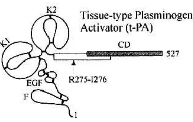
various stimuli such as stress, physical exercise and nicotinic acid. It is a 70-kDa protein secreted as a precursor in a single chain form [114]. Plasmin convert the precursor by cleaving the peptide bond Arg275-Ile276 to give an active two chain form held together by a single interchain disulphide bond. The tPA molecule contains 530 amino acids and has a molecular weight of about 67,000. The A-chain (Mr 38,000), is derived from the NH2-terminal portion, and the B- chain (Mr 30,000) containing the active site from the COOH-terminal portion [115]. The A-chain contains four domains; a finger do- main, a growth factor-like domain and two kringle domains. The finger and kringle 2 domains are involved in binding to fibrin. The human tPA gene is located on chromosome 8 [114], spans 33 kb and has 14 exons (Fig.I.13). tPA exists as two different isoforms (type I and type II) that differ on their glycosylation, displaying species, cell and site specific patterns of this post translation modification [116]
The main biological role of tPA is associated with fibrinolysis due to its high affinity for fibrin and activation by fibrin binding [117]. The finger domain present at the amino terminal enables tPA to have high affinity for fibrin making tPA a thrombolytic agent. Unlike uPA, tPA has got no specific receptors but it has a wide range of receptors including AnnexinA2 (AnxA2), α-enolase, HMG-1, LRP, NMDAR, etc.
I.4.1.4 The Plasminogen activator inhibitors
The Plasminogen activator inhibitors (PAI)s are members of serine protease inhibitor super family (SERPIN) and are the most potent inhibitor of both tissue type (t-PA) and urokinase type plasminogen activator (u-PA) and thus regulate fibrinolysis as well as proteolysis, cell migration, and tumor cell invasiveness[118]. They are stimulated by cytokines, lipopolysaccharide, very low density lipoprotein, TGF- β-1 etc. In pathological conditions, increased PAI-1 levels mainly result from release by endothelial cells or tumor cells [119]. There are several inhibitors of plasminogen activators: PAI-1, PAI-2, PAI-3 and TAFI
process and in the pathogenesis of many disorders including cancer [120]. It inhibits both uPA and tPA and also has high affinity for ECM proteins. PAI-1 not only binds to free uPA but also to uPAR – bound uPA [121]. uPA/PAI-1 complex interact with the transmembrane α2-macroglobulin receptor low-density lipoprotein (LDL) receptor- related protein (LRP), an endocytic receptor. By the combined action of uPAR ad LRP, the uPA/PAI-1 complex is internalized and degraded in lysosomes and the uPAR is recycled back to the cell surface [96, 122]. PAI-1 is believed to play a central role in cell adhesion mediated through integrins and the uPAR/uPA complexes [123]. Kwaan et al studied the action of PAI-1 in vascular smooth muscle cells and tumor cells and found out that PAI-1 inhibited apoptosis and contribute to tumor proliferation and to angiogenesis [124]. In vivo studies has shown that PAI-1 expression can be highly induced in both endothelial cells and activated platelets and it plays a role in inhibiting thrombolysis through rapid inhibition of tPA [125].
I.4.1.4.2 Plasminogen activator inhibitor type-2
proteolysis involved in apoptosis and inflammation. In experimental tumor mouse models PAI-2 has shown to decrease the tumor growth and metastasis. The physiological outcomes obtained by the administration of exogenous PAI-2suggested that inhibition of extracellular uPA activity is the mechanism underlying the reduction in tumor size [127]
I.4.2 The Plasminogen system in Cancer
PAI-1 are associated with poor prognosis [139]. In adenocarcinoma of esophagus, uPA and PAI-1 levels were significantly elevated in cancerous tissues. In PDAC, PAI-2 seemed most important prognostic factor as strong PAI-2 expression was significantly associated with higher survival compared to lower levels of PAI-2 [64]. In endometrial and lung cancer uPA, uPAR, PAI-1 and PAI-2 levels were higher in tumor tissue than compared to the normal ones[140, 141].
tPA has also been related to tumor progression and the role of this protease in cancer and PDAC will be described in another section (see point I.4.4).
I.4.3 The Plasminogen system and Angiogenesis
co-Studies by Pepper et al confirmed that when endothelial cells migrate they upregulate uPA, uPAR and PAI-1 [144]. A variety of angiogenic factors control the expression of these proteins in ECs, like VEGF and bFGF induces the expression of uPA, tPA, uPAR and PAI-1, while TGF-β down regulates uPA and enhances PAI-1 production [145]. Plasmin promotes tissue degradation and remodeling of the local extra cellular environment directly, by degrading ECM molecules and activating or releasing latent growth factors [146]. Plasmin also potentially activates pro–matrix metalloproteinases such as pro-MMP2 and pro-MMP9 [147] (Fig.I.14).
tumor growth in vivo is dependent on the generation of uPA mediated plasmin. Angiogenic endothelial cells require uPA and plasmin to degrade ECM components and migrate [152]. O’Reilly and Stack et al demonstrated that angiostatin can be generated by limited proteolysis of Plasminogen by plasmin, uPA, tPA and MMP [143, 153]. Studies in PAI-1 deficient mice have revealed an absolute requirement for PAI-1 in tumor angiogenesis. The absence of PAI-1 markedly impaired tumor invasion and vascularization [154].
Fig.I.14.- Plasmin activating the angiogenic factors. Adapted from Croucher et al, Nat Rev Cancer, 2010
implanted in integrin α1-deficient mice (which express higher levels of matrix metalloproteinases and plasma angiostatin), reduced tumor angiogenesis was observed [155]. Plasma from these mice inhibited endothelial cell proliferation, and this inhibition was decreased by administering an angiostatin-neutralizing antibody or matrix metalloproteinase inhibitors. Mice injected with colon cancer cells that overexpressed t-PA had a markedly lower number of liver metastases and a higher survival rate than mice injected with untransfected colon cancer cells [156]. A study of tumor specimens taken from patients with melanoma or with breast cancer showed that tumors expressing a high level of t-PA were associated with better prognosis [157]. Thus, it has been hypothesized that human plasma may serve as a rich source of biologically important angiogenesis inhibitors [158]. Increasing the antiangiogenic activity of plasma proteins by proteolytic cleavage may alter the behavior of a malignant tumor and could have clinically useful implications.
I.4.4 Tissue Plasminogen Activator in Cancer and PDAC
catalytic activity [159]. More recently, our group has also found similar mechanisms in PDAC [237]. tPA is overexpressed in several cancers, including melanoma [160, 161], hepatocellular carcinoma[162], ovarian[163], uterine [164] and pancreatic ductal adenocarcinoma [165-167]. In pancreatic cancer tPA is overexpressed in 95% of pancreatic ductal adenocarcinoma, while it is absent in normal pancreas [165, 166].
tPA contributes to cell invasion in in vitro studies of pancreatic cancer [165] via its interaction with AnnexinA2[168]. tPA induces Erk phosphorylation and cell proliferation[168], involving AnnexinA2 and EGFR [169, 237].
I.5 Galectins
Fig.I.15. - The galectin family. Many galectins are either bivalent or multivalent
with respect to their carbohydrate-binding activities. Adapted from Fu-Tong Liu
et al, Nat Rev, 2005.
On the cellular level galectins are involved in mechanisms both inside the cell and extracellularly. Galectins have been proposed to play important roles in inflammation, immunity and cancer based on whole animal experiments and by their effects in tissue culture [175, 176]. Intracellularly, galectins are found in the cytosol and nucleus where they are involved in targeting exocytosis, cell activation and differentiation[177-179]. Galectins are also secreted by different
cells via the non-classical pathway and can attach to sugar structures on the cell surface [180]. By binding to N-glycosylated proteins Galectins may reside in solution as serum proteins or, on the cell surface, where they can modulate cell adhesion [181].
I.5.1 Galectin Expression
which was originally identified as an eosinophil specific chemo attractant is also expressed in HUVECs [185]. Galectin-8 is expressed in lung both in basal cells, ciliated bronchial cells, chondrocytes, serous cells of the bronchial glands, smooth muscle cells and endothelial cells [186]
I.5.2 Galectin-1
I.5.3 Galectin-1 in Cancer
I.5.4 Galectin-1 in Pancreatic Cancer
Pancreatic tumours overexpresses Gal-1 and Gal-3 proteins in comparison to normal pancreatic tissue [220]. Gal-1 was mainly detected in pancreatic cancer tissue stromal fibroblasts and extracellular matrix [220]. Gal-1 protein was upregulated in pancreatic ductal adenocarcinoma in comparison to normal pancreatic tissue [221]. By making changes in the ECM, Gal-1 could be involved in tumor progression in pancreatic cancer. Schaffert et al looked at different pancreatic cancer lines and human pancreatic carcinoma tissue and saw a uniform and strong overexpression of Gal-1 in pancreatic cancer cells compared to normal controls. The expression pattern of Gal-1 in pancreatic cancer tissues indicated that Gal-1 plays a role in the desmoplastic reaction that occurs around pancreatic cancer cells [220]. Harsha et
al postulated that Gal-1 was overexpressed in the stroma of
pancreatic cancer tissue and in the neoplastic ductal cells [222].
interaction contributes to PDA progression involving both transformed epithelial cells and tumoral stroma [243].
I.5.5 Galectin-1 in Angiogenesis
OBJECTIVES
Previous data have shown a pro-angiogenic effect of tPA in in vivo models of PDAC [244, 168] although the molecular mechanisms are not well understood. The GENERAL AIM of this Thesis was to
elucidate the role of Tissue type Plasminogen Activator (tPA) in
the Angiogenesis of Pancreatic Cancer. To pursue this aim we
have focused on the following objectives:
1) To analyze whether tPA-mediated angiogenesis is an INDIRECT effect through the positive regulation of pro-angiogenic molecules (VEGF, TGF-β, interleukins, MMPs) produced by pancreatic tumoral cells.
2) To analyze whether tPA-mediated angiogenesis is a DIRECT effect mediated by tPA direct activation of EC proliferation, migration and tubulogenesis, and whether these effects are tPA-catalytic activity dependent or independent.
3) To elucidate the molecular mechanisms involved in tPA pro-angiogenic effects: analysis of ERK1/2, AKT and JNK signaling pathways.
RESULTS
R.1.- Effect of tPA on Angiogenic factors
As mentioned in the Introduction, VEGF, TGF-β and different cytokines can stimulate angiogenesis. We analyzed whether tPA pro-angiogenic effects can be indirectly mediated by the induction of these pro-angiogenic molecules in tumoral cells and in ECs. Pancreatic cancer cell lines (RWP1 and SKPC1) and an endothelial cell line (H5V) were exposed to 20µg/ml of tPA for 24 hours. RNA was extracted and checked for the expression of VEGF, TGFβ-1, TGFβ-2, TGFβ-3, IL1-α and IL-8 by semi-quantitative PCR, using β-actin levels as loading control. The levels of these molecules
remained unaltered after tPA treatment (Fig. R.1), indicating that tPA-proangiogenic effects are not mediated by them.
analyzed by RT-PCR for expression of VEGF, TGFβ-1, -2 and -3, IL-1α and IL-8. Levels were normalized using β-actin as loading control.then analyzed by RT-PCR for expression of cytokines. The RNA levels were normalized with β-actin.
protein data, MMP-2 RNA levels remained unaltered after tPA treatment in all cell lines. In contrast, MMP-9 RNA levels showed a slight increase only in HUVEC after tPA treatment and remained unaltered in pancreatic cells, suggesting that MM-9 increase obeserved after tPA-treatment in pancreatic cells should be mediated by post-transcriptional mechanisms (Fig. R.2C).
B
Fig R.2.- MMPs activity and expression was analyzed by gelatin zymography and RT-PCR analysis in pancreatic and endothelial cells after tPA treatment. A. Cell conditioned media were resolved in gelatin preimpregnated
polyacrylamide gels. Gels were developed to show clear bands of gelatinolysis, correlating with the latent and active isoforms of MMP-2 and -9. B. Densitometry analysis of the zymogram indicating the activity of MMP 9 in HUVEC. C. RT-PCR analysis of MMP-2 and MMP-9 mRNA from HUVEC and PDAC cells.
R.2.- tPA induces endothelial cell Proliferation, Migration and
Tubulogenesis
dimensional structure organization. The cells attach during the first hour and then, in the presence of angiogenic stimuli, migrate towards each other during the next 2-4 hours and form mature capillary-like tubes after 6-16 hours. HUVEC and H5V cells treated with tPA (0.5µg/ml) showed tube formation after 16 hours, indicating that tPA does promote tubule formation in vitro (Fig.R.3C). At 24-48 hours nearly all the cells were dead with complete destruction of tube structures (data not shown). VEGF (20µg/ml) treatment was used as positive control.
In order to test whether these effects were dependent on the catalytic activity of tPA, HUVEC and H5V cells were treated with a catalytically inactive mutant tPA (tPAS481A) and its effects in cell proliferation, migration and tubulogenesis was analyzed. tPAS481A increased EC proliferation and induced migration and tubulogenesis to the same extent than wild-type tPA, indicating that these pro-angiogenic tPA effects were directly mediated by tPA independently of its catalytic activity (data not shown).
Fig.R.3.- tPA effects in endothelial cell proliferation, migration and tubulogenesis. A. Proliferation of HUVEC (left) and H5V (right) cells untreated
(Neg), treated with 20 µg/ml of tPA (tPA) or with 5% FBS, as positive control (FBS), was measured by 5-bromo-2-deoxyuridine (BrdU) incorporation. B. HUVEC and H5V cells were grown to confluence, deprived in medium without FBS for 3 days, wounded and allowed to migrate in the absence or presence of tPA (20 mg/ml). FBS was used as positive control. C. Tubule formation in collagen sandwich model of HUVEC and H5V cells untreated (Neg), treated with 0.5 µg/ml of tPA (tPA) or with VEGF (20 µg/ml, positive control).
B
[image:77.499.97.443.73.428.2]R.3.- tPA induces catalytic-independent activation of ERK1/2,
AKT and JNK signalling pathways in ECs.
tPA has been described to have a mitogenic role on variety of cell types like aortic smooth muscle cells, pancreatic cells and neurons and this effect has been related to ERK 1/2 signalling pathway activation. In addition, ERK 1/2 kinases have been shown to regulate endothelial cell survival, migration and proliferation, particularly in conjuction with growth factor signaling. To analyse whether tPA effects in ECs described in the previous section (R.2) could be mediated by ERK1/2 activation, we examined the ability of tPA to activate the ERK1/2 cascade in HUVEC and H5V endothelial cells. tPA induced an strong increase in ERK1/2 activation, detected by Western blot using antibodies against phospho-ERK1/2. This activation was transient, reaching it maximum at 5 minutes and was maintained for 15 minutes in HUVEC (Fig. R.4A) and H5V (Fig.R.4 B) cells. The levels of total ERK1/2 remained unchanged indicating a specific effect in ERK1/2 phosphorylation/activation. Tubulin levels were also analysed as loading control.
Fig R.4. - tPA induces ERK activation in HUVEC and H5V cells. A)Western
blot analysis of phosphorylated extracellular signal-regulated kinase (p-ERK) and ERK in human umbilical vein endothelial cells (HUVEC) stimulated with tPA (20µg/ml) for 5, 15, and 30 min. (B) WB analysis of p-ERK in H5V endothelial cells treated with tPA (20µg/ml) for 5, 15 and 30 min. FBS (5%) treatment was used as positive control. Three independent experiments were performed and quantification is shown in the graph.
Recent report by An et al [242] demonstrated that tPA induces phosphorylation of AKT -a key pathway involved in cell survival- in the ischemic brain. To determine whether tPA can induce AKT activation in ECs, HUVEC and H5V were treated with tPA (20 µg/ml) and AKT activation was analyzed. tPA induced AKT phosphorylation in both HUVEC (Fig.R.5A) and H5V (Fig.R.5B) cells. AKT activation was detected at 15 minutes in HUVEC while in H5V it can be already observed at 2 minutes
Fig.R.5.- tPA induces AKT activation in HUVEC and H5V cells. A. Western
blot analysis of phosphorylated AKT (P-AKT) and total AKT (T-AKT) in HUVEC stimulated with tPA (20µg/ml) for 2, 5, 15, and 30 min. B. WB analysis of phosphorylated AKT (P-AKT) and total AKT (T-AKT) in H5V endothelial cells treated with tPA (20µg/ml) for 2, 5 and 15min. Three independent experiments were performed and quantification is shown in the graph
JNK signalling pathway has been also related to endothelial activation and angiogenesis, and recent data from our group has shown that tPA can activate JNK in microglial cells. We examined whether tPA can induce JNK activation in endothelial cells. Treatment with tPA induced JNK activation at 5 minutes and was maintained until 30 min in both HUVEC (Fig. R.6A) and H5V
(Fig.R.6B) cells. Total levels of JNK and tubulin were also
analyzed to rule out nonspecific effects in total protein levels.
Fig.R.6.- tPA induces JNK activation in HUVEC and H5V cells. A. Western
blot analysis of phosphorylated JNK (P-JNK) and total JNK (T-JNK) in HUVEC stimulated with tPA (20µg/ml) for 5, 15, and 30 min. B. WB analysis of phosphorylated JNK (P-JNK) and total JNK (T-JNK) in H5V endothelial cells treated with tPA (20µg/ml) for 5,15 and 30 min.Three independent experiments were performed and quantification is shown in the graph
Fig.R.7.- tPA-mediated phosphorylation of ERK kinases in endothelial cells does not require its catalytic activity. Mutant tPA S481A induces ERK1/2
activation similar to wild type tPA, indicating that proteolytic activity of tPA is not required for the activation of ERK signalling pathway.
R.4. - ERK1/2, AKT and JNK signalling pathways are responsible
for tPA-mediated EC Proliferation, Migration and Tubulogenesis
We next aimed to validate whether the intracellular signalling cascades triggered by tPA, in a catalytic independent way, are responsible for its effects in EC pro-angiogenic response. To address this point we analyzed HUVEC and H5V proliferation, migration and tubulogenesis after the treatment with tPA alone or in the presence of inhibitors for the different signalling pathways. Treatment of HUVEC and H5V cells with tPA in the presence of MEK/ERK1/2 inhibitor U0126 (10µM) resulted in a dramatic decrease in the cell proliferation, migration and tubulogenesis (Fig.R.8).
Fig.R.8. - Inhibition of ERK1/2 signalling pathway with U0186 abolishes tPA effects on endothelial cell proliferation, migration and tubulogenesis. A. tPA
(20 µg/ml) treatment in the presence of 10µM U0126 (tPA+U0126) failed to increase HUVEC and H5V proliferation, measured by BrdU incorporation. B. tPA (20 µg/ml) treatment in the presence of 10µM U0126 (tPA+U0126) failed to induce HUVEC and H5V migration in a wound healing assay. C. tPA (0.5µg/ml) treatment in the presence of 10µM U0126 (tPA+U0126) failed to induce HUVEC and H5V tubulogenesis in a collagen sandwich assay.
Similar results were obtained after tPA treatment in the presence of wortmannin (10µM) – an inhibitor of the AKT pathway- (Fig.R.9) or in the presence of a JNK inhibitor, JNKI1 (JNK inhibitor-1) (20µM) (Fig.R.10). Altogether these data demonstrate that activation of ERK1/2, AKT and JNK by tPA are required to induce endothelial cell proliferation, migration and angiogenesis.
(10µM) (tPA+Wo) failed to induce HUVEC and H5V migration in a wound healing assay. C. tPA (0.5 µg/ml) treatment in the presence of Wortmannin (10µM) (tPA+Wo) failed to induce HUVEC and H5V tubulogenesis in a collagen sandwich assay.
A
B
Fig. R.10.- Inhibition of JNK signalling pathway with JNKI1 abolishes tPA effects on endothelial cell proliferation, migration and tubulogenesis. A. tPA
treatment in the presence of JNKI1 (20µM) (tPA+ JNKI1) failed to increase H5V proliferation, measured by BrdU incorporation. B. tPA treatment in the presence of JNKI1 (tPA+ JNKI1) failed to induce H5V migration in a wound healing assay. C. tPA treatment in the presence of JNKI1 (tPA+ JNKI1) failed to induce H5V tubulogenesis in a collagen sandwich assay.
R.5. – AnnexinA2, EGFR and Galectin-1 are involved in
tPA-mediated signalling activation in endothelial cells
of siAnxA2 oligos. Treatment with double stranded oligonucleotide targeting an irrelevant transcript (siControl) was used as negative control. Downregulation of AnxA2 expression resulted in a complete abolishment of tPA-induced ERK1/2 activation in both HUVEC and H5V cells. As expected, transfection with siControl had no effect on activation of ERK1/2, indicating the specificity of siAnxA2 (Fig.R.11B). In contrast, FBS-mediated ERK1/2 activation remained unchanged by either siAnxA2 or siControl treatment, indicating the specificity of AnxA2 down regulation in the tPA-induced ERK1/2 signaling (Fig.R.11B). Contribution of AnxA2 to tPA-mediated JNK activation in endothelial cells was also analyzed. H5V cells were transfected with siRNA for AnxA2 (siAnxA2) or an irrelevant transcript (siControl) and JNK activation after tPA-treatment was studied by Western blot. AnxA2 downregulation led to an inhibition of JNK activation after tPA treatment, as shown in Fig.R.11C. Altogether these data indicate that AnxA2 is required for ERK1/2 and JNK activation induced by tPA in endothelial cells.
Fig. R.11.- AnxA2 is required for tPA-induced ERK1/2 and JNK signaling in HUVEC and H5V cells. A. Inhibition of AnxA2 expression using siRNA in
HUVEC and H5V cells. The cells were transiently transfected with siRNA against AnxA2 (siRNA) or an irrelevant target (siControl). Different concentrations of siRNA and siControl were used. Tubulin levels in cell lysates were assayed as a control. B. Depletion of AnxA2 using siRNA blocks tPA-induced ERK1/2 activation. HUVEC or H5V cells, untransfected (C) or transfected with siAnxA2 or siControl, were treated with or without tPA (20µg/ml) for 5 minutes or with FBS for 5 minutes. ERK1/2 activation was detected by WB using antibodies against phosphorylated ERK1/2. Three separate experiments were performed and quantification of reduction in tPA-mediated ERK1/2 activation by silencing of AnxA2 is shown in the graph. C. Depletion of AnxA2 using siRNA blocks tPA induced JNK activation. H5V cells, untransfected or transfected with siAnxA2 or siControl, were treated with or without tPA (20µg/ml) for 5 minutes or with FBS for 5 minutes. JNK activation was detected by WB using antibodies against phosphorylated JNK. Three separate experiments were performed and quantification of reduction in tPA-mediated JNK activation by silencing of AnxA2 is shown in the graph.
Fig.R.12.- EGFR is required for tPA-induced ERK1/2 signaling in HUVEC and H5V cells. ECs were treated with 20 µg/ml tPA for 15 minutes in the absence or in the presence of the EGFR inhibitor Gefitinib (10 µM). Cells were collected and lysates were analyzed by Western blotting using antibodies against phosphorylated (P-ERK1/2) and total ERK1/2 (T-ERK1/2). Treatment with FBS and EGF were used as positive control for ERK1/2 activation and Gefitinib effects, respectively. Inhibition of EGFR results in abolishment of tPA signaling.
(siControl) did not affect the Gal-1 levels. Downregulation of Gal-1 completely abolished ERK1/2 activation by tPA in both HUVEC and H5V cells whereas transfection with siControl had no effect on activation of ERK1/2 (Fig.R.13). In contrast FBS-induced ERK1/2 activation and total levels of ERK1/2 were unaffected by either siGal1 or siControl treatment indicating the specificity of Gal-1 knock down in tPA-induced ERK 1/2 signaling (Fig.R.13).
Fig. A
R.13.- Gal-1 is necessary to induce ERK 1/2 activation in endothelial cells. A.
Inhibition of Gal-1 expression using siRNA in HUVEC and H5V cells. The cells were transiently transfected with siRNA against Gal-1 (siRNA)(25nM, HUVEC; 1nM, H5V) or an irrelevant target (siControl). B. Depletion of Gal-1 using siRNA blocks tPA-induced ERK1/2 phosphorylation in HUVEC and H5V cells . Ecs, untransfected (C), transfected with an irrelevant siRNA (siControl) or with Gal1 siRNA (siGal1),were treated with tPA (20µg/ml) for 5 minutes or with FBS for 5 minutes. ERK1/2 activation was detected by WB using antibodies against phosphorylated ERK. Three separate experiments were performed and quantification of reduction in tPA-mediated ERK1/2 activation by silencing of Gal-1 is shown in the graph.
R.6. – TGF-β and IL-1a induce tPA in PDAC and HUVEC cells
induction of tPA expression in Hs766T cells (Fig.R.14B). Induction of tPA expression by IL-1 α, TGF-β or both cytokines was also determined at the RNA level using RT-qPCR. Fig. R.14C show that treatment of pancreatic cancer cells or HUVEC with IL-1 α, TGF-β or combined treatment (IL-1 α + TGF-β) resulted in a dose-dependent increase of tPA mRNA levels, indicating that the induction of tPA expression by IL-1 α and TGF-β is at the transcriptional level.
A
B
Fig.R.14. - Effect of IL1-α and TGF-β in tPA induction in pancreatic cancer cells and HUVEC A. Dose dependent stimulation of tPA protein by IL1-α and TGF-β in PDAC cells (SKPC-1 and Hs766T) and HUVEC. B. Combined treatment with IL1-α a and TGF-β at various concentrations in HUVEC cells lead to a sinergistic effect in tPA induction. C. tPA mRNA expression levels were analysed by RT-qPCR after treatment with IL1-α, TGF-β or combined IL1-
α+TGF- β in HUVEC and PDAC cells. Levels were normalised with β-actin.
R.7. – Hypoxia induces tPA in PDAC cells
These data suggest that tPA induction by hypoxia should be regulated post transcriptionally.
A
B
Fig.R.15.- Effect of hypoxia in tPA expression in PDAC cell lines. tPA was induced when SKPC-1 or Hs766T cells were
grown in hypoxia (1% oxygen). A. Western blot analysis of tPA in conditioned media from cells grown in normoxic or hypoxic conditions during 6, 12 and 24 hours. B. tPA activity was determined by caesin zymography of conditioned media from cells grown in normoxic or hypoxic conditions during 24 hours to check the activity of tPA during hypoxia in cell extract and conditioned medium. C. Analysis of tPA mRNA by semiquantitative PCR after 24 hours of cell culture in normoxic or hypoxic conditions. D. Analysis of tPA mRNA by RT-qPCR after 6, 12 and 24 h of cell culture in normoxic or hypoxic conditions.
DISCUSSION
D.1 tPA can have an indirect pro-angiogenic effect by inducing
MMP-9 expression in tumoral pancreatic and endothelial cells
D.2 tPA induces endothelial cell proliferation, migration and
tubulogenesis by activation of ERK1/2, AKT and JNK signaling
pathways and independently of tPA proteolytic activity
tPA acts as a cytokine also in endothelial cells. The key role of ERK1/2 activation in tPA effects in endothelial cell proliferation and migration has been demonstrated using U0126 MEK inhibitor, that blocks the induction in cell proliferation and migration in endothelial cells treated with tPA. We have also tested the role of tPA-mediated ERK1/2 activation in tube formation in ECs in 3-D collagen gels. Our experiments with endothelial cells cultured on collagen sandwich showed that tPA treatment can induce the cells to form tubular structures resembling or mimicking the blood vessel formation. Combination of tPA with the pharmacological inhibitor U0126 showed that tubule formation was completely abolished indicating the relevance of ERK1/2 pathway in this process. Previous data have shown that ECs stimulated with angiogenic factors (bFGF, VEGF, and PMA) can activate of ERK1/2, Akt, and PKC leading to increased migration but this is, at least to our knowledge, the first report for the involvement of tPA-induced ERK activation in tubulogenesis. Altoghether these experiments confirmed the role of ERK1/2 pathway in migration, proliferation and tubule formation of endothelial cells in response to tPA.
gastrointestinal cancers are well known [242]. In addition, activation of PI3K/ AKT by various growth factors, the modulation of downstream targets by AKT-induced phosphorylation as well as novel treatment strategies targeting this pathway in gastrointestinal tumors are widely studied. Activation of the AKT kinase orchestrates anumber of signaling pathways potentially involved in angiogenesis. The multiple downstream substrates of AKT not only converge to prevent the induction of apoptosis but may also interfere with numerous biological functions of the endothelial monolayer, which contribute to vascular remodeling and vessel integrity during the angiogenic process [231]. In our experiments we analzyed whether AKT pathway is induced in response to tPA. We found that AKT pathway was induced at 15 min and sustained till 30 min in HUVEC, while in H5V it was induced at 2 min and sustained till 30 min. Importantly, the effects of tPA in the induction of EC proliferation, migration and tubulogenesis were reduced to control levels when tPA was added in the presence of wortmanninm, a pharmacological inhibitor of AKT pathway. These data demonstrate that AKT signalling pathway is required for these events.
D.3 Annexin A2, EGFR and Galectin-1 are involved in binding
of tPA to initiate the activation of signaling pathways in
endothelial cells.
unaffected by the treatment with siAnxA2. Similarly, tPA treatment in the presence of an EGFR chemical inhibitor, Gefitinib, led to levels of phospho-ERK1/2 similar to basal levels. These data indicate that both AnxA2 and EGFR are required for ERK1/2 activation induced by tPA in ECs.
The work from Roda et al indentified that tPA exerts mitogenic effects on pancreatic cells through ERK1/2 activation via Gal-1 receptor [249]. In their experiments they showed that inhibition of Gal-1 expression using siGal-1 RNA markedly decreases tPA induced ERK1/2 activation and proliferation, confirming the crucial role of Gal-1 in tPA induced mitogenic process in pancreatic cancer cells. In our study we checked whether Gal-1 attenuation will affect the ERK1/2 and JNK signaling in endothelial cells treated with tPA. We found that downregulation of Gal-1 by siRNA transfection in HUVEC and H5V cells totally abolished the ERK1/2 signaling induced by tPA. These results indicated that Gal-1 is necessary for triggering ERK1/2 activation in tPA induced angiogenesis. One possible explanation for the activation of ERK1/2 via Gal-1 receptor might be the action of upstream element H-Ras, as it has been reported that Gal-1 can bind H-RasGTP promoting its membrane anchorage [248].
D.4 Cytokines (IL-1α and TGFβ) induce tPA expression in
Pancreatic cancer and Endothelial cell lines.
D.5 Hypoxia induces tPA expression in Pancreatic cancer cell
lines.
A hypoxic microenvironment is a characteristic of many solid
tumors including pancreatic cancer [76]. Hypoxia is a known inducer of many potent angiogenic factors like VEGF, EGF, FGF
which stimulate the initiation of angiogenesis in many types of
cancer. The prototypical angiogenic cytokine increased by hypoxia
is vascular VEGF and its receptor flt-1; however, other angiogenic
factors like angiopoietin-2, placental growth factor, and
platelet-derived growth factor B (PDGF-B) are also regulated by hypoxia
[79]. Another angiogenic cytokine, basic fibroblast growth factor (bFGF), exerts enhanced proliferative activity on endothelial cells
during hypoxia due to an increase of low-affinity heparan sulfate
bFGF binding sites. Hypoxia also induces a hypoxia-inducible
factor-1 (HIF-1α)-dependent bFGF autocrine loop that drives
angiogenesis in human endothelial cell [78]. The study by Sun et al showed that 70.7% of PDAC cells showed positive HIF-1α staining
[78]. Buchler et al demonstrated that hypoxia induced HIF-1α
dramatically transactivated VEGF gene expression in pancreatic
cancer cells in vitro and explored co-localisation of VEGF mRNA
Western blot analysis and casein zymography (Fig. R.15) we found
that tPA expression and activity was enhanced in a time-dependent
manner during hypoxia. To determine whether the cells were in
hypoxic conditions the levels of HIF-1α proteins were also checked
by Western blotting. tPA activity was more prominently seen from
conditioned media than from the cell extract thereby underling the
fact that tPA is secreted and is active during hypoxic conditions.
Interestingly, the levels of tPA mRNA in PDAC cells remained
unchanged during both normoxia and hypoxia conditions . These
data suggest that tPA overexpression during hypoxia is mediated by
posttranscriptional mechanisms (i.e. RNA translation, protein
stability, etc.) although additional experiments are required to
further characterize the specific molecular mechanisms involved in
this regulation.
In the light of our results regarding the role of inflammatory
cytokines and hypoxia in tPA induction by pancreatic and
endothelial cells, and considering our data about the mechanisms
involved in tPA pro-angiogenic effects, we propose the following
model: pancreatic tumoral cells and tumor microenviroment
(stroma), that will be in hypoxic conditions as a consequence of
tumor growth, will produce pro-angiogenic molecules like IL-1α
and TGFβ to create an angiogenic network in order to compensate
hypoxia. All these factors will induce tPA overexpression in both
tumoral and endothelial cells, which in turn is able to induce
angiogenesis both directly promoting EC proliferation, migration
way pro-angiogenic stimuli and tPA will collaborate in a feedback
[image:111.499.81.451.126.454.2]loop to promote tumoral angiogenesis (Fig.D.1)
Fig D.1 Proposed model for tPA angiogenic role in PDAC. Hypoxia and TGF-β/ IL-1α cytokines induce tPA overexpression in pancreatic tumoral cells and endothelial cells (ECs). tPA can then induce angiogenesis both indirectly, through MMP-9 induction and activation, or directly by interaction with EC cell
EC Proliferation, Migration and Tubulogenesis
Pancreatic
tumoral cells
ECs
tPA
AnxA2 /EGFR/ Gal-1
interaction
TGF-
β
, IL1-
α
Hypoxia
MMP-9
ANGIOGENESIS
ERK1/2
AKT
JNK
dir
ec
t
in
di
re
ct
EC Proliferation, Migration and Tubulogenesis
Pancreatic
tumoral cells
ECs
tPA
AnxA2 /EGFR/ Gal-1
interaction
TGF-
β
, IL1-
α
Hypoxia
MMP-9
ANGIOGENESIS
ERK1/2
AKT
JNK
dir
ec
t
in
di
CONCLUSIONS
1. The regulation of pro-angiogenic molecules like VEGF, TGF-β, IL-1α and IL-8 are not mediated by tPA, but the activation of the angiogenic molecule MMP-9 is mediated by tPA. These results indicating that MMP-9 upregulation can indirectly mediate tPA effects in angiogenesis.
2. tPA induce directly and in a catalytic-independent way endothelial cell proliferation, migration and tubule formation.
3. tPA induces activation of ERK1/2, AKT and JNK signaling pathways in endothelial cells and this activation is necessary to induce proliferation, migration and tubule formation .
4. AnnexinA2, EGFR and Galectin-1 cell membrane receptors are involved in tPA- mediated endothelial cell signaling