Functional analysis of Drosophila melanogaster
linker histone dH1
Olivera Vujatovic
TESI DOCTORAL UPF / 2012
Thesis supervisor: Dr Ferran Azorín Marin
Institute for Research in Biomedicine Barcelona
The Molecular Biology Institute of Barcelona
Thesis tutor: Dr. José Francisco Aramburu Beltrán
Department of Experimental and Health Sciences, University Pompeu Fabra
Acknowledgements
Dragi roditelji, najsrdacnije vam hvala za stalnu podrsku. Toliko vaseg razumevanja za
moju zelju da se bavim biologijom, svi uslovi i svestrana pomoc da se skolujem, ispunili
su moje snove. Tokom godina izrade doktorata jedno su rezultati, projekat, eksperimenti
i sva ta nauka. Postoji i mnogo vise od toga kao deo teze. Pridodala bih joj
prilagodjavanje na novu, cudnu zemlju, nostalgiju, padove motivacije sa jedne strane i
sa druge strane ushicenost zbog zanimljive konferencije, rezultata, nekog malog ili
velikog uspeha. I vi ste za njih hteli uvek da znate. Znam da je svaka moja sreca, tuga ili
briga bila makar duplo toliko i vasa i da nikada nisam bila zaista sama. Hrabrile su me vase reci ‘’plivaj Loli’’ ili su me smirivale ‘’tiha voda breg roni’’. Koliko sam samo
puta pomislila na njih kao na magicne reci koje bi mi spasile dan i volju. Priveli smo kraju moju doktorsku tezu zajedno!
Vlado, hvala ti za paznju koju si mi davao kad sam morala sa nekim da porazgovaram,
za sve savete kako da budem jaca, da se borim i cenim sebe. Jos od malena sam uvek
igrala igre koje si ti igrao, trenirala karate, samo da budem kao ti. Klonem li nekad
duhom zbog nostalgije, desavanja u nasoj tuznoj Srbiji ili zbog osecaja da sam
poslednji stranac, setim se tebe, kako si i ti preko i kako si posao daleko neizvesnijim
putem od mog. I tada, nema tu vise mesta samosazaljenju, znala sam da moram da se borim, kao ti.
I wish to thank to Dr. Ferran Azorín for giving me the opportunity to do PhD in his
laboratory and for supervising my thesis. I am very grateful for all the time you
dedicated me to talk about our project, to teach me about science in more general
terms, motivate me when I was feeling down and advise me with all the kindness when it
was necessary to work harder or do something better. Having a dedicated PhD
supervisor with so much interest in science was a great inspiration for me and essential for my decision to continue with scientific career. Many thanks for all this.
Albert Jordan and Dr. Travis Stracker. I am looking forward to hear your comments and questions about the thesis.
Dr. Jordi Casanova, Dr. José Ayté and, one more time, Dr. Alejandro Vaquero were
following progression of my thesis during all years in the form of Thesis Advisory
Committee. Dr. José Aramburu was also at these meetings as my thesis tutor. I appreciate all your suggestions, questions and discussions we had.
The Institute for Research in Biomedicine Barcelona (IRB Barcelona) where I did my
PhD, was a great place to work. It created stimulating environment with seminars,
conferences and other events that exposed me to a big scientific community. One could
see efforts of the institute to develop and grow in their constant innovations and prizes
the institute was winning. As IRB student I could count on help of the administration
related to any issue. I am the most grateful to Margarita Navia, Silvia Aiguadé, Maria
Rovira and Clara Caminal and all the rest of the people that make the institute a
wonderful place. The Molecular Biology Institute of Barcelona (IBMB) is also a part of this atmosphere.
I was fortunate to have Alex as a person who introduced me to the laboratory work.
Apart from his contribution to the project that was advancing very fast with him, his
sense of humour, advices for career and translations of conversations in Spanish he did for me at the beginning were of great help to me.
Many thanks to Jordi for his help in experiments, paper and interest in the progression
of my PhD. It was of big help to have somebody with knowledge needed to work with Drosophila melanogaster and broad knowledge about many other topics.
Thanks to Oscar Reina for the great contribution he had in our first paper, for efforts to
explain me the analysis he was doing, for having patience to go back to our project in different moments.
I owe much gratitude to different people that were working in our laboratory during my
PhD –to Esther and Estefania for so much help with flies; to Alicia for her help with
experiments, her tidiness and reliability; to Salva for being an enthusiastic student with
passion for experiments; to Marta Lloret for her readiness to help with any issue and
making me feel comfortable asking for any advice; to Katrin for help on the project in
experiments and discussions and for positivism she was bringing to the lab in the
mornings; to Roman for friendship, politeness and clear protocols; to Tomás for his
picturesque explications of scientific problems and for willingness to help with ideas in
the project; to Olga for help with cloning; to Joan Font for his critical point of view; to
Eva for scientific discussions and for irradiating enthusiasm for science; to Aleix for
sharing impressions about events in basketball; to Sergi for bringing leisure spirit to
the lab; to Marcia Lami for making cheerful atmosphere; to Dr. Josep Portugal for
interesting talks; to Sònia, Mònica, Lluisa, Anne, Lorena, Syvia Mansilla, people from Marian’s laboratory, Marta y David, Rute, Silvia Perez, Marc Bataller, Bet, Gemma,
Sani etc. There was not much time to get to know better all youngsters that spent few
months with us or the ones that came short time before I left – George, Joan, Ujue, Lucia etc.
I am thankful to many people from the institute: to Dr. Herbert Auer for help with micro
array analysis, protocols for real time PCR and for the interesting discussions; to the
group of Dr. Albert Jordan for the meetings where I learned a lot about H1; to Dr.
Julien Colombelli and Anna Llado from the microscope facility for development of the
software we used to analyse images related to PTMs in this thesis; to Luciano and
Angela from technical service for allowing me prepare special kind of food for flies that
we needed in the study; to Alberto Adeva for chemical synthesis of the peptides and all questions I had about them.
Zelim da zahvalim i drustvu i rodbini koji nisu bili direktno ukljuceni u projekat, ali su
mi itekako znacili: Isabella-i i Mariji, za nase druzenje, sale, nezaboravno putovanje,
interesovanje za to kako sam, mejlove podrske, misljenje u vezi svega i svacega,
caskanje o nauci i zivotu; Mirki Milanovic za nase razgovore na skajpu; tetki Rosi za
lepe poruke iznenadjenja i zato sto je mislila jesam li dobro i kako mi ide; Nikoli
Markovicu, Milosu Tatarskom i Saski Ivanovoj na druzenju; Ani Janic na prakticnim
savetima i pomoci; teta Ljubici i Nadi jer mi je znacilo kada su pitale za mene i htele da
The last days of my thesis preparation I did in Dr. Guillaume Filion’s laboratory. I
want to thank him and Heng-Chang for making me these days interesting and for giving
me the time I needed. I also met Roberto Blesa at the end of the thesis preparation. He
made me these days cheerful and was a big support to me. Thank you for the help with the resume and more importantly, for being with me when I needed it.
Contents
Abstract ... 1
Resumen ... 3
Prologue... 5
Introduction ... 7
Chromatin ... 7
Chromatin packing ... 8
Chromatin organisation ... 10
Types of heterochromatin – classical types ... 11
Types of heterochromatin – newly characterized types ... 12
Heterochromatin assembly ... 14
HP1-dependent and H3K9me-dependent heterochromatin assembly ... 14
RNAi-dependent heterochromatin assembly ... 15
Heterochromatin sequence... 16
Linker histones ... 19
Structural features of linker histones ... 19
Evolution of linker histones ... 21
H1 isoforms ... 23
Histone H1 in Drosophila melanogaster ... 26
Histone H1 protein ... 28
H1 deposition to chromatin ... 29
Histone H1 functions ... 30
H1 as a regulator of transcription ... 33
Posttranslational modifications ... 39
Posttranslational modifications of H1 ... 41
Papers and additional results ... 47
Bibliographic citation of the article 1 ... 49
Article 1 ... 51
Supplementary data of the first article ... 65
Article 2 ... 85
Additional results ... 101
Raising and purification of antibodies ... 102
Characterization of α2mK23dH1 and αpSer10dH1 ... 103
Characterization of α2mK27dH1 ... 103
Fuctional anaysis of α2mK27dH1 ... 106
Materials and Methods ... 111
Antibodies purification ... 111
Dot blot analysis ... 111
Peptide sequences ... 112
Immunostainng of S2 cells ... 112
Peptide competition assay used in immunostainig experiments ... 113
Immunostainng of polytene chromosomes ... 113
Analysis of cells overexpressing JMJD2A ... 114
Discussion ... 115
Extent of dH1 depletion in different tissues ... 115
Effect of dH1 on gene expression ... 116
Specificity of observed phenotypes to dH1 loss ... 116
dH1 affects expression of small portion of genes ... 116
dH1 mainly acts as a gene repressor ... 117
dH1 affects expression of greater portion of heterochromatic then euchromatic genes ... 118
dH1 silences TE expression ... 118
dH1 is necessary to maintain genome stability ... 119
dH1 loss causes DNA damage, stops cell proliferation and induces apoptosis ... 121
PTMs of dH1 ... 125
Conclusions ... 129
Abstract
We did functional characterisation of Drosophila melanogaster linker histone, dH1. In the mutant state for this protein, we observed structural changes in polytene chromosomes, chromocenter and nucleoli of mutant larvae. In addition, we performed a microarray analysis in H1 mutant background in order to determine contribution of dH1 to gene expression regulation. We determined effects of dH1 loss in different types of chromatin and we identified groups of differentially expressed (DE) genes, groups in sense of physical clusters of genes and genomic elements rather than groups of functionally related genes. We found that dH1 affects in greater extent expression of heterochromatin genes compared to its effect on euchromatin genes; that dH1 regulates transcription in a regional manner, since the genes physically nearest to the most DE genes tend to be upregulated as well; and that dH1 is negatively regulating expression of transposable elements and members of certain gene families. In addition, we found that dH1 is necessary for preserving genome stability. Among DE transposable elements we detected R1 and R2 retrotransposons, elements that are integrating specifically in rRNA locus. We showed that activation of their transcription is also upregulating expression of aberrant, transposon-inserted, rDNA units of the locus. In this regard we observed an accumulation of extra-chromosomal rDNA circles, increased
Resumen
Prologue
Histone H1 is evolutionary well conserved, very abundant protein, found in cells of all eukaryotic species while part of prokaryotic species contain proteins homologues to H1. These three characteristics are suggesting that H1 is a protein with important roles in a cell. During the relatively long history of studies of H1, there were changes in ideas about its functions.
The existence of H1 was reported for the first time in 1951, when it was detected in various tissues of different organisms (Stedman, 1951). It was described as a basic
nuclear protein similar to ″main histones″ (today termed as core histones) that however
number of genes whose expression H1 was affecting was limited in the majority of cases. There are couple of examples where even a molecular mechanism by which H1 is accomplishing it was determined (L.-jung Juan et al., 1997, Nishiyama et al., 2009). It was shown that those are infrequent examples so once again a role of H1 as a structural element is gaining importance against its role as a general regulator of transcription. Crystal structure of linker histone globular domain was determined (Ramakrishnan et al, 1993) and different models for the way H1 is binding to DNA to compact it are proposed. It is also becoming clear that posttranslational modifications of linker histone
can determine its functions just like it’s the case with core histones. However, much
more results were obtained for core histones modifications.
We decided to address the question of H1 functions in Drosophila melanogaster
because this is the only multicellular organism with a single H1 variant in the genome. We made a dH1 mutant condition and observed various phenotypes that led as to conclusions about different roles of dH1. We also decided to study a role of a particular modification of dH1 by raising antibodies that specifically recognize the modification.
Introduction
Chromatin
Genetic and epigenetic material of cells is placed in nucleus in the form of chromatin. Chromatin contains DNA, histones and other non-histone proteins. While DNA is a carrier of protein and non-coding RNA sequences, protein portion of chromatin is necessary for structuring and providing proper functioning of DNA. Only together, DNA and chromatin proteins, in a cell allow cell growth, division, specific differentiation, cell death etc. To accomplish these activities, chromatin needs to answer to stimuli from cell interior and environment by constant modulation of chromatin and its functions - gene expression, DNA replication, DNA repair and others.
Chromatin packing
Long DNA molecules have multiple levels of folding by chromatin proteins that allow placing into micronic-size nucleus. Those are nucleosomes (beads on the string structure), 30 nm chromatin fiber and other higher order chromatin structures.
Packing of DNA starts with DNA wrapping around histone octamers. Histone octamers contain two tetramers of core histones, each of them carrying H2A, H2B, H3 and H4. Approximately 146bp long DNA wrapped around the octamer forms a nucleosome. Crystal structure of the nucleosome has been determined at 7Å (Richmond et al, 1984) resolution when it was showed that it has a disc-like shape and in 1997 at 2.8Å
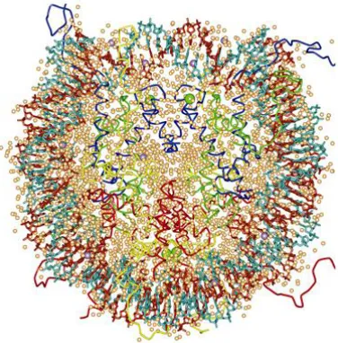
[image:16.595.204.392.497.688.2]resolution more details were obtained (Luger et al, 1997). For simplifying a model all core histone components were expressed and purified from bacteria so they were missing any posttranslational modification and DNA was of defined sequence. The main part of the structure was determined although part of histone C-terminus regions is not included in the model. Finally in 2002 researchers made a closer look to nucleosome with 1,9Å resolution (Davey et al., 2002) (Figure 1).
Array of nucleosomes forms 11nm wide, “beads on the string” structure (it is called like
that because the array resembles it when looked under an electron microscope). Between adjacent nucleosomes extends linker DNA where linker histone H1/H5 can bind and further compact DNA into 30 nm structure. Binding of linker histones is increasing spacing between nucleosomes, termed as nucleosome repeat length (NRL). There is a linear relationship between H1 stoichiometry and NRL (Woodcoc et al, 2006). H1 stoichiometry is variable from one cell type to another and in many of them not all nucleosomes are occupied by linker histone (splenocytes contain 0,79, thymocytes 0,83 H1 molecule per nucleosome (Fan et al., 2003)) which is possibly facilitating chromatin modulating and access to chromatin by regulatory complexes. Using in vitro conditions with high concentration of salt, 30 nm fiber structure can be assembled even in the absence of H1. The exact arrangement of DNA and histones even at this level of chromatin structure is still incomplete (reviewed in Li and Reinberg, 2011). There are two models trying to explain the positioning of chromatin elements at this level: 1) one-start helix (solenoid) and 2) two-start helix (zig-zag) model. Both models are based on studies on in vitro reconstituted chromatin to avoid obstacles of native chromatin: various types of DNA sequences, posttranslational histone modifications, histone variants and irregular nucleosome spacing.
Bases for proposing one-start helix (solenoid) structure model were obtained by electron microscope of long nucleosomal arrays of different repeat length with or without the presence of linker histone and salt. In this model nucleosomes are proposed to be connected with incurved linker DNA so that it delineates a complete circle within about six to eight nucleosomes.
The second model, two-start helix model, has been proposed based on electron microscope and X-ray structure at 9Å resolution obtained with tetranuleosomal array without the presence of linker histones, providing a proper salt concentration (Schalch et al., 2005). A longer, continuous chromatin fiber model was built by successively stacking tetranucleosomes one on another. In this model, linker DNA is connecting nucleosomes that are distributed in zig-zag mode.
existence of one-start helix model (Robinson & Rhodes, 2006). In the same study, in the absence of linker histone, the resulting fiber structure was highly disordered, more similar to the two-start helix model. This is suggesting that a general and unique model probably does not exist and that different types of chromatin exist even in the same cell, depending on the conditions in the local environment.
Little is known about the chromatin structure beyond 30nm fiber level. There are intra- and inter-fiber interactions between nucleosomes which in vitro conditions are observed in the presence of Mg2+. It has been suggested that interactions between adjacent nucleosomes, between histone H4 tail domain with H2A and H2B, can be achieved not only in the same fiber, but between adjacent fibers too, leading to higher order chromatin packing (Gordon et al, 2005).
Chromatin organization
Traditionally, in chromatin there are two main types - open, transcriptionally active part, euchromatin, and closed and transcriptionally silence part, heterochromatin. These two main types of chromatin differ in DNA sequence, in the presence of certain histone variants, degree of DNA methylation, presence of histone posttranslational modifications (PTMs) etc.
Euchromatin is not stained or is lightly stained in G-banding staining. This is an open fraction of the chromatin, available to transcriptional machinery and rich in genes. Besides being hypo DNA-methylated, euchromatin is marked by numerous histone modifications: di-methylation of Lys at position 4 in histone H3 (H3K4me2), tri- methylation at Lys79 in H3 (H3K79me3) and hyper acetylated core histones (H3K14, H4K16). During S phase, euchromatin is replicated before and faster than heterochromatic regions (Lima-De-Faria, 1959).
genes are placed in heterochromatin and are expressed (Smith et al, 2007). It is the part of the genome which is replicated later then euchromatin, at the end of S phase (Lima-De-Faria, 1959). In nuclei, heterochromatin is placed at the nuclear periphery and perinuclear area (Cremer & Cremer, 2001) (Towbin et al, 2009). For a long time it has been believed that this heterochromatic sequences have no role in cells and it has been
unfairly named as “junk DNA”. It turned out that this part of the genome has important
roles (Weiler & Wakimoto, 1995) in regulation of transcription , mostly gene silencing of repetitive DNA elements, by providing non coding RNAs. In addition, it has been demonstrated that it contributes to normal centromere and telomere functioning as well as to chromosome pairing during meiosis. The main histone modifications associated with heterochromatin include methylations: H3K9me3 (Peters et al., 2003), H3K27me3 and H4K20me3 and general histone hypo-acetylation. Besides these histone PTMs, DNA methylation is also heterochromatin mark. The main non-histone protein present in heterochromatin is heterochromatin protein 1 (HP1).
HP1 protein has been first identified in Drosophila melanogaster (James & Elgin, 1986), but their homologues have been determined in many other species, including humans. In D. melanogaster there are three types of HP1 protein, but only HP1a and HP1b can be found in heterochromatin while HP1c is found in euchromatin. HP1 molecules contain chromo domain that recognizes methyl group of H3K9me histones. HP1 is a dominant suppressor of position-effect variegation (PEV) (Wustmann et al, 1989). PEV is a phenomenon of euchromatin gene silencing once they are placed into heterochromatin by genetic manipulation (Muller, 1930). When proteins that are involved in heterochromatin formation and functioning (like HP1) are mutated, heterochromatin cannot silence the gene so that PEV effect is not happening and these proteins are called suppressors of PEV.
Types of heterochromatin – classical types
Facultative heterochromatin is silenced chromatin which however can be converted to open chromatin state when necessarily. Here belong genes whose expression is needed in particular periods during cell cycle, differentiation, development or only under certain environmental conditions. In different cell types of an organism these heterochromatic regions do not necessarily correspond because of their different differentiation programs.
Constitutive heterochromatin is permanently silenced through the cell cycle and is common to different cell types in an organism. It contains highly repetitive sequences, such as clusters of satellite sequences or transposable elements that are usually found around telomeres and centromeres.
Types of chromatin – newly characterized types
The recent genome-wide studies (Consortium 2011 and Filion et al., 2010) addressing a question of histone modifications marks and other non-histone chromatin proteins distribution along genes in genomes of different organisms, in D. melanogaster as well, put a new light on chromatin types. In this way, today we can distinguish even five main types of chromatin in Drosophila melanogaster instead of only two main known before (Filion et al., 2010).
contains genes that are determining cellular functions that are specific for certain cell types and that require more complex gene regulation.
This novel view on chromatin types distinguishes three distinct heterochromatin kinds: BLUE, GREEN and BLACK chromatin.
The most characteristic components of BLUE chromatin are Polycomb Group (PcG) proteins, proteins involved in regulation of developmental genes (Schuettengruber et al, 2007). For the first time they were discovered in Drosophila melanogaster as regulators of developmental, Hox genes, and their orthologs in mammals have been determined.
Hox genes expression is initiated early in embryonic development and PcG proteins are necessary for maintaining their expression in later embryonic stages. These proteins bind Polycomb response elements (PREs) and act as part of three complexes – PRC1, PRC2 and PhoRC. PRC2 has methyltransferase activity, it trimethylates Lys at H3K27 position. H3K27met3 is the main histone modification mark of BLUE chromatin.
GREEN chromatin is present in pericentric regions and on chromosome 4. The components that distinguish this chromatin from other chromatin types are SU(VAR)3-9, H3K9me2, HP1 and HP1 interacting proteins, LHR and HP6.
Heterochromatin assembly
HP1-dependent and H3K9me-dependent heterochromatin
assembly
[image:22.595.134.463.551.646.2]As already mentioned, HP1 is recruited to heterochromatin by recognition of methyl-group in H3K9me histones by its chromodomain. In Drosophila melanogaster HP1 is then recruiting SU(VAR)3-9, a methyltransferase that introduces H3K9 methylation mark. In this way a mechanism of positive feedback in heterochromatin assembly is initiated and can be spread, since there is a successive binding of HP1 for methylated histones and as a consequence recruitment of methyltransferase that introduces the same mark to the adjacent nucleosome until the appearance of boundary elements that stop the spreading (Figure 2). Similarly, in Saccharomyces pombe there is methylation of H3K9 with Clr4 (a yeast homologue of SU(VAR)3-9) and recruitment of Swi6 (yeast homologue of HP1) which provokes spreading of H3K9 methylation mark. Additional proteins that will form heterochromatin can be then recruited, like histone deacetylases (HDAC) that need to erase acetylation before the introduction of methylation mark by histone methyltransferases (HMTs) on target histones.
RNAi-dependent heterochromatin assembly
[image:23.595.160.434.273.597.2]RNAi-dependent assembly of heterochromatin (Figure 3) is mainly studied in S. pombe. One of the key players in this type of heterochromatin formation is RNA-induced transcriptional silencing (RITS) complex (Grewal, 2011).
Figure 3: RNAi-dependent heterochromatin assembly
This complex contains Argonaute 1 (Ago1), heterochromatin-associated chromodomain protein (Chp1), Targeting complex subunit 3 (Tas3) and siRNA that correspond to dg
Binding of RITS and Swi6 provides binding of RNA dependent polymerase 1 (Rdp1) that forms dsRNAs from nascent centromeric transcripts. These dsRNAs are then processed by Dicer1 (Dcr1) into siRNAs. It is not know how exactly siRNAs are promoting formation of heterochromatin, but it is possible that they recruit Clr4 that introduces H3K9me heterochromatin mark.
Heterochromatin sequence
Figure 4 Tandemly repeated sequences can provoke genome instability
Linker histones
Structural features of linker histones
Linker histone of metazoans contains central globular domain and long C- and short N-terminal tails. Several features of H1 structure allow this protein dynamical, regulatory prone interaction with linker DNA, which is necessary for chromatin compaction. Among these features are presence of numerous Lysine (Lys) rich in protein sequence, presence of AKP amino acid motifs, existence of winged helix motif and characteristics of N and C terminus of the protein.
The presence of numerous Lys in H1 protein sequence gives a positive electrical charge that allows interaction with negatively charged phospho groups exposed on DNA molecule surface. Lys provides a great advantage of this interaction, dynamism, since H1 can bind DNA, but if necessarily, it can easily dissociate and permit access of other proteins to the DNA. Lys showed to be much more useful in this sense than Arginine (Arg) (Kasinsky et al, 2001.). Arg is also positively charged amino acid enriched in some other DNA binding proteins, like protamines. The interaction that Arg accomplishes with DNA is much tighter and leads to more rigid interaction with DNA. To dissociate from DNA and allow decondensation, protamines need a presence of another protein deriving from egg. Lys on the other hand establishes more loose interactions with DNA which makes them more dynamic.
permit interaction with other H1 molecules and consequently their cooperation in chromatin folding.
Globular domain of linker histones has winged helix domain (WHD) (Figure 5) responsible for specific recognition of the four-way junction of DNA (4WJ)
(Varga-Weisz et al., 1993). WHD contains three α-helices and a ß-hairpin at the carboxyl terminus, with a short ß-strand situated between helices I and II. The name ‘’wings’’
[image:28.595.187.408.324.549.2]comes from the resemblance of large loops that connect structures of the proteins containing them to wings. It is believed that topology of DNA at the sites of entrance and exit into and from nucleosome resembles 4WJ structure (Lilley, 1992). In that way, linker histone recognizes DNA specifically at the sites where it enters and exits nucleosomes, at the sites of linker DNA.
Figure 5: Crystal structure of globular domain in H1
C-terminus of human H1.1 isoform are mutated, this leads to a decrease in the binding affinity and residence time of the isoform on chromatin (Hendzel et al, 2004). Nonetheless, single amino acid mutant in human H1.1 isoform affects more binding of the histone to chromatin than deletion of the region that contains numerous positively charged amino acids (Hendzel et al, 2004). This is suggesting that not only a charge neutralisation, but most probably, also formation of specific structure in C-terminus affects H1 binding to chromatin. In relation to this, it is important to mention that
C-terminus in mammals forms α-helical structure upon binding to chromatin (Vila, Ponte, Collado et al., 2001).
Little is known about posttranslational modifications that are decorating amino acids of N-terminus in linker histones. There are reports that phosphorylation of H1 is affecting its binding affinity to DNA (Dou et al., 1999), providing a fast way to change accessibility of DNA to various enzymes. Further text (section Posttranslational modifications) contains more information about PTMs in linker histones.
Evolution of linker histones
Appearance of the first proteins similar to linker histones in evolution happened in
Eubacteria. Several of them contain in their genomes genes for basic proteins that have similarities to C-terminus of typical metazoan linker histones. These proteins have high Ala and low Lys content compared to canonical H1.
Certain groups of protists (like Kinetoplastids) also have proteins that correspond only to a portion of H1, again only to its C-terminal domain. In some other groups of protists (Mycetozoans) for the first time in the evolution emerged winged helix domain (WHD) as a part of linker histone protein (this domain has already existed in other proteins, in some transcription factors).
As a possible event that led to the formation of more complete H1 protein in evolution, it has been suggested a fusion event between the carboxyl terminal domains of H1 related proteins and the proto-WHD of H1 related proteins.
Further evolution of H1 domains happened by different means. Globular domain has evolved by nucleotide substitutions while N- and C- terminus have evolved both by nucleotide substitutions and by accumulation of insertions and deletions events.
High incidence of insertion and deletions events are reflected in high variation in N- and C- terminus length through the evolution in organisms that contain all three H1 domains (40+-13 for N-terminus and 106+-17 for C-terminus compared to 79+-5 amino acids for globular domain). Significant simplicity of amino acids content and DNA sequence of the two termini of histone H1 (Ponte et al, 2003) can explain this high insertions and deletions events. Simple sequences (similarly to repetitive sequences) are prone to misalignement during occurrence of DNA functions – replication, repair and recombination (Pearson et al, 2005). The misalignment then can result in skipping or repeating replication of part of the sequence or in unequal crossing-over, all of them resulting in insertion or deletion events.
Evolution resulted in different outcomes in different species so today there are linker histones with two globular domains in Saccharomyces cerevisiae, linker histones with no globular domain at all in Tetrahymena thermophila etc. In majority of species it contains three domains and has numerous isoforms formed by process of gene duplication. Further text (section H1 isoforms) contains more information on this topic.
It is interesting that core histones have completely different origin from linker histones. In evolution, core histones appeared for the first time in Archaebacteria. There are suggestions that core and inker histones might unite in a single organism by lateral gene transfer, a common way of evolutional changes in Eubacteria and Archaebacteria
H1 isoforms
Linker histones have numerous subtypes in different species. While there are very few species with a single H1 variant (Saccharomyces cerevisiae, Tetrahymena thermophila, Drosophilamelanogaster), far most of the species have more H1 isoforms. Chicken for example contains six H1 isoforms while mice have five somatic in addition to two germ-line specific isoform, replacement linker histone H10. The variants can be specific for certain stage in development, for somatic or germ-line or for a tissue. Perhaps the most interesting example in this sense is spermatogenesis in mouse where there is a successive change of H1 isoforms all along maturation of gonad cells (Godde and Ura, 2009).
Humans have eleven different linker histone proteins. H1.X is ubiquitously expressed, five isoforms are present only in somatic cells (H1.1, H1.2, H1.3, H1.4 and H1.5) while the others are present only in certain tissues or organs (H1t, H1T2 and HILS1 present in testicle or H1oo present in oocytes) or in terminally differentiated cells of various tissues (replacement H1 subtype, H1.0). The subtypes differ among themselves at DNA and amino acid sequence level. The somatic type H1 genes (H1.1-H1.5) together with H1t gene are present in clusters with core histones genes, mainly on chromosome six. These genes are not simultaneously expressed. It has been proposed that this is accomplished by the specific chromosomal organization of the genes and by their different promoter structures (Doenecke et al, 1994).
In any of these two cases, it is likely that H1 variants do play specific roles in cells although a common role in chromatin compaction can be attributed to all of them. However, little is known about particular roles that each of them might be playing in a cell. What has been demonstrated so far is that the variants show different characteristics in various terms – difference in turn-over rate (Cole, 1987), different moments of synthesis (Higurashi et al, 1987) and different phosphorylation level (Talasz et al, 1996). In addition, it has been shown (Orrego et al., 2007) that mammalian H1 variants have different affinity for DNA (scaffold-associated region) and chromatin (native chromatin isolated from cells). These experiments consisted of competition assays where was examined relative affinity of six rat H1 subtypes to bind DNA or chromatin. H1a showed the lowest binding affinity, H1b and H1c intermediate and H1e, H1o and H1d the highest binding affinity. There is a high, 19 fold-change,
difference between the highest and the lowest binding affinity, suggesting a functional relevance of distinct binding abilities for H1 variants. Another study (Parseghian et al, 2000) has demonstrated that inactive and active chromatin are occupied by different ratio of human H1 variants. This group used the antibodies specific for four different H1 subtypes to immunoprecipitate genes that are known to be actively transcribed or silent. They have determined that the active chromatin contains less H1.2 and H1.4 and more H1.3; while inactive chromatin contains all four somatic H1 subtypes.
There are additional examples that encourage the assumption that H1 subtypes play specific roles in a cell. It has been determined that H1 subtypes differ in their ability to condense chromatin in vitro (Talasz et al, 1998). H1.3 could condense chromatin even when present in smaller quantities (1.5 times less then H1.2 or H1.4).
It was also found that H1t binding to chromatin has particularities. Namely, H1t binding to chromatin in vitro leaves it in more open state (which physiologically could have a significance in allowing recombination and facilitated replacement of H1t with protamines that should occur) (Talasz et al., 1998).
transition, there is a suppression only of oocyte specific genes. The suppression occurs at the same time when there is a change in H1 subtype in embryo, when germline-specific H1 (H1M or B4) is replaced by somatic H1 subtype. The experiments in which somatic H1 protein level was changed showed that this subtype is necessary for specific oocyte gene silencing (Kandolf, 1994) (Parseghian et al., 2000). It is also known that H1 in this case is blocking transcription by preventing nucleosome mobility and it seems that this can accomplish only a specific H1 subtype.
An obstacle that researches encounter in efforts to determine roles of specific H1 variants is that the variants are redundant. Isoforms play common roles in a cell and this as a consequence leads to an overexpression of the other subtypes and functional replacement of the missing variant (when it is eliminated by genetic manipulation). In this way, knock out experiments did not result in a real decrease in total H1 level so that the phenotypes could not be observed. For instance, knock out of H1c and H1e in mice did not give rise to any change in mice because of the compensatory effects of other H1 isoforms (Fan et al., 2003). Only with triple knock out problems could be observed. More successful try was in human breast cancer cells (Sancho et al, 2008). Namely, in the knock down of any particular H1 variant there was no compensatory increase in expression of other H1 subtypes. This study has pointed on the role of particular H1 subtypes. For example, H1.2 was shown to be the only variant to change a nucleosome repeat length. In addition, among all variants, H1.2 was the one causing the greatest downregulation in gene expression. H1.4 is the only variant essential for cell survival. It is noteworthy that depletion of specific H1 subtypes affected expression of mainly distinct group of genes, supporting the belief that H1 subtypes have distinct functions.
Nevertheless, presence of numerous H1 subtypes is a disadvantage of these model systems. It seems difficult to predict in which extent compensatory effects of the isoforms affect phenotypes one can detect and finally, if isoforms ever allow a sufficient H1 depletion to observe consequences of almost total linker histone loss. In this sense,
Histone H1 in Drosophila melanogaster
In Drosophila melanogaster, dH1 gene is placed in a gene cluster together with four core histones genes. This quintet gene unit (one linker and four core histones genes) is repeated about one hundred of times in a haploid genome (Matsuo et al, 1989). dH1 gene is consisting of TATA-less promoter, 839bp long sequence without introns and at distal stem-and-loop structure (Marzluff, 2005), while polyadenylation signal is absent.
It has been demonstrated that TATA-less promoter of dH1 gene is transcriptionally regulated by TRF2 (TBP (TATA-box-binding protein)-related factor 2) (Isogai et al, 2007). On the other hand, core histones genes, placed together with dH1 gene in the common gene cluster, have a distinct transcriptional regulator, TPB (TATA-box-binding protein) / TFIID. Specific (TATA-box-binding of TRF2 exclusively to dH1 gene promoter and TBP for core histones promoters have been proven by ChiP analysis. In addition, depletion of TRF2 affects negatively specifically level of dH1 mRNA (while not affecting mRNAs of core histones) and affects negatively expression of reported gene regulated by dH1 promoter. The existence of different regulators for linker and core histones is allowing a cell to provide distinct ratio of linker and core histones expression which is needed in different cells or tissues (Holmgren et al, 1985, Ner & Travers, 1994). It is also interesting that binding of PTB to core histone genes is increased in S phase of KC cells, while PTF2 is binding to dH1 promoter equally through the cell cycle (Isogai et al, 2007). Core histones also have a common negative regulator of transcription, Abnormal oocyte (Abo) protein. Again, this regulator of transcription is not shared between core and linker histones, since it was demonstrated that Abo is not controlling expression of dH1 gene, at least in early embryogenesis (Berloco et al, 2001).
means a genetic separation for a time long even to accumulate gene polymorphisms. However, as mentioned, open reading frame of dH1 gene is completely monomorphic. This could be interpreted by a particular localisation that histone genes are occupying in
Drosophila melanogaster genome. Namely, they are on chromosome II, locus 39D/E, very close to the centromere heterochromatin, where recombination rate is very low. To this day, poorness of recombination preserved the original dH1 gene sequence that was present in a common ancestor of all Drosophila melanogaster lines from any genetic change. In Drosophila virilis, on the other hand, histone genes are located in a region more prone to recombinations, which during evolution resulted in genetic polymorphism of histone genes, gene duplications and resulting existence of three linker histone subtypes. A particularity of almost all histone genes, dH1 gene among them, is that they do not contain introns. Only some germ line specific H1 isoforms contain them and they are present in rare examples, like in Xenopus laevis linker histone B4 (Cho, 1994).
A typical metazoan gene at the end contains a sequence which is a signal for polyadenylation. The signal is recognized by polyadenylate polymerase that introduces polyA tail (stretch of 20-250 adenosine monophosphates) at the end of mRNA transcribed from the gene. PolyA tail protects it from degradation and promotes mRNA export from the nucleus to cytoplasm and its translation.
In contrast to this, the majority of histone genes (all except from H3.3, H2a.Z, H1o, H3-cid and macroH2a genes) and linker histone genes among them contain at the end of the gene a stem-and-loop structure (Marzluff, 2005). The stem-and-loop contains a six-base stem, four-base loop and 25-26bp AC-rich sequence (histone downstream element, HDE). After synthesis of H1 mRNA stem-loop binding protein (SLBP) binds stem loop and helps binding of U7 snRNP to complementary HDE. U7 is them recruiting other
necessary proteins (ZP100, Lsm10, Lsm11) that results in 3’ cleavage of HDE part of
histone mRNA. After fast transport of mRNA to cytoplasm, translation occurs.
There is a strong regulation of the level of histone mRNA level mediated by
in histone mRNA processing. In this way level of SLBP is regulating the level of mature mRNA. SLBP is synthesised before entering S-phase and degraded very fast at the end of S-phase of cell cycle.
Histone H1 protein
In early Drosophila melanogaster embryo there are maternally inherited mRNA for dH1, but they are not translated before nuclear division seven (Sarbjit S Ner & Travers, 1994) so that the first stages of Drosophila melanogaster development are occurring in the absence of linker histones. In this period of development there are numerous replications and fast condensation and decondensation is needed. The presence of dH1 would be an obstacle to these cyclic changes of condensation level. On the other hand HMG-D proteins are more useful in this sense. HMG-D mRNAs are maternally inherited and HMG-D proteins can function as linker histones since they protect linker DNA from nucleases (Ner & Travers, 1994). It has been demonstrated that they alter nucleosome repeat length (NRL) when incorporated into reconstituted chromatin (Ner et al., 2001). The level of NRL extension by HMG-D is smaller than level of extension by histone H1 and this could determine the level of chromatin compaction that these two proteins can provide. Namely, it has been suggested that there is a correlation between nucleosome spacing and level of chromatin folding (Blank & Becker, 1996).
Increased nucleosome spacing (longer nucleosome repeat length) is a characteristic of more compacted chromatin. That could mean that HMG-D containing chromatin is less compacted and could provide faster decondensation than dH1 containing chromatin.
First information about genomic localisation of H1 came from immunolocalization experiments in polytene chromosomes in Drosophila melanogaster (Jamrich et al., 1977). Staining of the chromosomes showed that histone H1 is present in gene poor, bands regions where RNA polymerase is absent.
Much more precise genomic position of linker histones in Drosophila melanogaster
was determined much later, by usage of DamID (Braunschweig et al., 2009) and ChiP techniques. Project of Encyclopedia of DNA Elements (modENCODE) (Consortium et al., 2011) employed ChiP analysis to determine locations of many chromatin proteins, including dH1. Project aimed to determine binding sites of the proteins across a developmental time course and in multiple cell lines. The results are showing that dH1 is present along the great portion of the genome and that it is one of the main constituents in repressive chromatin. Scientific audience had to wait for a release of data about dH1 distribution, because of the doubts in quality of the results since dH1 binds at the majority of genomic locations and is not enriched at many regions. Rather, at least in the data obtained with DamID technique, regions where dH1 is depleted can be defined more easily (so called H1 dips).
H1 deposition to chromatin
Proper deposition and removal of linker histone is essential for providing its function in chromatin organization.
In vitro studies identified nucleophosmin 1 (NPM1) as a chaperon that interacts with H1 and deposits it onto dinucleosomal templates (Gadad et al., 2011).
most probably it is affecting assembly of H1 once the chromatin structure has already been established, after DNA replication.
Histone H1 functions
A great abundance of histone H1 molecules in nucleus suggests an important role of these molecules in a cell. Linker histones doubtless have structural role in packing DNA molecule into nucleus as described previously in the text. As mentioned, linker histones regulate spacing between nucleosomes and contribute to formation of 30 nm fiber and higher order chromatin structure. In addition to this, histone H1 plays many important roles in cells. Some of the roles are discovered for the moment only in specific specie, but even so, this information is contributing to the knowledge about all linker histones. The roles of H1 in many cases were determined by following phenotypes in various species with H1 mutant condition.
Saccharomyces cerevisiae mutant for H1 homologue, Hho1p, do not show any significant phenotype. The mutants are viable, without change in growth rate. At the level of chromatin neither could be observed significant changes – there are no changes in nucleosome repeat length neither in MNase accessibility of Hho1p depleted chromatin (Patterton et al, 1998). Compared to numerous phenotypes observed in higher eukaryotes (specified in the further text) the absence of any obvious phenotype is surprising fact in yeast mutants. However, Hho1p is only a putative H1 in yeast since it contains very different features, like structure, to H1 in higher eukaryotes. Instead of N- and C- terminus and globular domain, yeast H1 contains two globular domains separated by Lysine-, Alanine- and Proline-rich domain. Some more recent research identified a role for Hho1p in inhibition of recombination at rRNA locus (Li et al, 2008) and determined that H1 is playing this role independently from Sir2, another protein involved in suppression of rRNA recombination in yeast.
effect on viability neither in growth-rate. However, an increase in nucleus volume has been reported as well as a change in transcription level of specific genes (Shen & Gorovsky, 1996).
In Caenorhabditis elegans H1.1 isoform is shown to be a specific regulator of gene silencing in germ-line cells (Jedrusik & Schulze, 2001). Depletion of only this specific variant results in reduced fertility of the mutants, formation of less germ nuclei and production of severely disordered gonads with lower number of differentiated oocytes. The same research group (Jedrusik & Schulze, 2007) in more recent report determined an increase in H3K4me and decrease in H3K9 level in the mutants, which could point on a role of H1 in establishing repressive chromatin.
There are many papers addressing the question of H1 role in regulation of transcription of rRNA genes in Xenopus laevis (described in section H1 isoforms). Apart from it, roles of embryonic histone H1 (B4) have been studied (Maresca et al, 2005). It has been determined that the loss of B4 provokes defects in mitotic chromosomes. They have elongated arms, they seem to be buckled and twisted and finally they cannot be properly aligned in the metaphase plate. Consequently, problems of chromosome segregation in anaphase occur. On the interphase chromatin on the other hand, no phenotype could be observed. These results are suggesting an essential role of B4 in metaphase chromosomes alignment and anaphase segregation.
chromatin. All these results indicate the involvement of dH1 in heterochromatin structuring (by contributing to unique chromocenter establishment) and functioning as a regulator of expression of heterochromatic genes or genes placed in heterochromatin by genetic manipulation (as proven to be a transcriptional activator of genes active inside of heterochromatin and as being suppressor of PEV, respectively). Apart from the effect on heterochromatin, histone dH1 is essential for the normal polytene chromosomes structure since in dH1 mutants these chromosomes are fragile, have problems of endoreplicated sister chromatids alignment and DNA replication.
Higher eukaryotes are characterised by the existence of genes for numerous H1 subtypes in their genome. These subtypes are redundant and as a consequence, an elimination of a single (or even more) variants is compensated by the increase in expression of other H1 subtypes. The real decrease in total H1 amount can be achieved only by elimination of more variants.
In the case of chicken DT40 B lymphocyte cell line, elimination of ten out of twelve in total alleles coding for six H1 subtypes, did not change total level of H1 in the cells (Hashimoto et al., 2010). Only elimination of eleventh allele decreased the total H1 mRNA level to 50%. These mutants show change in protein patterns in 2D-PAGE gels and the increase in HMG proteins, but they show no changes at the level of global chromatin structure, neither significant change in growth rate. Elimination of the last allele results in increased chromosomal aberration rate, increased nuclear volume, decrease in NRL and affected gene expression globally, mainly reduction in their expression. Growth rate is also affected, as proven not by stop in any part of the cell cycle, but most probably as a consequence of longer cell cycle. Another study in the same cell type determined a role of H1R isoform in DNA damage response (Hashimoto et al., 2007). Cells lacking this specific isoform are showing increased sensitivity to alkylating agent MMS and increased sensitivity to infra red radiation. The authors suggest involvement of H1R in Rad54-mediated homologous recombination.
they die by midgestation. At the chromatin level, occurs a reduction in NRL. Latter studies concerned with gene expression in these mice showed that only a small portion of the genes is changing expression and interestingly, among them are many genes regulated by DNA methylation (Fan et al., 2005). This is suggesting that H1 might be contributing to the formation or maintenance of DNA methylation patterns in mice.
In human breast cancer cell line, T47D, H1.2 depletion is provoking problems in cell growth, cell cycle arrest in G1 phase, decrease in NRL and repression of limited number of genes, many of which are involved in regulation of cell-cycle progression (which can explain G1 arrest). Loss of only H1.4 subtype is provoking cell lethality.
In HeLa cells linker histone was identified as a factor important in DNA repair. It was determined that H1 enhances non-homologous end joining (NHEJ) by DNA Ligase III and DNA Ligase IV (Rosidi et al., 2008).
H1 as a regulator of transcription
Gene expression analysis was done in some of the H1 mutants mentioned in the previous pages to check for the role of linker histones in regulation of transcription. Expression of genes was not drastically affected and H1 most probably has only a limited effect on gene expression. It is interesting that initial experiments that were examining this question got quite different conclusion.
First experiments addressing the question of linker histone role in chromatin were done using in vitro transcriptional system. At the beginning the system consisted of naked DNA used as a template for RNA polymerase activity. With time, naked DNA was changed for chromatin template that besides DNA contains core histones and can contain linker histones. Transcriptional models that were used very often were nucleosomal arrays of mouse mammary tumor virus (MMTV) and 5S rDNA chromatin reconstituted from Xenopus cells.
demonstrated that dissociation of histone H1 is necessary for the transcriptional activation to occur.
Expression of 5S rDNA is also regulated by linker histone. Histone H1 is preventing transcription from oocyte-type 5S rRNA genes by positioning nucleosomes at the site where key positive regulatory elements for transcription are found. In this way binding of transcription factors is prevented and there is no transcription of the genes (Pennings et al, 1994).
In these first experiments H1 has been identified as a general negative regulator of transcription and this epithet has been attributed to H1 for a long time. However, it has been demonstrated that H1 in some cases can act as a positive regulator of transcription since their overexpression leads to transcriptional induction of some genes (Brown et al, 1996). In addition, in vivo studies are giving less importance to histone H1 as a transcriptional regulator. Namely, in knockout models for histone H1 in different species the loss of H1 has only a minor effect on gene transcription.
In mice for example 50% decrease in total H1 content affected only 0.56% of examined genes with two-fold change in expression level (Fan et al., 2005). In chicken DT40 cells complete loss of H1 results in more than two-fold change in expression of only 4.2% of analyzed genes (Hashimoto et al., 2010).
In addition, ways by which H1 regulates transcription have been explained at the molecular level. Linker histones are regulating transcription by different means: by restricting nucleosome mobility, by regulating accessibility of different transcriptional factors to regulatory sequences, by forming a part of transcriptional complexes, by interacting with transcription factors and chromatin remodelling complexes, by regulating acetylation of core histones, in association with ribosomal proteins etc. I will make a brief description by making examples of these types of H1 transcriptional regulation.
access to DNA (Ura et al, 1995). In a sea urchin H1 is also limiting TFs access to rDNA (Pennings et al, 1994). Since H1 can easily associate and disassociate from chromatin and in this way prohibit or allow gene expression, H1 contributes to chromatin dynamics. The dynamics then depends on chromatin affinity for binding of H1 or possibility for redistribution of H1 on chromatin. In the context of H1 as a protein that
restricts mobility of nucleosomes it’s interesting to mention that in vitro has been demonstrated that H1 modulates remodelling of nucleosomes by SWI/SNF. On DNA molecule, SWI/SNF would slide nucleosomes to the end of the template while the presence of H1 changes the sliding to the central part of the template (Ramachandran et al, 2003).
Another example where linker histone regulates binding of TFs is in nucleosomes from HeLa cells where H1 prevents binding of USF (Juan et al, 1994). The same authors do not observe repression in the same extend for Gal4-AH TF binding and propose existence of differential repression rate by which H1 prevents binding of different TFs. It has been show that H1 in this case is actually stabilizing interaction of core histones with DNA and leaving less exposure time of DNA for binding of TF (Juan et al., 1997). Acetylation of core histones negatively affects H1 binding and consequently abolishes H1 repression effect.
Next, H1 can act on transcription by interaction with chromatin remodelling complexes. For instance, H1 is interacting with chromodomain-helicase- DNA-binding 8 (CHD 8), an ATP dependent chromatin remodelling factor. In this way occurs repression of
transcription of targets of two TFs, p53 and β–catenin (Nishiyama et al., 2009). In these
experiments, done on mice by the same group for both p53 and β–catenin, the authors have proven that the presence of H1 is necessary in trimeric complexes (CHD8, H1 and
p53 or CHD8, H1 and β-catenin) for transcriptional repression of specific target genes since either triple knock-out of three H1 isoforms or expression of dominant negative mutant of H1 abolishes the transcriptional inhibition.
isoform, when it modulates affinity of H1.5 binding to IL2 gene promoter. Depletion of H1.5 abolishes IL2 transcriptional inhibition, demonstrating a necessity of linker histone for the inhibition. However, FoxP3 – H1 interaction is not always negatively contributing to transcription of the target genes. For example, in the case of CTLA 4 gene promoter, the interaction of FoxP3 with H1 enhances its expression when H1 binding is weaker.
Joint binding of H1 and ribosomal protein L22 to chromatin is regulating expression of specific genes (Ni et al, 2006). These two proteins have been shown to interact and depletion or overexpression of one of them results in changes in expression in the same direction for common set of genes, suggesting interplay of H1 with ribosomal proteins in regulation of transcription.
Linker histone has been implicated in regulation of transcription by interplay with core histone acetylation. Data suggest that linker histones affect core histones acetylation and
vice versa. Acetylation level of core histones is tightly regulated in a cell by two main groups of enzymes: histone acetyltransferases (HAT) and histone deacetylases (HDAC) that introduce and erase the mark, respectively. This strong regulation of core histone acetylation is suggesting that the modification plays an important role in chromatin functioning. Indeed, there are many evidences that correlate H3 lysine acetylation with transcriptional activation (Parekh & Maniatis, 1999). By affecting core histone acetylation, linker histones are affecting gene expression. The interplay between H1 and core histone acetylation is mutual.
On one hand, in vitro analysis have shown that acetylation of core histones is altering the capacity of H1 to form higher order structures (Ridsdale et al, 1990) and that it decreases H1 repression of transcription factor binding on reconstituted nucleosomes (Juan et al, 1994). However, there are contrary in vitro results arguing that repressive effect of H1 on transcription and reduction effect of H1 on nucleosome mobility depends on core histones acetylation (Ura et al, 1997).
Posttranslational modifications
Proteins can change their properties and functions by introduction of simple chemical groups on their amino acids. Some of the groups for instance contain a lot of electrical charge that affects structure of a protein and consequently, its function. Good example for this is greatly negatively charged phospho-group that in numerous enzymes is a regulatory modification for their activity. For a cell these, so called, posttranslational modifications (PTMs) are very practical since they can change the functionality of the proteins without a need to invert energy and time to synthesise a completely new protein.
In histones, groups that are introduced on amino acids of N-terminal tails and effect of
these PTMs on histones’ properties are studied the most. Majority of them are reversible
domain of WDR5 recognizes H3R2 and H3K4me2 etc. Determination and studying PTMs of histones is then a very interesting task since these marks are regulating some of the most basal functions of chromatin and most crucial functions of a cell. As a good example of PTMs studies importance stand new medical approaches in which enzymes that regulate histone PTMs are targeted. By imitating their role in a cell gene expression can be regulated and this can have many consequences.
From recently, there are genome-wide data about chromatin proteins distribution and genome sequences in different species. Their availability is unrevealing that there is regularity in the distribution of histone with particular modifications in different chromatin types and even along single genes. It is known that euchromatin is decorated with H3K4me, H3K36me, that heterochromatin contains H3K9me, H4K20me etc. In addition, active and silenced genes have a special pattern of histone modifications along their sequence (Figure 6). Sequences upstream from active gene promoters have H3K9me2/3, H3K27me2/3 and H4K20me3. Promoters of these genes have nucleosomes with H3K4me2, H3K4me3, H3K9ac and in less extent H3K4me1 and general histone acetylation. Downstream from TSS, active genes contain H3K79me1/2/3 followed by H2BK5me1, H3Kme1, H3K27me1 and H4K20me1 and further, closer to the gene end are H3K36me3 and completely distally are H3K9me2/3, H3K27me2/3 and H3K20me3. On the other hand, silenced genes have H3K9me2/3 and H4K20me3 upstream their promoters, H3K27me3 and H3K4me 3 around TSS and in less extent along the whole gene as well.
Mass spectrometry techniques have contributed much in detection of histone posttranslational modifications. Histone modification maps for chicken, mouse and human (Wood et al., 2009) and other species are becoming more complete and functions for some of these new observed modifications are being established.
Posttranslational modifications of dH1
ubiquitination is TAFII250 (Pham et al., 2000), but the exact position of the modification is not determined yet.
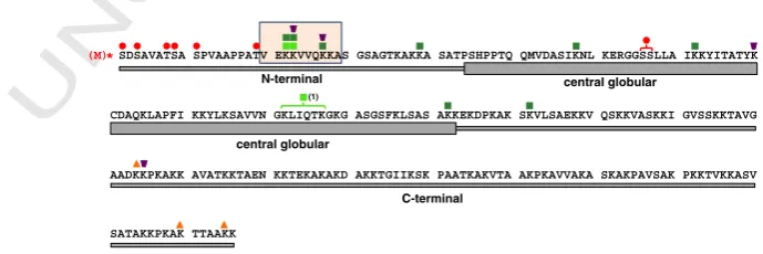
It is interesting that there are several potential targets for methylation in dH1 (e.g. Lysines at positions 22, 23, 27 and 28 in N-terminus) and that in human H1 residues with methyl groups have been determined (Garcia et al, 2004). It is known that H1K26me is included in heterochromatin formation and HP1 protein in vitro recognizes this modification (Daujat et al., 2005) Methyltransferase and demethylase that regulate the mark have been determined (Trojer et al., 2009). Even so, methylation in dH1 has not been reported yet. Mass-spectrometry has limitations in detection of small methyl groups and particularly in the case of Drosophilamelanogaster there is a case where an amino acid polymorphism produces spectrometric peaks that can be ambiguous since the same peak can be corresponding to an amino acid which is methylated or, on the other hand, the same peak might correspond to another, unmodified amino acid (Villar et al, 2007). Therefore analysis of peaks that contain methyl-groups can be equivocal and lead to wrong interpretation. Additional approaches besides classical mass-spectrometry are needed for confident determination of methylated residues.
Drosophila melanogaster as a model system
Drosophila melanogaster is widely used model system in biology. Apart from having a short life cycle and being easy to cultivate, there is a great pool of genetic tools that are developed for this specie, due to a very long history of laboratory work on this fly. There are only four chromosomal pairs in this specie and for each of them there are chromosomal markers that can be followed phenotypically and in this way inheritance of the chromosomes can be easily followed from generation to generation. There is a great collection of mutants available for this fly. Recently, D. melanogaster genome has been sequenced and genomic maps for numerous chromatin proteins have been determined as well. There are three cell lines of the specie. Gal4 system has been described (Brand & Perrimon, 1993) and widely used to induce RNAi response in
[image:51.595.92.490.557.699.2]Drosophila melanogaster. In this system, Gal4 gene, which is a yeast gene, not present in wild type fruit fly, is introduced into D. melanogaster genome (or in another genome) and expressed by control of different promoters. Gal4 protein has an affinity for binding upper activating sequence (UAS) and in this way activates expression of the downstream gene (Figure7).
It means that the expression of the hairpin can be controlled by usage of a specific promoter controlling expression of Gal4. Some promoters are active in all cell types (like actin or tubulin promoter), some others only in particular cell types or tissue compartments. Some promoters are expressed early in embryogenesis, some others later or only during specific developmental stages. In other words, by making careful choice one can induce expression of Gal4 and the hairpin in specific part of fly life stage and part of the body. The hairpin is cut by Dicer in short pieces that will activate other elements in RNAi machinery pathway. This will result in degradation of mRNA corresponding to the hairpin sequence and consequently, decrease in the protein level. To accomplish stronger mRNA depletion, one can overexpress Dicer2 to stimulate RNAi machinery pathway.
Wing imaginal discs are Drosophila larval organs that after metamorphosis give rise to adult wings. They are very often used in studies. They can be easily isolated from larvae and there are optimized protocols for different techniques applied on them. This organ contains only a membrane and two layers of cells, which is very useful for immunostaining experiments because it is not necessary to make thinner cuts. Many signalling pathways are studied in discs and lot is known about them and about phenotypes that are produced as a consequence in of impairment in some of these
pathways. In this way, by following phenotypes in wing’s shape, vein morphology, state