10.1128/MCB.00087-06.
2006, 26(14):5214. DOI:
Mol. Cell. Biol.
Geha
Miguel A. de la Fuente, Lalit Kumar, Bao Lu and Raif S.
Ligation
Cells, but Not T Cells, to Antigen Receptor
http://mcb.asm.org/content/26/14/5214
Updated information and services can be found at:
These include:
REFERENCES
http://mcb.asm.org/content/26/14/5214#ref-list-1
at:
This article cites 59 articles, 37 of which can be accessed free
CONTENT ALERTS
more»
articles cite this article),
Receive: RSS Feeds, eTOCs, free email alerts (when new
http://journals.asm.org/site/misc/reprints.xhtml
Information about commercial reprint orders:
http://journals.asm.org/site/subscriptions/
To subscribe to to another ASM Journal go to:
on August 26, 2013 by Harvard Libraries
http://mcb.asm.org/
MOLECULAR ANDCELLULARBIOLOGY, July 2006, p. 5214–5225 Vol. 26, No. 14 0270-7306/06/$08.00⫹0 doi:10.1128/MCB.00087-06
Copyright © 2006, American Society for Microbiology. All Rights Reserved.
3BP2 Deficiency Impairs the Response of B Cells, but Not T Cells,
to Antigen Receptor Ligation
Miguel A. de la Fuente,
1Lalit Kumar,
1Bao Lu,
2and Raif S. Geha
1*
Division of Immunology1and Ina Sue Perlmutter Laboratory,2Children’s Hospital and Departments of Pediatrics and Medicine, Harvard Medical School, Boston, Massachusetts 02115
Received 13 January 2006/Returned for modification 20 February 2006/Accepted 19 April 2006
The adapter protein 3BP2 is expressed in lymphocytes; binds to Syk/ZAP-70, Vav, and phospholipase C-␥ (PLC-␥); and is thought to be important for interleukin-2 gene transcription in T cells. To define the role of 3BP2 in lymphocyte development and function, we generated 3BP2-deficient mice. T-cell development, prolif-eration, cytokine secretion, and signaling in response to T-cell receptor (TCR) ligation were all normal in 3BP2ⴚ/ⴚmice. 3BP2ⴚ/ⴚmice had increased accumulation of pre-B cells in the bone marrow and a block in the
progression of transitional B cells in the spleen from the T1 to the T2 stage, but normal numbers of mature B cells. B-cell proliferation, cell cycle progression, PLC-␥2 phosphorylation, calcium mobilization, NF-ATp dephosphorylation, and Erk and Jnk activation in response to B-cell receptor (BCR) ligation were all impaired. These results suggest that 3BP2 is important for BCR, but not for TCR signaling.
3BP2 is a protein originally characterized as an Abl SH3-interacting protein (44). It consists of an N-terminal pleckstrin homology domain, a proline-rich central region, and a C-ter-minal SH2 domain. 3BP2 mRNA is expressed in hematopoi-etic tissues. 3BP2 protein is expressed in T cells, B cells, natural killer (NK) cells, monocytic cell lines, osteoclasts, and the rat basophilic leukemia cell line RBL-2H3 (11, 37). 3BP2 associ-ates with Syk and ZAP-70 (11, 36). Association of 3BP2 with Syk was shown to require the SH2 domain of 3BP2 and the catalytic activity of Syk. The SH2 domain of 3BP2 has also been shown to bind to the adapter protein LAT in T cells and mast cells after T-cell receptor (TCR) and FcεRI liga-tion, respectively (11, 46). Phospholipase C-␥(PLC-␥) and Vav were also identified as binding partners of 3BP2 (22). Other partners of 3BP2 include 14-3-3, Grb2, Cbl, and Fyn (11, 14, 15).
TCR stimulation induces a significant translocation of 3BP2 to the membrane and detergent-insoluble (cytoskeleton) frac-tions, suggesting a role for 3BP2 in TCR-mediated signal trans-duction (11). Transient overexpression of 3BP2 in Jurkat T cells induces transcriptional activation of the interleukin-2 (IL-2) gene promoter and its NF-AT and AP-1 elements and cooperates with TCR ligation and ionomycin in activating NF-AT/AP-1-driven transcription. Overexpression of 3BP2 re-sulted in calcineurin-dependent dephosphorylation of NF-ATc and stimulated AP-1 activity independently of NF-AT (11). The SH2 and PH domains of 3BP2, but not its proline-rich domain, are required for NF-AT activation. Furthermore, overexpression of the SH2 domain of 3BP2 inhibited TCR-mediated NF-AT activation (11). Activation of NF-AT by 3BP2 in T cells required ZAP-70, because overexpression of 3BP2 in a ZAP-70-deficient Jurkat cell clone failed to activate NF-AT (11).
Overexpression of 3BP2 enhances NK cell-mediated cyto-toxicity (22). FcεRI ligation induces the phosphorylation of 3BP2 and its association with LAT in rat basophilic leukemia RBL-2H3 cells (46). Overexpression of the SH2 domain of 3BP2 in these cells suppresses FcεRI-induced signaling (46). Recently, it was shown that 3BP2 is tyrosine phosphorylated following B-cell receptor (BCR) ligation in B cells and is a substrate for Syk and Fyn, but not Btk (14). 3BP2 was shown to interact with several components of the BCR signaling path-way, including Syk, PLC-␥, and Vav, and cooperated with Vav to activate NF-AT after BCR ligation. To investigate the role of 3BP2 in lymphocyte development and function, we gener-ated 3BP2-null mice by gene targeting.
MATERIALS AND METHODS
Generation of 3BP2-deficient mice.The3bp2gene was cloned from isogenic 129 genomic DNA library (Stratagene) using the full-length cDNA as a probe. The 5⬘5.4-kb arm (PstI-EcoRV) and the 3⬘8-kb arm (HindIII-HindIII) frag-ments were subcloned into pScrambler vector (Stratagene), which contains the neomycin phosphotransferase (neo) cDNA driven by the PGK promoter. The construct was introduced by electroporation into embryonic stem (ES) cells using standard protocols (2), and cells were then selected in medium containing 200 g/ml G418 and 1M ganciclovir. ES clones containing the homologous recom-binant event were identified by Southern blot analysis and by a 5.6-kb PCR product using forward primer a (5⬘-TGAGTCACTGGATCATGGGAG-3⬘) lo-cated upstream of the 5⬘arm and reverse primer b (5⬘-CGCATGCTCCAGAC TGCCTTG-3⬘) located in theneogene. A 0.3-kb product is amplified by PCR from the WT allele using forward primercin exon 9 (5⬘-ACAGGCTGACACT GGCGA-3⬘) and reverse primerdin exon 10 (5⬘-CGCAAGACTCTGTCGTG T-3⬘). Two ES clones (no. 3 and 7) with the recombinant allele were injected in C57BL/6 blastocysts, and 3BP2⫺/⫺mice were obtained by standard methods
(54). 3BP2 mRNA expression was analyzed by reverse transcription-PCR (RT-PCR) using a forward primer, e, that hybridizes to exon 2 (5⬘GCTGGTTACC TGCATAAG3⬘) and a reverse primer, f, that hybridizes to the 5⬘half of exon 6 (5⬘ATAGGTCGCTCAACTGCA3⬘). 3BP2 protein expression was examined by Western blotting using an antibody raised against amino acids 359 to 462 of the protein.
FACS analysis.Single-cell suspensions from spleen, bone marrow, thymus, and peritoneal cavity were isolated on a density gradient of Lympholyte-M (Cedarlane Laboratories). Cells were stained with phycoerythrin- or fluorescein isothiocyanate (FITC)-labeled monoclonal antibodies (MAbs) from PharMingen and analyzed on a FACSCalibur flow cytometer (Becton Dickinson) as previ-ously described (2). Fluorescence-activated cell sorter (FACS) analysis was
per-* Corresponding author. Mailing address: Division of Immunology, Children’s Hospital, 300 Longwood Ave., Boston, MA 02115. Phone: (617) 919-2484. Fax: (617) 730-0528. E-mail: raif.geha@childrens.harvard .edu.
5214
on August 26, 2013 by Harvard Libraries
http://mcb.asm.org/
formed on cells from mice between 6 and 10 weeks of age. Annexin-FITC staining and propidium iodide (PI) staining were performed using a kit from Biovision Inc.
Antibodies.The MAbs were obtained from the following suppliers: anti-CD3ε (KT3) was from Serotec, anti-CD28 was from eBioscience, antiactin was from Chemicon, anti-ZAP-70 and anti-PY 4G10 were from UpstateBiotechnology, and anti-CD40 was from R&D. The polyclonal antibodies were obtained from the following suppliers: anti-Syk was from Abcam; anti-Vav1, -PLC-␥1, and -PLC-␥2 were from Santa Cruz Biotechnology; anti-NF-ATp was a gift from A. Rao; anti-cyclin D2 was from Biosource; and anti-cyclin D3, -cdk4, -cdk6, -p27, and -pRb antibodies were from Cell Signaling. All antibodies to mitogen-acti-vated protein (MAP) kinases were from Cell Signaling, except antibody to phospho-Jnk, which was from Biosource.
Cell purification, proliferation of splenic T and B cells, and Ig synthesis.
Splenic T cells were purified (⬎95% CD3⫹cells) by negative selection using CD3 enrichment columns (R&D) and cultured at 1⫻105
/well for 64 h in wells coated with 2g/ml anti-CD3 MAb, with or without 2g/ml anti-CD28 MAb or IL-2 (R&D Systems) at 40 ng/ml. Phorbol myristate acetate was used at 15 ng/ml, and ionomycin was used at 0.5M. Proliferation was assessed by the incorpo-ration of [3
H]thymidine. Concentrations of IL-2 in supernatants were measured by enzyme-linked immunosorbent assay (ELISA; R&D). B cells were purified from splenocytes labeled with biotin-conjugated MAbs to CD43 and CD11b (BD Pharmingen) and negatively sorted with streptavidin-conjugated magnetic beads (Dynal). B cells (⬎85% CD43-negative cells) were cultured at 1⫻105
/well in medium alone or in the presence of goat F(ab⬘)2anti-mouse immunoglobulin M
(IgM) (Jackson Immunoresearch) without or with IL-4 (40 ng/ml; R&D Sys-tems), anti-CD40 (2 g/ml; R&D), or lipopolysaccharide (LPS; 10 g/ml [Sigma]), and 48 h later, the cells were pulsed with 1Ci [3
H]thymidine and counted. B and T cells were also incubated with 5M 5,6-carboxyfluorescein succinimidyl ester (CFSE) at 37°C for 10 min and then washed in phosphate-buffered saline (PBS) before culture at 37°C with different stimuli. After 72 h, cells were analyzed by FACS and percentages of cells for each cell division were calculated. For Ig synthesis, B cells were cultured in complete medium alone or in the presence of 10g/ml LPS (Sigma-Aldrich) and IL-4 without or with goat F(ab⬘)2anti-mouse IgM as previously described (21). After 6 days, supernatants
were assayed for IgM, IgG1, and IgE by ELISA.
For in vitro survival, purified B cells were cultured at 5⫻105cells/ml in the
presence or absence of 200 and 500 ng/ml BAFF (Biosource). At various time points, viable cells were counted in a light microscope using a Neubauer hemo-cytometer and the trypan blue exclusion test. B cells were also washed, incubated with 10g/ml merocyanine 540 (Sigma-Aldrich) in PBS–0.1% bovine serum albumin, and analyzed by FACS.
Immunoprecipitation and Western blotting.Cells were lysed in ice-cold lysis buffer containing 1% Triton X-100, and lysates were centrifuged for 15 min at 4°C and precleared for 1 h at 4°C with protein G-Sepharose (Amersham Phar-macia Biotech). Immunoprecipitation was performed overnight at 4°C with antibody preadsorbed onto protein G-Sepharose. Beads were washed five times with modified lysis buffer containing 0.2% Triton X-100. Bound proteins were eluted, run on sodium dodecyl sulfate-polyacrylamide gel electrophoresis, and analyzed by Western blotting with the indicated antibodies followed by goat anti-mouse or rabbit antibodies conjugated to horseradish peroxidase (Amer-sham) and enhanced chemiluminescent (ECL) detection (Perkin-Elmer). As indicated, Western blot images were scanned and quantified using NIH Image (version 1.63) software.
Calcium mobilization.Measurement of calcium flux was done as described previously (30). Briefly, 2⫻106
purified T or B cells/ml were incubated with 4 g/ml Fluo-4-AM (Molecular Probes). T cells were preloaded with 2g/ml anti-CD3 antibody and then stimulated with 10g/ml goat F(ab⬘)2anti-rat IgG.
B cells were stimulated with 5 to 20g/ml goat F(ab⬘)2anti-mouse IgM. Cells
were analyzed by FACS. Intracellular Ca2⫹concentration was measured by
FACS as Fluo-4AM fluorescence intensity using the FL1 channel.
Serum Ig levels and antibody responses.Serum Ig levels were quantified by ELISA. Immunizations with keyhole limpet hemocyanin (KLH), 2,4,6-trinitrophenyl (TNP)–LPS, and TNP-Ficoll were performed, and the serum antibody responses to KLH and TNP were determined by ELISA as previously described (54).
Statistical analysis.Statistical analysis of the data using Student’sttest was performed with Prism software.
RESULTS
Generation of 3BP2ⴚ/ⴚmice.A targeting construct, in which
the 3⬘half of exon 6 plus all of exons 7 through 13 of murine
3bp2were replaced with a neomycin-resistant gene, was intro-duced into ES cells (Fig. 1A). The deletion includes the pro-line-rich region and the SH2 domain of 3BP2. ES clones with targeted disruption of3bp2were identified by Southern blot-ting and genomic PCR (data not shown). Of 250 ES clones analyzed, 5 were found to have a disrupted allele and 2 were used to generate 3BP2⫺/⫺mice. Mice with a disrupted3bp2
gene were identified by PCR on tail genomic DNA, as evi-denced by the amplification of a novel 5.6-kb fragment derived from the targeted allele compared to the 0.3-kb fragment de-rived from the WT allele (Fig. 1B). Lysates and immunopre-cipitates from thymus and spleen probed by Western blotting using an antibody raised against an amino acid sequence im-mediately upstream of the SH2 domain of 3BP2 (amino acids 359 to 462) revealed no detectable 3BP2 protein in 3BP2⫺/⫺
mice (Fig. 1C) (data not shown). Because there are no avail-able antibodies directed against the N-terminal region of 3BP2, we looked for the presence of a transcript encoded by the N-terminal exons preserved in the targeted allele. RT-PCR analysis of mRNA from splenocytes with primers that hybrid-ize to sequences in exon 2 and exon 6, which are preserved in the targeted allele, amplified a 392-bp product in WT mice, but no detectable product in 3BP2⫺/⫺mice (Fig. 1D). 3BP2⫺/⫺
mice did not display apparent differences from WT littermates in growth, weight, or health. Results of all analyses presented below were similar in the two independently derived 3BP2⫺/⫺
mouse strains.
Lymphoid development in 3BP2ⴚ/ⴚ
mice.Cellularity of the thymus, bone marrow, spleen, and peritoneum was normal in 3BP2⫺/⫺ mice. Detailed FACS analysis of thymocytes from
3BP2⫺/⫺ mice and WT littermates at 6 to 10 weeks of age
revealed no obvious difference in the percentages of CD4⫹and CD8⫹cells or of CD3⫹, TCR-␣⫹, TCR-␥␦⫹, and CD2⫹cells (data not shown). Bone marrow cells from 3BP2⫺/⫺mice had
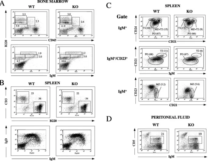
a normal percentage of the B220⫹CD43⫹pro-B-cell popula-tion (Fig. 2A, upper panel), but an increased percentage of B220loIgM⫺population (Fig. 2A, lower panel) that includes
pro-B cells and pre-B cells, indicating that pre-B cells were increased (approximately twofold). The percentages of B220lo IgM⫹ immature B cells (Fig. 2A, lower panel) and B220hi IgM⫹/B220hi CD43⫺ mature recirculating B cells (Fig. 2A,
upper and lower panels) were normal. The absolute numbers of pro-B, pre-B, immature, and recirculating B cells in bone marrow from femurs of 3BP2⫺/⫺mice and WT controls were
determined using triple staining for IgM, B220, and CD43. The results revealed a significant increase in the numbers of pre-B cells in 3BP2⫺/⫺ mice and no significant difference in the
numbers of the other B-cell populations (Table 1). There were no obvious differences in the numbers and percentages of Thy1⫹CD4⫹, CD8⫹, B220⫹IgM⫹, and IgD⫹cells in spleens and lymph nodes of 3BP2⫺/⫺mice and WT littermates (Fig.
2B) (data not shown).
Immature IgMhi HSA/CD24⫹ bone marrow B cells are
transported into the spleen, where they start to express the complement receptor CD21, a part of the CD19-CD21-CD81 coreceptor complex, and the low-affinity IgE receptor CD23. Splenic transitional B cells have been divided into IgM⫹CD24hi CD21⫺CD23⫺T1 cells, which mature into IgMhiCD21⫹CD23⫹
T2 cells (8). T2 cells mature into IgMloCD21⫹CD23⫹follicular
B cells, which recirculate. Marginal zone (MZ) B cells are a
on August 26, 2013 by Harvard Libraries
http://mcb.asm.org/
sessile population of IgM⫹ CD21⫹ CD23⫺ cells generated from T2 cells and possibly from follicular B cells and are located close to the marginal sinus. MZ B cells play an impor-tant role in the response to TI antigens (1, 7). Detailed analysis of spleen B cells was performed by simultaneously staining for IgM, CD21, and CD24 or IgM, CD21, and CD23 (Fig. 2C). 3BP2⫺/⫺mice had an increased percentage of IgM⫹CD24hi CD23⫺transitional T1 cells (17.2%⫾3.2% of splenic B cells in 3BP2⫺/⫺mice versus 11.4%⫾1.3% in controls;P⬍0.05,
n⫽ 3), a decreased percentage of IgMhiCD21⫹CD23⫹T2
cells (5.3%⫾2.0% of splenic B cells in 3BP2⫺/⫺mice versus
12.4% ⫾3.3% in controls; P⬍ 0.05, n ⫽3), and a normal percentage of IgMlo CD21⫹ CD23⫹ follicular B (FO) cells
(66.5% ⫾ 10.7% of splenic B cells in 3BP2⫺/⫺ mice versus
68.7%⫾ 8.4% in controls; n ⫽3). There was no significant difference in the percentage of IgM⫹CD21⫹CD23⫺MZ B cells (3.7%⫾ 2% in 3BP2⫺/⫺ mice versus 4.2%⫾ 1.3% in
controls; n ⫽ 5). There was a significant decrease of CD23 expression on IgM⫹splenic B cells from 3BP2⫺/⫺mice (mean
fluorescence intensity [MFI], 222 ⫾ 76.4 in 3BP2⫺/⫺ mice
versus 773⫾268.4 in controls;P⬍0.0001,n⫽9). A similar decrease in CD23 expression was observed on peripheral blood B cells (data not shown). The percentage of IgM⫹CD5⫹B1 cells in the peritoneum of 3BP2⫺/⫺mice was normal (Fig. 2D).
These results suggest that 3BP2 plays a role in the progression from pre-B cells to immature B cells and from T1 to T2 B cells.
Normal T-cell function in 3BP2ⴚ/ⴚmice.The proliferation
of T cells from 3BP2⫺/⫺mice in response to plates coated with
anti-CD3 was similar to that of T cells from WT littermates over the range of anti-CD3 concentrations tested (0.3 to 10
g/ml) (Fig. 3A). Similar proliferation of 3BP2⫺/⫺and WT T
cells was also observed when cells were stimulated with anti-CD3 plus anti-CD28 or anti-anti-CD3 plus IL-2 (Fig. 3B). Consis-tent with the results from [3H]thymidine incorporation, similar percentages of T cells from 3BP2⫺/⫺mice and WT controls
underwent a comparable number of rounds of cell division in response to anti-CD3, with or without anti-CD28, as deter-mined by CFSE dilution assay (Fig. 3C). 3BP2⫺/⫺ T cells
normally upregulated CD69 and CD25 surface expression fol-lowing TCR ligation (data not shown) and secreted normal amounts of IL-2 following stimulation with CD3 plus anti-CD28 (Fig. 3D). These results suggest that 3BP2 is not essen-tial for T-cell activation in response to TCR ligation.
TCR signaling is normal in 3BP2ⴚ/ⴚ mice. TCR ligation
results in protein tyrosine phosphorylation that includes phos-phorylation of ZAP-70, Vav, and PLC-␥1. Total protein ty-rosine phosphorylation and phosphorylation of ZAP-70, Vav, and PLC-␥1 were normal in 3BP2⫺/⫺T cells (Fig. 4A and B).
PLC-␥1 activation results in IP3 generation and calcium mo-bilization. Calcium mobilization after TCR ligation was normal in T cells from 3BP2⫺/⫺mice (Fig. 4C). Phosphorylation of the
FIG. 1. Generation of 3BP2-deficient mice (A). Genomic structure of the murine3bp2gene, targeting vector, and predicted structure of the targeted allele after homologous recombination. Exons are represented by boxes. Neo, neomycin resistance gene; TK, thymidine kinase gene. (B) PCR analysis of tail genomic DNA from 3BP2⫹/⫹, 3BP2⫹/⫺, and 3BP2⫺/⫺littermates. Two sets of primers were used in the same reaction and are shown in Fig. 1A. Primers a and b amplify a 5.6-kb band in the rearranged allele. Primers c and d amplify a 0.3-kb band in the WT allele. (C) Western blot (WB) analysis of 3BP2 immunoprecipitates from thymocytes of two 3BP2⫺/⫺mice (knockout [KO] mice 3 and 7) derived from different founders and a WT control (PS, preimmune rabbit serum). (D) RT-PCR analysis of mRNA from splenocytes of WT and 3BP2⫺/⫺mice
using primers e and f, which hybridize to exon 2 and the 5⬘region of exon 6 that is preserved in the targeted allele. These two primers amplify a 392-bp product from cDNA. GAPDH (glyceraldehyde-3-phosphate dehydrogenase) is used as a positive control.
5216 DE LA FUENTE ET AL. MOL. CELL. BIOL.
on August 26, 2013 by Harvard Libraries
http://mcb.asm.org/
MAP kinases Erk, Jnk, and p38 was also normal in these cells (Fig. 4D).
Impaired B-cell proliferation in response to BCR ligation in 3BP2ⴚ/ⴚ mice. Primary mouse splenic B cells express 3BP2
(data not shown). Highly purified B cells (⬎90% B220⫹) from 3BP2⫺/⫺mice showed a significant defect in proliferation in
response to BCR cross-linking with anti-IgM over the range of concentrations tested (0.8 to 12g/ml), as assessed by [3
H]thy-midine incorporation (P⬍0.05,n⫽6) (Fig. 5A). This defect was partially corrected by IL-4, although the proliferation of B cells from 3BP2⫺/⫺mice to anti-IgM plus IL-4 remained
sig-nificantly lower than that of WT B cells (Fig. 5B). Consistent with results from [3H]thymidine incorporation, CFSE dilu-tion assays showed that after anti IgM treatment a lower percentage of B cells from 3BP2⫺/⫺mice entered cell
divi-sion compared to WT B cells (Fig. 5C, middle panel). Cell division was also delayed in 3BP2⫺/⫺ B cells treated with
anti-IgM plus IL-4 (Fig. 5C, right panel). 3BP2⫺/⫺B cells
proliferated normally in response to anti-CD40⫾IL-4 and LPS⫾IL-4, as determined by [3H]thymidine incorporation (Fig. 5D) and CFSE dilution assay (data not shown). 3BP2⫺/⫺B cells normally upregulated surface expression of
the major histocompatibility complex class II and CD86 in response to all stimuli tested, including anti-IgM (data not shown).
Normal B-cell survival in response to BCR ligation, but impaired expression of cell cycle regulatory proteins in B cells in 3BP2ⴚ/ⴚ
mice.To determine whether the impaired B-cell
FIG. 2. Detailed FACS analysis of B cells in bone marrow (A), spleen (B and C), and peritoneum (D). In panel C, T1 cells were identified as CD24hiCD21⫺cells in the IgM⫹gate (top panel), T2 cells as IgMhiCD21⫹cells in the IgM⫹/CD23⫹gate (middle panel), and follicular B cells as CD24⫹
CD21⫹cells in the IgM⫹gate (top panel) and as IgMloCD21⫹in the CD23⫹gate (middle panel). MZ B cells were identified as CD21⫹CD23⫺
cells in the IgM⫹gate (lower panel). The percentages shown in the middle panel were adjusted to depict the percentage of total IgM⫹cells. KO, knockout.
TABLE 1. Absolute numbers of B lineage cells in bone marrow
Mouse genotype
No. of B lineage cells (106)a
Total
B220lo
CD43⫹ (pro-B)
B220lo
CD43⫺ IgM⫺ (pre-B)
B220lo
CD43⫺ IgM⫹ (immature)
B220hiIgM⫹
(recirculating)
WT 15.51⫾1.21 0.37⫾0.03 0.63⫾0.08 0.44⫾0.07 0.30⫾0.02 3BP2⫺/⫺ 17.62⫾1.81 0.49⫾0.05 1.56⫾0.11* 0.38⫾0.06 0.35⫾0.04
aThe means⫾standard deviation of the absolute numbers are shown for the
indicated populations in one femur each from WT and 3BP2⫺/⫺mice (n⫽6
each). *,Pⱕ0.05.
on August 26, 2013 by Harvard Libraries
http://mcb.asm.org/
proliferation in response to anti-IgM was due to impaired survival, we performed dual staining with annexin V and PI. We found no difference in the percentage of annexin V⫹ apop-totic cells and annexin V⫹/PI⫹necrotic cells between B cells from 3BP2⫺/⫺ mice and WT littermates at 24 h and 48 h
poststimulation with anti-IgM⫾IL-4 or LPS (Fig. 6A). These results suggest that 3BP2 is not essential for the survival of B cells following BCR ligation.
Cyclins and cyclin-dependent kinases (Cdks) as well as Cdk inhibitors control cycle progression. With entry into the cell cycle, the levels of cyclins D2 and D3 and of their partners Cdk4 and Cdk6 are upregulated, the level of the Cdk inhibitor p27 decreases, and the Cdk target retinoblastoma tumor sup-pressor protein Rb is phosphorylated on Ser807 and Ser811 (5, 32, 51). We examined these events in WT and 3BP2⫺/⫺B cells
stimulated with anti-IgM. Stimulation of WT B cells with anti-IgM resulted in upregulation of the levels of cyclins D2 and D3 and of their partners Cdk4 and Cdk6, decreased p27 levels, and Rb phosphorylation (Fig. 6B). 3BP2⫺/⫺B cells
exhibited defective upregulation of cyclins D2 and D3, Cdk4, and, to a lesser extent, Cdk6; no detectable decrease in p27 levels; and no detectable Rb phosphorylation after stimulation with anti-IgM (Fig. 6B). These defects were partially rescued by IL-4. 3BP2⫺/⫺B cells responded
nor-mally to LPS. Taken together, these results suggest that 3BP2 is important for the progression of B cells through the cell cycle following BCR ligation.
BAFF is important for B-cell survival (45). We examined the survival response of B cells from 3BP2⫺/⫺mice to BAFF (200
ng/ml and 500 ng/ml). Cells were analyzed every 24 h by trypan blue exclusion for 5 days. No significant difference in the per-centages of viable cells was found between WT and 3BP2⫺/⫺
mice (n⫽4 each) at any time point. At 5 days, the viability of WT B cells was 4%⫾2% in medium versus 41%⫾5% in 200 ng/ml BAFF. At that time point, the viability of 3BP2-deficient B cells was 6%⫾2% in medium versus 38%⫾6% in BAFF (data not shown). Similar results were obtained when the cells were analyzed by FACS for exclusion of merocyanine 540 (data not shown).
Impaired BCR signaling in 3BP2ⴚ/ⴚ
mice. 3BP2 has been shown to associate with Syk, Vav, and PLC-␥2, which are phosphorylated and activated following BCR ligation (11, 14). There was no detectable decrease in the tyrosine phosphory-lation of Syk and Vav1 after BCR ligation in 3BP2⫺/⫺B cells
(Fig. 7A). In contrast, PLC-␥2 phosphorylation was decreased (Fig. 7A). Calcium mobilization after BCR ligation was im-paired in B cells from 3BP2⫺/⫺mice over several
concentra-tions of anti-IgM (5, 10, 15, and 20g/ml) tested (Fig. 7B)
FIG. 3. T-cell proliferation and cytokine secretion in 3BP2⫺/⫺mice. (A and B) Proliferation of purified T cells from 6- to 10-week-old WT mice
and 3BP2⫺/⫺littermates (knockout [KO] mice) to increasing concentrations of anti-CD3 (␣-CD3) used to coat wells (A) and to anti-CD3 (2g/ml) with or without soluble anti-CD28 (␣-CD28; 2g/ml) or IL-2 (40 ng/ml), and to phorbol myristate acetate and ionomycin (P⫹Io) (B), as assessed by [3H]thymidine incorporation. Med, medium. (C) Proliferation of purified T cells to anti-CD3⫾anti-CD28 as assessed by CFSE dilution assay.
(D) Secretion of IL-2 in supernatants. Results shown are representative of three experiments.
5218 DE LA FUENTE ET AL. MOL. CELL. BIOL.
on August 26, 2013 by Harvard Libraries
http://mcb.asm.org/
(data not shown), suggesting impaired activation of PLC-␥2. B cells from 3BP2⫺/⫺mice responded with normal calcium influx
to ionomycin.
Calcium mobilization plays a critical role in calcineurin-dependent dephosphorylation of the transcription factor ATp, which allows it to translocate to the nucleus (35). NF-ATp dephosphorylation after BCR ligation, as indicated by a downward shift in molecular weight, was readily apparent in WT cells but barely detectable in 3BP2⫺/⫺B cells (Fig. 7C).
3BP2⫺/⫺B cells responded normally to ionomycin with
com-plete dephosphorylation of NF-ATp. BCR stimulation of splenic B cells induces NF-AT-dependent upregulation of sur-face CD5 expression (4, 53). B cells from 3BP2⫺/⫺mice were
impaired in their ability to upregulate CD5 in response to BCR ligation (Fig. 7D).
BCR ligation results in the phosphorylation and activa-tion of the MAP kinases Erk, Jnk, and p38, which are im-plicated in BCR-mediated proliferation (23, 39). Phosphor-ylation of ERK1/2 and JNK after BCR ligation was reduced in 3BP2⫺/⫺cells, whereas phosphorylation of p38 was
un-affected (Fig. 7E). Phosphorylation of MEK1/2, which is upstream of ERK, was also decreased after BCR ligation in 3BP2⫺/⫺B cells (Fig. 8).
BCR ligation also results in phosphatidylinositol 3-kinase activation and subsequent phosphorylation and activation of Akt. Akt contributes to NF-B activation, which is important for B-cell survival (25, 41, 43). Phosphorylation of Akt and of the NF-B inhibitor IB following BCR ligation was normal in 3BP2⫺/⫺B cells (Fig. 8).
Serum immunoglobulin levels and serum antibody re-sponses in 3BP2ⴚ/ⴚmice.Serum levels of IgM, IgG subclasses,
and IgA were not significantly different between 6- and
8-week-old 3BP2⫺/⫺mice and WT littermates (data not shown). To
determine the role of 3BP2 in the antibody response, we im-munized 3BP2⫺/⫺mice and WT littermates with the
T-depen-dent (TD) antigen TNP-KLH, the type I T-indepenT-depen-dent (TI) antigen TNP-LPS, and the type II TI antigen TNP-Ficoll. IgM and IgG anti-TNP antibody responses of 3BP2⫺/⫺ mice to
all three antigens were comparable to those of WT controls (Fig. 9A). In addition, 3BP2⫺/⫺mice made a normal response
to KLH (data not shown). Furthermore, the size and number of germinal centers in the spleen, as determined by staining for bcl-6, were comparable in 3BP2⫺/⫺ mice and WT controls
(data not shown).
Activation of B cells by TI antigens, and particularly type II TI antigens, is dependent on BCR cross-linking and activation (38). Given their defective BCR signaling, the normal antibody response of 3BP2⫺/⫺ mice to TI antigens was unexpected.
BCR cross-linking is known to inhibit IgM synthesis and iso-type switching (26). This raised the possibility that decreased sensitivity to BCR inhibition of Ig synthesis may compensate for impaired BCR activation in the antibody response of 3BP2⫺/⫺mice. To test this hypothesis, we examined the effect
of BCR cross-linking on IgM secretion and isotype switching to IgG1 and IgE in 3BP2⫺/⫺B cells. Increasing concentrations of
anti-IgM resulted in a dodependent inhibition of IgM se-cretion and of isotype switching to IgG1 and IgE in LPS/IL-4-stimulated WT B cells (Fig. 9B). B cells from 3BP2⫺/⫺mice
secreted significantly more IgM, IgG1, and IgE than B cells from WT controls in the presence of anti-IgM. At all the concentrations used, anti-IgM exerted no inhibitory effect on the proliferation of WT or 3BP2⫺/⫺B cells to LPS plus IL-4
(data not shown).
FIG. 4. TCR signaling in 3BP2⫺/⫺mice. Purified splenic T cells were stimulated with anti-CD3 (␣-CD3; 3g/ml) and then cross-linked with 10g/ml goat F(ab⬘)2anti-rat Ig. (A) Protein tyrosine phosphorylation. pY, phosphotyrosine. (B) Phosphorylation of ZAP-70, Vav1, and PLC-␥1.
IP, immunoprecipitation. (C) Calcium flux measured as intensity of Fluo-4-AM. Io, ionomycin. (D) MAP kinase phosphorylation.
on August 26, 2013 by Harvard Libraries
http://mcb.asm.org/
DISCUSSION
This study demonstrates that the adapter protein 3BP2 is important for BCR, but not TCR, signaling.
3BP2-deficient mice were viable and exhibited no apparent anatomical defects. Hemizygous point mutations of 3BP2 in humans have been associated with cherubism, which is char-acterized by multiple symmetric cysts in the jaw bones filled with multinucleated osteoclasts, stromal cells, and mast cells (20, 33, 55). All mutations in cherubism are located in exon 9 between the central proline-rich region and C-terminal SH2 domain and affect 3 amino acids within a 6-amino-acid se-quence. The mutation in cherubism affects only one of the two 3BP2 alleles, and overexpression of the mutant in RBL-2H3 cells inhibits FcεRI-induced mast cell activation (37). Thus, the mutant gene product may function as a dominant-negative or gain-of-function mutant, rather than exerting its effect due to haploinsufficiency. Our observation that the gross and radio-graphic appearance of the jaws in 3BP2⫹/⫺and 3BP2⫺/⫺mice
is normal (our unpublished observations) is consistent with this hypothesis.
Based on studies using overexpression of 3BP2 and 3BP2 mutants in Jurkat cells, it has been suggested that 3BP2 plays an important role in TCR-mediated activation (11).
Unexpect-edly, our studies in 3BP2⫺/⫺mice indicate that 3BP2 is not
essential for either T-cell development or activation via TCR. T cells from 3BP2⫺/⫺mice proliferated normally and secreted
normal amounts of IL-2 in response to TCR ligation in vitro (Fig. 3A to D). Biochemical studies revealed that ZAP-70, PLC-␥1, Vav1, and MAP kinases are normally phosphorylated in 3BP2⫺/⫺T cells (Fig. 4). Thus, although these three proteins
interact with 3BP2, their phosphorylation after TCR ligation proceeds in the absence of 3BP2.
3BP2⫺/⫺mice had a twofold increase in pre-B cells in the
bone marrow (Fig. 2A and Table 1). Signaling via the pre-BCR is critical for the transition of pre-B cells to immature B cells and involves signaling pathways similar to those activated by the BCR (18). Defective signaling via the pre-BCR may have resulted in the accumulation of pre-B cells in 3BP2⫺/⫺mice. In
one study, pro-B cells were increased in mice with a disrupted
5 gene, suggesting that pre-BCR signaling also influences pro-B-cell–to–pre-B-cell transition (29). However, pro-B cells were normal in 3BP2⫺/⫺mice.
3BP2⫺/⫺mice had a twofold increase in splenic T1 B cells
and a twofold decrease in splenic T2 B cells (Fig. 2C), consis-tent with a role for BCR signaling in the development of T1 cells into T2 cells, as suggested by Syk- and Ig␣-deficient mice
FIG. 5. B-cell proliferation in 3BP2⫺/⫺mice. (A to C) Purified splenic B cells from WT mice and 3BP2⫺/⫺littermates (knockout [KO] mice) were stimulated with anti-IgM without or with or IL-4 and examined for proliferation as assessed by [3H]thymidine incorporation (A and B) or
CFSE dilution (C). div., divisions. (D) Proliferation of B cells to anti-CD40 and LPS without or with IL-4. Bars represent standard deviation.ⴱ,P⬍
0.05.
5220 DE LA FUENTE ET AL. MOL. CELL. BIOL.
on August 26, 2013 by Harvard Libraries
http://mcb.asm.org/
(34). B-cell survival in response to BAFF, which is important for the transition from T1 cells (48), was normal. Although BCR signaling also plays a role in the maturation of T2 cells into follicular cells (34), the percentage of follicular cells was normal in 3BP2⫺/⫺mice, possibly because recirculating
mature B cells in these mice proliferate normally in re-sponse to CD40 and Toll receptor engagement and accu-mulate (Fig. 5D).
An inverse relationship between the strength of BCR sig-naling and the development of MZ B cells has been suggested (9). We did not detect a difference in the percentage of IgM⫹ CD21⫹CD23⫺MZ B cells in the spleens of 3BP2⫺/⫺mice.
Low numbers of T2 precursors in 3BP2⫺/⫺mice may negate
the potentially enhancing effect of weak BCR signaling on the development of MZ B cells (6). BCR signaling is important for the generation and maintenance of peritoneal CD5⫹B1 cells, as the numbers of these cells are decreased in several gene knockouts that interfere with BCR signaling (10, 12, 17, 24, 28, 47, 52, 58, 59). Residual BCR signaling in 3BP2⫺/⫺mice may
be sufficient to sustain the development and maintenance of normal numbers of CD5⫹B1 cells in these mice.
B cells from 3BP2⫺/⫺mice exhibited an activation defect
restricted to BCR ligation. 3BP2-deficient B cells had impaired proliferation in response to BCR ligation as determined by [3H]thymidine incorporation and CFSE dilution assay (Fig. 5A to C). B cells from 3BP2⫺/⫺ mice stimulated via the BCR
failed to upregulate cyclins D2 and D3 and the Cdks Cdk4 and Cdk6, to degrade the Cdk inhibitor p27, and to phosphorylate Rb protein (Fig. 6B), all of which are necessary for progression
through the cell cycle. However, phosphorylation of Akt and IB, which results in activation of NF-B and is important for B-cell survival (25, 41, 43), was normal in 3BP2⫺/⫺mice (Fig.
8), consistent with the normal survival of these cells in re-sponse to BCR ligation (Fig. 6A). Tyrosine phosphorylation of Syk and its substrate Vav1 following BCR ligation was normal in 3BP2⫺/⫺B cells (Fig. 7A). This suggests that activation of
upstream src kinases that phosphorylate Syk is independent of 3BP2. These kinases include Lyn and Fyn, which have been shown to interact with 3BP2 (11). Tyrosine phosphorylation of PLC-␥2, the major isoform of PLC-␥in B cells, was impaired in 3BP2⫺/⫺B cells following BCR ligation (Fig. 7A). PLC-␥2
phosphorylation following BCR ligation depends on src ki-nases, Syk, and the Tec kinase Btk (31, 50). It has been shown that PLC-␥2 interacts via its SH2 domain with pY183 of 3BP2 (14, 22). The interaction of Syk and 3BP2 involves a phospho-tyrosine residue in the bridge region of Syk and the SH2 domain of 3BP2 (11), while interaction of the src kinase Lyn with 3BP2 involves the SH2 and SH3 domains of Lyn with pY486 and the proline-rich region of 3BP2, respectively (36). Furthermore, the SH3 domain of the Tec kinase Itk is a can-didate for interaction with the proline-rich region of 3BP2 (49). This raises the possibility that 3BP2 may serve as a scaf-fold to promote the phosphorylation of PLC-␥2 by Syk, src kinases, and Tec kinases.
Calcium mobilization after BCR ligation was impaired in B cells from 3BP2⫺/⫺mice (Fig. 7B). This probably reflects
de-creased activation of PLC-␥2. More importantly, Ca2⫹
/cal-cineurin-dependent dephosphorylation of the transcription factor NF-ATp after BCR ligation was impaired in B cells from 3BP2⫺/⫺mice (Fig. 7C). Furthermore, NF-AT-dependent
in-duction of CD5 expression after BCR ligation was deficient in these B cells (Fig. 7D). This suggests that 3BP2 is important for the calcium-dependent activation of NF-AT transcriptional ac-tivity after BCR ligation. Since NF-AT activation is important for B-cell proliferation following BCR ligation (3, 56), defec-tive NF-AT activation probably contributes to impaired pro-liferation of 3BP2⫺/⫺B cells in response to anti-IgM.
Activation of the MAP kinase Erk and of its upstream kinase MEK1/2 after BCR ligation was impaired in B cells from 3BP2⫺/⫺mice (Fig. 7E and Fig. 8). This is possibly a
conse-quence of impaired PLC-␥2 activation and decreased genera-tion of diacylglycerol, which binds to RasGRP and activates the Ras pathway (13, 40). Since the MEK1/2-Erk pathway is important for cyclin D2 expression, progression into the cell cycle and proliferation in response to BCR ligation (42), de-creased Erk activation following BCR ligation may contribute to the proliferative defect in 3BP2⫺/⫺B cells. Decreased
phos-phorylation of Jnk after BCR ligation in 3BP2⫺/⫺B cells is
compatible with the observations that overexpression of 3BP2 results in the stimulation of AP-1 activity, independently of NF-AT (11), and that a GCSF-R mutant that fails to bind 3BP2 and Grb2 is impaired in its ability to activate Jnk (27).
In a number of knockouts that selectively disrupt BCR sig-naling, the antibody response to TD antigens is preserved, while the antibody response to TI type II antigens is impaired (16, 19, 24, 28, 57, 59). In some of these knockouts, the total serum IgG3 level is decreased (28, 59), while in others only the IgG but not the IgM response to type II TI antigens is affected (19). Serum Igs were normal in 3BP2⫺/⫺mice, and these mice
FIG. 6. B-cell survival and progression through the cell cycle. Pu-rified B cells were stimulated with anti-IgM (␣-IgM) without or with or IL-4 or LPS for 24 h. (A) Annexin V and PI staining. Similar results were obtained at 48 h. (B) Lysates at 36 h were Western blotted for cyclins D2 and D3, cdk4, cdk6, p27, pRb, and actin as a control. Results in panels A and B are representative of three experiments. KO, knockout.
on August 26, 2013 by Harvard Libraries
http://mcb.asm.org/
mounted a normal antibody response and germinal center for-mation in response to TD antigens (Fig. 9A) (data not shown). The normal response of these mice to TI antigens (Fig. 9A), particularly to type II TI antigens, which activate B cells by cross-linking the BCR, was unexpected. This may be simply explained by the fact that the BCR signaling defect is incom-plete in 3BP2⫺/⫺mice. However, the magnitude of this defect
is comparable to that seen in BLNK⫺/⫺and Bam32⫺/⫺mice,
which have impaired antibody response to type II TI antigens (19, 58). BCR ligation inhibits IgM secretion and isotype switching in normal B cells (26). We demonstrated that 3BP2⫺/⫺B cells are less susceptible to the inhibitory effect of
BCR ligation on IgM secretion and isotype switching (Fig. 9B). The effects of impaired BCR activation and decreased BCR-mediated inhibition of immunoglobulin production may cancel out each other in 3BP2⫺/⫺mice, resulting in a normal antibody
response to TI antigens.
FIG. 7. BCR signaling in 3BP2⫺/⫺mice. Purified B cells were stimulated with anti-IgM (␣-IgM). (A) Lysates were immunoprecipitated with anti-Syk, anti-PLC-␥2, or anti-Vav1 antibodies and probed with antiphosphotyrosine (pY) MAb 4G10 and reprobed with anti-PLC-␥2, anti-Syk, or anti-Vav1 antibodies as loading controls. WB, Western blotting. (B) Calcium mobilization measured as Fluo-4-AM intensity. Io, ionomycin. (C) NFATp dephosphorylation. deP, dephosphorylation. (D) CD5 surface expression at 72 h. Unst., unstimulated. (E) MAP kinase phosphory-lation. Lysates were probed with phospho-specific antibodies to ERK1/2, Jnk, and p38. Blots were reprobed with antibodies to Erk1/2, Jnk, and p38 as loading controls. Results in panels A to E are representative of three experiments. Numbers in panels A and E reflect the relative intensity of bands representing phosphorylated protein in relation to total protein.
FIG. 8. Phosphorylation of MEK1/2, Akt, and IB in anti-IgM-stimulated B cells. Purified B cells were anti-IgM-stimulated with anti-IgM (␣-IgM) at different time points. Lysates were probed with phospho-specific antibodies to MEK1/2, I, and Akt, and with antiactin MAb as a loading control. WB, Western blotting.
5222 DE LA FUENTE ET AL. MOL. CELL. BIOL.
on August 26, 2013 by Harvard Libraries
http://mcb.asm.org/
Our results clearly show that 3BP2 is important for BCR signaling, but redundant for TCR signaling. 3BP2 may be re-dundant in T cells because of the expression of a functional homologue. This homologue may not be expressed or may be poorly expressed in B cells. The observation that TCR signal-ing is inhibited by overexpression of the isolated SH2 domain of 3BP2 in Jurkat cells (11) can be explained by inhibition of the function of both 3BP2 and its homologue. The presence of a 3BP2 homologue would be consistent with the observation that 3BP2 sequence was normal in 3 out of 12 families with cherubism (55).
ACKNOWLEDGMENTS
We thank Tatyana Sannikova for excellent technical assistance. We thank John P. Manis for helpful discussions and advice and Hans C. Oettgen for critical review of the manuscript.
This work is supported by USPHS grant 35714.
REFERENCES
1.Allman, D., B. Srivastava, and R. C. Lindsley.2004. Alternative routes to maturity: branch points and pathways for generating follicular and marginal zone B cells. Immunol. Rev.197:147–160.
2.Anton, I. M., M. A. de la Fuente, T. N. Sims, S. Freeman, N. Ramesh, J. H. Hartwig, M. L. Dustin, and R. S. Geha.2002. WIP deficiency reveals a differential role for WIP and the actin cytoskeleton in T and B cell activation. Immunity16:193–204.
3.Antony, P., J. B. Petro, G. Carlesso, N. P. Shinners, J. Lowe, and W. N. Khan.2004. B-cell antigen receptor activates transcription factors NFAT (nuclear factor of activated T-cells) and NF-kappaB (nuclear factor kappaB) via a mechanism that involves diacylglycerol. Biochem. Soc. Trans.32:
113–115.
4.Berland, R., and H. H. Wortis. 1998. An NFAT-dependent enhancer is necessary for anti-IgM-mediated induction of murine CD5 expression in primary splenic B cells. J. Immunol.161:277–285.
5.Blanchard, D. A., M. T. Affredou, and A. Vazquez.1997. Modulation of the p27kip1 cyclin-dependent kinase inhibitor expression during IL-4-mediated human B cell activation. J. Immunol.158:3054–3061.
6.Cariappa, A., M. Tang, C. Parng, E. Nebelitskiy, M. Carroll, K. Georgopoulos, and S. Pillai.2001. The follicular versus marginal zone B lymphocyte cell fate decision is regulated by Aiolos, Btk, and CD21. Immunity14:603–615. 7.Carsetti, R.2000. The development of B cells in the bone marrow is
con-FIG. 9. Antibody production in 3BP2⫺/⫺mice. (A) TNP-specific IgM and IgG antibody responses of WT and 3BP2⫺/⫺(knockout [KO]) mice (n⫽6 each) to TNP-KLH, TNP-LPS, and TNP-Ficoll as determined by ELISA. (B) Effect of BCR cross-linking on IgM, IgG1, and IgE secretion in LPS/IL-4-stimulated B cells. Because of the higher sensitivity of IgM secretion to inhibition by BCR cross-linking, lower concentrations of anti-IgM were required to reveal the dose dependence of its inhibition. Cultures were done in triplicate. Bars represent standard deviation.ⴱ,P⬍
0.05. Results are representative of three experiments.
on August 26, 2013 by Harvard Libraries
http://mcb.asm.org/
trolled by the balance between cell-autonomous mechanisms and signals from the microenvironment. J. Exp. Med.191:5–8.
8.Carsetti, R., M. M. Rosado, and H. Wardmann.2004. Peripheral develop-ment of B cells in mouse and man. Immunol. Rev.197:179–191. 9.Casola, S., K. L. Otipoby, M. Alimzhanov, S. Humme, N. Uyttersprot, J. L.
Kutok, M. C. Carroll, and K. Rajewsky.2004. B cell receptor signal strength determines B cell fate. Nat. Immunol.5:317–327.
10.Clayton, E., G. Bardi, S. E. Bell, D. Chantry, C. P. Downes, A. Gray, L. A. Humphries, D. Rawlings, H. Reynolds, E. Vigorito, and M. Turner.2002. A crucial role for the p110delta subunit of phosphatidylinositol 3-kinase in B cell development and activation. J. Exp. Med.196:753–763.
11.Deckert, M., S. Tartare-Deckert, J. Hernandez, R. Rottapel, and A. Altman.
1998. Adaptor function for the Syk kinases-interacting protein 3BP2 in IL-2 gene activation. Immunity9:595–605.
12.Doody, G. M., S. E. Bell, E. Vigorito, E. Clayton, S. McAdam, R. Tooze, C. Fernandez, I. J. Lee, and M. Turner. 2001. Signal transduction through Vav-2 participates in humoral immune responses and B cell maturation. Nat. Immunol.2:542–547.
13.Ebinu, J. O., D. A. Bottorff, E. Y. Chan, S. L. Stang, R. J. Dunn, and J. C. Stone.
1998. RasGRP, a Ras guanyl nucleotide-releasing protein with calcium- and diacylglycerol-binding motifs. Science280:1082–1086.
14.Foucault, I., S. Le Bras, C. Charvet, C. Moon, A. Altman, and M. Deckert.
2005. The adaptor protein 3BP2 associates with VAV guanine nucleotide exchange factors to regulate NFAT activation by the B-cell antigen receptor. Blood105:1106–1113.
15.Foucault, I., Y. C. Liu, A. Bernard, and M. Deckert.2003. The chaperone protein 14-3-3 interacts with 3BP2/SH3BP2 and regulates its adapter func-tion. J. Biol. Chem.278:7146–7153.
16.Fournier, E., S. J. Isakoff, K. Ko, C. J. Cardinale, G. G. Inghirami, Z. Li, M. A. Curotto de Lafaille, and E. Y. Skolnik.2003. The B cell SH2/PH domain-containing adaptor Bam32/DAPP1 is required for T cell-indepen-dent II antigen responses. Curr. Biol.13:1858–1866.
17.Fruman, D. A., S. B. Snapper, C. M. Yballe, L. Davidson, J. Y. Yu, F. W. Alt, and L. C. Cantley.1999. Impaired B cell development and proliferation in absence of phosphoinositide 3-kinase p85alpha. Science283:393–397. 18.Gauld, S. B., J. M. Dal Porto, and J. C. Cambier.2002. B cell antigen
receptor signaling: roles in cell development and disease. Science296:1641– 1642.
19.Han, A., K. Saijo, I. Mecklenbrauker, A. Tarakhovsky, and M. C. Nussenzweig.
2003. Bam32 links the B cell receptor to ERK and JNK and mediates B cell proliferation but not survival. Immunity19:621–632.
20.Imai, Y., K. Kanno, T. Moriya, S. Kayano, H. Seino, Y. Matsubara, and A. Yamada.2003. A missense mutation in the SH3BP2 gene on chromosome 4p16.3 found in a case of nonfamilial cherubism. Cleft Palate Craniofac. J.
40:632–638. [Online.] http://cpcj.allenpress.com/cpcjonline.
21.Jabara, H., D. Laouini, E. Tsitsikov, E. Mizoguchi, A. Bhan, E. Castigli, F. Dedeoglu, V. Pivniouk, S. Brodeur, and R. Geha.2002. The binding site for TRAF2 and TRAF3 but not for TRAF6 is essential for CD40-mediated immunoglobulin class switching. Immunity17:265–276.
22.Jevremovic, D., D. D. Billadeau, R. A. Schoon, C. J. Dick, and P. J. Leibson.
2001. Regulation of NK cell-mediated cytotoxicity by the adaptor protein 3BP2. J. Immunol.166:7219–7228.
23.Johnson, G. L., and R. Lapadat.2002. Mitogen-activated protein kinase pathways mediated by ERK, JNK, and p38 protein kinases. Science298:
1911–1912.
24.Jumaa, H., B. Wollscheid, M. Mitterer, J. Wienands, M. Reth, and P. J. Nielsen. 1999. Abnormal development and function of B lymphocytes in mice deficient for the signaling adaptor protein SLP-65. Immunity
11:547–554.
25.Kaisho, T., K. Takeda, T. Tsujimura, T. Kawai, F. Nomura, N. Terada, and S. Akira.2001. IkappaB kinase alpha is essential for mature B cell develop-ment and function. J. Exp. Med.193:417–426.
26.Kearney, J. F., M. D. Cooper, and A. R. Lawton.1976. B lymphocyte differ-entiation induced by lipopolysaccharide. III. Suppression of B cell matura-tion by anti-mouse immunoglobulin antibodies. J. Immunol.116:1664–1668. 27.Kendrick, T. S., R. J. Lipscombe, O. Rausch, S. E. Nicholson, J. E. Layton, L. C. Goldie-Cregan, and M. A. Bogoyevitch.2004. Contribution of the mem-brane-distal tyrosine in intracellular signaling by the granulocyte colony-stimu-lating factor receptor. J. Biol. Chem.279:326–340.
28.Khan, W. N., A. Nilsson, E. Mizoguchi, E. Castigli, J. Forsell, A. K. Bhan, R. Geha, P. Sideras, and F. W. Alt.1997. Impaired B cell maturation in mice lacking Bruton’s tyrosine kinase (Btk) and CD40. Int. Immunol.9:395–405. 29.Kitamura, D., A. Kudo, S. Schaal, W. Muller, F. Melchers, and K. Rajewsky.
1992. A critical role of lambda 5 protein in B cell development. Cell
69:823–831.
30.Kumar, L., V. Pivniouk, M. A. de la Fuente, D. Laouini, and R. S. Geha.
2002. Differential role of SLP-76 domains in T cell development and func-tion. Proc. Natl. Acad. Sci. USA99:884–889.
31.Kurosaki, T., S. A. Johnson, L. Pao, K. Sada, H. Yamamura, and J. C. Cambier.1995. Role of the Syk autophosphorylation site and SH2 domains in B cell antigen receptor signaling. J. Exp. Med.182:1815–1823. 32.Lam, E. W., J. Glassford, L. Banerji, N. S. Thomas, P. Sicinski, and G. G.
Klaus. 2000. Cyclin D3 compensates for loss of cyclin D2 in mouse B-lymphocytes activated via the antigen receptor and CD40. J. Biol. Chem.
275:3479–3484.
33.Lo, B., M. Faiyaz-Ul-Haque, S. Kennedy, R. Aviv, L. C. Tsui, and A. S. Teebi.
2003. Novel mutation in the gene encoding c-Abl-binding protein SH3BP2 causes cherubism. Am. J. Med. Genet.121:37–40.
34.Loder, F., B. Mutschler, R. J. Ray, C. J. Paige, P. Sideras, R. Torres, M. C. Lamers, and R. Carsetti.1999. B cell development in the spleen takes place in discrete steps and is determined by the quality of B cell receptor-derived signals. J. Exp. Med.190:75–89.
35.Loh, C., K. T. Shaw, J. Carew, J. P. Viola, C. Luo, B. A. Perrino, and A. Rao.
1996. Calcineurin binds the transcription factor NFAT1 and reversibly reg-ulates its activity. J. Biol. Chem.271:10884–10891.
36.Maeno, K., K. Sada, S. Kyo, S. M. Miah, K. Kawauchi-Kamata, X. Qu, Y. Shi, and H. Yamamura.2003. Adaptor protein 3BP2 is a potential ligand of Src homology 2 and 3 domains of Lyn protein-tyrosine kinase. J. Biol. Chem.
278:24912–24920.
37.Miah, S. M., T. Hatani, X. Qu, H. Yamamura, and K. Sada.2004. Point mutations of 3BP2 identified in human-inherited disease cherubism result in the loss of function. Genes Cells9:993–1004.
38.Mond, J. J., A. Lees, and C. M. Snapper.1995. T cell-independent antigens type 2. Annu. Rev. Immunol.13:655–692.
39.Niiro, H., and E. A. Clark. 2002. Regulation of B-cell fate by antigen-receptor signals. Nat. Rev. Immunol.2:945–956.
40.Oh-hora, M., S. Johmura, A. Hashimoto, M. Hikida, and T. Kurosaki.2003. Requirement for Ras guanine nucleotide releasing protein 3 in coupling phospholipase C-gamma2 to Ras in B cell receptor signaling. J. Exp. Med.
198:1841–1851.
41.Pasparakis, M., M. Schmidt-Supprian, and K. Rajewsky.2002. IkappaB kinase signaling is essential for maintenance of mature B cells. J. Exp. Med.
196:743–752.
42.Piatelli, M. J., C. Doughty, and T. C. Chiles.2002. Requirement for a hsp90 chaperone-dependent MEK1/2-ERK pathway for B cell antigen receptor-induced cyclin D2 expression in mature B lymphocytes. J. Biol. Chem.
277:12144–12150.
43.Pogue, S. L., T. Kurosaki, J. Bolen, and R. Herbst.2000. B cell antigen receptor-induced activation of Akt promotes B cell survival and is dependent on Syk kinase. J. Immunol.165:1300–1306.
44.Ren, R., B. J. Mayer, P. Cicchetti, and D. Baltimore.1993. Identification of a ten-amino acid proline-rich SH3 binding site. Science259:1157–1161. 45.Rolink, A. G., J. Tschopp, P. Schneider, and F. Melchers.2002. BAFF is a
survival and maturation factor for mouse B cells. Eur. J. Immunol.32:2004– 2010.
46.Sada, K., S. M. Miah, K. Maeno, S. Kyo, X. Qu, and H. Yamamura.2002. Regulation of FcepsilonRI-mediated degranulation by an adaptor protein 3BP2 in rat basophilic leukemia RBL-2H3 cells. Blood100:2138–2144. 47.Saijo, K., I. Mecklenbrauker, A. Santana, M. Leitger, C. Schmedt, and A.
Tarakhovsky.2002. Protein kinase C beta controls nuclear factor kappaB activation in B cells through selective regulation of the IkappaB kinase alpha. J. Exp. Med.195:1647–1652.
48.Schiemann, B., J. L. Gommerman, K. Vora, T. G. Cachero, S. Shulga-Morskaya, M. Dobles, E. Frew, and M. L. Scott.2001. An essential role for BAFF in the normal development of B cells through a BCMA-independent pathway. Science293:2111–2114.
49.Songyang, Z., S. E. Shoelson, J. McGlade, P. Olivier, T. Pawson, X. R. Bustelo, M. Barbacid, H. Sabe, H. Hanafusa, T. Yi, R. Ren, D. Baltimore, S. Ratnofsky, R. A. Feldman, and L. C. Cantley.1994. Specific motifs recog-nized by the SH2 domains of Csk, 3BP2, fps/fes, GRB-2, HCP, SHC, Syk, and Vav. Mol. Cell. Biol.14:2777–2785.
50.Takata, M., and T. Kurosaki.1996. A role for Bruton’s tyrosine kinase in B cell antigen receptor-mediated activation of phospholipase C-gamma 2. J. Exp. Med.184:31–40.
51.Tanguay, D. A., T. P. Colarusso, C. Doughty, S. Pavlovic-Ewers, T. L. Rothstein, and T. C. Chiles.2001. Cutting edge: differential signaling requirements for activation of assembled cyclin D3-cdk4 complexes in B-1 and B-2 lymphocyte subsets. J. Immunol.166:4273–4277.
52.Tedford, K., L. Nitschke, I. Girkontaite, A. Charlesworth, G. Chan, V. Sakk, M. Barbacid, and K. D. Fischer.2001. Compensation between Vav-1 and Vav-2 in B cell development and antigen receptor signaling. Nat. Immunol.
2:548–555.
53.Teutsch, M., M. Higer, D. Wang, and H. W. Wortis.1995. Induction of CD5 on B and T cells is suppressed by cyclosporin A, FK-520 and rapamycin. Int. Immunol.7:381–392.
54.Tsitsikov, E. N., J. C. Gutierrez-Ramos, and R. S. Geha.1997. Impaired CD19 expression and signaling, enhanced antibody response to type II T independent antigen and reduction of B-1 cells in CD81-deficient mice. Proc. Natl. Acad. Sci. USA94:10844–10849.
55.Ueki, Y., V. Tiziani, C. Santanna, N. Fukai, C. Maulik, J. Garfinkle, C. Ninomiya, C. doAmaral, H. Peters, M. Habal, L. Rhee-Morris, J. B. Doss, S. Kreiborg, B. R. Olsen, and E. Reichenberger.2001. Mutations in the gene encoding c-Abl-binding protein SH3BP2 cause cherubism. Nat. Genet.28:
125–126.
5224 DE LA FUENTE ET AL. MOL. CELL. BIOL.
on August 26, 2013 by Harvard Libraries
http://mcb.asm.org/
56.Venkataraman, L., D. A. Francis, Z. Wang, J. Liu, T. L. Rothstein, and R. Sen.1994. Cyclosporin-A sensitive induction of NF-AT in murine B cells. Immunity1:189–196.
57.Xu, S., and K. P. Lam.2002. Delayed cellular maturation and decreased immunoglobulin kappa light chain production in immature B lymphocytes lacking B cell linker protein. J. Exp. Med.196:197–206.
58.Xu, S., J. E. Tan, E. P. Wong, A. Manickam, S. Ponniah, and K. P. Lam.
2000. B cell development and activation defects resulting in xid-like immu-nodeficiency in BLNK/SLP-65-deficient mice. Int. Immunol.12:397–404. 59.Yamazaki, T., K. Takeda, K. Gotoh, H. Takeshima, S. Akira, and T. Kurosaki.
2002. Essential immunoregulatory role for BCAP in B cell development and function. J. Exp. Med.195:535–545.

![FIG. 3. T-cell proliferation and cytokine secretion in 3BP2 ⫺/⫺ mice. (A and B) Proliferation of purified T cells from 6- to 10-week-old WT mice and 3BP2 ⫺/⫺ littermates (knockout [KO] mice) to increasing concentrations of anti-CD3 ( ␣-CD3) used to coat we](https://thumb-us.123doks.com/thumbv2/123dok_es/5979691.167394/6.877.132.749.101.580/proliferation-secretion-proliferation-purified-littermates-knockout-increasing-concentrations.webp)

![FIG. 5. B-cell proliferation in 3BP2 ⫺/⫺ mice. (A to C) Purified splenic B cells from WT mice and 3BP2 ⫺/⫺ littermates (knockout [KO] mice) were stimulated with anti-IgM without or with or IL-4 and examined for proliferation as assessed by [ 3 H]thymidine](https://thumb-us.123doks.com/thumbv2/123dok_es/5979691.167394/8.877.109.763.98.601/proliferation-purified-littermates-knockout-stimulated-proliferation-assessed-thymidine.webp)


![FIG. 9. Antibody production in 3BP2 ⫺/⫺ mice. (A) TNP-specific IgM and IgG antibody responses of WT and 3BP2 ⫺/⫺ (knockout [KO]) mice (n ⫽ 6 each) to TNP-KLH, TNP-LPS, and TNP-Ficoll as determined by ELISA](https://thumb-us.123doks.com/thumbv2/123dok_es/5979691.167394/11.877.66.813.101.677/antibody-production-specific-antibody-responses-knockout-ficoll-determined.webp)
