Predicting outcome in primary biliary cirrhosis
Willem J. Lammers,* Kris V. Kowdley,** Henk R. van Buuren*
* Dept. of Gastroenterology and Hepatology, Erasmus University Medical Centre, Rotterdam, The Netherlands. ** Liver Center of Excellence, Digestive Disease Institute, Virginia Mason Medical Center, Seattle, WA, USA.
ABSTRACT
Primary biliary cirrhosis (PBC) is a slowly progressive autoimmune liver disease that may ultimately result in liver failure and premature death. Predicting outcome is of key importance in clinical management and an essential requirement for patients counselling and timing of diagnostic and therapeutic interventions. The following factors are associated with progressive disease and worse outcome: young age at diagnosis, male gender, histological presence of cirrhosis, accelerated marked ductopenia in relation to the amount of fi-brosis, high serum bilirubin, low serum albumin levels, high serum alkaline phosphatase levels, esophageal varices, hepatocellular carcinoma (HCC) and lack of biochemical response to ursodeoxycholic acid (UDCA). The prognostic significance of symptoms at diagnosis is uncertain. UDCA therapy and liver transplantation have a significant beneficial effect on the outcome of the disease. The Mayo risk score in PBC can be used for estimating individual prognosis. The Newcastle Varices in PBC Score may be a useful clinical tool to predict the risk for development of esophageal varices. Male gender, cirrhosis and non-response to UDCA therapy in particular, are risk factors for development of HCC.
Key words. Prognostic factors. Prediction models. Liver transplant-free survival. Esophageal varices. Hepatocellular carcinoma.
Correspondence and reprint request: Willem J. Lammers, M.D. Erasmus University Medical Center
Department of Gastroenterology and Hepatology
‘s-Gravendijkwal 230, Room Ca-413. 3015 CE Rotterdam, the Netherlands Tel.: +31 10 703-3040 Fax: +31 10 703-2908
E-mail: [email protected]
Manuscript received: May 19, 2014. Manuscript accepted: May 19, 2014.
INTRODUCTION
Primary biliary cirrhosis (PBC) is an
autoim-mune liver disease characterized by chronic
nonsup-purative destructive cholangitis, typically affecting
middle-aged women.
1,2The disease is relatively rare
with reported incidence rates varying from 0.33 to
5.8 per 100,000 persons/year and prevalence rates
ranging from 1.91 to 40.2 per 100,000 persons.
3Fa-tigue and pruritus are the most prevalent symptoms
and have a major impact on quality of life,
particu-larly in young patients.
4,5The only accepted medical
treatment is ursodeoxycholic acid (UDCA), while
liver transplantation is a lifesaving option in
per-sons who have progressed to end-stage disease.
6,7PROGNOSIS
PBC usually has a slowly progressive course and
contrary to the name, generally considered a
classi-cal misnomer, cirrhosis is only manifest in the late
stages of the disease. The life expectancy of affected
patients is worse compared with the general
popula-tion, but on an individual basis the course of the
disease and the prognosis vary greatly. Currently,
patients are more likely to be asymptomatic and
di-agnosed at earlier stages of the disease.
8-10Table 1
summarizes studies published in the last fifteen
years reporting 5- and 10- year liver transplant-free
survival rates based on Kaplan Meier estimates. The
reported differences in outcome are probably
attrib-utable to differences in study populations and
varia-bility with respect to duration and dose of treatment
with UDCA.
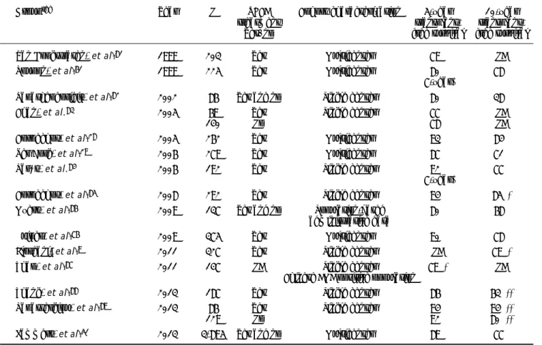
Table 1. Reported prognosis in primary biliary cirrhosis.
Groupref Year N UDCA Cohort characterization 5-year 10-year
treatment transplant-
transplant-Yes/no free survival free survival
Van Hoogstraten, et al.81 1999 203 Yes Multicenter 79% NA
Poupon, et al.61 1999 225 Yes Multicenter 80% 78%
(7-year)
Papatheodoridis, et al.82 2002 86 Yes and no Single center 80% 38%
Chan, et al.83 2005 69 Yes Single center 77% NA
140 No 78% NA
Corpechot, et al.28 2005 262 Yes Multicenter 93% 84%
Ter Borg, et al.29 2006 279 Yes Multicenter 87% 71%
Parés, et al.84 2006 192 Yes Single center 92% 77%
(7-year)
Corpechot, et al.85 2008 292 Yes Single center 94% 85%*
Myers, et al.86 2009 137 Yes and no Population-based 80% 68%
administrative data
Kuiper, et al.76 2009 375 Yes Multicenter 90% 78%
Floreani, et al.9 2011 327 Yes Single center NA 79%*
Zhao, et al.87 2011 147 NA Single center 79%* NA
Chinese AMA positive population
Zhang, et al.88 2013 187 Yes Single center 86% 63%**
Papastergiou, et al.89 2013 86 Yes Single center 94% 94%**
129 No 92% 80%**
Lammers, et al.51 2013 3,895 Yes and no Multicenter 89% 77%
* Cumulative probability of survival. ** Liver transplant-free survival and survival free of complications of cirrhosis.
with PBC, including predictive scoring models for
two of the most serious clinical complications,
namely esophageal variceal bleeding and
hepatocel-lular carcinoma (HCC).
FACTORS DETERMINING PROGNOSIS
Histological stage
Severity of disease in PBC is based on the
Scheu-er
14and Ludwig
1histologic scoring systems, both
recognizing 4 stages. Early histological stages are
as-sociated with favourable prognosis. The last phase,
or cirrhotic phase, is irreversible and classically only
this stage is associated with an increased risk of liver
decompensation and development of HCC.
6,11Thus,
liver histology is a strong prognostic factor.
A particular variant form of PBC, the premature
ductopenic variant, is characterized by rapid,
exces-sive bile duct loss in relation to the amount of
fibro-sis. In individuals with this subtype, severe
cholestasis with progressive jaundice and marked
hypercholesterolemia may require liver
transplanta-tion well before the development of cirrhosis.
12Histological progression of PBC was assessed in
patients originally included in a clinical trial of
D-penicillamine.
13Since this agent does not delay
his-tological progression,
14this study is considered as
representative of histological progression in
treat-ment-naïve PBC patients. Approximately 80% of
pa-tients had histological progression of at least one
stage during a median follow-up of 3 years, and 31%
with stage I disease progressed to cirrhosis within 4
years. Another study followed-up 183 patients
treat-ed with UDCA and reporttreat-ed a 4% incidence of
cir-rhosis at 5 years in patients with stage I disease,
15suggesting that UDCA delays histological
progres-sion.
Several other histologic features have been
de-scribed as important prognostic parameters of worse
outcome in PBC, such as central and periportal
cholestasis,
11,16periportal cell necrosis and
piece-meal necrosis,
15,16interface hepatitis,
15and
Many of these histological features are not
sys-tematically included in the Ludwig and Scheuer
his-tological scoring systems; in fact, an expert panel on
PBC, working under the auspices of the American
Association for the Study of Liver Disease (AASLD),
agreed that histology should neither be included in
prognostic scoring models nor used as a primary
endpoint in clinical trials.
18A recently proposed
his-tologic scoring system taking into account several of
the histological features discussed awaits further
validation.
19Efficacy of treatment
Treatment options in PBC are limited. Liver
trans-plantation is the only curative treatment for PBC with
excellent survival rates,
20but is an option only for
pa-tients with end-stage liver disease. UDCA is the only
approved treatment for PBC,
6,7although several
meta-analyses have failed to show a beneficial effect of
UDCA in PBC.
21-23However, only a few of the
includ-ed studies lastinclud-ed longer than 24 months, a very short
period to demonstrate effects on transplant-free
surviv-al, and most studies were clearly underpowered. In
contrast, a pooled analysis of individual patient data
from the 3 largest placebo-controlled double-blind
studies which included longer follow-up data from one
center, showed an improvement in survival with
UDCA after four years of treatment.
24Another
meta-analysis showed that the use of UDCA in studies that
incorporated placebo control, long-term follow-up
(more than 2 years) or larger numbers of patients
(more than 100 patients) were associated with both
improved serum liver biochemical tests and reduced
in-cidence of liver transplantation or death.
25Several studies extending the follow-up of earlier
published randomized, placebo-controlled UDCA
tri-als showed that UDCA not only improves some
his-tological features, but can delay hishis-tological
progression. Two separate studies from the U.S. and
France demonstrated a delay in histological
progres-sion after a minimum of four years of UDCA
treat-ment.
17,26A combined analysis, which also used data
from a Canadian and Spanish trial, showed that
his-tologic progression was delayed after 2 years
treat-ment, but that UDCA treatment was not associated
with regression of fibrosis.
27Several studies have shown that UDCA-treated
patients with early stage disease have survival rates
comparable with a standardized general
popula-tion.
28,29For UDCA-treated patients with advanced
disease survival was diminished compared with an
age- and sex-matched controlled population.
In summary, there is strong evidence to support
the use of UDCA to delay the progression of PBC
and currently it remains the only licensed medical
therapy.
Gender and age at time of diagnosis
Data on the prognostic significance of factors
such as gender or age are scare. A recent landmark
study from the UK PBC consortium clearly showed
the impact of important disease subgroups in a
study cohort including 2,353 PBC patients.
4Impor-tantly, male patients were less likely to respond to
UDCA treatment and were at higher risk of worse
outcome. Another important finding was an inverse
relationship between age and likelihood to respond
to UDCA. Thus, gender and age appear important in
predicting prognosis in PBC.
Presence of symptoms at time of diagnosis
Risk stratification according to the presence of
symptoms at time of diagnosis has been the subject
of many studies over the past decades.
16,30-35Of
note, most studies did not use validated symptom
as-sessment measures, which is essential for assessing
the impact of subjective parameters, such as fatigue
or pruritus. Therefore interpretation of such studies
may be difficult.
Most studies have reported that asymptomatic
patients have earlier histologic stage of disease
com-pared with symptomatic patients, in addition to better
liver enzyme profiles and lower bilirubin and higher
albumin levels.
36Several studies showed that a
substantial proportion of asymptomatic patients will
develop symptoms over time.
31,34,36-38The vast
majori-ty (95%) of asymptomatic patients followed for up to
20 years will become symptomatic.
8Once symptoms
appear, survival of initially asymptomatic patients is
comparable with survival of patients who initially
presented with symptoms.
33,36Therefore asymptomatic
PBC patients rather appear to represent an earlier
stage of the disease than a separate clinical entity.
Serological prognostic factors
Antimitochondrial antibodies (AMA) are highly
specific for PBC and a cornerstone for establishment
of the diagnosis. Up to 95% of PBC patients have
positive AMA titers,
39and patients having positive
status nor AMA titer has been shown to be
correlat-ed with prognosis.
40,41AMA subtypes were found to
be associated with a progressive course in some
studies,
42but this was not confirmed by others.
43,44Approximately half of PBC patients also have
anti-nuclear antibodies (ANA) detectable in serum.
In particular, ANA against Sp100 and
anti-gp210 antigens are highly specific for PBC and
therefore useful to establish the diagnosis of PBC in
AMA-negative patients.
45It has been suggested that
patients with initially positive anti-gp210 have more
active disease and are more likely to develop liver
failure.
46,47Biochemical prognostic factors
From a diagnostic point of view increased serum
alkaline phosphatase values (ALP) with or without
increased gamma-glutamyltranspeptidase (
γ
-GT) are
important, and both are considered as early
mark-ers of cholestasis in contrast to elevated serum total
bilirubin values, which are clearly suggestive of
more advanced disease.
48It has been known for several decades that serum
bilirubin is one of the most powerful predictors of
prognosis in PBC and this variable has been
incor-porated in most scoring and prediction models. A
classical study demonstrated a two-phase pattern of
bilirubin during the course of the disease;
49a first
phase in which serum bilirubin remains stable for
many years and a second phase of rapidly increasing
values, the so called ‘acceleration phase’. Repeated
measurements of serum bilirubin > 2.0mg/dL was a
sign of late stage disease and preceded death within
a few years.
49A French study showed that
persist-ent abnormal bilirubin levels were predictive for
ex-tensive fibrosis, with a positive predictive value of
90%.
15In patients in whom serum bilirubin
normal-izes upon treatment with UDCA, transplant-free
survival was found to be comparable with that in
placebo-treated patients with initial normal serum
bilirubin levels.
50The same applied to survival of
pa-tients without normalization of bilirubin and
place-bo-using patients with abnormal bilirubin values at
baseline. In other words, serum bilirubin values
re-tain prognostic utility irrespective of treatment,
un-derlining the utility of serum bilirubin as a useful
surrogate endpoint of outcome.
Albumin is regarded as another important and
powerful biochemical predictor of liver
decompensa-tion. Low serum albumin and high bilirubin values
were shown to be independent predictors of the
de-velopment of cirrhosis
15and mortality.
29Recently, a
global study including almost 5,000 subjects with
PBC not only confirmed the strong prognostic
im-portance of serum bilirubin, but also demonstrated
that serum ALP values have significant independent
and additional prognostic value in prediction of
transplant-free survival.
51Angulo and colleagues were the first to report on
the prognostic impact of changes in ALP values
upon treatment with UDCA, showing that ALP
val-ues
≥
2x upper limit of normal (ULN) after 6
months of treatment predicted future treatment
failure.
52Several recent studies have also clearly
demonstrated that quantitative decreases in
bilirubin, albumin, ALP, aspartate aminotransferase
(AST) and/or
γ
-GT levels 6 months, 1 year or 2
years UDCA treatment, are predictive for improved
transplant-free survival (Table 2). Responders
according to these criteria were likely to have
sur-vival rates comparable with a general population.
These biochemical response criteria are useful and
now generally accepted tools for stratification
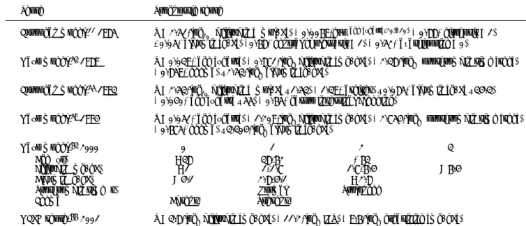
pur-Table 2. Biochemical response criteria for risk stratification in UDCA treated patients.
Criteriaref Definition of biochemical response Evaluation time point N
Mayo criterion, 199952 ALP < 2.0xULN 6 months 180
Barcelona criterion, 200684 > 40% decrease of ALP or normalization 1 year 192
Paris-1 criterion, 200885 ALP < 3.0xULN, AST < 2.0xULN and bilirubin ≤ 1mg/dL 1 year 292
Rotterdam criterion, 200976 Normalization of abnormal bilirubin and/or albumin 1 year 375
Toronto criterion, 201090,91 ALP ≤ 1.67xULN 2 years 69
Paris-2 criterion,* 201192 ALP ≤ 1.5xULN, AST ≤ 1.5xULN and bilirubin ≤ 1mg/dL 1 year 165
Ehim criterion,** 201193,94 ≥ 70% decrease of γ-GT 6 months 138
Momah/Lindor criterion, 201195 ALP ≤ 1.67xULN and bilirubin ≤ 1mg/dL 1 year 73
poses and for identifying patients in need of
addi-tional treatment.
PREDICTION MODELS OF
TRANSPLANT-FREE SURVIVAL
Mathematical prediction models, either time-fixed
or time-dependent, have been developed to predict
the probability of survival using biochemical,
clini-cal and/or histologiclini-cal features. Serum bilirubin and
age are the main components of almost all proposed
models.
11,16,53-56Roll, et al. showed that age at time of diagnosis,
presence of hepatomegaly and increased serum
bi-lirubin were all independently associated with
sur-vival.
16Notably, portal fibrosis was an independent
predictor of prolonged survival in this study. Other
studies identified (log)bilirubin,
32,56.57variceal
bleed-ing
32albumin, age and ascites
57as independent
pre-dictors of outcome.
Bonsel, et al. constructed a prognostic model
in-corporating nine variables: log(bilirubin), age,
albu-min, HBsAg, neurological complications, varices,
ascites, clinical icterus and Quick-time
prolonga-tion.
54Two well defined and cross-validated models, the
European Model and the Mayo risk score, are
sum-marized in table 3. The European model was
pub-lished in 1985 by Christensen, et al. based on data
from 248 patients, originally included in an
azathio-prine placebo-controlled trial.
11This time-fixed
mod-el included age at time of diagnosis, bilirubin,
albumin, cirrhosis, central cholestasis and usage of
azathioprine at baseline. In 1993 this group
pub-lished two time-dependent models; one included only
clinical and biochemical variables (bilirubin, ascites,
albumin, age and gastrointestinal bleeding) and one
extended version, included additionally IgM and two
histological variables (central cholestasis and
cir-rhosis).
The Mayo risk score is the most frequently used
model in PBC to predict the short-term survival
probability. This model was published in 1989 and
cross-validated in independent cohorts.
53,58The
fol-lowing clinical and biochemical variables were
in-cluded: age of the patient, serum bilirubin, serum
albumin, prothrombin time (PT) and severity of
edema. A great advantage of this model was that
liver histology was not required to calculate the
risk score. The original model was based on
base-line characteristics and less useful to predict
sur-vival over time. An adapted Mayo model was
proposed in 1994 using the same variables (INR
in-stead of PT) to predict short-term (< 2 years)
sur-vival or time to transplantation at any time point
during follow-up.
59Data on the predictive value of the Mayo risk
score after the introduction of UDCA treatment is
conflicting. Kilmurry, et al. showed that in a group
of 222 patients originally included in an UDCA trial,
the Mayo risk score remained a useful tool for
pre-diction of survival when calculations are repeated
after 6 months treatment.
60Later studies suggested
Table 3. Important prediction models in primary biliary cirrhosis.
Score Prognostic score
European model,11 1985 R = 2.51*log
10 (bilirubin [μmol/L]) + 0.0069*exp(age [years]-20)/10) + 0.88*(cirrhosis = 1)
- 0.05*(albumin [g/L]) +0.68*(central cholestasis = 1) + 0.52*(azathioprine = 0)
Mayo model,53 1989 R = 0.039*(age [years]) + 0.871*log
e(bilirubin [mg/dL]) + 2.38*loge (prothrombin time [sec])
+ 0.859*(edema) – 2.53*loge(albumin [g/dL]) European model,55 1993 R = 2.53*log
10(bilirubin [μmol/L] – 1.53) + 1.39*(ascites) – 0.085*(albumin [g/L] – 34.3)
+ 0.040*(age [years] – 55) + 0.65*(gastro intestinal bleeding)
Mayo model,59 1994 R = 0.051*(age [years]) + 1.209*log
e(bilirubin [mg/dL]) + 2.754*loge (prothrombin time [sec])
+ 0.675*(edema) – 3.304*loge(albumin [g/dL])
Mayo model,63 2000 0 1 2 3
Age (yr) < 38 38-62 ≥ 63
Bilirubin (mg/dL) < 1 1-1.7 1.7-6.4 > 6.4
Albumin (g/dL) > 4.1 2.8-4.1 < 2.8
Prothrombin time (s) Normal Prolonged
Edema Absent Present
MELD score,66 2001 R = 3.8*log
that the Mayo risk score overestimated the risk of
death when applied before the start of
treat-ment.
29,61,62In a general sense the Mayo risk score
is a useful tool to stratify patients for survival and
possibly for clinical trials.
A simplified model of the Mayo risk score was
proposed by Kim, et al.,
63and web based
applica-tions are available for the Mayo risk score, which
facilitate its usage in clinical practice.
In addition, more general prediction liver scores
are used in PBC, such as the Model of End-Stage
Liver Disease (MELD) score and the
Child-Turcotte-Pugh-score.
64,65The MELD score is based on serum
bilirubin, serum creatinine and INR. This score was
originally proposed as a prognostic marker for the
outcome after placement of a transjugular
intrahe-patic portosystemic shunt (TIPSS),
66and currently
used for liver organ allocation. We believe that the
MELD score does not perform well in PBC and may
result in excessive waiting time.
PREDICTION OF PORTAL HYPERTENSION
AND ESOPHAGEAL VARICES
Esophageal varices may develop in the cirrhotic
and pre-cirrhotic stages of PBC.
68,69Survival of PBC
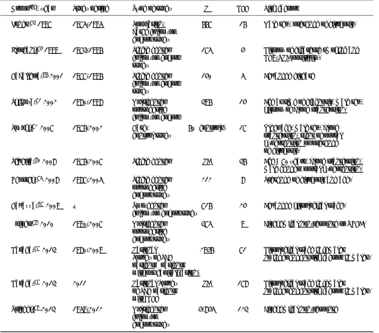
Table 4. Risk factors for development of hepatocellular carcinoma.
Groupref, year Study period Type of study N HCC Risk factors
Jones,73 1997 1975-1995 Population- 667 16 Male sex, presence of cirrhosis.
based follow-up cohort study
Floreani,74 1999 1973-1996 Single center 175 4 History of cigarette smoking and
follow-up cohort HCV-RNA positivity.
study
Caballería,96 2001 1977-1996 Single center 140 5 Advanced disease
follow-up cohort study
Shibuya,71 2002 1980-1998 Multicenter 396 14 Age at time of diagnosis, male sex,
prospective history of blood transfusion.
follow-up cohort
Suzuki,72 2007 1976-2002 Case- 60 (controls) 17 Older age, male sex, blood
control study transfusion, signs of portal
hypertension or presence of cirrhosis.
Silveira,80 2008 1976-2007 Single center NA 36 Age > 70 years, blood transfusion,
male gender, portal hypertension.
Deutsch,75 2008 1987-2005 Single center 212 8 Presence of cirrhosis and age.
prospective cohort study
Cavazza,97 2009 – Two-center 716 24 Advanced histological stage.
follow-up cohort study
Kuiper,78 2010 1990-2007 Multicenter 375 9 Biochemical non-response to UDCA.
prospective cohort study
Harada,77 2013 1980-2009 National 2946 71 Histological stage in females
Survey of PBC no independent risk factors in males.
patients (patients without capitalization)
Harada,77 2013 2011 National Survey NA 178 Histological stage in females
of PBC patients no independent risk factors in males.
with HCC
Trivedi,79 2013 1959-2012 Multicenter 4,845 123 Biochemical non-response
patients who develop esophageal varices has been
reported to be poor.
67,68Patanwala, et al. reported a
5-year survival rate of 63% and 91% for patients
with and without esophageal varices, respectively.
The poor prognosis associated with esophageal
varices may partly reflect the advanced stage of the
disease in the majority of cases who develop varices,
but may also be related to mortality associated with
variceal bleeding. Therefore tools for timely
diagno-sis of varices and institution of prophylactic
treat-ment are of obvious clinical importance.
A Mayo risk score
≥
4.0 was seen in 93% of
pa-tients who developed esophageal varices,
52while
an-other study identified a Mayo risk score
≥
4.5
together with a platelet count of < 140.000/mm as
independent risk factors for development of
esopha-geal varices.
69The current AASLD guideline on
PBC recommends surveillance for esophageal
varic-es of patients with a platelet count of < 140.000/
mm
3or Mayo risk score > 4.1.
6Recently the Newcastle Varices in PBC Score was
proposed to predict esophageal varices,
68based on a
retrospective study including 330 PBC patients. This
score was validated externally in two independent
co-horts. Low albumin, low platelet count, abnormal
ALP values and splenomegaly were independent
pre-dictors of varices development. An adapted score was
proposed excluding splenomegaly to improve the
usa-bility in clinical practice and an online calculator is
available
(http://www.uk-pbc.com/resources/uk-pbc-varice-prediction-tool.html),
PREDICTION OF
HEPATOCELLULAR CARCINOMA (HCC)
A recent systematic review and meta-analysis
demonstrated a pooled relative risk of the
develop-ment of HCC of 18.80 (95% CI, 10-81-26.79) for PBC
patients compared with a general population, which
makes HCC the most prevalent cancer in PBC.
70The
outcome of patients with HCC is poor.
HCC is less frequently seen in patients who
ini-tially present with early stage disease.
71,72Jones, et
al. followed-up 667 patients with early (stage I or II)
and late (stage III or IV) stage disease, and both
groups over the same period of time. All 16 HCC
cases in this study were found in patients with
ad-vanced disease (stage III or IV) and not in patients
with early disease (stage I or II).
73A similar finding
was reported by Floreani, et al.
74Additional Greek
and Dutch studies clearly showed that despite the
differences in disease stages at baseline, all HCC
cases had advanced disease at time of HCC
diagno-sis.
75,76However, a study from Japan of 178 HCC
cases, described HCC cases among all four
histologi-cal stages,
77especially in males. Histological stage
at time of PBC diagnosis was independently
associ-ated with development of HCC for females, but not
for males. These findings suggest that once cirrhosis
occurs, risk of HCC development increases for
fe-males, but males may be at risk at any histological
stage of disease. The Japanese study also showed a
10-year incidence of HCC for males versus females of
6.5% versus 2.0% (P < 0.0001). Several other
stud-ies also have demonstrated that in general males are
more likely to develop HCC than females.
71-73Estro-gens are considered as having possibly protective
ef-fect on HCC development.
Male gender and advanced disease are the most
frequently reported risk factors for HCC in PBC
(Ta-ble 4). Japanese researchers proposed a highly
accu-rate prediction model (area under the curve of 0.95)
to predict development of HCC. Patients with older
age, male sex, history of blood transfusion and any
signs of portal hypertension or cirrhosis were more
likely to develop HCC.
72These intriguing results
await confirmation by other studies. Recently,
ab-sence of biochemical response in UDCA-treated PBC
patients was proposed as another important risk
fac-tor for HCC.
78A large international cohort study
in-volving 4845 PBC patients and 123 HCC cases
confirmed these findings and indicated that
biochemi-cal non-response to UDCA therapy is the strongest
predictive risk factor for development of HCC.
79Surveillance strategies resulting in early
diagno-sis of HCC may improve outcome.
80Clearly, routine
screening of all PBC patients on a regular basis is
not practical. The current AASLD PBC guideline
suggests that surveillance of HCC in PBC should be
performed in cirrhotic patients and older men.
6Pos-sibly, the recently reported overwhelming prognostic
importance of biochemical response to UDCA may
prompt future modifications of present guidelines.
ABBREVIATIONS
• AMA: antimitochondrial antibody.
• ANA: anti-nuclear antibodies
• AST: aspartate aminotransferase
• Gamma-GT: gamma-glutamyltranspeptidase.
• HCC: hepatocellular carcinoma.
• MELD: Model of End-Stage Liver Disease.
• PBC: primary biliary cirrhosis.
GRANTS AND FINANCIAL SUPPORT
None.
REFERENCES
1. Ludwig J, Dickson ER, McDonald GS. Staging of chronic nonsuppurative destructive cholangitis (syndrome of pri-mary biliary cirrhosis). Virchows Arch A Pathol Anat His-tol 1978; 379: 103-12.
2. Kaplan MM, Gershwin ME. Primary biliary cirrhosis. N Engl J Med 2005; 353: 1261-73.
3. Boonstra K, Beuers U, Ponsioen CY. Epidemiology of prima-ry sclerosing cholangitis and primaprima-ry biliaprima-ry cirrhosis: a systematic review. J Hepatol 2012; 56: 1181-8.
4. Carbone M, Mells GF, Pells G, Dawwas MF, Newton JL, He-neghan MA, Neuberger JM, et al. Sex and age are determi-nants of the clinical phenotype of primary biliary cirrhosis and response to ursodeoxycholic acid. Gastroenterology
2013; 144: 560-9.
5. Mells GF, Pells G, Newton JL, Bathgate AJ, Burroughs AK, Heneghan MA, Neuberger JM, et al. Impact of primary bi-liary cirrhosis on perceived quality of life: the UK-PBC na-tional study. Hepatology 2013; 58: 273-83.
6. Lindor KD, Gershwin ME, Poupon R, Kaplan M, Bergasa NV, Heathcote EJ, American Association for Study of Liver Di-sease. Primary biliary cirrhosis. Hepatology 2009; 50: 291-308.
7. European Association for the Study of the Liver. EASL Cli-nical Practice Guidelines: management of cholestatic liver diseases. J Hepatol 2009; 51: 237-67.
8. Prince MI, Chetwynd A, Craig WL, Metcalf JV, James OF. Asymptomatic primary biliary cirrhosis: clinical features, prognosis, and symptom progression in a large population based cohort. Gut 2004; 53: 865-70.
9. Floreani A, Caroli D, Variola A, Rizzotto ER, Antoniazzi S, Chiaramonte M, Cazzagon N, et al. A 35-year follow-up of a large cohort of patients with primary biliary cirrhosis seen at a single centre. Liver Int 2011; 31: 361-8.
10. Prince MI, James OF. The epidemiology of primary biliary cirrhosis. Clin Liver Dis 2003; 7: 795-819.
11. Christensen E, Neuberger J, Crowe J, Altman DG, Popper H, Portmann B, Doniach D, et al. Beneficial effect of aza-thioprine and prediction of prognosis in primary biliary cirrhosis. Final results of an international trial. Gastroen-terology 1985; 89: 1084-91.
12. Vleggaar FP, van Buuren HR, Zondervan PE, ten Kate FJ, Hop WC, Dutch Multicentre PBC Study Group. Jaundice in non-cirrhotic primary biliary cirrhosis: the premature ductopenic variant. Gut 2001; 49: 276-81.
13. Locke GR, 3rd, Therneau TM, Ludwig J, Dickson ER, Lindor KD. Time course of histological progression in primary bi-liary cirrhosis. Hepatology 1996; 23: 52-6.
14. Gong Y, Frederiksen SL, Gluud C. D-penicillamine for pri-mary biliary cirrhosis. Cochrane Database Syst Rev 2004: CD004789.
15. Corpechot C, Carrat F, Poupon R, Poupon RE. Primary bi-liary cirrhosis: incidence and predictive factors of cirrho-sis development in ursodiol-treated patients.
Gastroenterology 2002; 122: 652-8.
16. Roll J, Boyer JL, Barry D, Klatskin G. The prognostic impor-tance of clinical and histologic features in asymptomatic and symptomatic primary biliary cirrhosis. N Engl J Med
1983; 308: 1-7.
17. Corpechot C, Carrat F, Bonnand AM, Poupon RE, Poupon R. The effect of ursodeoxycholic acid therapy on liver fibro-sis progression in primary biliary cirrhofibro-sis. Hepatology
2000; 32: 1196-9.
18. Silveira MG, Brunt EM, Heathcote J, Gores GJ, Lindor KD, Mayo MJ. American Association for the Study of Liver Di-seases endpoints conference: design and endpoints for clinical trials in primary biliary cirrhosis. Hepatology
2010; 52: 349-59.
19. Hiramatsu K, Aoyama H, Zen Y, Aishima S, Kitagawa S, Nak-anuma Y. Proposal of a new staging and grading system of the liver for primary biliary cirrhosis. Histopathology
2006; 49: 466-78.
20. Liermann Garcia RF, Evangelista Garcia C, McMaster P, Neuberger J. Transplantation for primary biliary cirrhosis: retrospective analysis of 400 patients in a single center.
Hepatology 2001; 33: 22-7.
21. Goulis J, Leandro G, Burroughs AK. Randomised controlled trials of ursodeoxycholic-acid therapy for primary biliary cirrhosis: a meta-analysis. Lancet 1999; 354: 1053-60. 22. Rudic JS, Poropat G, Krstic MN, Bjelakovic G, Gluud C.
Ur-sodeoxycholic acid for primary biliary cirrhosis. Cochrane Database Syst Rev 2012; 12: CD000551.
23. Gong Y, Huang Z, Christensen E, Gluud C. Ursodeoxycholic acid for patients with primary biliary cirrhosis: an upda-ted systematic review and meta-analysis of randomized clinical trials using Bayesian approach as sensitivity analy-ses. Am J Gastroenterol 2007; 102: 1799-807.
24. Poupon RE, Lindor KD, Cauch-Dudek K, Dickson ER, Poupon R, Heathcote EJ. Combined analysis of randomized con-trolled trials of ursodeoxycholic acid in primary biliary cirrhosis. Gastroenterology 1997; 113: 884-90.
25. Shi J, Wu C, Lin Y, Chen YX, Zhu L, Xie WF. Long-term effects of mid-dose ursodeoxycholic acid in primary biliary cirrhosis: a meta-analysis of randomized controlled trials.
Am J Gastroenterol 2006; 101: 1529-38.
26. Angulo P, Batts KP, Therneau TM, Jorgensen RA, Dickson ER, Lindor KD. Long-term ursodeoxycholic acid delays his-tological progression in primary biliary cirrhosis. Hepato-logy 1999; 29: 644-7.
27. Poupon RE, Lindor KD, Pares A, Chazouilleres O, Poupon R, Heathcote EJ. Combined analysis of the effect of treat-ment with ursodeoxycholic acid on histologic progression in primary biliary cirrhosis. J Hepatol 2003; 39: 12-6. 28. Corpechot C, Carrat F, Bahr A, Chretien Y, Poupon RE,
Poupon R. The effect of ursodeoxycholic acid therapy on the natural course of primary biliary cirrhosis. Gastroen-terology 2005; 128: 297-303.
29. ter Borg PC, Schalm SW, Hansen BE, van Buuren HR, Dutch PBCSG. Prognosis of ursodeoxycholic Acid-treated pa-tients with primary biliary cirrhosis. Results of a 10-yr co-hort study involving 297 patients. Am J Gastroenterol
2006; 101: 2044-50.
30. Nyberg A, Loof L. Primary biliary cirrhosis: clinical featu-res and outcome, with special reference to asymptomatic disease. Scand J Gastroenterol 1989; 24: 57-64.
31. Balasubramaniam K, Grambsch PM, Wiesner RH, Lindor KD, Dickson ER. Diminished survival in asymptomatic primary biliary cirrhosis. A prospective study. Gastroenterology
1990; 98: 1567-71.
32. Rydning A, Schrumpf E, Abdelnoor M, Elgjo K, Jenssen E. Factors of prognostic importance in primary biliary cirr-hosis. Scand J Gastroenterol 1990; 25: 119-26.
34. Prince M, Chetwynd A, Newman W, Metcalf JV, James OF. Survival and symptom progression in a geographically based cohort of patients with primary biliary cirrhosis: follow-up for up to 28 years. Gastroenterology 2002; 123: 1044-51.
35. Quarneti C, Muratori P, Lalanne C, Fabbri A, Menichella R, Granito A, Masi C, et al. Fatigue and pruritus at onset identify a more aggressive subset of primary biliary cirrhosis. Liver Int 2014 doi: 10.1111/liv.12560.
36. Mitchison HC, Lucey MR, Kelly PJ, Neuberger JM, Williams R, James OF. Symptom development and prognosis in pri-mary biliary cirrhosis: a study in two centers. Gastroen-terology 1990; 99: 778-84.
37. Metcalf JV, Mitchison HC, Palmer JM, Jones DE, Bassendi-ne MF, James OF. Natural history of early primary biliary cirrhosis. Lancet 1996; 348: 1399-402.
38. Springer J, Cauch-Dudek K, O’Rourke K, Wanless IR, Heath-cote EJ. Asymptomatic primary biliary cirrhosis: a study of its natural history and prognosis. Am J Gastroenterol
1999; 94: 47-53.
39. Oertelt S, Rieger R, Selmi C, Invernizzi P, Ansari AA, Coppel RL, Podda M, et al. A sensitive bead assay for antimito-chondrial antibodies: Chipping away at AMA-negative pri-mary biliary cirrhosis. Hepatology 2007; 45: 659-65. 40. Invernizzi P, Crosignani A, Battezzati PM, Covini G, De
Valle G, Larghi A, Zuin M, et al. Comparison of the clinical features and clinical course of antimitochondrial antibo-dy-positive and -negative primary biliary cirrhosis. Hepa-tology 1997; 25: 1090-5.
41. Joshi S, Cauch-Dudek K, Heathcote EJ, Lindor K, Jorgen-sen R, Klein R. Antimitochondrial antibody profiles: are they valid prognostic indicators in primary biliary cirrho-sis? Am J Gastroenterol 2002; 97: 999-1002.
42. Klein R, Pointner H, Zilly W, Glassner-Bittner B, Breuer N, Garbe W, Fintelmann V, et al. Antimitochondrial antibody profiles in primary biliary cirrhosis distinguish at early stages between a benign and a progressive course: a prospective study on 200 patients followed for 10 years.
Liver 1997; 17: 119-28.
43. Vleggaar FP, van Buuren HR. No prognostic significance of antimitochondrial antibody profile testing in primary bilia-ry cirrhosis. Hepatogastroenterology 2004; 51: 937-40. 44. Palmer JM, Yeaman SJ, Bassendine MF, James OF. M4 and
M9 autoantigens in primary biliary cirrhosis—a negative study. J Hepatol 1993; 18: 251-4.
45. Invernizzi P, Selmi C, Ranftler C, Podda M, Wesierska-Ga-dek J. Antinuclear antibodies in primary biliary cirrhosis.
Semin Liver Dis 2005; 25: 298-310.
46. Nakamura M, Kondo H, Mori T, Komori A, Matsuyama M, Ito M, Takii Y, et al. Anti-gp210 and anti-centromere antibo-dies are different risk factors for the progression of pri-mary biliary cirrhosis. Hepatology 2007; 45: 118-27. 47. Muratori P, Muratori L, Ferrari R, Cassani F, Bianchi G,
Lenzi M, Rodrigo L, et al. Characterization and clinical im-pact of antinuclear antibodies in primary biliary cirrhosis.
Am J Gastroenterol 2003; 98: 431-7.
48. Corpechot C, Poujol-Robert A, Wendum D, Galotte M, Chretien Y, Poupon RE, Poupon R. Biochemical markers of liver fibrosis and lymphocytic piecemeal necrosis in UDCA-treated patients with primary biliary cirrhosis. Liver Int
2004; 24: 187-93.
49. Shapiro JM, Smith H, Schaffner F. Serum bilirubin: a prog-nostic factor in primary biliary cirrhosis. Gut 1979; 20: 137-40.
50. Bonnand AM, Heathcote EJ, Lindor KD, Poupon RE. Clinical significance of serum bilirubin levels under ursodeoxycho-lic acid therapy in patients with primary biliary cirrhosis.
Hepatology 1999; 29: 39-43.
51. Lammers WJ, van Buuren HR, Janssen HLA, Invernizzi P, Battezatti PM, Floreani A, Hirschfield GM, et al. Validation of alkaline phosphatase and bilirubin values as a surrogate endpoint in primary biliary cirrhosis - an international, collaborative study. Hepatology 2013; 58: 250A.
52. Angulo P, Lindor KD, Therneau TM, Jorgensen RA, Malin-choc M, Kamath PS, Dickson ER. Utilization of the Mayo risk score in patients with primary biliary cirrhosis receiving ursodeoxycholic acid. Liver 1999; 19: 115-21. 53. Dickson ER, Grambsch PM, Fleming TR, Fisher LD,
Lan-gworthy A. Prognosis in primary biliary cirrhosis: model for decision making. Hepatology 1989; 10: 1-7.
54. Bonsel GJ, Klompmaker IJ, van’t Veer F, Habbema JD, Slooff MJ. Use of prognostic models for assessment of value of liver transplantation in primary biliary cirrhosis. Lancet
1990; 335: 493-7.
55. Christensen E, Altman DG, Neuberger J, De Stavola BL, Tygstrup N, Williams R. Updating prognosis in primary bi-liary cirrhosis using a time-dependent Cox regression mo-del. PBC1 and PBC2 trial groups. Gastroenterology 1993; 105: 1865-76.
56. Krzeski P, Zych W, Kraszewska E, Milewski B, Butruk E, Habior A. Is serum bilirubin concentration the only valid prognostic marker in primary biliary cirrhosis? Hepatology
1999; 30: 865-9.
57. Hughes MD, Raskino CL, Pocock SJ, Biagini MR, Burroughs AK. Prediction of short-term survival with an application in primary biliary cirrhosis. Stat Med 1992; 11: 1731-45. 58. Grambsch PM, Dickson ER, Kaplan M, LeSage G, Fleming TR,
Langworthy AL. Extramural cross-validation of the Mayo primary biliary cirrhosis survival model establishes its ge-neralizability. Hepatology 1989; 10: 846-50.
59. Murtaugh PA, Dickson ER, Van Dam GM, Malinchoc M, Grambsch PM, Langworthy AL, Gips CH. Primary biliary cirrhosis: prediction of short-term survival based on repeated patient visits. Hepatology 1994; 20: 126-34. 60. Kilmurry MR, Heathcote EJ, Cauch-Dudek K, O’Rourke K,
Bailey RJ, Blendis LM, Ghent CN, et al. Is the Mayo model for predicting survival useful after the introduction of ur-sodeoxycholic acid treatment for primary biliary cirrho-sis? Hepatology 1996; 23: 1148-53.
61. Poupon RE, Bonnand AM, Chretien Y, Poupon R. Ten-year survival in ursodeoxycholic acid-treated patients with primary biliary cirrhosis. The UDCA-PBC Study Group. He-patology 1999; 29: 1668-71.
62. Koulentaki M, Moscandrea J, Dimoulios P, Chatzicostas C, Kouroumalis EA. Survival of anti-mitochondrial antibody-positive and -negative primary biliary cirrhosis patients on ursodeoxycholic acid treatment. Dig Dis Sci 2004; 49: 1190-5.
63. Kim WR, Wiesner RH, Poterucha JJ, Therneau TM, Benson JT, Krom RA, Dickson ER. Adaptation of the Mayo primary biliary cirrhosis natural history model for application in liver transplant candidates. Liver Transpl
2000; 6: 489-94.
64. Child CG, Turcotte JG. Surgery and portal hypertension.
Major Probl Clin Surg 1964; 1: 1-85.
66. Kamath PS, Wiesner RH, Malinchoc M, Kremers W, Ther-neau TM, Kosberg CL, D’Amico G, et al. A model to predict survival in patients with end-stage liver disease. Hepato-logy 2001; 33: 464-70.
67. Gores GJ, Wiesner RH, Dickson ER, Zinsmeister AR, Jor-gensen RA, Langworthy A. Prospective evaluation of eso-phageal varices in primary biliary cirrhosis: development, natural history, and influence on survival. Gastroentero-logy 1989; 96: 1552-9.
68. Patanwala I, McMeekin P, Walters R, Mells G, Alexander G, Newton J, Shah H, et al. A validated clinical tool for the prediction of varices in PBC: The Newcastle Varices in PBC Score. J Hepatol 2013; 59: 327-35.
69. Levy C, Zein CO, Gomez J, Soldevila-Pico C, Firpi R, Morelli G, Nelson D. Prevalence and predictors of esophageal vari-ces in patients with primary biliary cirrhosis. Clin Gas-troenterol Hepatol 2007; 5: 803-8.
70. Liang Y, Yang Z, Zhong R. Primary biliary cirrhosis and cancer risk: a systematic review and meta-analysis. Hepa-tology 2012; 56: 1409-17.
71. Shibuya A, Tanaka K, Miyakawa H, Shibata M, Takatori M, Sekiyama K, Hashimoto N, et al. Hepatocellular carcinoma and survival in patients with primary biliary cirrhosis. He-patology 2002; 35: 1172-8.
72. Suzuki A, Lymp J, Donlinger J, Mendes F, Angulo P, Lindor K. Clinical predictors for hepatocellular carcinoma in pa-tients with primary biliary cirrhosis. Clin Gastroenterol Hepatol 2007; 5: 259-64.
73. Jones DE, Metcalf JV, Collier JD, Bassendine MF, James OF. Hepatocellular carcinoma in primary biliary cirrhosis and its impact on outcomes. Hepatology 1997; 26: 1138-42. 74. Floreani A, Baragiotta A, Baldo V, Menegon T, Farinati F,
Naccarato R. Hepatic and extrahepatic malignancies in primary biliary cirrhosis. Hepatology 1999; 29: 1425-8. 75. Deutsch M, Papatheodoridis GV, Tzakou A, Hadziyannis SJ.
Risk of hepatocellular carcinoma and extrahepatic malig-nancies in primary biliary cirrhosis. Eur J Gastroenterol Hepatol 2008; 20: 5-9.
76. Kuiper EM, Hansen BE, de Vries RA, den Ouden-Muller JW, van Ditzhuijsen TJ, Haagsma EB, Houben MH, et al. Dutch PBCSG. Improved prognosis of patients with primary bilia-ry cirrhosis that have a biochemical response to ur-sodeoxycholic acid. Gastroenterology 2009; 136: 1281-7. 77. Harada K, Hirohara J, Ueno Y, Nakano T, Kakuda Y,
Tsu-bouchi H, Ichida T, et al. Incidence of and risk factors for hepatocellular carcinoma in primary biliary cirrhosis: na-tional data from Japan. Hepatology 2013; 57: 1942-9. 78. Kuiper EM, Hansen BE, Adang RP, van Nieuwkerk CM,
Tim-mer R, Drenth JP, Spoelstra P, et al. Dutch PBCSG. Relati-vely high risk for hepatocellular carcinoma in patients with primary biliary cirrhosis not responding to ur-sodeoxycholic acid. Eur J Gastroenterol Hepatol 2010; 22: 1495-502.
79. Trivedi PJ, Lammers WJ, van Buuren HR, Janssen HLA, In-vernizzi P, Battezatti PM, Floreani A, et al. Effective stratification of hepatocellular carcinoma risk in primary biliary cirrhosis: results of a multi-centre international study. J Hepatol 2014; 60: S55.
80. Silveira MG, Suzuki A, Lindor KD. Surveillance for hepato-cellular carcinoma in patients with primary biliary cirrho-sis. Hepatology 2008; 48: 1149-56.
81. van Hoogstraten HJ, Hansen BE, van Buuren HR, ten Kate FJ, van Berge-Henegouwen GP, Schalm SW. Prognostic fac-tors and long-term effects of ursodeoxycholic acid on liver biochemical parameters in patients with primary biliary
cirrhosis. Dutch Multi-Centre PBC Study Group. J Hepatol
1999; 31: 256-62.
82. Papatheodoridis GV, Hadziyannis ES, Deutsch M, Hadzi-yannis SJ. Ursodeoxycholic acid for primary biliary cirrho-sis: final results of a 12-year, prospective, randomized, controlled trial. Am J Gastroenterol 2002; 97: 2063-70. 83. Chan CW, Gunsar F, Feudjo M, Rigamonti C,
Vlachogian-nakos J, Carpenter JR, Burroughs AK. Long-term ur-sodeoxycholic acid therapy for primary biliary cirrhosis: a follow-up to 12 years. Aliment Pharmacol Ther 2005; 21: 217-26.
84. Pares A, Caballeria L, Rodes J. Excellent long-term survival in patients with primary biliary cirrhosis and biochemical response to ursodeoxycholic Acid. Gastroenterology
2006; 130: 715-20.
85. Corpechot C, Abenavoli L, Rabahi N, Chretien Y, Andreani T, Johanet C, Chazouilleres O, et al. Biochemical response to ursodeoxycholic acid and long-term prognosis in prima-ry biliaprima-ry cirrhosis. Hepatology 2008; 48: 871-7.
86. Myers RP, Shaheen AA, Fong A, Burak KW, Wan A, Swain MG, Hilsden RJ, et al. Epidemiology and natural history of primary biliary cirrhosis in a Canadian health re-gion: a population-based study. Hepatology 2009; 50: 1884-92.
87. Zhao DT, Liao HY, Liu YM, Zhao Y, Feng X, Yan HP. Prog-nostic factors and survival analysis of antimitochondrial antibody-positive primary biliary cirrhosis in Chinese pa-tients. Dig Dis Sci 2011; 56: 2750-7.
88. Zhang LN, Shi TY, Shi XH, Wang L, Yang YJ, Liu B, Gao LX et al. Early biochemical response to ursodeoxycholic acid and long-term prognosis of primary biliary cirrhosis: Re-sults of a 14-year cohort study. Hepatology 2013; 58: 264-72.
89. Papastergiou V, Tsochatzis EA, Rodriquez-Peralvarez M, Thalassinos E, Pieri G, Manousou P, Germani G, et al. Bio-chemical criteria at 1 year are not robust indicators of response to ursodeoxycholic acid in early primary biliary cirrhosis: results from a 29-year cohort study. Aliment Pharmacol Ther 2013; 38: 1354-64.
90. Kumagi T, Guindi M, Fischer SE, Arenovich T, Abdalian R, Coltescu C, Heathcote EJ, et al. Baseline ductopenia and treatment response predict long-term histological progression in primary biliary cirrhosis. Am J Gastroenterol 2010; 105: 2186-94.
91. Lammert C, Juran BD, Schlicht E, Chan LL, Atkinson EJ, de Andrade M, Lazaridis KN. Biochemical response to ur-sodeoxycholic acid predicts survival in a North American cohort of primary biliary cirrhosis patients. J Gastroente-rol 2013.
92. Corpechot C, Chazouilleres O, Poupon R. Early primary bi-liary cirrhosis: biochemical response to treatment and prediction of long-term outcome. J Hepatol 2011; 55: 1361-7.
93. Azemoto N, Abe M, Murata Y, Hiasa Y, Hamada M, Matsuu-ra B, Onji M. Early biochemical response to ursodeoxycho-lic acid predicts symptom development in patients with asymptomatic primary biliary cirrhosis. J Gastroenterol
2009; 44: 630-4.
94. Azemoto N, Kumagi T, Abe M, Konishi I, Matsuura B, Hiasa Y, Onji M. Biochemical response to ursodeoxy-cholic acid predicts long-term outcome in Japanese pa-tients with primary biliary cirrhosis. Hepatol Res
2011; 41: 310-7.
cli-nical trials in primary biliary cirrhosis. Liver Int 2012; 32: 790-5.
96. Caballeria L, Pares A, Castells A, Gines A, Bru C, Rodes J. Hepatocellular carcinoma in primary biliary cirrhosis: simi-lar incidence to that in hepatitis C virus-related cirrhosis.
Am J Gastroenterol 2001; 96: 1160-3.
97. Cavazza A, Caballeria L, Floreani A, Farinati F, Bruguera M, Caroli D, Pares A. Incidence, risk factors, and survival of hepatocellular carcinoma in primary biliary cirrhosis: comparative analysis from two centers. Hepatology