The Official Journal of the Mexican Association of Hepatology, the Latin-American Association for Study of the Liver and
the Canadian Association for the Study of the Liver
XXV ANNUAL MEETING OF THE LATIN AMERICAN
ASSOCIATION FOR THE STUDY OF THE LIVER
(ALEH, for its acronym in Spanish)
September 20-23, 2018
Punta Cana, República Dominicana
POSTER
DOI:10.5604/01.3001.0012.7214
01 AUTOINMUNE
P-48
AUTOIMMUNE HEPATITIS IN ELDERLY COLOMBIAN
PATIENTS, DIFFERENTIAL CHARACTERISTICS AND
OUTCOMES
DÍAZ-RAMÍREZ GS,* DÍAZ-RAMÍREZ GS,* DÍAZ-RAMÍREZ GS,* DÍAZ-RAMÍREZ GS,*
DÍAZ-RAMÍREZ GS,*,,,,,** MARTÍNEZ-CASAS OY,* MARÍN-ZULUAGA** MARTÍNEZ-CASAS OY,* MARÍN-ZULUAGA** MARTÍNEZ-CASAS OY,* MARÍN-ZULUAGA** MARTÍNEZ-CASAS OY,* MARÍN-ZULUAGA** MARTÍNEZ-CASAS OY,* MARÍN-ZULUAGA JI,**
JI,** JI,** JI,**
JI,**,,,,,*** DONADO-GÓMEZ JH,***** DONADO-GÓMEZ JH,***** DONADO-GÓMEZ JH,***** DONADO-GÓMEZ JH,***** DONADO-GÓMEZ JH,**,,,,,**** MUÑOZ-MAYA O,****** MUÑOZ-MAYA O,****** MUÑOZ-MAYA O,****** MUÑOZ-MAYA O,****** MUÑOZ-MAYA O,**,,,,,*** SANTOS-*** SANTOS-*** SANTOS-*** SANTOS-*** SANTOS-SÁNCHEZ O,**
SÁNCHEZ O,** SÁNCHEZ O,** SÁNCHEZ O,**
SÁNCHEZ O,**,,,,,*** RESTREPO-GUTIÉRREZ JC***** RESTREPO-GUTIÉRREZ JC***** RESTREPO-GUTIÉRREZ JC***** RESTREPO-GUTIÉRREZ JC***** RESTREPO-GUTIÉRREZ JC**,,,,,*************** * CLINICAL HEPATOLOGY, UNIVERSIDAD DE ANTIOQUIA, MEDELLÍN, COLOMBIA. ** GRUPO DE GASTROHEPATOLOGÍA, UNIVERSIDAD DE ANTIOQUIA, MEDELLÍN,
COLOMBIA.
*** HEPATOLOGY AND LIVER TRANSPLANT UNIT, HOSPITAL PABLO TOBÓN URIBE, MEDELLÍN, COLOMBIA.
**** EPIDEMIOLOGY UNIT, HOSPITAL PABLO TOBÓN URIBE, MEDELLÍN, COLOMBIA.
Introduction. Elderly patients with autoimmune hepatitis
(AIH) are a special population due a well-recognized HLA (Hu-man Leukocyte Antigen) predisposition and an indolent and progressive course, however current information comes from
non-Latin American population. Aim. To analyze
compara-tively the characteristics of AIH between patients above and be-low 65 years regarding its clinical presentation, diagnostic
features, response to treatment and outcomes. Material and
methods. A retrospective cohort study of 214 AIH patients
di-agnosed using the simplified criteria, between January 2010 and December 2016. Patients with acute liver failure, drug induced AIH and AIH diagnosed < 16 years were excluded. Statistical analysis was performed using SPSS statistical program version 20.1. A P-value < 0.05 was considered statistically significant
and was two-tail calculated. Results. Twenty-nine (13.5%)
pa-tients were ≥ 65 years. Elderly patients more commonly had
hy-pertension (34.5% vs. 15.1%, p = 0.011), dyslipidemia (20.7%
vs. 5.9%, p = 0.006) and cardiovascular diseases (17.2% vs.
2.7%, p = 0.001). The most common type of clinical
presenta-tion was the acute hepatitis in both groups (31% vs. 32.4%, p =
0.881). When evaluating radiological and histologically the liver fibrosis degree elderly patients had a higher frequency of
cirrho-sis (55.1% vs. 33.5%, p = 0.024). Older patients had a higher
bi-ochemical remission (100% vs. 83.9%, p = 0.022) after
treatment. There were no differences regarding the laboratory test, autoantibodies, type of pharmacologic treatment, relapses, treatment related adverse effects, liver transplantation
require-ment and death. Conclusion. AIH affects Colombian adults of
all ages and should be considered in the diagnostic work-up of liver disease in elderly because these patients had higher fre-quency of cirrhosis at time of diagnosis. Early diagnosis of AIH in elderly is important since pharmacologic treatment is effective and well tolerated.
P-99
AUTOINMUNE HEPATITIS-PRIMARY BILIARY
COLANGITIS OVERLAP SYNDROME: LONG-TERM
OUTCOMES OF A RETROSPECTIVE COHORT IN A
UNIVERSITY HOSPITAL
MARTÍNEZ CASAS OY,* DÍAZ-RAMÍREZ GS,* MARÍN-ZULUAGA JI,** MARTÍNEZ CASAS OY,* DÍAZ-RAMÍREZ GS,* MARÍN-ZULUAGA JI,**MARTÍNEZ CASAS OY,* DÍAZ-RAMÍREZ GS,* MARÍN-ZULUAGA JI,** MARTÍNEZ CASAS OY,* DÍAZ-RAMÍREZ GS,* MARÍN-ZULUAGA JI,** MARTÍNEZ CASAS OY,* DÍAZ-RAMÍREZ GS,* MARÍN-ZULUAGA JI,**,,,,,***************
SANTOS-SÁNCHEZ O,** SANTOS-SÁNCHEZ O,**SANTOS-SÁNCHEZ O,**
SANTOS-SÁNCHEZ O,**SANTOS-SÁNCHEZ O,**,,,,,*** MUÑOZ-MAYA O,***** MUÑOZ-MAYA O,***** MUÑOZ-MAYA O,***** MUÑOZ-MAYA O,***** MUÑOZ-MAYA O,**,,,,,*** DONADO-GÓMEZ*** DONADO-GÓMEZ*** DONADO-GÓMEZ*** DONADO-GÓMEZ*** DONADO-GÓMEZ JH,**** RESTREPO-GUTIÉRREZ JC**
JH,**** RESTREPO-GUTIÉRREZ JC**JH,**** RESTREPO-GUTIÉRREZ JC** JH,**** RESTREPO-GUTIÉRREZ JC**JH,**** RESTREPO-GUTIÉRREZ JC**,,,,,***************
* HEPATOLOGÍA CLÍNICA, UNIVERSIDAD DE ANTIOQUIA, MEDELLÍN, COLOMBIA. ** GRUPO GASTROHEPATOLOGÍA, UNIVERSIDAD DE ANTIOQUIA. MEDELLÍN,
COLOMBIA.
*** UNIDAD DE HEPATOLOGÍA Y TRASPLANTE HEPÁTICO, HOSPITAL PABLO TOBÓN URIBE, MEDELLÍN, COLOMBIA.
**** UNIDAD DE EPIDEMIOLOGÍA, HOSPITAL PABLO TOBÓN URIBE. MEDELLÍN, COLOMBIA.
Background. Autoimmune hepatitis with characteristics of
primary biliary cholangitis is known as overlap syndrome. Its prevalence and prognosis have not been properly determined
comparatively yet with those with classic AIH. Material and
methods. A retrospective cohort study was conducted
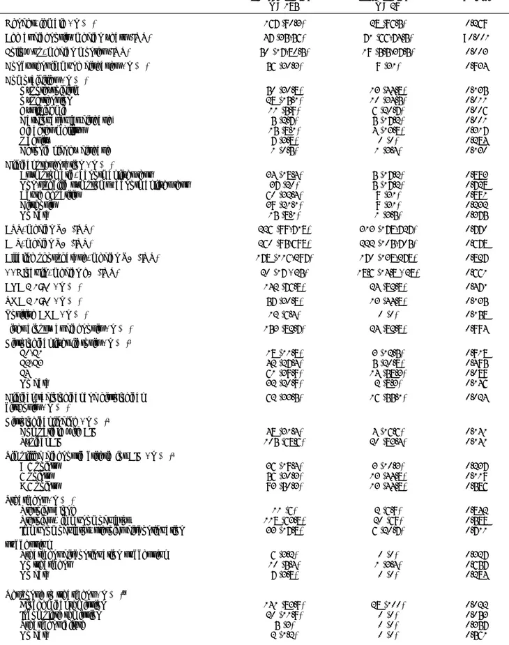
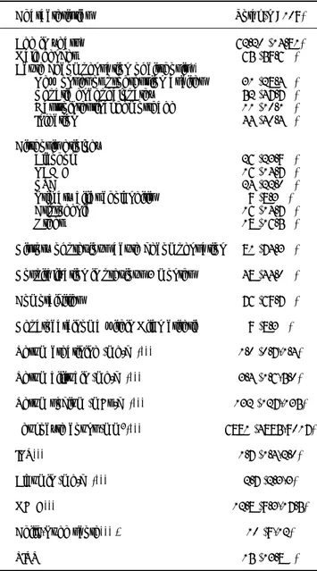
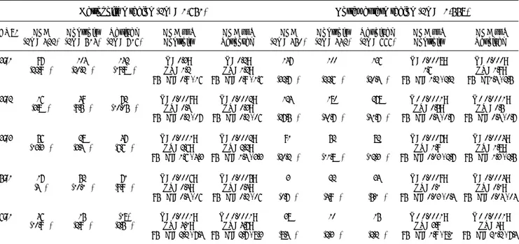
per-Table 1 (P-48). Patient demographic, clinical, laboratory, treatment and outcomes data.
Younger patients Older patients P-Value
n = 185 n = 29
Gender, female - n (%) 167 (90.3) 28 (96.5) 0.269
Age at diagnosis, median years (IQR) 47 (35-56) 71 (66-74.5) <0.001
Follow-up, median months (IQR) 50 (17-80.5) 19 (5.5-37.5) 0.003
Concurrent immune diseases - n (%) 56 (30.3) 9 (31) 0.934
Comorbidities - n (%)
Hypothyroidism 50 (30.8) 13 (44.8) 0.135
Hypertension 28 (15.1) 10 (34.5) 0.011
Dyslipidemia 11 (5.9) 6 (20.7) 0.006
Cardiovascular disease* 5 (2.7) 5 (17.2) 0.001
Diabetes mellitus 15 (8.1) 4 (13.8) 0.317
Obesity 7 (3.8) 0 (0) 0.284
Chronic kidney disease 1 (0.5) 1 (3.4) 0.130
Clinical presentation - n (%)
Asymptomatic, abnormal liver test 34 (18.4) 5 (17.2) 0.883
Non-specific symptoms, abnormal liver test 37 (20) 5 (17.2) 0.728
Acute hepatitis 60 (32.4) 9 (31) 0.881
Cirrhosis 39 (21.1) 9 (31) 0.232
No data 15 (8.1) 1 (3.5) 0.375
AST, median U/L (IQR) 226 (99-718) 313 (178-727) 0.770
ALT, median U/L (IQR) 260 (95-698) 222 (105-705) 0.678
Alkaline phosphatase, median U/L (IQR) 178 (116-297) 170 (138-278) 0.827
γ - Globulin, median g/L (IQR) 20 (17 - 25) 18.6 (14.8 - 28) 0.661
ANA ≥ 1:40 - n (%) 142 (76.8) 24 (82.8) 0.471
SMA ≥ 1:40 - n (%) 57 (30.8) 13 (44.8) 0.135
Positive AMA - n (%) 12 (6.4) 0 (0) 0.158
Liver biopsy at diagnosis - n (%) 153 (82.7) 24 (82.8) 0.994
Histological liver fibrosis - n (%)†
F0-F1 18 (11.8) 3 (12.5) 0.918
F2-F3 42 (27.4) 5 (20.8) 0.495
F4 61 (39.9) 14 (58.3) 0.089
No data 32 (20.9) 2 (8.3) 0.146
Clinical, radiological and histological 62 (33.5) 16 (55.1) 0.024
cirrhosis - n (%)
Histological finding - n (%)‡
Compatible with AIH 48 (31.4) 4 (16.6) 0.141
Typical AIH 105 (68.6) 20 (83.4) 0.141
Simplified diagnostic criteria for AIH - n (%)‡
< 6 points 36 (19.4) 3 (10.3) 0.237
6 points 56 (30.3) 13 (44.8) 0.119
> 6 points 93 (50.3) 13 (44.8) 0.586
Treatment - n (%)
Steroids alone 11 (6) 2 (6.9) 0.842
Steroids + immunomodulator 118 (63.8) 20 (69) 0.588
Immunomodulator, steroids discontinuation 33 (17.8) 6 (20.7) 0.711
successful
Treatment discontinuation successful 6 (3.2) 0 (0) 0.327
No treatment 10 (5.4) 1 (3.4) 0.657
No data 7 (3.8) 0 (0) 0.284
Response to treatment - n (%)§
Biochemical remission 141 (83.9) 28 (100) 0.022
Incomplete remission 20 (11.9) 0 (0) 0.053
Treatment failure 5 (3) 0 (0) 0.357
Relapses - n (%)§ 35 (18.9) 2 (6.9) 0.111
Cirrhosis development during follow-up - n (%)|| 18 (14.6) 2 (16.6) 0.873
Liver transplantation - n (%) 12 (6.5) 1 (3.4) 0.522
Recurrence after transplantation- n (%)¶ 2 (16.6) 1 (100) 0.057
Re-transplantation - n (%) 1 (8.3) 0 (0) 0.764
Death - n (%) 10 (5.4) 0 (0) 0.200
IQR: interquartile range. ANA: antinuclear antibodies. SMA: anti-smooth muscle antibodies. AMA: anti-mitochondrial antibodies. * Ischemic heart disease, heart failure, peripheral vascular disease, stroke. † Calculated using patients with liver biopsy at diagnosis in each group. ‡ According to International Autoim-mune Hepatitis Group (IAIHG) recommendations. § Calculated using patients who received treatment. || Calculated using patients who were non-cirrhotic at AIH diagnosis. ¶ Calculated using patients underwent liver transplantation. Declaration of conflict of interest: None.
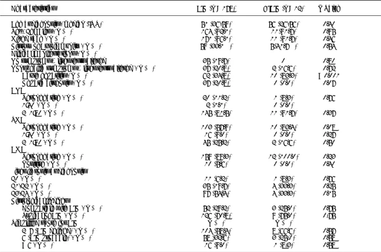
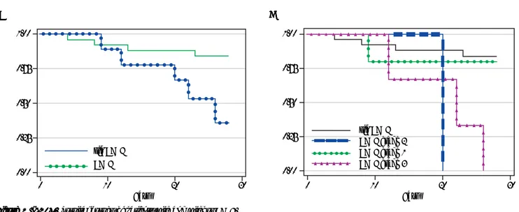
Figure 1 (P-99). Figure 1 (P-99). Figure 1 (P-99). Figure 1 (P-99).
Figure 1 (P-99). Differential survival in patients with AIH and overlap syndrome (Panel A), transplant-free survival in AIH and overlap syndrome. (G1: HAI, G2: overlap), relapse-free survival in those who achieved biochemical or partial remission (Panel C) and ci-rrhosis-free survival in those with F3 fibrosis or not reported (D).
formed with the statistical software SPSS version 20.1 (SPSS
Inc.). Results. Of 362 potential patients by ICD-10 diagnostic
code for autoimmune hepatitis, after exclusions, 210 patients were included (195 women, mean age 48.5 years). Of these, 32 (15.2%) had AIH-PBC overlap syndrome. At diagnosis, no
sig-nificant differences were found by demographic profile, positive
autoantibodies (ANA, ASMA) except AMA (81.2% vs. 3.9%, P
< 0.001) and histological grade of fibrosis. The most frequent clinical presentation were nonspecific symptoms in AIH-PBC and acute hepatitis in AIH. Although there were no significant
0 50 100 150 200 250
Seguimiento en Meses En negro
01 178 111 71 35 20 10 5 3
02 32 20 10 6 5 2 2 2 2 2
0 50 100 150 200
Tiempo a cirrosis
0 50 100 150 200 250
Seguimiento en Meses En negro
01 178 114 74 33 20 10 5 3
02 32 22 14 6 3 2 2 2
0 24 50 75 100 125
Tiempo a recaída P = 0.711
HAI Superposición
HAI Superposición C
C C C
C DDDDD
A A A A
A BBBBB
100
80
60
40
20
0
Porcentaje de supervivencia
100
80
60
40
20
0
Porcentaje de supervivencia
100
80
60
40
20
0
Porcentaje de supervivencia
100
80
60
40
20
0
differences, AIH showed greater biochemical response to
immu-nosuppressive management (87.3% vs. 74.2%, P = 0.061) and
greater number of relapses in those who achieved partial or
com-plete remission during treatment (12.4% vs. 7.6.3%; P = 0.727).
Patients with AIH-PBC had greater progression to cirrhosis
(22.2% vs. 13.1%, P = 0.038), even in those who achieved
par-tial or complete biochemical remission without relapse, with greater indication of OLT (P = 0.009), but not
retransplanta-tion (P = 0.183); there were no differences in mortality.
Con-clusions. The AIH-PBC overlap syndrome constitutes a not
insignificant proportion among those with AIH, with greater progression to cirrhosis, indication of liver transplantation and possibly retransplantation. This higher risk of adverse outcomes suggests that these patients must have a stricter follow-up and probably with follow up until confirmed histopathological re-mission (Figure 1).
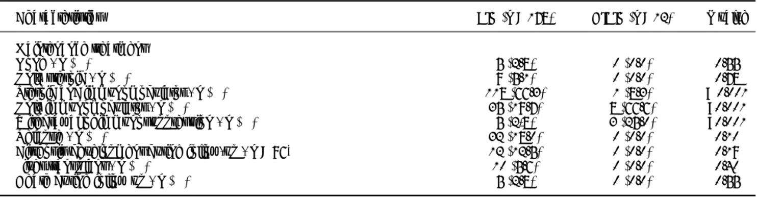
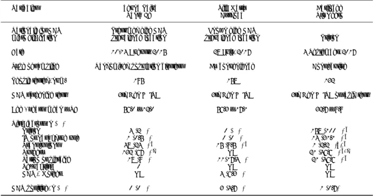
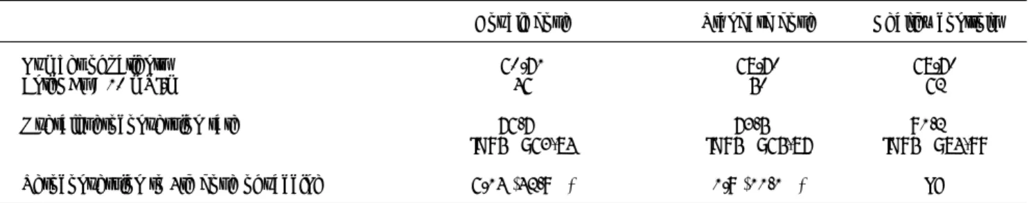
Table 1 (P-100). Demographic, clinical, analytical characteristics to the diagnosis of AIH and DIAIH.
Characteristics AIH (n = 178) DIAIH (n = 12) P Value
Age at diagnosis - median (IQR) 51 (36-59) 56 (26-56) 0.40
Sex, Females - n (%) 164 (92.1) 11(91.7) 0.95
Mixed race - n (%) 171 (96.1) 11 (91.7) 0.46
History of Autoimmunity - n (%) 59 (33.1%) 5(41.7%) 0.54
Clinical manifestations - n (%)
No symptoms + liver tests altered 35 (19.7) 0 0.80
Nonspecific symptoms + liver tests altered - n (%) 37 (20.8) 2 (16.6) 0.72
Acute hepatitis - n (%) 62 (34.8) 10 (83.3) < 0.001
Hepatic cirrhosis - n (%) 37 (20.8) 0 (0.0) 0.07
ANA
Seronegative - n (%) 20 (11.2) 1 (8.3) 0.76
1:40 - n (%) 2 (1.1) 0 (0.0)
≥ 1:80 - n (%) 145 (81.5) 11 (91.7) 0.37
SMA
Seronegative - n (%) 103 (57.9) 10 (83.4) 0.08
1:40 - n (%) 16 (9.0) 0 (0.0) 0.27
≥ 1:80 - n (%) 45 (25.2) 2 (16.6) 0.50
AMA
Seronegative - n (%) 159 (89.3) 12 (100.0) 0.23
Positive - n (%) 10 (5.6) 0 (0.0) 0.40
Liver fibrosis at diagnosis
F0 - n (%) 11 (6.2) 1 (8.3) 0.76
F1-F2 - n (%) 35 (19.7) 4 (33.3) 0.25
F3-F4 - n (%) 97 (54.4) 4 (33.3) 0.15
Histological findings
Compatible with AIH - n (%) 52 (29.2) 3 (25.0) 0.75
Typical of AIH - n (%) 126 (70.8) 9 (75.0) 0,75
Simplified score for AIH n (%) n (%)
≥ 7 (AIH defined) - n (%) 104 (58.4) 8 (66.6) 0.57
6 (AIH probable) - n (%) 58 (32.6) 3 (25,0) 0.58
< 6 - n (%) 16 (9.0) 1 (8,3) 0.58
† There are no data about state or degree of hepatic fibrosis in the biopsy report. ‡The diagnosis of these cases was given by AIH criteria and response to
treatment. *Mann-Whitney U test was used to establish differences. IQR, interquartile range. METAVIR: F0, absence of fibrosis; F1, mild fibrosis; F2, mode-rate fibrosis; F3, severe fibrosis; F4, cirrhosis. AIH: autoimmune hepatitis. AMA: antimitochondrial antibodies. ANA: antinuclear antibodies. ASMA: anti-smooth muscle antibodies. DIAIH: drug-induced autoimmune hepatitis.
P-100
DIFFERENTIAL CHARACTERISTICS IN
DRUG-INDUCED AUTOIMMUNE HEPATITIS
MARTÍNEZ-CASASN OY,* DÍAZ-RAMÍREZ GS,* MARÍN-ZULUAGA MARTÍNEZ-CASASN OY,* DÍAZ-RAMÍREZ GS,* MARÍN-ZULUAGA MARTÍNEZ-CASASN OY,* DÍAZ-RAMÍREZ GS,* MARÍN-ZULUAGA MARTÍNEZ-CASASN OY,* DÍAZ-RAMÍREZ GS,* MARÍN-ZULUAGA MARTÍNEZ-CASASN OY,* DÍAZ-RAMÍREZ GS,* MARÍN-ZULUAGA JI,**
JI,** JI,** JI,**
JI,**,,,,,*** SANTOS-SÁNCHEZ O,***** SANTOS-SÁNCHEZ O,***** SANTOS-SÁNCHEZ O,***** SANTOS-SÁNCHEZ O,***** SANTOS-SÁNCHEZ O,**,,,,,*** MUÑOZ-MAYA O,**,*** DONADO-*** MUÑOZ-MAYA O,**,*** DONADO-*** MUÑOZ-MAYA O,**,*** DONADO-*** MUÑOZ-MAYA O,**,*** DONADO-*** MUÑOZ-MAYA O,**,*** DONADO-GÓMEZ JH,**** RESTREPO-GUTIÉRREZ JC**
GÓMEZ JH,**** RESTREPO-GUTIÉRREZ JC**GÓMEZ JH,**** RESTREPO-GUTIÉRREZ JC** GÓMEZ JH,**** RESTREPO-GUTIÉRREZ JC** GÓMEZ JH,**** RESTREPO-GUTIÉRREZ JC**,,,,,*************** * HEPATOLOGÍA CLÍNICA, UNIVERSIDAD DE ANTIOQUIA, MEDELLÍN, COLOMBIA.
** GRUPO GASTROHEPATOLOGÍA, UNIVERSIDAD DE ANTIOQUIA. MEDELLÍN, COLOMBIA.
*** UNIDAD DE HEPATOLOGÍA Y TRASPLANTE HEPÁTICO, HOSPITAL PABLO TOBÓN URIBE, MEDELLÍN, COLOMBIA.
**** UNIDAD DE EPIDEMIOLOGÍA, HOSPITAL PABLO TOBÓN URIBE. MEDELLÍN, COLOMBIA.
Background and aim. Drug-induced autoimmune hepatitis
(DIAIH) is an adverse effect associated with several drugs that usually occurs acutely, with variable latency, and it may poten-tially be mortal. There are a few reports and studies about
DI-AIH. Material and methods. This is an analytical study of a
signifi-cant. Calculations were performed with the SPSS statistical
package versión 20.1 (SPSS Inc., Chicago, USA). Results. A
to-tal of 190 patients were selected for the analysis, 12 (6.3%) with DIAIH. The two main drugs related to DIAIH were nitro-furantoin, n = 8 (67%), and NSAID, in = 2 (17%), constitut-ing 84% of the cases. There were no significant differences in seropositivity between AIH with DIAIH in antinuclear anti-bodies (ANA) and smooth muscle antianti-bodies (SMA) antianti-bodies,
with 82.6% vs. 82.6% and 34% vs. 16%, respectively. The
fibro-sis stages were similar, except for the F4 stage, in a greater pro-portion in AIH. None of the patients with DIAIH had cirrhosis or developed it during follow-up, but it was present in 42.1% of the AIH cases at diagnosis (P = 0.003). Biochemical remission with management was higher in DIAIH but not significant
(91.7% vs. 80.9%, P = 0.35). The definitive withdrawal of
im-munosuppression was successfully performed in 25% of those with DIAIH without relapses but was only possible in 2.8% in
AIH (P < 0.001) with 32 cases of relapses. Conclusion. DIAIH
constitutes a minor proportion of AIH. The clinical and histo-logical characteristics may be similar; DIAIH patients have a greater chance of having treatment suspended with a low risk of relapse, progression to cirrhosis, or need for liver transplant.
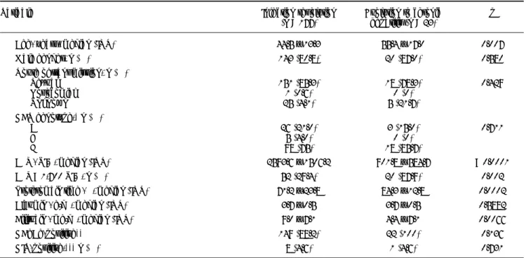
Table 2 (P-100). Treatment, follow-up and outcomes in patients with AIH and DIAIH.
Characteristics AIH (n = 178) DIAIH (n = 12) P value
Maintenance treatment
None - n (%) 5 (2.8) 0 (0.0) 0.55
Only steroid - n (%) 9 (5.1) 0 (0.0) 0.78
Steroid and immunomodulator - n (%) 118 (66.3) 1 (8.3) < 0.001
Only immunomodulator - n (%) 35 (19.7) 8 (66.6) <0.001
Withdrawal of immunosuppression - n (%) 5 (2,8) 3 (25.0) <0.001
Relapse - n (%) 32 (18.0) 0 (0.0) 0.10
Cirrhosis development during follow-up - n = 96* 12 (12.5) 0 (0.0) 0.19
Liver transplant - n (%) 10 (5.6) 0 (0.0) 0.40
Death during follow up - n (%) 5 (2.8) 0 (0.0) 0.55
* Patients with cirrhosis at diagnosis were excluded for the calculation.
P-149
SEQUENTIAL OVERLAP SYNDROME: CLINICAL
CHARACTERISTICS OF 10 PATIENTS
PINTO C,* LAGOS I,** BRAHM J* PINTO C,* LAGOS I,** BRAHM J*PINTO C,* LAGOS I,** BRAHM J* PINTO C,* LAGOS I,** BRAHM J* PINTO C,* LAGOS I,** BRAHM J*,,,,,***************
* DEPARTMENT OF GASTROENTEROLOGY. HOSPITAL CLÍNICO UNIVERSIDAD DE CHILE.
** DEPARTMENT OF INTERNAL MEDICINE, HOSPITAL CLÍNICO UNIVERSIDAD DE CHILE. *** DEPARTMENT OF GASTROENTEROLOGY, CLÍNICA LAS CONDES; SANTIAGO,
CHILE (A GASTROENTEROLOGY RESIDENT; B INTERNAL MEDICINE RESIDENT).
Introduction. The sequential overlap syndrome (SOS) is the
consecutive presentation of two autoimmune chronic liver dis-eases, in most cases association of Autoimmune Hepatitis (AIH) and Primary Biliary Cholangitis (PBC) or Primary Sclerosing Cholangitis (PSC). It constitutes a clinical challenge, since there are no universally accepted diagnostic criteria. Given its low
prevalence, only small series of cases have been reported.
Ob-jectives. Characterization of 10 patients with SOS diagnosis.
Material and methods. Retrospective descriptive study of
pa-tients from 2 centers diagnosed with SOS, followed-up by one hepatologist. Review of clinical records and analysis of biochem-ical, serologbiochem-ical, radiological and histological parameters and therapy used at the time of diagnosis of the initial and
subse-quent liver disease. Results. 10 patients had SOS, average age
49 years (23-74), 90% female. Five patients developed overlap between AIH and PSC, as well as AIH and PBC. In the group with overlap between AIH/PSC, 4 female patients initially
pre-Table 1 (P-149). Characteristics of patients with sequential overlap syndrome (n = 10).
Initial Sequential Patients Time Age at Biochemical characteristics at diagnostic of SOS
diagnosis diagnosis (n) to SOS diagnosis
average of SOS AST ALT ALP GGT Bilirubin
years average (x ULN) (x ULN) (x ULN) (x ULN) (mg(dL)
(range) average average average average average
AIH PSC 4 8.5 35 (23-40) 2.6 2.1 2.4 8.2 N
AIH PBC 3 5.3 63 (53-74) 1.7 N 1.4 5.4 N
PSC AIH 1 1.0 41 18 13 3.1 2.7 8.4
PBC AIH 2 7.5 59.5 (56-63) 13 18 2.3 10.3 8.1
sented AIH, developing PSC after 8.5 years average (average age 35 years). All patients had compatible MR cholangiography, bi-opsy with chronic cholangitis, and one case developed ulcerative colitis. Only 1 male patient initially presented PSC and subse-quently AIH, presenting high ANA titers and the appearance of ASMA. The 5 patients with AIH/PBC overlap were female. Three patients were initially diagnosed with AIH, developing PBC after 5.2 years (average age 63 years). Only 1 had positive AMA. Two patients with initial PBC subsequently developed AIH, after 7.5 years average. The 10 patients received combined treatment with immunosuppressants and UDCA at the time of
diagnosis of SOS. Conclusion. SOS is a rare entity that should
be suspected when a patient with an autoimmune liver disease does not follow the normal clinical phenotype and expected re-sponse to therapy. Its adequate diagnosis modifies the treatment and prognosis.
02 BASIC STUDIES
P-15
IMMUNE RESPONSE IN THE PATHOGENESIS OF CHC:
T CELL POPULATIONS AND CYTOKINE MILIEU IN
LIVER AND PERIPHERAL BLOOD
RÍOS D,RÍOS D,RÍOS D,
RÍOS D,RÍOS D,1,1,1,1,1, VALVA P, VALVA P, VALVA P, VALVA P, VALVA P,1,1,1,1,1, GIADANS C, GIADANS C, GIADANS C, GIADANS C, GIADANS C,11111 VISTARINI C, VISTARINI C, VISTARINI C, VISTARINI C, VISTARINI C,22222 DE MATTEO E,
DE MATTEO E, DE MATTEO E, DE MATTEO E,
DE MATTEO E,11111 CASCIATO P, CASCIATO P, CASCIATO P, CASCIATO P, CASCIATO P,33333 BRODERSEN C, BRODERSEN C, BRODERSEN C, BRODERSEN C, BRODERSEN C,44444 PIETRANTONIO A,
PIETRANTONIO A,PIETRANTONIO A,
PIETRANTONIO A,PIETRANTONIO A,22222 CALDIROLA MS, CALDIROLA MS, CALDIROLA MS, CALDIROLA MS, CALDIROLA MS,55555 GAILLARD MI, GAILLARD MI, GAILLARD MI, GAILLARD MI, GAILLARD MI,55555 AMEIGEIRAS B,
AMEIGEIRAS B, AMEIGEIRAS B, AMEIGEIRAS B,
AMEIGEIRAS B,22222 PRECIADO MV PRECIADO MV PRECIADO MV PRECIADO MV PRECIADO MV11111
1 MULTIDISCIPLINARY INSTITUTE FOR INVESTIGATION IN PEDIATRIC PATHOLOGIES (IMIPP), CONICET-GCBA, LABORATORY OF MOLECULAR BIOLOGY, PATHOLOGY
DIVISION, HOSPITAL DE NIÑOS “RICARDO GUTIÉRREZ”; GALLO 1330 C1425EFD, CIUDAD AUTÓNOMA DE BUENOS AIRES, ARGENTINA.
2 LIVER UNIT, HOSPITAL “RAMOS MEJÍA”; URQUIZA 609, CP1221, CIUDAD AUTÓNOMA DE BUENOS AIRES, ARGENTINA.
3 LIVER UNIT, HOSPITAL ITALIANO DE BUENOS AIRES; JUAN D PERÓN 4190 C1181ACH, CIUDAD AUTÓNOMA DE BUENOS AIRES, ARGENTINA.
4 LIVER UNIT, HOSPITAL GENERAL DE AGUDOS “CARLOS G. DURAND”; AVDÍAZ VÉLEZ 5044, C1405DCS, CIUDAD AUTÓNOMA DE BUENOS AIRES, ARGENTINA. 5 MULTIDISCIPLINARY INSTITUTE FOR INVESTIGATION IN PEDIATRIC PATHOLOGIES
(IMIPP), CONICET-GCBA, LABORATORY OF IMMUNOLOGY, HOSPITAL DE NIÑOS “RICARDO GUTIÉRREZ”; GALLO 1330 C1425EFD, CIUDAD AUTÓNOMA DE BUENOS
AIRES, ARGENTINA. EQUALLY CONTRIBUTED.
Introduction. In chronic hepatitis C (CHC) the immune
sys-tem is involved in liver damage; but, the role of each immune cell is unknown. We aimed to evaluate T cell populations and cytokine milieu in liver and peripheral blood (PB) to elucidate
the immune system role in CHC liver disease. Material and
methods. Liver biopsies and concomitant PB samples from 48
untreated adult CHC patients were analyzed. CTL (CD8), Th (CD4), Th17 (IL17A/CD4-IL17A), Treg
(Foxp3/CD4-CD25hi-Foxp3) and Th1 (Tbet/CD4-IFNγ) cell frequencies
were evaluated by immunohistochemistry on formalin-fixed
bi-opsies and by flow cytometry in PB. TGFβ, IFNγ, IL6, IL1β,
IL8, IL10, IL23, IL21 levels were evaluated by RT-qPCR in fresh liver and by CBA in plasma. PB samples from uninfected donors were included. Results were related to hepatitis and
fi-brosis severity. Results. Liver infiltrates showed Th
predomi-nance, high Treg and Th1 but low Th17 frequency. Th17 cells and Th17/Treg ratio showed fibrosis association (both p =
0.04). TGFβ (p < 0.05, r = 0.49), IL8 (p < 0.01; r = 0.49) and
IL6 (p < 0.05, r =-0.43) displayed correlations with Th17
fre-quency. While TGFβ, IL23, IL1β were associated with hepatitis
severity (all p < 0.05), IL8 was associated with advanced fibrosis (p = 0.004). IL10 correlated to IL6, IL21 and IL23 (all p < 0.05; r = 0.43, r = 0.66, r = 0.39, respectively) and was higher in se-vere hepatitis cases. The PB lymphocyte profile in CHC patients was similar to donors, but cytokines pattern showed higher
lev-els in patients, being IL6 and TGFβ significantly elevated (p =
0.03; p = 0.04). Conclusion. The liver immune
microenviron-ment in CHC depicted a complex cytokine milieu that allows the Th17 and Treg interplay. Although Treg was not directly involved in liver damage, high IL10 levels might reflect a differ-ent Treg activation status throughout disease progression. Th17 and IL8 might have a key role in fibrogenesis. While CHC pe-ripheral lymphocyte frequency showed no alterations, the cy-tokine profile delineated an activated scenario.
P-132
MOLECULAR GENETIC ANALYSIS OF MARKER
RS738409 GENE PNPLA3 IN THE YAKUT
POPULATION
KHARITON-ALEKSEEVICH K, NADEZHDA-IVANOVNA P, KHARITON-ALEKSEEVICH K, NADEZHDA-IVANOVNA P, KHARITON-ALEKSEEVICH K, NADEZHDA-IVANOVNA P, KHARITON-ALEKSEEVICH K, NADEZHDA-IVANOVNA P, KHARITON-ALEKSEEVICH K, NADEZHDA-IVANOVNA P,
ALEKSANDRA-TIMOFEEVNA D, MARINA-ALEKSEEVNA V TIMOFEEVNA D, MARINA-ALEKSEEVNA V TIMOFEEVNA D, MARINA-ALEKSEEVNA V TIMOFEEVNA D, MARINA-ALEKSEEVNA V TIMOFEEVNA D, MARINA-ALEKSEEVNA V
DEPARTMENT OF MOLECULAR GENETICS, FEDERAL STATE BUDGETARY SCIENTIFIC INSTITUTION “YAKUT SCIENCE CENTER OF COMPLEX MEDICAL PROBLEMS”,
YAKUTSK, RUSSIA
Introduction. Non-alcoholic fatty liver disease (NAFLD) is an
emerging health concern, with increasing prevalence world-wide. Recently, a non-synonymous genetic variation (rs738409) in the human patatin-like phospholipase domain-containing 3 gene (PNPLA3), was found to be associated with NAFLD among Hispanics, African Americans, and European Americans. Studies have shown that the G allele (risk allele) of rs738409 in PNPLA3 gene was associated with increased propensity of
stea-tosis and severe fibrosis. Objectives. In this study, we
investi-gated the distribution of PNPLA3 genotypes among Yakut. Understanding the prevalence of PNPLA3 genetic variation among various ethnic populations could provide useful infor-mation for the improvement of care of patients at risk for
devel-oping hepatic steatosis and advanced liver damage. Material
and methods. 179 samples, a population cohort of the Yakuts,
were analyzed. Study participants were enrolled from YSC CMP hospital-Yakutsk City. The study was approved by YSC CMP Ethics Committee. All study participants gave written informed
consent. SNP (rs738409)
PNPLA3. Results. As a result of genotyping of the
population sample of the Yakuts for the PNPLA3 gene, the prevalence of the GG genotype (61.5%) was revealed. The allele G frequency was 76.8%. The distribution of genotypes of poly-morphism rs738409 was in the Hardy-Weinberg equilibrium in
the sample studied (p > 0.05). Conclusions. The high
03 CIRRHOSIS
P-04
A DIFFERENT GUT MICROBIOME LINKED TO
INFLAMMATION FOUND IN CIRRHOTIC PATIENTS
WITH AND WITHOUT HEPATOCELLULAR
CARCINOMA
PIÑERO F,* PIÑERO F,* PIÑERO F,* PIÑERO F,*
PIÑERO F,*,†,•,†,•,†,•,†,•,†,• VÁZQUEZ M, VÁZQUEZ M, VÁZQUEZ M, VÁZQUEZ M, VÁZQUEZ M,‡,§,•‡,§,•‡,§,•‡,§,•‡,§,• BARÉ P, BARÉ P, BARÉ P, BARÉ P, BARÉ P,§,||§,||§,||§,||§,|| ROHR C, ROHR C, ROHR C, ROHR C, ROHR C,‡‡‡‡‡ MENDIZÁBAL MENDIZÁBAL MENDIZÁBAL MENDIZÁBAL MENDIZÁBAL M,*
M,* M,* M,*
M,*,†,†,†,†,† SCIARA M, SCIARA M, SCIARA M, SCIARA M, SCIARA M,‡‡‡‡‡ ALONSO C,* ALONSO C,* ALONSO C,* ALONSO C,* ALONSO C,*,†,†,†,†,† FAY F, FAY F, FAY F, FAY F, FAY F,‡‡‡‡‡ SILVA M* SILVA M* SILVA M* SILVA M* SILVA M*,†,†,†,†,† * HOSPITAL UNIVERSITARIO AUSTRAL, LIVER TRANSPLANT AND HEPATOLOGY UNIT.
AUSTRAL UNIVERSITY SCHOOL OF MEDICINE, ARGENTINA. † LATIN AMERICAN LIVER RESEARCH, EDUCATION AND AWARENESS NETWORK
(LALREAN).
‡ HERITAS. ROSARIO, SANTA FE. ARGENTINA. § CONICET, ARGENTINA.
|| INSTITUTO DE MEDICINA EXPERIMENTAL (IMEX), CONICET, ACADEMIA NACIONAL DE MEDICINA. BUENOS AIRES. ARGENTINA.
• CONTRIBUTED EQUALLY TO THIS WORK AS FIRST AUTHORS.
Introduction. Increasing interest has been focused during the
last years regarding microbiome and human diseases including cirrhosis, alcoholic liver disease, fatty liver and fibrosis progres-sion. Changes in gut microbiome have been observed with pro-gression of liver disease including a reduced abundance of taxa
considered benign, such as Lachnospiraceae, Ruminococcaceae and
Clostridialies and a higher abundance of non-benefitial taxa such as Enterobacteriaceae and Bacteriodaceae. A renewed novel research has focused on microbiome and cancer development. However, no specific microbiome in patients with hepatocellular
carcino-ma (HCC) has been reported to date. Aims. We sought to
compare the gut microbiome found in cirrhotic patients with and without HCC, in order to try to identify a specific gut
microbiome profile among cirrhotic patients with HCC.
Mate-rial and methods. This observational case-control study was
nested on a prospective longitudinal cohort of patients with cir-rhosis who were followed-up in our Liver Unit at Austral Uni-versity Hospital, School of Medicine, in colaboration with HERITAS (Rosario), CONICET and the National Academy of Medicine from Argentina. This study was carried out between December 2015 and October 2016 in accordance with interna-tional recommendations for observainterna-tional studies. From 407 pa-tients with Child Pugh A/B cirrhosis prospectively followed, 25 with HCC (cases) were matched with 25 without HCC (wo-HCC) in a 1:1 ratio according to age, gender, etiology, Child Pugh and severity of portal hypertension. In addition results were also compared with 25 healthy subjects. Plasma cytokines were quantified including interleukin-6 (IL-6) and tumor
necrosis factor α (TNF-α). Faecal stool samples were sequenced
for the V3-V4 region of the microbial 16S rRNA (Illumina
MiSeq Platform). Results. We found a differential abundance
in family members of Firmicutes with a 3-fold increased of
Ery-sipelotrichaceae and a 5-fold decrease in family Leuconostocaceae in HCC when compared to wo-HCC controls. Genus
Fusobacte-rium was found 5-fold decreased in HCC vs. wo-HCC. The
ra-tio bacteriodes/prevotella was increased in HCC. Three
operational taxonomic units (OTUs), genus Odoribacter and Butyricimonas were more abundant in HCC, whereas a
de-creased abundace in Lachnospiraceae family genus Dorea was
ob-served in HCC patients. This pattern has been previously associated with an inflammatory milieu with a putative
in-creased activation of NOD-like receptor signalling pathways.
Conclusions. A pattern of gut microbiome linked to
inflam-mation was observed in patients with HCC, opening the dis-cussion whether or not microbiota has a physiopathologic role. The differential abundance in 3 specific OTUs might be further explored as a new tumor biomarker.
P-10
THE IMPACT OF INFECTIONS ON THE MORTALITY
OF HOSPITALIZED PATIENTS WITH LIVER
CIRRHOSIS
DÍAZ-HERNÁNDEZ HA,* VÁZQUEZ-ANAYA G,* CASTRO-NARRO GE* DÍAZ-HERNÁNDEZ HA,* VÁZQUEZ-ANAYA G,* CASTRO-NARRO GE* DÍAZ-HERNÁNDEZ HA,* VÁZQUEZ-ANAYA G,* CASTRO-NARRO GE* DÍAZ-HERNÁNDEZ HA,* VÁZQUEZ-ANAYA G,* CASTRO-NARRO GE* DÍAZ-HERNÁNDEZ HA,* VÁZQUEZ-ANAYA G,* CASTRO-NARRO GE* * DEPARTMENT OF GASTROENTEROLOGY, NATIONAL INSTITUTE OF MEDICAL
SCIENCES AND NUTRITION “SALVADOR ZUBIRÁN”, MEXICO CITY, MEXICO.
Introduction. Bacterial infections are common complications
in patients with cirrhosis and are associated with a poor progno-sis. However, there are no studies that analyze the impact of the different infectious complications on the mortality of patients
with cirrhosis. Objective. To evaluate the impact of infectious
complications in the short-term mortality of hospitalized
pa-tients with cirrhosis. Material and methods. We performed a
case-control study in adult patients from both sexes with cir-rhosis who had been hospitalized from 2014 to 2017 and with a follow-up for at least one year. We recorded demographic data, prognostic scales, infectious complications and mortality at 30, 90 and 365 days. Demographic data are presented as numbers with percentages and medians and inter-quartile ranges as ap-propriate. The primary outcome was mortality. For the survival analysis, hazard ratios were calculated with 95% confidence in-tervals by Cox-regression in univariate and multivariate models.
For the comparison between the groups the χ2 test, Fisher’s
ex-act test and Mann-Whitney U test were used as appropriate.
Results. We included 500 patients. The median age was 58
years (47-65), the predominant sex was woman (52%) and the most common infections were urinary tract infections (UTI) (35%), pneumonias (28.2%) and spontaneous bacterial peritoni-tis (SBP) (18%). In the univariate analysis, infections in general, SBP, pneumonia and central nervous system (CNS) infections had an increased mortality at the three follow up periods, but in the multivariate analysis with the prognostic scales, only pneu-monia (HR 2.03, CI 95% [1.06-3.86]) and CNS infections (HR 4.84, CI 95% [1.38-16.93]) remained with increased mortality
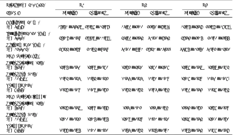
(Table 1). Conclusions. Some infectious complications as
pneumonia and CNS infections independently increase
short-term mortality in hospitalized patients with cirrhosis. Conflict
Table 1 (P-10).
Univariate and multivariate analysis of the impact of infectious complications in the mortality of hospitalized patients with
cirrhosis at 30, 90 and 365 days.
Mortality in hospitalized patients with cirrhosis Variable
3 0 days 90 days 365 days Univariate Multivariate Univariate Multivariate Univariate Multivariate HR p H R p HR p H R p HR p H R p (CI 95%) (CI 95%) (CI 95%) (CI 95%) (CI 95%) (CI 95%) Child-Pugh C 5.27 < 0.001 6.12 < 0.001 6.22 < 0.001 (3.33-8.36) (4.04-9.27) (4.33-8.94) MELD >15 2.19 0.096 2.14 0 .064 2.70 0 .0 0 9 (0.87-5.52) (0.95-4.78) (1.28-5.69) Infections 3.24 < 0.001 0.94 0 .9 2 4 4.25 < 0.001 1.38 0 .5 6 6 5.21 < 0.001 1.70 0 .3 3 2 (1.92-5.46) (0.30-2.96) (2.58-7.01) (0.45-4.17) (3.26-8.34) (0.58-4.99) Bacterial 3.03 < 0.001 0.81 0 .6 6 5 3.43 < 0.001 0.69 0 .3 6 3 4.35 < 0.001 0.91) 0.818 infections (1.85-4.95) (0.33-2.02) (2.19-5.35) (0.31-1.52) (2.85-6.62) (0.43-1.92 Spontaneous 1.72 0.017 1.17 0 .7 1 5 1.67 0 .012 1.15 0 .7 1 8 1.67 0 .0 0 5 1.01 0 .9 7 6 Bacterial (1.10-2.70) (0.49-2.77) (1.12-2.49) (0.53-2.47) (1.16-2.39) (0.51-2.00) peritonitis Urinary 1.14) 0.515 1.29 0 .148 1.40 0 .0 3 4 0.98 0 .9 5 1 tract (0.76-1.71 (0.91-1.85) (1.02-1.92) (0.52-1.83) infections Pneumonia 3.54 <0.001 1.57 0 .2 8 0 4.30 < 0.001 2.06 0 .0 5 3 4.77 < 0.001 2.03 0 .0 3 1 (2.38-5.27) (0.69-3.61) (3.01-6.12) (0.99-4.28) (3.47-6.54) (1.06-3.86) Abdominal 1.12 0.768 1.11 0 .751 0.96 0 .9 1 4 sepsis (0.52-2.42) (0.56-2.19) (0.50-1.83) Soft tissue 0.84 0.712 0.77) 0.547 1.29) 0.395 infections (0.34-2.07) (0.34-1.76 (0.71-2.32 Central 3.57 0.013 4.84 0 .0 1 4 3.13 0 .025 4.39 0 .0 1 9 2.73 0 .0 4 7 4.01 0 .0 2 5 nervous (1.31-9.71) (1.38-16.93) (1.15-8.50) (1.28-15.04) (1.01-7.40) (1.19-13.57)
system infections Upper
0.32 0.265 0.24 0 .164 0.78 0 .6 2 5 respiratory (0.04-2.33) (0.03-1.77) (0.28-2.10)
tract infections Osteomyelitis
P-11
EARLY PREDICTORS OF AKI DEVELOPMENT/
PROGRESSION IN PATIENTS WITH CIRRHOSIS AND
ASCITES ADMITTED WITH A BACTERIAL INFECTION
XIMENES RO,* DE SOUZA HP, XIMENES RO,* DE SOUZA HP, XIMENES RO,* DE SOUZA HP, XIMENES RO,* DE SOUZA HP,
XIMENES RO,* DE SOUZA HP,††††† BARBEIRO DF, BARBEIRO DF, BARBEIRO DF, BARBEIRO DF, BARBEIRO DF,††††† MENDES LSC, MENDES LSC, MENDES LSC, MENDES LSC, MENDES LSC,‡‡‡‡‡ MARTINELLI AMC,
MARTINELLI AMC, MARTINELLI AMC, MARTINELLI AMC,
MARTINELLI AMC,§§§§§ MAZO DFC,* MAZO DFC,* MAZO DFC,* MAZO DFC,* MAZO DFC,*,||,||,||,||,|| ÁLVARES-DA-SILVA MR, ÁLVARES-DA-SILVA MR, ÁLVARES-DA-SILVA MR, ÁLVARES-DA-SILVA MR, ÁLVARES-DA-SILVA MR,¶¶¶¶¶ CIARLEGLIO M,** YANHONG D,
CIARLEGLIO M,** YANHONG D, CIARLEGLIO M,** YANHONG D, CIARLEGLIO M,** YANHONG D,
CIARLEGLIO M,** YANHONG D,†††††††††† CARRILHO FJ,* GARCIA-TSAO G, CARRILHO FJ,* GARCIA-TSAO G, CARRILHO FJ,* GARCIA-TSAO G, CARRILHO FJ,* GARCIA-TSAO G, CARRILHO FJ,* GARCIA-TSAO G,†††††††††† FARIAS AQ*
FARIAS AQ* FARIAS AQ* FARIAS AQ* FARIAS AQ*
* DEPARTMENT OF GASTROENTEROLOGY,
† EMERGENCY DEPARTMENT, UNIVERSITY OF SAO PAULO, SAO PAULO, BRAZIL. ‡ DEPARTMENT OF GASTROENTEROLOGY, HOSPITAL DE BASE DO DF, BRASILIA,
BRAZIL.
§ DEPARTMENT OF GASTROENTEROLOGY, UNIVERSITY OF SAO PAULO, RIBEIRAO PRETO, BRAZIL.
|| DEPARTMENT OF GASTROENTEROLOGY, STATE UNIVERSITY OF CAMPINAS, CAMPINAS, BRAZIL.
¶ DEPARTMENT OF GASTROENTEROLOGY, FEDERAL UNIVERSITY OF RIO GRANDE DO SUL, PORTO ALEGRE, BRAZIL.
** DEPARTMENT OF STATISTICS, YALE UNIVERSITY, NEW HAVEN, USA. †† SECTION OF DIGESTIVE DISEASES, YALE UNIVERSITY, NEW HAVEN, USA.
Introduction. Acute kidney injury (AKI) is a common
compli-cation of bacterial infections in patients with cirrhosis and as-cites. AKI is associated with high mortality, especially in patients
in whom AKI p rogresses. Objective. Identifying factors
(in-cluding urinary NGAL) that predict development/progression
of AKI in patients admitted with bacterial infection. Material
and methods. Multicenter study including adult patients with
cirrhosis and ascites admitted with a bacterial infection between June 2013 and April 2017. Baseline characteristics were
com-pared between patients who developed (vs. those who did not
develop) the outcome: AKI development in those without AKI on admission; progression of AKI in those with AKI on
admis-sion. Results. 179 patients were included (72 without and 107
with AKI at admission), median age was 57 years, 70% were male and spontaneous bacterial peritonitis was the most fre-quent infection (53%). Of those without AKI, 25 (35%) devel-oped it during admission. Independent predictors of AKI development were female gender (OR: 3.50 [1.19-10.34], p = 0.023) and overt hepatic encephalopathy (OHE) (OR: 4.25 [1.29-14.04], p = 0.018). Of those with AKI on admission, it progressed in 39 (36%). The only independent predictor of AKI progression was urinary NGAL (OR 1.46 [1.03-2.07], p = 0.035). Although female gender and Child were associated with AKI progression on univariate analysis, they were of borderline significance in the multivariate model (p = 0.0502 and p =
0.059 respectively). Conclusions. Patients with cirrhosis and
ascites admitted with an infection and OHE are at increased risk of AKI development, which might reflect some degree of renal dysfunction that does not yet fulfill AKI criteria. These patients should have renal function closely monitored. Females seem to be more prone to AKI development and progression. High uri-nary NGAL levels appear to identify patients with increased risk of AKI progression that should be prioritized for timely
treat-ment of AKI. Conflicts of interest. This study received grants
from Sao Paulo Research Foundation (FAPESP). Ciarleglio M, Yanhong D and Garcia-Tsao G are funded from Yale Liver Center NIH P30 DK34989.
Table 1 (P-11).
AKI development AKI Progression
Yes (n = 25) No (n = 47) p Yes (n = 39) No (n = 68) p
Age 53.0 56.0 0.75 59.0 58.5 0.80
(51.0-60.0) (44.0-63.0) (53.0-63.0) (49.0-65.5)
Gender (female) 13 (52%) 12 (26%) 0.025 14 (36%) 14 (21%) 0.08
Overt hepatic 10 (40%) 7 (15%) 0.017 16 (41%) 22 (32%) 0.37
encephalopathy
Serum creatinine 0.8 (0.7-0.9) 0.8 (0.7-1.0) 0.97 2.2 (1.8-3.0) 1.8 (1.5-2.3) 0.014*
Child
- A 0 (0%) 1 (2%) 0.87 0 (0%) 0 (0%) 0.039
- B 10 (40%) 21 (45%) 6 (15%) 23 (34%)
- C 15 (60%) 25 (53%) 33 (85%) 45 (66%)
MELD 16.0 15.0 0.92 27.0 24.5 0.06
(14.0-19.0) (13.0-21.0) (23.0-30.0) (20.0-30.0)
Urinary NGAL, μg/L 94.5 76.3 0.34 333.0 163.1 0.008
(49.0-366.8) (51.3-192.4) (182.9-1158.3) (102.1-435.7)
P-17
GOOD PERFORMANCE OF LIVER FIBROSIS
ASSESSMENT WITH A NEW DEVICE FROM
TRANSIENT ELASTOGRAPHY
VALENZUELA M,* VALENZUELA M,*VALENZUELA M,* VALENZUELA M,*
VALENZUELA M,*,||,||,||,||,|| GAJARDO AIJ,** GAJARDO AIJ,** GAJARDO AIJ,** GAJARDO AIJ,** GAJARDO AIJ,**,||,||,||,||,|| PÉREZ-LUCO T, PÉREZ-LUCO T, PÉREZ-LUCO T, PÉREZ-LUCO T, PÉREZ-LUCO T,‡‡‡‡‡ BELMAR F, BELMAR F, BELMAR F, BELMAR F, BELMAR F,‡‡‡‡‡ NAVEA C,*
NAVEA C,* NAVEA C,* NAVEA C,*
NAVEA C,*,||,||,||,||,|| CARREÑO L, CARREÑO L, CARREÑO L, CARREÑO L, CARREÑO L,§§§§§ VERA DB,* SANDOVAL A,* PONIACHIK J,* VERA DB,* SANDOVAL A,* PONIACHIK J,* VERA DB,* SANDOVAL A,* PONIACHIK J,* VERA DB,* SANDOVAL A,* PONIACHIK J,* VERA DB,* SANDOVAL A,* PONIACHIK J,* GONZÁLEZ K*
GONZÁLEZ K* GONZÁLEZ K* GONZÁLEZ K* GONZÁLEZ K*
* GASTROENTEROLOGY SECTION. UNIVERSITY OF CHILE CLINICAL HOSPITAL, SANTIAGO, CHILE.
† DEPARTMENT OF INTERNAL MEDICINE. UNIVERSITY OF CHILE CLINICAL HOSPITAL, SANTIAGO, CHILE.
‡ FACULTY OF MEDICINE. UNIVERSITY OF CHILE CLINICAL HOSPITAL, SANTIAGO, CHILE.
§ DEPARTMENT OF PATHOLOGICAL ANATOMY, UNIVERSITY OF CHILE CLINICAL HOSPITAL, SANTIAGO, CHILE.
|| RESIDENT GASTROENTEROLOGY. UNIVERSITY OF CHILE CLINICAL HOSPITAL, SANTIAGO, CHILE.
Introduction. Transient elastography (TE) is a non-invasive
method that quantifies liver fibrosis (LF), being liver biopsy
(LB) the gold standard. Objective. To evaluate the diagnostic
performance of TE against LB in LF assessment in patients with
chronic liver disease (CLD). Material and methods.
Diagnos-tic test study. CLD patients with LB performed from August 2016 to November 2017 were enrolled (n = 39). TE was
per-formed with Fibrotouch®HISKY/FT1000 device by a single
op-erator. 69% were women, age average 57 ± 11 years old, time between TE and LB was 4 ± 2 months; GPT 76 ± 58 IU/L; CLD etiologies: Autoimmune Hepatitis 44%, Non-alcoholic st-eatohepatitis 28%, Chronic cholestasia 15%, other etiologies 13%. LF assessed by LB was classified according to METAVIR. TE results were classified according to the presence of mild or no LF (F0 or F0/F1) and in advanced LF (F3, F3/F4 or F4). Statis-tical analysis was done with ROC curve and area under curve (AUC), sensitivity (Se), specificity (Sp), positive predictive value
(PPV), negative predictive value (NPV) were calculated.
Re-sults. TE showed an AUC = 0.77 for mild or no LF (F0 or F1
in LB), Se: 57.14%; Sp: 96%, PPV 100%, NPV 88.89%. On the other hand, for the diagnosis of advanced LF (F3 or F4 in LB), TE showed an AUC = 0.92, Se: 100%, Sp: 83.33%, PPV:
85.71%; NPV: 100%. Conclusion. TE with a new device, has a
good diagnostic performance to discard LF, as well as to confirm advanced LF comparate with LB. Therefore, in patients with a known etiology, TE could substitute the LB for follow-up of LF.
P-18
EVALUATION OF CXCL-8 AND IL-6 IN PATIENTS
WITH CHRONIC LIVER DISEASE
ROSIQUE-ORAMAS D,* MEDINA-ÁVILA Z,* GARCÍA-LÓPEZ B,* ROSIQUE-ORAMAS D,* MEDINA-ÁVILA Z,* GARCÍA-LÓPEZ B,* ROSIQUE-ORAMAS D,* MEDINA-ÁVILA Z,* GARCÍA-LÓPEZ B,* ROSIQUE-ORAMAS D,* MEDINA-ÁVILA Z,* GARCÍA-LÓPEZ B,* ROSIQUE-ORAMAS D,* MEDINA-ÁVILA Z,* GARCÍA-LÓPEZ B,* RAMÍREZ-MENDOZA A,* GUZMÁN C,* PÉREZ-HERNÁNDEZ JL,** RAMÍREZ-MENDOZA A,* GUZMÁN C,* PÉREZ-HERNÁNDEZ JL,**RAMÍREZ-MENDOZA A,* GUZMÁN C,* PÉREZ-HERNÁNDEZ JL,** RAMÍREZ-MENDOZA A,* GUZMÁN C,* PÉREZ-HERNÁNDEZ JL,**RAMÍREZ-MENDOZA A,* GUZMÁN C,* PÉREZ-HERNÁNDEZ JL,**
HIGUERA-DELATIJERA F,** GUTIÉRREZ-REYES G* HIGUERA-DELATIJERA F,** GUTIÉRREZ-REYES G*HIGUERA-DELATIJERA F,** GUTIÉRREZ-REYES G* HIGUERA-DELATIJERA F,** GUTIÉRREZ-REYES G* HIGUERA-DELATIJERA F,** GUTIÉRREZ-REYES G* * LABORATORIO HÍGADO, PÁNCREAS Y MOTILIDAD (HIPAM), DPTO. MEDICINA
EXPERIMENTAL, UNAM, MÉXICO CITY, MÉXICO.
** CLÍNICA DE HÍGADO, HOSPITAL GENERAL DE MÉXICO “DR. EDUARDO LICEAGA”, MÉXICO CITY, MÉXICO.
Background. Cytokines play a critical role in cell
communica-tion and activacommunica-tion, the liver is a source of cytokines and chem-okines, are key molecules that participate in the development of most liver diseases. There is evidence about the participation of neutrophils that are recruited by CXCL-8 in acute liver
diseas-es, and IL-6 has a hepatoprotector effect in alcoholic liver dis-ease (ALD), however, the participation of CXCL-8 and IL-6 in
other liver diseases still not clear. Objective. To evaluate the
concentration of CXCL-8 and IL-6 in alcoholic subjects, alco-holic liver cirrhotic patients, chronic hepatitis C and control
subjects. Material and methods. We included alcoholic
pa-tients that were seen at the Liver Clinic of the Hospital General de Mexico. Alcoholism was defined according to the WHO criteria (70 g/day for men and 50 g/day for women over the last 5 years) and the alcoholic patients were classified as those with-out cirrhosis of the liver (OH) and those with liver cirrhosis (COH). A group of patients with chronic hepatitis C (CHC) and a control group (CT) were also included. The CT subjects consumed 10 g/day of alcohol and had negative viral serology. CXCL-8 was determined in serum through Luminex technolo-gy (Biorad). The Mann-Whitney U test was employed in the
statistical analysis. Results. 81 alcoholic patients were included:
19 without cirrhosis of the liver (OH) and 62 with cirrhosis of the liver (COH). They were compared with 108 CHC patients
and 100 CT subjects (Table 1). Conclusion. CXCL-8
concen-tration in alcoholics with and without COH was 3 times higher compared with the CHC patients and 15 times higher compared with the control subjects. While the concentration of IL-6 in al-coholics was 2 times higher compared to controls and in COH patients was 100 times compared with OH and CHC patients. Our results showed that CXCL-8 and IL-6 participate actively maintaining the inflammatory process even in liver cirrhosis. This work has been partially funded by CONACYT: SALUD-2016-272579.
P-21
EVOLUTION OF DIAGNOSTIC CRITERIA FOR ACUTE
KIDNEY INJURY IN DECOMPENSATED CIRRHOTIC
PATIENTS: PROSPECTIVE STUDY IN A TERTIARY
UNIVERSITY HOSPITAL
VAZ NF,* CUNHA VNR,* CUNHA-SILVA M,* SEVÁ-PEREIRA T,* ALMEIDA VAZ NF,* CUNHA VNR,* CUNHA-SILVA M,* SEVÁ-PEREIRA T,* ALMEIDA VAZ NF,* CUNHA VNR,* CUNHA-SILVA M,* SEVÁ-PEREIRA T,* ALMEIDA VAZ NF,* CUNHA VNR,* CUNHA-SILVA M,* SEVÁ-PEREIRA T,* ALMEIDA VAZ NF,* CUNHA VNR,* CUNHA-SILVA M,* SEVÁ-PEREIRA T,* ALMEIDA
JRS,* MAZO DF* JRS,* MAZO DF* JRS,* MAZO DF* JRS,* MAZO DF* JRS,* MAZO DF*,,,,,**********
* DIVISION OF GASTROENTEROLOGY (GASTROCENTRO), SCHOOL OF MEDICAL SCIENCES, UNIVERSITY OF CAMPINAS, CAMPINAS, BRAZIL.
** DIVISION OF CLINICAL GASTROENTEROLOGY AND HEPATOLOGY, DEPARTMENT OF GASTROENTEROLOGY, UNIVERSITY OF SÃO PAULO SCHOOL OF MEDICINE, SAO
PAULO, BRAZIL.
Introduction. Acute kidney injury (AKI) was traditionally
di-agnosed as serum creatinine (sCr) ≥ 1.5 mg/dL. Recently,
changes in AKI diagnostic criteria have been proposed
(ICA-AKI criteria). Objectives. To evaluate the prevalence of AKI in
cirrhotics; to evaluate the agreement between traditional and ICA-AKI criteria; to assess its etiologies, risk factors, responses to treatment, progression for liver transplantation and mortality.
Material and methods. Prospective cohort study in cirrhotic
patients non-electively admitted from October 2016 to August 2017. Kappa coefficient was used to evaluate the agreement be-tween the two AKI criteria. The total number of hospitaliza-tions was evaluated using the PWP statistical model for
recurring events; p value < 0.05 was considered significant.
Re-sults. 154 admissions of 75 patients were included. The mean
substantial agreement between both AKI classifications (Kappa 0.7293). The main etiology of AKI was pre-renal (69.66%), fol-lowed by renal (26.96%) and hepatorenal syndrome (10.11%). Risk factors for ICA-AKI in univariate analysis were a higher MELD, infection, lower serum sodium and diuretics. In multi-variate analysis, MELD (p = 0.0188) and furosemide (p = 0.0014) were associated to ICA-AKI. In-hospital mortality in
univariate analysis was associated with sCr ≥ 1.5 mg/dL (p =
0.0373), MELD (p = 0.0296), bilirubin (p = 0.0064), shock (p = 0.0003) and peak stage3 ICA-AKI (p = 0.0221), while in multivariate analysis, peak stage3 ICA-AKI (p = 0.0252, RR: 10.499,95%CI: 1.339-82.335). Among hospitalizations with AKI, death was significantly associated with peak stage3 ICA-AKI, non-response to treatment and -dialysis. Stage1A-AKI had
a better prognosis than stage ≥ 1B-AKI. Conclusions. AKI
strongly impacted mortality. ICA-AKI stage3 led to a 10.49 times higher risk of death. Substantial agreement between AKI definitions was observed, and sCr > 1.5 mg/dL still remained an ominous finding.
P-23
RENAL DYSFUNCTION IN PATIENTS HOSPITALIZED
WITH CIRRHOSIS: A STUDY OF RETROSPECTIVE
COHORTE
ABARCA J, CAMACHO G ABARCA J, CAMACHO G ABARCA J, CAMACHO G ABARCA J, CAMACHO G ABARCA J, CAMACHO G
GASTROENTEROLOGY SERVICE, EUGENIO ESPEJO SPECIALTY HOSPITAL, QUITO, ECUADOR
Introduction. Renal dysfunction in cirrhotic patients increases
the probability of death during hospitalization. Predictors of mortality were examined, particularly the increase in serum
cre-atinine as well as other causes of decompensation. Objectives.
To know the clinical and epidemiological characteristics of pa-tients with liver cirrhosis of the Eugenio Espejo Hospital. To as-sess the impact of renal dysfunction in hospitalized cirrhotic
patients. Material and methods. Observational analytical
study of a retrospective cohort. We analyzed 119 cirrhotic pa-tients hospitalized from January 1, 2017 to March 31, 2018. The information was collected from the virtual medical records, obtaining epidemiological data, causes of hospital admission, presence of diseases associated with renal dysfunction such as ar-terial Hypertension and Diabetes Mellitus. In addition to imag-ing and laboratory reports. Serum creatinine measurements were standardized in the hospital laboratory and the kidney function
scale AKIN (Acute Kidney Injury Network) was used. Results.
A total of 119 patients with liver cirrhosis were included, from which 64 (53.8%) were men, with an average age of 60.5 ± 12.8 years. The cryptogenic etiology predominated with 53% and from this group, 55.56% were female. Stage I acute kidney dysfunction was 10.93%, stage II was 2.52%, and stage III was 2.52%. 8.4% of the patients had pre-established chronic kidney disease. There were 11 deaths with a higher percentage in the
group of patients with acute renal injury (AKI) 5.88% vs. 3.36%
in patients without AKI (< 0.01). Conclusion. Mortality in
patients with liver cirrhosis and acute renal injury was more fre-quent than in the group without renal dysfunction.
P-24
RISK FACTORS TO PRESENT A FIRST EPISODE OF
SPONTANEOUS BACTERIAL PERITONITIS (SPB) IN
THE HEPATOPATH PATIENTS ENTERED IN THE “DR.
SALVADOR B. GAUTIER”
JIMÉNEZ M, CORDONES E JIMÉNEZ M, CORDONES EJIMÉNEZ M, CORDONES E JIMÉNEZ M, CORDONES EJIMÉNEZ M, CORDONES E
INTERNAL MEDICINE SERVICE, “DR. SALVADOR B. GAUTIER” HOSPITAL, SANTO DOMINGO, DOMINICAN REPUBLIC.
Introduction. Since its description in 1964, a large number of
studies, guidelines and national and international consensus have emerged that have made a great advance in the diagnosis and treatment of this disease and have significantly changed its
prognosis.Objectives. This study was carried out with the
ob-jective of identifying the predictors or risk factors for the ap-pearance of a first episode of SBP in hepatopath patients with
ascites. Material and methods. Is an observational,
cross-sec-tional, descriptive study of prospective follow-up. From August 2013 to January 2014, we collected a total of 30 hepatopath
pa-tients with ascites who presented a first episode of SBP.
Re-sults. 16 variables were obtained at the time of admission
(including total ascitic fluid protein concentrations, serum bi-lirubin, severity of liver disease by the Child Pugh scale, serum creatinine, transaminase levels, prothrombin time, history of use of PPI, among others), in relation to its value in the prediction of development in a first episode of SPB. In the univariate anal-ysis, four variables reached statistical significance (P < 0.05) as predictive factors for the development of the first episode of SBP. These were serum albumin less than 2.8 g/dL, advanced liver disease, ascitic fluid total protein concentration < 1.5 g/dL
and elevated serum creatinine levels. Conclusions. The
proba-bility of a first episode of SBP is significantly influenced by the concentration of ascitic fluid proteins and the severity of liver disease.
P-25
UTILITY OF REAGENTS STRIPS FOR THE EARLY
DIAGNOSIS OF SPONTANEOUS BACTERIAL
PERITONITIS IN CIRRHOTIC PATIENTS WITH
ASCITIS
BEJARANO J,* GARZÓN M,** CASTELLANOS J,* BELTRÁN O,** PONCE BEJARANO J,* GARZÓN M,** CASTELLANOS J,* BELTRÁN O,** PONCE BEJARANO J,* GARZÓN M,** CASTELLANOS J,* BELTRÁN O,** PONCE BEJARANO J,* GARZÓN M,** CASTELLANOS J,* BELTRÁN O,** PONCE BEJARANO J,* GARZÓN M,** CASTELLANOS J,* BELTRÁN O,** PONCE DE LEÓN E,** CEBALLOS J,** HERNÁNDEZ G,** SALINAS C,** VARÓN DE LEÓN E,** CEBALLOS J,** HERNÁNDEZ G,** SALINAS C,** VARÓN DE LEÓN E,** CEBALLOS J,** HERNÁNDEZ G,** SALINAS C,** VARÓN DE LEÓN E,** CEBALLOS J,** HERNÁNDEZ G,** SALINAS C,** VARÓN DE LEÓN E,** CEBALLOS J,** HERNÁNDEZ G,** SALINAS C,** VARÓN
A * * A * *A * * A * *A * *
* INTERNAL MEDICINE RESIDENT, UNIVERSIDAD DEL ROSARIO, BOGOTÁ, COLOMBIA. ** DEPARTMENT OF GASTROENTEROLOGY-HEPATOLOGY, CARDIOINFANTIL
FOUNDATION-INSTITUTE OF CARDIOLOGY, BOGOTÁ, COLOMBIA.
Introduction. Spontaneous bacterial peritonitis (SBP) requires
an early diagnosis for the initiation of antibiotic therapy. The diagnosis is based on cytochemical examination of ascitic fluid, which takes hours to perform and has limited availability 24 h a
day, especially in first level hospitals and in rural areas.
Objec-tive. Evaluate the utility and diagnostic accuracy of Multistix
10SG reagent strip for diagnosis of SBP in cirrhotic patients with ascites admitted in Cardioinfantil Foundation –Cardiology
Institute. Material and methods. Diagnostic test study in
of SBP (polymorphonuclear (PMN) ≥ 250 cells/mm3).
Re-sults. Of 106 patients with ascites (51.8% men, mean age 59
years) 16 patients were diagnosed with SBP. The sensitivity, specificity, positive predictive value, negative predictive value, positive likelihood ratio and negative likelihood ratio taking grade ++ as cut off were 93.7%, 95.5%, 78.9%, 98%, 21 and
0.06, respectively. Conclusion. Multistix 10SG reagent strip
has a high sensitivity and specificity for the diagnosis of PBE compared to PMN count method. It is an inexpensive and easy technique, requires minimal time for interpretation and is avail-able, even in remote areas. This test may be useful for the diag-nosis of SBP in country like Colombia.
P-29
SURVIVAL AND CUMULATIVE INCIDENCE OF
COMPLICATIONS IN CUBAN PATIENTS WITH
COMPENSATED LIVER CIRRHOSIS: A PROSPECTIVE
LONG-TERM STUDY
CASTELLANOS-FERNÁNDEZ MI,* BERTOT LC,* CASTELLANOS-FERNÁNDEZ MI,* BERTOT LC,* CASTELLANOS-FERNÁNDEZ MI,* BERTOT LC,* CASTELLANOS-FERNÁNDEZ MI,* BERTOT LC,*CASTELLANOS-FERNÁNDEZ MI,* BERTOT LC,*,,,,,*** VILAR-GÓMEZ E,*,***** VILAR-GÓMEZ E,*,***** VILAR-GÓMEZ E,*,***** VILAR-GÓMEZ E,*,***** VILAR-GÓMEZ E,*,** TORRES-GONZÁLEZ AL,* DORTA-GURIDI Z,* INFANTE-VELÁZQUEZ M,* TORRES-GONZÁLEZ AL,* DORTA-GURIDI Z,* INFANTE-VELÁZQUEZ M,* TORRES-GONZÁLEZ AL,* DORTA-GURIDI Z,* INFANTE-VELÁZQUEZ M,* TORRES-GONZÁLEZ AL,* DORTA-GURIDI Z,* INFANTE-VELÁZQUEZ M,* TORRES-GONZÁLEZ AL,* DORTA-GURIDI Z,* INFANTE-VELÁZQUEZ M,*
ARÚS-SOLER E,* RUENES-DOMECH C,* ALBÁN-ORTEGA L* ARÚS-SOLER E,* RUENES-DOMECH C,* ALBÁN-ORTEGA L* ARÚS-SOLER E,* RUENES-DOMECH C,* ALBÁN-ORTEGA L* ARÚS-SOLER E,* RUENES-DOMECH C,* ALBÁN-ORTEGA L* ARÚS-SOLER E,* RUENES-DOMECH C,* ALBÁN-ORTEGA L* * DEPARTMENT OF HEPATOLOGY, NATIONAL INSTITUTE OF GASTROENTEROLOGY,
HAVANA, HAVANA, CUBA.
** INDIANA UNIVERSITY SCHOOL OF MEDICINE, DIVISION OF GASTROENTEROLOGY AND HEPATOLOGY, INDIANAPOLIS, IN, UNITED STATES.
*** UNIVERSITY OF WESTERN AUSTRALIA, SCHOOL OF MEDICINE AND PHARMACOLOGY, NEDLANDS, WA, AUSTRALIA.
Introduction. Natural history of subjects with compensated
liver cirrhosis from Latin-American and Caribbean’s countries
has been deficiently study. Objectives. Assess liver- related
complications and survival in Cuban patients with compensated liver cirrhosis according to D’Amico stage prospectively followed
for 10 years. Material and methods. Among 424 subjects
re-cruited from a tertiary referral care center in Havana, Cuba (2007 to 2016), 256 were into stage 1(absence of esophageal varices] and 168 in stage 2 (presence of esophageal varices). The cumulative probability for survival and clinical outcomes [as de-fined by the first occurrence of at least one of conditions: as-cites, variceal hemorrhage, hepatic encephalopathy or hepatocelular carcinoma (HCC)] were analyzed by the standard Kaplan-Meier method (95% CI), differences between stages were compared using log-rank test. Univariate and
multivaria-ble analysis were done for prognoses factors. Results. Over a
median of 288 (16-272) weeks, liver-related outcomes occurred in 152 (35.8%) patients, higher in stage 2 (50.6%)/stage 1(26.2%) (p < 0.001). The cumulative 6-year incidence of as-cites, variceal haemorrhage, encephalopathy and hepatocelular carcinoma was 60.3%, 45.3%, 16.7% and 12.5%. The overall cumulative 10-year incidence of gastroesophageal variceal
haemorrhage in stage 2 was 29.8% vs. 5.5% in stage 1 (p <
0.001). The viral cause (HCV/HBV) was related to the develop-ment of liver-related outcomes [HR: 1.7 (95% CI: 1,2-2,4)], higher in HCV. The overall survival at 3, 5 and 10 years were 92.9%, 88.5% and 77.7% and they were not different according to the D’Amico stage stratification (Log Rank [Mantel - Cox] p = 0.12). Prothrombin time (PT) and albumin were associated
with survival (p < 0.001). Conclusion. Stage 2 compensated
cirrhotics had significantly higher complications, mainly variceal
haemorrhage. Hepatic dysfunction as indicated by low albumin and prolonged PT were the only predictors of mortality.
P-31
HIGH INCIDENCE OF SPONTANEOUS BACTERIAL
PERITONITIS RECURRENCE IN PATIENTS UNDER
SECONDARY PROPHYLAXIS WITH NORFLOXACIN
MARCIANO S,* MARCIANO S,* MARCIANO S,* MARCIANO S,*
MARCIANO S,*,†,†,†,†,† DIRCHWOLF M, DIRCHWOLF M, DIRCHWOLF M, DIRCHWOLF M, DIRCHWOLF M,‡‡‡‡‡ DÍAZ JM,* BERMÚDEZ C,* DÍAZ JM,* BERMÚDEZ C,* DÍAZ JM,* BERMÚDEZ C,* DÍAZ JM,* BERMÚDEZ C,* DÍAZ JM,* BERMÚDEZ C,* GUTIÉRREZ-ACEVEDO MN,* BARCÁN LA,
GUTIÉRREZ-ACEVEDO MN,* BARCÁN LA, GUTIÉRREZ-ACEVEDO MN,* BARCÁN LA, GUTIÉRREZ-ACEVEDO MN,* BARCÁN LA,
GUTIÉRREZ-ACEVEDO MN,* BARCÁN LA,§§§§§ SMUD A, SMUD A, SMUD A, SMUD A, SMUD A,§§§§§ GIUNTA D, GIUNTA D, GIUNTA D, GIUNTA D, GIUNTA D,†,||†,||†,||†,||†,|| GADANO A*
GADANO A* GADANO A* GADANO A* GADANO A*,†,†,†,†,†
* SECCIÓN DE HEPATOLOGÍA, HOSPITAL ITALIANO DE BUENOS AIRES. † DEPARTAMENTO DE INVESTIGACIÓN, HOSPITAL ITALIANO DE BUENOS AIRES.
‡ UNIDAD DE HÍGADO, HOSPITAL PRIVADO DE ROSARIO. § SECCIÓN INFECTOLOGÍA, HOSPITAL ITALIANO DE BUENOS AIRES. || ÁREA DE INVESTIGACIÓN DE MEDICINA INTERNA, HOSPITAL ITALIANO DE BUENOS
AIRES.
Background. Few studies evaluated spontaneous bacterial
peritonitis (SBP) recurrence in patients receiving secondary anti-biotic prophylaxis, all performed more than 20 years ago. Changes in the bacteriology of SBP over the last years might
have a negative effect on secondary prophylaxis. Aim. To
esti-mate the incidence of SBP recurrence in patients with cirrhosis receiving secondary prophylaxis with norfloxacin, and to
ex-plore factors associated with SBP recurrence. Material and
methods. Retrospective cohort study of patients with cirrhosis
receiving norfloxacin for secondary prophylaxis of SBP from (March/2003-March/2016). Secondary prophylaxis had to be started no later than one week after the resolution of the first SBP. Patients with a history of transplantation or advanced hepatocellular carcinoma were excluded. Patients who died or were transplanted during SBP treatment (before secondary prophylaxis was started) were excluded. Follow up consisted on 365 days since secondary prophylaxis started. SBP recurrence was defined as any episode of SBP diagnosed during follow-up.
Results. The characteristics of the 115 patients included at the
time of their first SBP is shown in the table. Overall, 70.96% (95% CI 51.96%-85.77%) had a history of quinolone-resistant SBP and 12.90% (95% CI 3.63%-29.83%) of multiresistant SBP. The cumulative incidence of SBP recurrence was 28.53% (95%CI 20.15%-37.45%) at 365 days. In the bivariate analyses (table), the only variable that significantly differed among tients with and without SPB recurrence was gender: male pa-tients presented a crude sHR of SBP recurrence of 2.52 (95%CI
1.07-5.91, p = 0.034). Conclusions. Almost one third of the
Puntaje
Table 1 (P-31).
Variable Without With P1
recurrence recurrence
(N = 85) (N = 30)
Male gender, num (%) 44 (52) 23 (77) 0.035
Age-years 58 ± 14 55 ± 14 0.626
Cirrhosis etiology, num (%)
Viral hepatitis 29 (34) 8 (27)
Alcohol 16 (19) 10 (33)
Non-alcoholic steatohepatitis 4 (5) 3 (10) 0.321
Cryptogenic 11 (13) 2 (7)
Autoimmune hepatitis 10 (12) 5 (17)
Primary biliary cholangitis 12 (14) 1 (3)
Other 3 (3) 1 (3)
Diabetes, num (%) 8 (9) 6 (20) 0.145
Child-Pugh score 10.49 ± 1.76 10.73 ± 1.13 0.653
MELD score 20.04 ± 6.63 21.10 ± 4.68 0.369
Ascitic fluid proteins- mg/dL2 1.25 ± 0.69 1.30 ± 0.84 0.749
Leukocyte count2 (x103/mm3) 8.36 ± 4.06 8.36 ± 5.41 0.915
Creatinine2 (mg/dL) 1.20 ± 0.68 1.08 ± 0.33 0,265
Total bilirubin2 (mg/dL) 6.54 ± 6.79 5.81 ± 4.47 0.518
Albumin2 (g/dL) 2.43 ± 0.43 2.57 ± 0.49 0.233
International Normalized Ratio2 1.86 ± 0.66 1,90 ± 0.44 0.742
Sodium2 (mEq/L) 131 ± 6 131 ± 5 0.784
Ascitic fluid proteins-mg/dL2 1.25 ± 0.69 1.30 ± 0.84 0.749
Systemic inflammatory response syndrome3, num (%) 33 (48) 17 (63) 0.256
Acute-on-chronic liver failure3, num (%) 18 (26) 3 (11) 0.114
Culture-positive SBP4, num (%) 23 (32) 8 (29) 0.772
Antibiotic susceptibility (n = 31)5, num (%)
Quinolone-resistant 18 (78) 4 (50) 0.154
Multiresistant 3 (13) 1 (12) 0.994
Nosocomial or healthcare-related infection6 31 (43) 13 (43) 0.981
Adequate initial antibiotic treatment5 (n = 31) 20 (87) 7 (87) 0.851
Concomitant medication, num (%)
Rifaximin 32 (37) 12 (40) 0.965
Beta-blockers 52 (61) 16 (53) 0.557
Proton-pump inhibitors 71 (84) 24 (80) 0.370
All variables were collected at the time of the first SBP episode, except for concomitant medications which were collected during the follow up. 1 Hypothesis testing was performed with bivariate Fine and Gray models. 2 Available in 107 patients. 3 Available in 100 patients. 4 Available in 98 patients. 5 Data only for patients with culture positive SBP (n = 31). 6 Available in 98 patients.
P-32
THE ETIOLOGY OF LIVER DISEASE AS A RISK
FACTOR IN THE DEVELOPMENT OF FIBROSIS: A
STUDY WITH TRANSITIONAL ELASTOGRAPHY
PÉREZ-HERNÁNDEZ JL, SANTANA-VARGAS AD, MANJARREZ-MARTÍN PÉREZ-HERNÁNDEZ JL, SANTANA-VARGAS AD, MANJARREZ-MARTÍN PÉREZ-HERNÁNDEZ JL, SANTANA-VARGAS AD, MANJARREZ-MARTÍN PÉREZ-HERNÁNDEZ JL, SANTANA-VARGAS AD, MANJARREZ-MARTÍN PÉREZ-HERNÁNDEZ JL, SANTANA-VARGAS AD, MANJARREZ-MARTÍNDA, PÉREZ-SOTO F, HIGUERA-DE LA TIJERA MF DA, PÉREZ-SOTO F, HIGUERA-DE LA TIJERA MF DA, PÉREZ-SOTO F, HIGUERA-DE LA TIJERA MF DA, PÉREZ-SOTO F, HIGUERA-DE LA TIJERA MF DA, PÉREZ-SOTO F, HIGUERA-DE LA TIJERA MF GASTROENTEROLOGY. HOSPITAL GENERAL DE MÉXICO “DR. EDUARDO LICEAGA”.
CIUDAD DE MÉXICO
Introduction. The chronic liver disease is increasingly
preva-lent worldwide, the etiology includes it is diverse. The develop-ment and progression of hepatic fibrosis, although multifactorial and not linear, is not similar in the different causes of liver
dis-ease. Objective. Determine for each etiology the risk of
devel-oping advanced fibrosis, measured with transitional elastography
(TE). Material and methods. TE was performed in patients
with a risk factor for the development of hepatic fibrosis in the following groups: alcohol liver disease, autoimmune disease, non-alcoholic fatty liver, hepatitis C virus, cholestatic diseases. Statistic analysis. Comparisons were made between the different etiologies to evaluate differences in each of them, between the degrees of fibrosis and steatosis independently. Binary logistic regressions were carried out to determine which etiology has the highest risk of developing grade F4 fibrosis and grade S3 steato-sis. A level of alpha significance less than 0.05 was considered.
Results. Included 446 patients (61% women) with 52 ± 10.9