Annals of Hepatology 7(1) 2008: 52-58
52
www.medigraphic.com
Annals of Hepatology 2008; 7(1): January-March: 52-58
Annals of Hepatology
Original Article
Clinicopathologic features of dual chronic hepatitis B
and C infection: A comparison with single hepatitis B,
C and delta infections
Hakan Senturk;1 Veysel Tahan;2 Billur Canbakan;3 Faysal Dane;2 Yakup Ulger;4 Resat Ozaras;5 Fehmi Tabak;6 Ali Mert;6
Gulsen Ozbay7
1Professor Istanbul University Cerrahpasa Medical Faculty
Department of Gastroenterology, Istanbul.
2Fellow, Marmara University School of Medicine Department of
Internal Medicine, Istanbul.
3Associate Professor Istanbul University Cerrahpasa Medical
Faculty Department of Gastroenterology, Istanbul.
4Fellow, Istanbul University Cerrahpasa Medical Faculty
Department of Gastroenterology, Istanbul.
5Associate Professor Istanbul University Cerrahpasa Medical
Faculty Department of Infectious Diseases, Istanbul.
6Professor Istanbul University Cerrahpasa Medical Faculty
Department of Infectious Diseases, Istanbul.
7Professor Istanbul University Cerrahpasa Medical Faculty
Department of Pathology, Istanbul, TURKEY.
Address for correspondence: Veysel Tahan, MD
Marmara University School of Medicine Department of Internal Medicine, Altunizade, Uskudar, 34662 Istanbul –TURKEY. Phone + 90 216 3267073 Mobile: +90 532 3629602, E-mail: [email protected]
Manuscript received and accepted: 23 September and 29 October 2007
Abstract
There is controversial data in the literature about the characteristics or features of dual hepatitis B and C fection. Several studies have reported that the dual in-fection has a more severe histological picture; faster progression leading to cirrhosis and a higher risk for hepatocellular carcinoma compared with the single in-fections. These findings have not yet been supported. We assessed the patients with dual hepatitis B and C in-fection with respect to their different features in our country. Method: the chronic hepatitis patients of our clinics were tested, and both HBsAg and anti-HCV pos-itive patients with chronic hepatitis were enrolled to the study. All patients were tested for the biochemical pa-rameters and the presence of HBV-DNA and HCV-RNA. Results: Of the 1950 patients, 51 (2.6%) were both HBsAg and HCV positive and 67 were anti-delta positive. Patients were followed up for 5.4 ± 2.1 years. Of the 51 dual hepatitis patients, 6 had no HBV-DNA and HCV-RNA detectable by PCR, 36 were only
HCV-RNA positive, 9 were only HBV-DNA positive and 3 were both HBV-DNA and HCV-RNA positive. Domi-nant infection in ¾ of the patients was hepatitis C. Clini-cal and histologiClini-cal properties of the cases with dual Hepatitis B and C infection showed no significant dif-ferences compared to the single infections. In conclu-sion, regarding the prognosis, no significant differenc-es were found between such dual and single infections. Dual infection with hepatitis B virus and delta virus is a significantly more severe condition than the dual infec-tion with hepatitis B and C viruses.
Key words: Chronic Hepatitis B and C, hepatitis delta virus, liver cirrhosis, hepatocellular carcinoma.
Chronic viral hepatitides cause serious health prob-lems in terms of their prevalence, morbidity and mortali-ty. A subgroup of patients may have hepatitis B and C in-fections concomitantly. The rate of occurrence of the two hepatitides occurring at the same time has been reported from different countries and varies from a range of 1 to 15 percent.1-7 It has been suggested that the real
preva-lence is much higher in the regions where Hepatitis B is moderate to highly endemic, and that the presence of HBV-DNA can be detected in serum and/or liver tissue with sensitive methods in a large proportion of patients with chronic hepatitis C, despite the absence of detect-able HbsAg.8-10 It is generally considered that the course
of delta infection is more severe than that of single hepa-titis B and C infections.11 However, delta infections with
very benign course have been reported in some areas of the world.12 Moreover, there is controversial data in the
literature on the features of dual hepatitis B and C infec-tions. Whereas several studies have reported that the dual hepatitis B and C infections have a more severe histolog-ical picture, faster progression to cirrhosis and have a higher risk for hepatocellular carcinoma (HCC) com-pared with any of the single Hepatitis B or C infec-tion,2,3,5,13 others have not supported these findings.14,15 In
our country, yet there has not been any comprehensive study of hepatitis B and C coinfection. We assessed 51 patients who had been diagnosed with dual Hepatitis B
Artemisa
www.medigraphic.com
and C infection with respect to their different features. We compared their status and features at the time of diag-nosis with the status of patients having single hepatitis B, C and delta infections.
Patients and methods
The study included patients who were diagnosed as carriers of inactive hepatitis B, chronic viral hepatitis or related cirrhosis and those who were screened for HBsAg and anti-HCV with third generation ELISA [Abbott Lab-oratories (EIA), North Chicago, IL, USA], in the Depart-ment of Internal Medicine, Cerrahpasa Medical Faculty, University of Istanbul, between January 1991 and Febru-ary 2006. An informed consent in writing was obtained from each patient and the study protocol conformed to the ethical guide-lines of the 1975 Declaration of Helsin-ki as reflected in a priori approval by our institutional re-view committee. Serum HBV-DNA was tested with mo-lecular hybridization technique without amplification (Digene Hybrid-capture; Murex Diagnostics, Dartford, UK). The patients who had acute hepatitis and HCC were excluded from the study, since none of the patients diag-nosed with acute hepatitis B were screened for HCV-RNA by RT- polymerase chain reaction (PCR) (Roche, Amplicor, Basel, Switzerland), a marker of acute hepati-tis C infection, and HCC was the subject of another simi-lar study. Of total 1950 patients, 51 (2.6%) were both
HBsAg and anti-HCV positive (Table I) and the routine
biochemical and hematological analyses were performed for this subgroup of patients. The diagnosis of inactive carriage of hepatitis B virus was based on a normal physi-cal examination together with HBsAg positivity, all liver function tests being within normal limits, and serum HBV-DNA negativity by HBeAg [Abbott Laboratories (EIA), North Chicago, IL, USA] and hybridization assays. The diagnosis of chronic hepatitis B was made by the el-evation of serum ALT level for at least 6 months, a serum HBV-DNA level detectable by the hybridization (HBeAg positive or negative), and a histologic activity index score ≥ 4 on liver biopsy. The diagnosis of chronic hep-atitis C was based on the presence of anti-HCV and HCV-RNA in the serum, and the evidence of chronic hepatitis in the liver. The patients who were diagnosed with dec-ompensated cirrhosis had ascites, marked esophageal
va-rices or hepatic encephalopathy. All patients who were positive for HBsAg and anti-HCV were tested for the presence of HBV-DNA by hybridization assay, and the samples that gave negative results were retested by PCR. All patients were tested for HCV-RNA using PCR and the genotype by the acid-guanidium-phenol-chloroform method.16 Patients were followed up for 5.4 ± .2.1years
(mean ± SD; range: 3-10). Various parameters of 36 chronic hepatitis patients who had dual B and C infec-tion and were HCV-RNA positive were compared with those of 38 chronic hepatitis B, 38 hepatitis C, and 38 delta hepatitis patients who were followed up during the same period of time.
Statistical evaluations were performed using the Stu-dent’s , Mann-Whitney U, chi-square, or Kaplan-Meier tests when applicable. A p value < 0.05 was considered significant.
Results
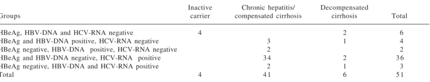
Of 51 patients with hepatitis B and C co-infection, 6 had neither HBV-DNA nor HCV-RNA detectable by PCR, of whom 4 had normal physical examination find-ings, biochemical liver function tests and imaging re-sults, and 2 had decompensated cirrhosis. Four patients were positive for HBeAg and HBV-DNA, while being negative for HCV-RNA. Two patients were positive for anti-HBe/HBV-DNA while being negative for HCV-RNA. Three out of 51 patients were positive for both HBV-DNA and HCV-RNA. In the remaining 36 patients,
only HCV-RNA were found positive. Tables I, II and III
show the status of patients with dual infection according to viral markers.
The above mentioned 4 patients who were negative for both HBV-DNA and HCV-RNA and in whom all re-sults were within normal ranges seemed to be carriers of hepatitis B, and showed no evidence of HCV replication, although they were exposed to hepatitis C virus. Two of these patients underwent liver biopsy, and their hepatitis activity index (HAI) scores were found to be 1 and 2. In fact, liver biopsy is not indicated for such patients ac-cording to the standard guidelines. These patients were those who were found to be positive for anti-HCV among 718 inactive hepatitis B carriers. As a conclusion, among the inactive hepatitis B carriers who underwent anti-HCV
Table I. Serologic markers and nucleotide positivity in the patients with dual infection.
Inactive Chronic hepatitis/ Decompensated
Groups carrier compensated cirrhosis cirrhosis Total
HBeAg, HBV-DNA and HCV-RNA negative 4 2 6
HBeAg and HBV-DNA positive, HCV-RNA negative 3 1 4
HBeAg negative, HBV-DNA positive, HCV-RNA negative 2 2
HBeAg and HBV-DNA negative, HCV-RNA positive 3 4 2 3 6
HBeAg negative, HBV-DNA and HCV-RNA positive 2 1 3
www.medigraphic.com
detection, the proportion of those found to be positive was 0.55%. During follow up, none of these patients showed elevations in the ALT and AST enzyme levels, and HBV-DNA and HCV-RNA continued to remain nega-tive. The rate of anti-delta positivity was 0.27% among inactive hepatitis B carriers.
Overall, six patients who had normal enzyme levels during the 6-month follow up period were positive for both anti-HCV and HCV-RNA but negative for HBV-DNA. Only these six patients representing 17.6% of 34 patients with HCV-RNA positive chronic hepatitis un-derwent liver biopsy, which showed minimal hepatitis in two patients, mild activity and mild fibrosis in three pa-tients, and moderate activity and moderate fibrosis in one patient. Among our 378 HBsAg negative patients moni-tored for chronic hepatitis C, 12% had persistent normal ALT enzyme serum levels, whereas only 28 HCV-RNA positive patients had chronic hepatitis accompanied by elevations in the transaminases serum levels.
The mean ALT and AST enzyme levels in the patients with chronic hepatitis accompanied by high ALT en-zyme levels were found 93 ± 97 U/L and 97 ± 130 U/L, respectively. The chronic hepatitis patients diagnosed with hepatitis B, C and delta hepatitis had mean serum ALT enzyme levels of 121 ± 99 U/L, 110 ± 63 U/L and 144 ± 90 U/L, and mean serum AST enzyme levels of 94 ± 96 U/L, 86 ± 73 U/L and 110±63 U/L, respectively. The patients with delta hepatitis had significantly higher mean ALT enzyme serum levels compared to those with dual hepatitis B and C infection (p = 0.01). The propor-tion of male patients among the chronic hepatitis/com-pensated cirrhosis patients with hepatitis B and C dual infection (81%) was significantly greater than among those with chronic delta hepatitis (p = 0.046) and chronic hepatitis C (p = 0.014) (Table IV). Inflammatory activity
was mild in 30% of the patients, moderate in 50%, and severe in 20%, with no fibrosis in 10%, stage 1 fibrosis in 30%, stage 2 in 45%, and stage 3 in 15%. There was no significant difference for the average level of fibrosis be-tween dual infection and single hepatitis B or C virus in-fection. Four out of 36 patients (11%) who were positive for HBsAg and HCV-RNA with high levels of liver en-zymes and underwent liver biopsy had cirrhosis, and this rate indicated no significant difference from that of those with single hepatitis B or C virus infection (Table V). Cir-rhosis was detected in 28.9% (11/38) of the patients with delta hepatitis; this rate was significantly higher than for those with single hepatitis B or C infection, and dual hepatitis B and C infection.
The numbers of the patients who were diagnosed with single chronic hepatitis B, single chronic hepatitis C, and delta hepatitis during the same period were 482, 378, and 38, respectively, while six patients were diagnosed with decompensated cirrhosis. Among those patients who had been screened for HBsAg and anti-HCV during the same period, 166 were HBsAg positive, 92 were anti-HCV positive, 27 were anti-delta positive decompensat-ed cirrhosis cases. The percentage of decompensatdecompensat-ed cir-rhosis in delta hepatitis was significantly higher than in the other hepatitides (p = 0.001). Clinical status of the patients are summarized in Table I.
During the follow-up period, in one of the patients who were followed up with the diagnosis of initial chron-ic hepatitis (HBV-DNA- and HCV-RNA positive), HCC was identified, and eventually decompensation devel-oped in the same patient. This patient refused transplan-tation and died. One patient was admitted with the diag-nosis of decompensated cirrhosis and presented with ter-minal hepatic insufficiency, liver transplantation was not done because the patient had no social security; and this Table II. Demographic data and nucleotide positivity in several studies concerning dual chronic hepatitis infections.
Study N Age Male % HBe (+) % HBVDNA (+) % HCVRNA (+) % Both (+) % Country
Zarski et al. (2) 2 3 4 0 8 7 3 9 100 France
Mathurin et al. (3) 3 4 4 5 8 2 4 2 5 9 France
Jardi et al. (4) 2 5 5 1 6 0 2 8 8 8 8 0 Spain
Sagnelli et al. (5) 2 7 4 6 8 5 1 9 4 4 1 9 Italy
Gaeta et al. (6) 5 9 5 0 7 5 0 3 6 2 4 8 Italy
Villa et al. (7) 3 0 3 3 7 3 2 0 100 Italy
Present study 4 1 4 7 8 1 7 1 2 8 3 5 Turkey
General 239 4 5 7 5
Table III. Comparison of the patients with dual infection with those with single agent viral infections (Mean ± SD).
Type (n) Age Female/Male ALT (U/l) AST (U/l)
B and C (36) 4 7 ± 1 2 7/29 9 3 ± 9 8 9 7 ± 132
B (38) 3 8 ± 1 2 12/26 121 ± 9 9 9 4 ± 9 6
C (38) 5 1 ± 1 1 18/20 110 ± 6 3 8 6 ± 7 3
www.medigraphic.com
patient also died. This patient was found negative for HBV-DNA and HCV-RNA. A 55 year old female patient was admitted with the complaints of diffuse joint pain, fatigue and loss of appetite for the last two weeks. On ex-aminations prior to mastectomy performed 18 months be-fore, she was positive for HBsAg, HBeAg and anti-HCV, and moderate activity and moderate fibrosis was detect-ed on liver biopsy performdetect-ed six months before. Physical examination showed tense ascites. Her laboratory results were as follows: ALT 1410 U/L, AST 1215 U/L, alkaline phosphatase 326 U/L, GGT 168 U/L, bilirubin 12.2 mg/ dL, Albumin 3.1 g/dL, and prothrombin time 19.3 sec (control, 12 sec). It was observed that her previous sero-logical results remained unchanged, HBV-DNA was posi-tive, but the HCV-RNA was negative. Anti-HAV IgM and anti-delta negativity was found. A short period of time af-ter hospitalization, she developed hepatic encephalopa-thy, and died on a hepatic coma within 10 days, despite supportive therapy. One female patient who was fol-lowed up with the diagnosis of decompensated cirrhosis and positive only for HCV-RNA also died due to hepatic insufficiency during the follow up period. One HBV-DNA positive and HCV-RNA negative male patient with an initial diagnosis of decompensated cirrhosis under-went liver transplantation.
Another HBeAg-positive patient in our study present-ed with fatigue. This patient had an ALT enzyme level of 277 U/L and AST enzyme level of 228 U/L, and showed mild activity and no fibrosis on liver biopsy. A 23-year old male patient on renal replacement therapy was posi-tive for HBsAg, HBeAg and anti-HCV, and negaposi-tive for HCV-RNA, with HBV-DNA levels being above 2000 pg/ mL. On liver biopsy, HAI score was 2 with no fibrosis.
Finally a 68-year female patient also was found
pos-itive for HBsAg, HBeAg and anti-HCV, and negative for HCV-RNA, with a HBV-DNA level of 749 pg/mL. Her liver biopsy showed an HAI score of 14 and stage 1 fibrosis.
Of the two HBV-DNA negative and HCV-RNA posi-tive patients with an initial diagnosis of chronic hepati-tis, one developed HCC and was followed for 6 years. This patient initially presented with moderate activity and moderate fibrosis with a HBV-DNA level of 678 pg/ mL, and was found positive for HCV-RNA by PCR. Lami-vudine therapy was administered for 2 years, and with vi-ral relapse after the discontinuation of lamivudine. HCV-RNA remained positive on follow-up visits. He initially presented with an alpha fetoprotein level of 27 IU/mL, which was decreased to 13 IU/mL with lamivudine thera-py, and then increased to 32 IU/mL 6 months after the discontinuation of lamivudine therapy, with no mass on imaging findings. He was a non-compliant, hard to fol-low-up patient for two years. When he presented and HCC was detected with esophageal variceal hemorrhage.
His initial platelet count of 102.000/mm3 showed
pro-gressively decrease over time, including the course of lamivudine therapy. A retrospective assessment suggest-ed that this patient actually had cirrhosis, which could not be detected by histological examination. For this pa-tient, alcohol consumption and the presence of hepatic stenosis with a body mass index (BMI) value of 31.2 kg/
m2 was considered as co-carcinogenic factors. Another
HBV-DNA negative and HCV-RNA positive patient was a female who was diagnosed when she was 53 years old. She was found asymptomatic at initial diagnosis. Her liv-er biopsy showed a HAI score of 5 with no evidence of fi-brosis. During the four years of follow up, her serum ALT enzyme level remained constantly below 1.3 the
border-Table IV. Histological features of the dual and single viral infections in several studies (Mean ± SD).
Study P Cirrhosis B Cirrhosis Cirrhosis
(Reference) Fibrosis B and C Fibrosis C Fibrosis B and C (%) C (%) B (%) P
Zarski et al. (2) 1.8 ± 1.1 1.3 ± 1.2 NA NS 43.5 8.7 S*
Mathurin et al. (3) 2.2 ± 1.3 1.8 ± 1.1 NA NS 25.0 8.8 S*
Sagnelli et al. (5) 2.1 ± 1.2 1.5 ± 1.1 1.9 ± 1.2 S* 18.5 8.6 14.0 NS
Present study 1.8 ± 1.4 1.4 ± 1.2 1.7 ± 1.1 NS 11.1 6.7 10.1 NS
NA: Not available, S: Significant, p < 0.05; NS: Not significant; * B and C vs C.
Table V. The distribution of the clinical pictures in the dual, single, delta and triple infections.
Inactive carrier Chronic hepatitis/
Compensated cirrhosis Decompensated cirrhosis Total
B and C 4 4 1 6 5 1
B 712 482 166 1360
Delta 2 3 8 2 7 6 7
C 378 9 2 470
B, C and Delta 2 2
www.medigraphic.com
line limit of normal. Mean 5-year survival was 92%. Al-though it was 98 % when only patients with chronic hep-atitis were considered. This survival rate was 75% for dual hepatitis B and delta infection, which was signifi-cantly lower compared with dual hepatitis B and C infec-tion (p = 0.01).
In 2 patients a triple infection (e.g. HBsAg, anti-delta and anti-HCV positivity) was seen during the 15 years period. In both of them HBV-DNA and HCV-RNA nega-tivity and HDV-RNA posinega-tivity were found, so the domi-nant infection was delta hepatitis. In one of these pa-tients, serum ALT and AST enzyme levels were found 240 U/L and 306 U/L and in the other 50 U/L and 41 U/ L, respectively.
Discussion
Co-existence of the parenterally-transmitted hepatitis viruses, i.e. the occurrence of multiple infections, is not rare. Coexistence of hepatitis B and C viruses is called co-infection or dual infection, and these viruses that usu-ally show a casual co-existence suppress each other’s rep-lication.3,4 The presence of hepatitis B virus is essential
for delta virus to cause infection. Rarely, all three viruses may occur together in the same patient, a condition called triple infection.
In the present study, we mainly focused on the hep-atitis B and C co-infection, but also compared the pa-rameters with those of single hepatitis B, C and delta infection.
Dual infection is typically characterized by HBsAg and anti-HCV positivity. However, several studies have reported a silent hepatitis B infection in some hepatitis C patients and the presence of HBV-DNA in their liver and
sometimes serum, despite HBsAg negativity in serum.8-10
In a study from Japan, 65 patients with HBsAg negative hepatitis C were evaluated, of whom 34 had HBV-DNA in the liver tissue.8 Of these patients, 23% and 28% were
found to be positive for anti-HBs and anti-HBc, respec-tively. Silent hepatitis B infection has been reported to be more frequent in genotype 1b.
In a study published in Korea, 70% of the patients di-agnosed with HCC had the markers of hepatitis B tion, whereas only 8% had those of hepatitis C
infec-tion.9 However, 18% of the HCC patients with HCV
vire-mia were positive for HBsAg, while 37.5% were negative for HBsAg with HBV-DNA positivity detected by PCR and most of them were anti-HBs/anti-HBc positive. Therefore, it has been concluded that the presence of HB-sAg positivity is not an absolute finding for the patients with HBV/HCV co-infection accompanied with HCV viremia to be diagnosed with dual infection, and at least two times more patients had findings of a previous HBV infection accompanied by HBV-DNA positivity. Taking up the subject in this point of view and considering that 5% of our population are HBsAg positive and additional
30% are anti-HBc/anti-HBs positive, it can be suggested that the patients we considered within the scope of co-in-fection were those who had over co-inco-in-fection, there might be an even higher number of patients with silent co-infection than the number of these patients, and the reason that co-infection was not observed as a more mor-bid condition than single hepatitis B and C virus infec-tions might be the result of omitted patients with silent co-infection.
Sagnelli et al. have detected anti-HBc positivity in 88 of 205 patients (47.6%) who are anti-HCV/HCV-RNA positive and HBsAg/anti-HBs negative. Of the pa-tients who are anti-HBc positive, 68% are anti-HBe pos-itive, and 36.3% HBV-DNA positive. Histological liver damage has been reported to be higher in the anti-HBc positive patients than in the anti-HBc negative patients,
independently from the HBV-DNA status.10 It has been
reported that, in proportion of these patients, HBV-DNA was lost from circulating blood as a result of the sup-pression of HBV by HCV, but its replication still con-tinued in the liver.
Cacciola et al. have detected HBV-DNA sequences in the liver tissue in 33% of 200 HBsAg negative chronic hepatitis C patients. Cirrhosis has been detected in 33% of 66 patients with HBV-DNA sequences and in 19 % of 134 patients without HBV-DNA sequences (p < 0.05). It is interesting that the average rate of cirrhosis (27%) ob-served in this study is aproximatly four times the rate that has been previously observed in chronic hepatitis C.17
Sagnelli et al. have found the mean fibrosis score to be significantly greater in the patients with dual infection
than in those with single infection.8 Similarly, in our
study, the fibrosis score tended to be greater in the pa-tients with dual infection than in those with single infec-tion, although the difference was not statistically signifi-cant. Several previous studies have also reported non-significant differences2,3,18,19(Table V).
In the study by Sagnelli, the fibrosis score in HCV-RNA negative dual infection has been found to be great-er than in HCV-RNA positive dual infection. In this case, the aggressivity of HBV, which becomes free from the suppression of HCV, may play a role. A study has report-ed that despite the loss of HCV-RNA from circulating blood depending on the suppression of HBV in dual in-fection, replication was more aggressively in the liver.20
www.medigraphic.com
ESTE DOCUMENTO ES ELABORADO POR MEDIGRA-PHIC
the dual positivity of HBV-DNA and HCV-RNA in two patients, and Hepatitis C was preponderant in our series. Only 12% of the patients were positive for only HBV-DNA (of whom 4 being HBsAg positive), whereas 70% were positive for only HCV-RNA. The percentage of cir-rhosis patients was 28.8% in the series mentioned above, whereas it was 11.1% in our series. In another study pub-lished in France, among 33 patients with dual infection, 14 were reported to have HBV replication, and 19 to have HCV replication, with no data given for either of
them, the HBV-DNA and HCV-RNA patients.2 The results
of that study were similar to those of our series in terms of viral markers. In a study from Italy, only 6 of 30 pa-tients with dual infection were found positive for HBV-DNA.7 In that series, the dominant virus was HCV, as
ob-served in our series. In another Italian study, interesting-ly, 35 HBsAg positive patients were identified among 82 anti-HCV positive patients, and HBV genome was detect-ed in the liver tissue in 24 patients. Thereby, more than half of the anti-HCV positive patients appear to have
dual infection, representing an unusual rate.20 In two
studies from Taiwan, dual infection was reported in a substantial proportion (10-20%) of the patients diag-nosed with fulminant hepatitis.22,23 In our series, we did
not observe such patients. We could have erroneously di-agnosed the female patient who had acute hepatitis en-zyme levels and died after a fulminant course with fulmi-nant hepatitis, any data from her examinations performed 18 months ago and biopsy performed 6 months ago was not available. However, it is known that sometimes such exacerbations during the course of chronic hepatitis B may result in a fulminant course. We don’t know whether accompanying anti-HCV positivity in this patient had an unfavorable effect on outcome.
In a review published by Liaw,24 the frequency of HCV
positivity among the HBsAg carriers through the world has been reported to be more than 10%.
Zarski et al. detected HBsAg in 23 (1.8%) of the 1248 hepatitis C patients with higher transaminase levels (all
HCV-RNA positive).2 Four patients were HBeAg positive
and 19 were anti-HBe positive, and in nine patients HBV-DNA was detected and in fourteen was not. In our series, the rate of the patients with HBsAg positive among chronic hepatitis B patients with higher enzyme levels was substantially higher (9.5%) than the men-tioned rate levels.
In the above mentioned series, the percentage of HBV-DNA positivity was 0.7% which was similar to the one of our series. The explanation of the first finding is not difficult, because the prevalence of the HBsAg posi-tivity in our country is five times higher than that in France. In the above mentioned study, no relationship was found between the type of HCV genotype (1 or non-1) and HBV-DNA positivity and negativity. Although more than half of the patients enrolled in this study were infected with HCV genotype non-1, all the
pa-tients included in our series were HCV genotype 1 and this situation supports the thesis that such a relation-ship does not exist.
When dual HBV and HCV infection is compared with dual HBV and HDV infection, it was seen that the second dual infection is more aggressive than the first one. Among 73 patients admitted with dual HBV and HDV in-fections between the years of 1991 and 2006, decompen-sated cirrhosis and HCC was found at the time of diagno-sis in 27 and 6 patients, respectively. However, only in 6 patients among 51 patients who had dual B and C infec-tion patients (p = 0.013), decompensated cirrhosis was determined and there was no patient admittance with the diagnosis of HCC (p = 0.041).
In the first patient who was on renal replacement ther-apy severe inflammation activity and severe fibrosis was identified. The other patient had inflammatory activity and moderate fibrosis. It appears that the dominant infec-tion was delta infecinfec-tion in triple infecinfec-tions.
Finally, co-positivity of HBsAg and Anti-HCV in our country is not a common situation. The rate among inac-tive hepatitis B carriers, chronic viral hepatitis/compen-sated cirrhosis and viral decompenhepatitis/compen-sated cirrhosis was found as 0.55%, 4.4% and 2.1%, respectively. Dominant infection in ¾ of the patients was hepatitis C. Clinical and histological properties of the cases with dual B and C infection showed no significant differences compared to the single infections. Regarding the prognosis, no sig-nificant differences were found between such dual and single B or C infection. However, dual infection with hepatitis B virus and delta virus is a significantly more severe condition than the dual infection with hepatitis B and hepatitis C viruses.
References
1. Stroffolini T, Sagnelli E, Mele A, Craxi A, Almasio P and Italian Hospitals Collaborating Group. The etiology of Chronic Hepati-tis in Italy: Results from a multicentre national study. Dig Liver Dis 2004; 36: 829-33.
2. Zarski J-P, Bohn B, Bastie A, et al. Characteristics of patients with dual infection by hepatitis B and C viruses. J Hepatol 1998; 28: 27-33.
3. Mathurin P, Thibault V, Kadidja, et al. Replication status and histological features of patients with triple (B, C, D) and dual (B, C) hepatic infections. J Viral Hep 2000; 7: 15-22.
4. Jardi R, Rodriguez F, Buti M, et al. Role of hepatitis B, C, and D viruses in dual and triple infection: Influence of viral genotypes and hepatitis B precore and basal core promoter mutations on viral replicative interference. Hepatology 2001; 34: 404-10. 5. Sagnelli E, Pasquale G, Coppola N, et al. Influence of chronic
infection with hepatitis B and C virus on liver histology. Infection 2004; 32: 144-8.
6. Gaeta GB, Stornaiuolo G, Precone DF, et al. Epidemiological and clinical burden of chronic hepatitis B virus/hepatitis C virus infection. A multicenter Italian study. J Hepatol 2003; 39: 1036-41.
www.medigraphic.com
2973-7.8. Fukuda R, Ishimura N, Nigaki M, et al. Serologically silent hepa-titis B virus coinfection in patients with hepahepa-titis C virus associ-ated chronic liver disease: Clinical and virological significance. J Med Virol 1999; 58: 201-7.
9. Lee D-S, Huh K, Lee E-H, Lee D-H, Hong K-S, Sung Y-C. HCV and HBV coexist in HBsAg-negative patients with HCV viraemia: Possibility of coinfection in these patients must be considered in HBV-high endemic area. J Gastroenterol Hepatol 1997; 12: 855-61.
10. Sagnelli E, Coppola N, Scolastico C, Mogavero AR, Filippini P, Piccinino F. HCV genotype and silent HBV coinfection: Two main risk factors for a more severe liver disease. J Med Virol 2001; 64: 350-5.
11. Farci P. Delta hepatitis: an update. J Hepatol 2003; 39: 212-9. 12. Hadziyannis SJ. Hepatitis delta: an overview. In Rizzetto M, Purcell
RH, Gerin JL, editors. Viral hepatitis and Liver disease. Turin, Italy: Minerva Medica; 1997: 283-9.
1 3 . Kubo S, Nishiguchi S, Hirohashi K, et al. Clinical signifi-cance of prior hepatitis B virus infection in patients with hepatitis C virus-related hepatocellular carcinoma. Cancer 1999; 86: 793-8.
14. Ruiz J, Sangro B, Cuende JI, et al. Hepatitis B and C viral infec-tions in patients with hepatocellular carcinoma. Hepatology 1992; 16: 637-41.
15. Shiratori Y, Shiina S, Zhang PY, et al. Does dual infection by hepatitis B and C viruses play an important role in the pathogen-esis of hepatocellular carcinoma in Japan? Cancer 1997; 80:
2060-7.
16. Chomcynski P, Sacci N. Single step method of RNA isolation by acid-guanidium thiocyanate-phenol-chloroform axtraction. Anal Biochem 1984; 162: 156-9.
17. Cacciola I, Pollicino T, Squadrito G, Cerenzia G, Orlanda ME, Raimondo G. Occult hepatitis B virus infection in patients with chronic hepatitis C liver disease. N Engl J Med 1999; 341: 22-6. 18. Pontisso P, Ruvoletto MG, Fattovich G, et al. Clinical and viro-logical profiles in patients with multiple hepatitis B virus infec-tion. Gastroenterology 1993; 105: 1529-33.
19. Villari D, Pernice M, Spinella S, et al. Chronic hepatitis in patients with active hepatitis B virus and hepatitis C virus combined infec-tions: A histological study. Am J Gastroenterol 1995; 90: 955-8. 20. Dries V, Von Both I, Muller M, et al. Detection of hepatitis C in parafin-embedded liver biopsies of patients negative for viral RNA in serum. Hepatology 1999; 29: 223-9.
21. Squadrioto G, Orlando ME, Pollicino T, et al. Virological pro-files in patients with chronic hepatitis C and overt or occult HBV infection. Am J Gastorenterol 2002; 97: 1518-23.
22. Wu JC, Chen CL, Hou MC, Chen TZ, Lee SD, Lo KJ. Multiple viral infection as the most common cause of fulminant and subfulminant viral hepatitis in an endemic area for hepatitis B: Application and limitation of the polymerase chain reaction. Hepatology 1994; 19: 836-40.
23. Chu CM, Sheen IS, Liaw YF. The role of hepatitis C virus in fulminant viral hepatitis in an endemic area of hepatitis A and B. Gastroenterology 1994; 107: 189-95.