Annals of Hepatology 7(2) 2008: 168-173
168
www.medigraphic.com
Annals of Hepatology 2008; 7(2): April-June: 168-173
Annals of Hepatology
Case Report
Intraductal oncocytic papillary neoplasm
of the liver: Case and review of a rare variant
James H. Tabibian;1,2 Charles R. Lassman;3 Daniel J. Margolis;4 Carmen Landaverde;1,2 Ronald W. Busuttil;1
Francisco A. Durazo1,2
1 Dumont-UCLA Liver Transplant Center.
2 UCLA Departments of Internal Medicine, Division of Digestive
and Liver Diseases.
3 Pathology.
4 Radiology. Los Angeles, CA, USA.
Address for correspondence:
James H. Tabibian, 1440 Veteran Ave. # 601. Los Angeles, CA 90024
Phone: (310) 849-9488 Fax: (310) 206-1269 [email protected]
Conflicts of interest, Declarations, Copyright transfer withholdings: none
Abbreviations:
Intrahepatic cholangiocarcinoma (ICCA), cholangiocarcinoma (CCA), intraductal oncocytic papillary neoplasm (IOPN), orthotopic liver transplantation (OLT), intraductal papillary mucinous neoplasms of the pancreas (IPMN), computerized tomography (CT), international normalized ratio (INR), partial thromboplastin time (PTT), alpha-fetoprotein (AFP), carcino-embryonic antigen (CEA), carbohydrate antigen 19-9 (CA19-9), pulmonary embolism (PE), magnetic resonance (MR)
Manuscript received and accepted: 29 April and 7 May 2008 Abstract
Intrahepatic cholangiocarcinoma (ICCA) comprises 10% of all cholangiocarcinoma (CCA). It can be divid-ed into three macroscopic subtypes, the least common of which is characterized by intraductal growth and be-lieved to be more amenable to good outcomes with sur-gical resection compared to other ICCA. Recently, the rare finding of oncocytic differentiation has been de-scribed in this subtype and termed «intraductal onco-cytic papillary neoplasm» (IOPN), but it remains un-clear if the presence of oncocytes confers a different tu-mor behavior. We present the eighth reported case of IOPN, which to our knowledge, is the first such case that, due to its location and vascular compromise, re-quired orthotopic liver transplantation (OLT). This case adds to the little that is known about the behavior of IOPN and supports the observation that resection, or OLT when resection is not possible, is a valid treat-ment option.
Key words: Cholangiocarcinoma, intrahepatic bile ducts, liver transplantation, oncocyte.
Introduction
Cholangiocarcinoma (CCA) is the second most com-mon neoplasm of the human liver after hepatoma.1-4
In-trahepatic cholangiocarcinoma (ICCA) is a primary ade-nocarcinoma arising from the epithelium of the second or more distal branches of the intrahepatic ducts and is hence also termed peripheral CCA.4-6 It comprises
ap-proximately 10% of all CCA, while the remaining 90% originate from the extrahepatic or hilar ducts.4,6-8
Surgical resection is the primary treatment for ICCA.7,9
Resectable ICCAs are divided into 3 fundamental macro-scopic subtypes: 1) mass-forming, 2) periductal-infiltrat-ing, and 3) intraductal growth.10,11 Intraductal ICCA is the
least common of these subtypes, comprising only 8-14% of all ICCA cases.10-12 It is characterized by papillary
growth in the bile duct lumen with no or micro-invasion beyond the bile duct walls, and is believed to be analo-gous to intraductal papillary mucinous neoplasms of the pancreas (IPMN).13-16 The few available reports on
intra-ductal ICCA have suggested less aggressive tumor be-havior and the potential for better outcome with resec-tion compared to other ICCAs despite the typically large tumor size (> 5 cm) at presentation.17 In 2002, the finding
of oncocytic differentiation was described as a variant of this uncommon tumor subtype; it remains to be seen if the presence of oncocytes alters the course of disease.17
We present a patient who was diagnosed with a large intraductal ICCA with the rare finding of oncocytic dif-ferentiation, hence an intraductal oncocytic papillary neoplasm (IOPN) of the liver. This is the first case, to our knowledge, of bilobar tumor which was treated with orthotopic liver transplantation (OLT). We hope this case will add to the little known about intraductal ICCA, and more specifically, IOPN.
Case report
The patient is a 50-year-old Japanese-American fe-male who presented to her primary care physician with
Artemisa
www.medigraphic.com
several days of progressive weakness, anorexia, non-pro-ductive cough, right shoulder and pleuritic chest pain, and lower extremity edema. She was a smoker, non-drinker with no past medical history except Grave’s dis-ease which was treated with thyroid radioablation 15 years prior. An outpatient chest X-ray revealed bilateral foci of opacification, while the computerized tomogra-phy (CT) of the chest and abdomen revealed extensive enhancing liver lesions, the largest involving the left and right lobes and measuring 10.3 cm in greatest dimension, large air fluid levels in the right lobe of the liver, left por-tal vein and right and middle hepatic vein thrombi ex-tending into the inferior vena cava, multiple pulmonary nodules, a large lingular mass with air fluid levels, and moderate right-sided pleural effusion. The patient was scheduled for liver biopsy to aid in diagnosis, but her condition rapidly deteriorated to where she could not care for herself anymore, thus requiring hospitalization.
She was an alert and oriented, thin women in mild res-piratory distress. Vital signs were blood pressure 120/80 mm/Hg, pulse 90 beats per minute, temperature 36.5 ºC, respiratory rate 20 breaths per minute, and arterial oxy-gen saturation 95% on 2 L of oxyoxy-gen by nasal cannula. Head and neck examination was unremarkable, includ-ing absence of lymphadenopathy. Cardiopulmonary ex-amination was significant for decreased breath sounds over the lower lung fields, particularly the right. The ab-domen was somewhat distended and tympanitic, but without tenderness, rebound, or hepatosplenomegaly. The remainder of the physical examination was unre-markable, except for 3+ pitting edema involving the bi-lateral lower extremities. Pertinent laboratory results in-cluded white blood cells 38.0*103/μL with 81%
neutro-phils, hemoglobin 9.4 g/dL, and platelets 268*103/μcL.
Chemistries were significant for a sodium of 131 mmol/L and creatinine of 1.2 mg/dL. Liver tests were remarkable for alanine aminotransferase of 49 U/L (normal 5-50 U/L) and alkaline phosphatase (ALK) of 484 U/L (normal 35-110 U/L). International normalized ratio (INR) was 1.3 and partial thromboplastin time (PTT) was low at 22.4 (normal 24.6 to 32.4 seconds), and a hypercoagulability panel revealed an elevated homocysteine level at 13.1
μmol/L (normal 3.3-10.2 μmol/L) but negative Factor V R506Q mutation, Prothrombin G20210A mutation, and Antiphospholipid antibodies as well as normal Anti-thrombin III activity. Hepatitis A, B, and C serologies were negative. Tumor markers revealed normal Alpha-fe-toprotein (AFP), Carcino-embryonic antigen (CEA), and Carbohydrate antigen 19-9 (CA19-9).
Given the patient’s clinical presentation and imaging data, she was empirically started on antibiotics as well as anticoagulation due to concern for possible pulmonary embolism (PE). On the day after admission, the patient underwent left lobe liver biopsy with placement of two drains. Purulent fluid was noted to be draining, and cul-ture revealed viridans Streptococci. Based on the biopsy
material, a diagnosis of «oncocytic papillary carcinoma of the biliary tract» was made. Given the clinical infor-mation, it was concluded that the patient had a primary intrahepatic biliary neoplasm complicated by infection and abscess formation, with consequent septic emboli to the lungs.
With continued antibiotic therapy and anticoagula-tion, the patient experienced some improvement in ener-gy, appetite, cough, shoulder and pleuritic chest pain, and lower extremity edema. Repeat CT scan one week af-ter admission revealed inaf-terval development of a cavi-tary lesion in the right upper lobe likely representing pneumonia or a septic embolus, a small left-sided pleural effusion, an increase in the size of the right-sided pleural effusion, and PE in the right upper lobe pulmonary ar-tery. The lingular lesion and right lobe cavitary fluid-containing lesions were unchanged, as were the portal and hepatic venous defects (Figure 1a). The patient re-mained in the hospital for a total of 16 days, after which she was discharged home on subcutaneous enoxaparin injections and antibiotics through a peripherally-inserted central venous catheter, with hepatology, surgery, and in-fectious disease follow-up.
Abdominal magnetic resonance (MR) angiogram ob-tained as an outpatient showed multiple thrombosed venous vessels and a stenotic left hepatic vein. It was felt that the patient’s hepatic blood supply would be exces-sively compromised by performing a partial hepatecto-my. Despite the large size of the tumor, recognizing its slow growth rate and the low likelihood of metastasis, the possibility of liver transplantation was raised.
The patient was referred to our liver transplantation center. On presentation, she had normal vital signs and was in no acute distress. Physical examination was re-markably normal except for a large, non-tender mass in the right upper quadrant and epigastrium that moved with deep inspiration but did not produce a hepatic bruit. CBC complete blood count and chemistries were normal. Liver tests were remarkable only for (ALK) of 205 U/L. AFP, CEA, and CA-19-9 tumor markers were not elevat-ed. The patient underwent MR of the abdomen with and without contrast, which revealed one enhancing cavitary mass measuring approximately 11.3 by 11.5 cm span-ning the left and right lobes and resolution of the other liver lesions previously seen on CT. The right hepatic vein was thrombosed, the left hepatic vein was stenotic, the right portal vein was compressed, the left portal vein was either compressed by the mass or thrombosed, and there was a small persistent IVC thrombus, as on prior im-aging (Figures 1b, 1c, 1d).
www.medigraphic.com
complicated by acute renal failure which resolved with-out requiring dialysis. Gross examination of the speci-men revealed an approximately 13 cm mass which ob-scured the porta hepatis (Figure 2a). The mass consisted of a number of closely-approximated, well-circumscribed nodules which appeared to be intraluminal. Histologic examination revealed dilated ducts filled with a papil-lary neoplasm composed of thin fibrous septae lined by
multiple layers of cells punctuated by sharply-defined round lumens (Figure 2b). The cells were large and had abundant granular, esosinophilic cytoplasm characteris-tic of oncocycharacteris-tic differentiation (Figure 2b). The nuclei were round with mild to moderate atypia. The ducts were lined either by similar appearing cells or by flattened bile duct epithelial cells. Multiple sections of the inter-face between the tumor and the surrounding liver
paren-Radiological studies:
A
B
Figure 1a. Axial computed tomography demonstrating necrotic/ infected portion of large liver mass with fluid level (long white arrow), junction of compressed left hepatic vein and inferior vena cava (black arrow), and compressed vessels that are likely portal (double white arrows).
Figure 1b. Gadopentetate dimeglumine (contrast) enhanced 2D gradient T1 magnetic resonance image demonstrating large liver tumor, thrombosed inferior vena cava (black arrow), and stenotic or compressed left hepatic vein.
D
Figure 1d. Magnetic resonance angiography demonstrating thrombosed inferior vena cava (white arrow) and non-visualiza-tion of the left portal system.
C
www.medigraphic.com
chyma demonstrated hyalinized stroma with small foci of microinvasive tumor. There was no extension into the ad-jacent liver parenchyma. Surrounding bile ducts were di-lated and frequently contained neurophils and necrotic debris. Immunohistochemistry was performed, and the re-sults, while nonspecific, were characteristic of intraduc-tal oncocytic papillary neoplasm of the bile duct. The pa-tient was discharged 18 days after OLT, and at 6-months post-OLT follow-up continues to do well, without recur-rence of malignancy.
Discussion
Intraductal ICCA is the newest-described macroscop-ic subtype of ICCA.10,11 Several conditions involving
chronic inflammation of the bile ducts are believed to predispose to ICCA, including PSC, hepatotropic trema-todal infection, and hepatolithiasis, although most pa-tients, including those with the intraductal subtype, have no identifiable risk factors.9,18 The average age at
presen-tation is 55 years, and patients typically do not present until the disease is relatively advanced.9,18
Surgical resection, which is the primary treatment for ICCA, is indicated for patients without vascular in-vasion and extrahepatic metastasis and if the tumor is not too large to compromise liver function if resected, although post-operative outcome remains poor due to recurrence of disease, with median survival being only 3 years.7,9 Post-operative survival rates may, however,
be more favorable in the cohort of patients with early-staged tumor, well-differentiated tumor, absence of physical findings, presence of mucobilia, negative his-tologic margins, and the intraductal ICCA sub-type.9,12,18,19
In 2002 Martin et al. reported 3 cases and reviewed another 39 cases of intraductal ICCA reported in the world literature. Since then, there have been several more case reports and series with a total of over 200 pa-tients.13,15,20-26 In the cases where there was sufficient
in-formation, follow-up was usually on the order of 1-3 years, and nearly all patients were doing well and dis-ease-free at the time of follow up. Given the encouraging survival rates, it has been hypothesized that the intradu-catal subtype of ICCA has good prognostic implications over the mass-forming & periductal-infiltrating sub-types.16,27
Comprising only a small fraction of intraductal ICCA is the variant containing oncocytic differentiation, IOPN. The term oncocyte was coined in 1931 by Hamperl, refer-ring to epithelial cells with abundant, brightly eosino-philic and granular cytoplasm, and high numbers of mito-chondria.28 These cells occur in a variety of organs, both
in benign as well as malignant tumors.28,29 Oncocytic
dif-ferentiation in the biliary tract was first described by Wolf et al. in 1992 in a cystadenocarcinoma.30 The first
report of IOPN was not until 2002, and since then only 7 of all the cases of intraductal ICCA in the English lan-guage literature were of the IOPN variant.13,17,20,22,25 Our
patient represents the 8th known case with this rare
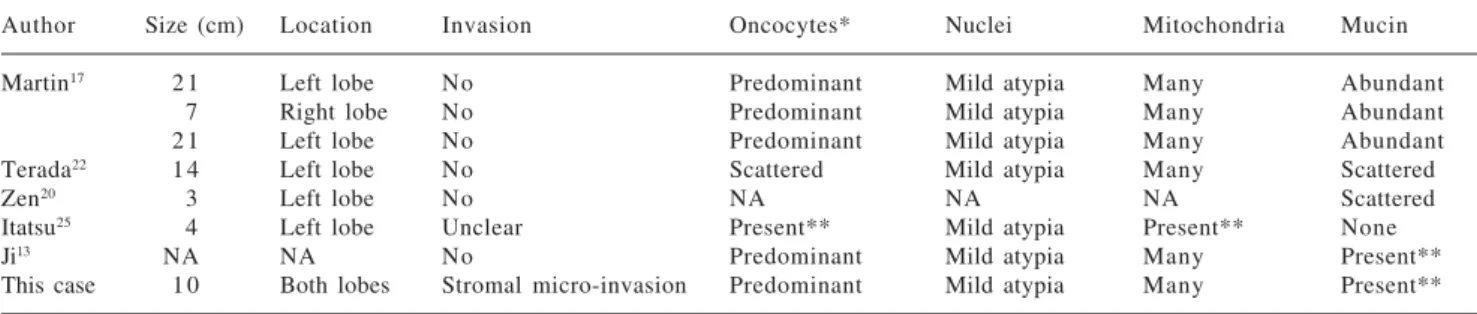
histo-logic finding. A synopsis of all 8 cases in chronohisto-logical order is provided in tables 1a and 1b.
Although there are few cases and insufficient follow up from which to draw firm conclusions, it appears there is little, if any, clinical significance to oncoyctic differ-entiation in intraductal ICCA. The biologic behavior and prognosis of IOPN seems similar to that of intraductal ICCA, in whom the 5-year survival after resection is near Figure 2a. Section of gross hepatectomy specimen demonstrating
well-circumscribed tumor nodules in the center of the liver.
A
B
www.medigraphic.com
ESTE DOCUMENTO ES ELABORADO POR MEDI-GRAPHIC
80%.16,27 In our case, because resection was not an
op-tion, OLT was performed. This has not been done previ-ously in cases of IOPN, but was deemed appropriate in our patient given the encouraging results with resection in the literature and the patient’s overall wellbeing. At her 6-month post-operative follow-up appointment, the patient was feeling well and had no evidence of tumor re-currence. Her long term outcome remains to be seen, al-though based on previous reports, it is expected to be fa-vorable.
In conclusion, intraductal ICCA is an uncommon sub-type of cholangiocarcinoma that includes a rare variant, IOPN. Similar to intraductal ICCA without oncocytic dif-ferentiation, long-term survival or cure may be achieved with complete resection of IOPN. In cases where resec-tion is not possible due to tumor size or bilobar tumor, we believe OLT is a valid option given the slow growth and minimal metastasis of IOPN. Accumulation of future data on this variant will help further characterize its clini-cal features, treatment, and prognosis.
References
1. Harrison LE, Fong Y, Klimstra DS, Zee SY, Blumgart LH. Surgi-cal treatment of 32 patients with peripheral intrahepatic cholangiocarcinoma. Br J Surg 1998; 85(8): 1068-70. 2. Okuda K. Primary liver cancer. Quadrennial review lecture. Dig
Dis Sci 1986; 31(9 Suppl): 133S-146S.
3. Primary liver cancer in Japan. Clinicopathologic features and results of surgical treatment. Liver Cancer Study Group of Japan.
Ann Surg 1990; 211: 277-287.
4. Edmondson HA. Tumors of the liver and intrahepatic bile ducts.
Atlas of tumor pathology. Sec 7, fasc 25. Washington, DC: Armed Forces Institute of Pathology, 1958.
5. Soyer P, Bluemke DA, Reichle R, Calhoun PS, Bliss DF, Scherrer A, Fishman EK. Imaging of intrahepatic cholangiocarcinoma: 1. Peripheral cholangiocarcinoma. AJR Am J Roentgenol 1995; 165(6): 1427-31.
6. Ros PR, Buck JL, Goodman ZD, Ros AM, Olmsted WW. Intrahe-patic cholangiocarcinoma: radiologic-pathologic correlation.
Radiology 1988; 167(3): 689-93.
7. Chen MF. Peripheral cholangiocarcinoma (cholangiocellular car-cinoma): clinical features, diagnosis and treatment. J Gastroenterol Hepatol 1999; 14(12): 1144-9.
8. Liver Cancer Study Group of Japan. The General Rules for the Clinical and Pathological Study of Primary Liver Cancer. 3rd edn. (in Japanese). Tokyo: Kanehara, 1992.
9. Gores GJ. Liver mass lesions. In: Hauser SC, Pardi DS, Poterucha JJ, eds. Mayo Clinic Gastroenterology and Hepatology Board Review. Rochester: Scientific Publications, 2006.
10. Liver cancer study group of Japan. Classification of Primary Liver Cancer. Tokyo: Kanehara-Shuppan, 1997: 6-7.
11. Yamamoto J, Kosuge T, Shimada K, Takayama T, Yamasaki S, Ozaki H, Makuuchi M. [Intrahepatic cholangiocarcinoma: pro-posal of new macroscopic classification]. Nippon Geka Gakkai Zasshi 1993; 94(11): 1194-200.
12. Leong TY, Leong AS-Y. Prognostication in intrahepatic cholangiocarcinoma. Advances in Anatomic Pathology 2006; 13(2): 99-100.
13. Ji Y, Fan J, Zhou J, Wang BS, Liu HB, Wu ZW, Tan YS. Intraduc-tal papillary neoplasms of bile duct. A distinct entity like its counterpart in pancreas. Histol Histopathol 2008; 23(1): 41-50. 14. Shibahara H, Tamada S, Goto M, Oda K, Nagino M, Nagasaka T, Batra SK, Hollingsworth MA, Imai K, Nimura Y, Yonezawa S. Pathologic features of mucin-producing bile duct tumors: two histopathologic categories as counterparts of pancreatic intra-ductal papillary-mucinous neoplasms. Am J Surg Pathol 2004; 28(3): 327-38.
Table 1a. Patient data.
Author Age Sex Presenting Sx Surgical treatment Outcome
Martin17 4 6 M RUQ Pain & Rigors Left & caudate lobectomy Alive at 36 months despite local recurrence
5 0 M Vague abdominal pain Left lobectomy Free of disease at 18 months
3 8 M Painless jaundice Right lobectomy Free of disease at 24 months
Terada22 6 3 M Abdominal pain, jaundice Left lobectomy Free of disease at 30 months
Zen20 8 3 M RUQ pain Left lobectomy Free of disease at 15 months
Itatsu25 6 5 F NA Left lobectomy NA
Ji13 NA NA NA Resection NA
This case 5 0 F Fatigue, edema, PE OLT Free of disease at 6 months
Table 1b. Tumor data.
Author Size (cm) Location Invasion Oncocytes* Nuclei Mitochondria Mucin
Martin17 2 1 Left lobe No Predominant Mild atypia Many Abundant
7 Right lobe No Predominant Mild atypia Many Abundant
2 1 Left lobe No Predominant Mild atypia Many Abundant
Terada22 1 4 Left lobe No Scattered Mild atypia Many Scattered
Zen20 3 Left lobe No NA NA NA Scattered
Itatsu25 4 Left lobe Unclear Present** Mild atypia Present** None
Ji13 NA NA No Predominant Mild atypia Many Present**
This case 1 0 Both lobes Stromal micro-invasion Predominant Mild atypia Many Present**
www.medigraphic.com
15. Zen Y, Fujii T, Itatsu K, Nakamura K, Minato H, Kasashima S, Kurumaya H, Katayanagi K, Kawashima A, Masuda S, Niwa H, Mitsui T, Asada Y, Miura S, Ohta T, Nakanuma Y. Biliary papillary tumors share pathological features with intraductal papillary muci-nous neoplasm of the pancreas. Hepatology 2006; 44(5): 1333-43. 16. Kim HJ, Kim MH, Lee SK, Yoo KS, Park ET, Lim BC, Park HJ, Myung SJ, Seo DW, Min YI. Mucin-hypersecreting bile duct tumor characterized by a striking homology with an intraductal papillary mucinous tumor (IPMT) of the pancreas. Endoscopy
2000; 32(5): 389-93.
17. Martin RC, Klimstra DS, Schwartz L, Yilmaz A, Blumgart LH, Jarnagin W. Hepatic intraductal oncocytic papillary carcinoma.
Cancer 2002; 95(10): 2180-7.
18. Jan YY, Yeh CN, Yeh TS, Hwang TL, Chen MF. Clinicopatho-logical factors predicting long-term overall survival after hepate-ctomy for peripheral cholangiocarcinoma. World J Surg 2005; 29(7): 894-8.
19. Jarnagin WR, Fong Y, DeMatteo RP, Gonen M, Burke EC, Bodniewicz BS J, Youssef BA M, Klimstra D, Blumgart LH. Staging, resectability, and outcome in 225 patients with hilar cholangiocarcinoma. Ann Surg 2001; 234(4): 507-17; discus-sion 517-9.
20. Zen Y, Fujii T, Itatsu K, Nakamura K, Konishi F, Masuda S, Mitsui T, Asada Y, Miura S, Miyayama S, Uehara T, Katsuyama T, Ohta T, Minato H, Nakanuma Y. Biliary cystic tumors with bile duct communication: a cystic variant of intraductal papillary neoplasm of the bile duct. Mod Pathol 2006; 19(9): 1243-54. Epub 2006 Jun 2.
21. Güllüoglu MG, Ozden I, Poyanli A, Cevikbas U, Ariogul O. Intraductal growth-type mucin-producing peripheral
cholangiocarcinoma associated with biliary papillomatosis. Ann Diagn Pathol 2007; 11(1): 34-8.
22. Terada T, Taniguchi M. Intraductal oncocytic papillary neo-plasm of the liver. Pathol Int 2004; 54(2): 116-23.
23. Chen TC, Nakanuma Y, Zen Y, Chen MF, Jan YY, Yeh TS, Chiu CT, Kuo TT, Kamiya J, Oda K, Hamaguchi M, Ohno Y, Hsieh LL, Nimura Y. Intraductal papillary neoplasia of the liver associ-ated with hepatolithiasis. Hepatology 2001; 34(4 Pt 1): 651-8. 24. Neto AG, Dainiak C, Qin L, Salem RR, Jain D. Intraductal
papil-lary cholangiocarcinoma associated with von Meyenberg com-plexes: a case report. Dig Dis Sci 2007; 52(10): 2643-5. Epub 2007 Mar 30.
25. Itatsu K, Fujii T, Sasaki M, Zen Y, Nakanuma Y. Intraductal papillary cholangiocarcinoma and atypical biliary epithelial le-sions confused with intrabiliary extension of metastatic colorectal carcinoma. Hepatogastroenterology 2007; 54(75): 677-80. 26. Yeh TS, Tseng JH, Chiu CT, Liu NJ, Chen TC, Jan YY, Chen MF.
Cholangiographic spectrum of intraductal papillary mucinous neoplasm of the bile ducts. Ann Surg 2006; 244(2): 248-53. 27. Suh KS, Roh HR, Koh YT, Lee KU, Park YH, Kim SW.
Clinico-pathologic features of the intraductal growth type of peripheral cholangiocarcinoma. Hepatology 2000; 31(1): 12-7.
28. Hamperl H. Beiträge zur normalen und pathologischen Histologie menschlicher Speicheldrüsen. Ztschr f mikr-anat. Forsch 1931; 27: 1-55.
29. Ghadially F. Diagnostic Electronmicroscopy of Tumours. Ed 2. p 153. London, Butterworths, 1985.