Evaluation of liver diseases in
Iranian patients with primary antibody deficiencies
Farzaneh Motamed,* AsgharAghamohammadi,*, Mahmoud Soltani,* Mahboubeh Mansouri,§Nima Rezaei,*, Shahram Teimourian, Nima Pouladi, Sina Abdollahzadeh,|| Nima Parvaneh*,
* Pediatrics Center of Excellence, Childrens Medical Center, Tehran University of Medical Sciences, Tehran, Iran. Growth and Development Research Center, Tehran University of Medical Sciences, Tehran, Iran.
Pasteur Institute of Iran, Tehran, Iran.
§ Mofid Hospital, Shahid Beheshti University of Medical Sciences, Tehran, Iran. || Students Scientific Research Center, Tehran University of Medical Sciences, Tehran, Iran.
ABSTRACT
Introduction. Patients with primary antibody deficiency (PAD) can complicate with liver disease. This study was per-formed in order to study the prevalence and causes of hepatobiliary diseases in Iranian patients with PAD. Material and methods. Sixty-two patients with PAD were followed-up and signs and symptoms of liver disease were recorded. All patients were screened for hepatitis C virus (HCV-RNA) and those patients with any sign of liver disease or gas-trointestinal complaints were tested for Cryptosporidium parvum. Results. Clinical evidences of liver disease, includ-ing hepatomegaly, were documented in 22 patients (35.5%). Eight patients (13%) had clinical and/or laboratory criteria of chronic liver disease. Only one patient was HCV-RNA positive; he had stigmata of chronic liver disease and pathologic evidence of chronic active hepatitis with cirrhosis. Cryptosporidium parvum test was positive for one pa-tient with hyper-IgM syndrome. In liver biopsy of papa-tients with liver involvement, one had histological findings related to sclerosing cholangitis, and five had mild to moderate chronic active hepatitis with unknown reason. Conclusions.
Chronic active hepatitis is the most common pathologic feature of liver injury in Iranian patients with PAD. Liver dis-ease in PAD usually accompanies with other organ involvements and could incrdis-ease the mortality of PAD. Whether this high rate of liver disease with unknown origin (75%) is the result of an unidentified hepatotropic virus or other mecha-nisms such as autoimmunity, is currently difficult to understand.
Key words. Liver disease. Primary antibody deficiency. HCV infection. Chronic active hepatitis.
Correspondence and reprint request: Asghar Aghamohammadi, MD Department of Pediatrics, Childrens Medical Center
62 Qarib St, Keshavarz Blvd, Tehran 14194, Iran. Tel.: +98 (21) 6643-8622 Fax: +98 (21) 6692-3054
Email: [email protected]
Manuscript received: February 4, 2009 Manuscript accepted: April 19, 2009
INTRODUCTION
Primary immunodeficiency diseases (PID) include a heterogeneous group of disorders that render the host to be susceptible to a wide set of infections. Pri-mary antibody deficiencies (PAD) are the most
com-mon forms of PID in human,1-3 with a wide
spectrum, ranging from severe reduction of all serum immunoglobulins with absence of B cells to a selective antibody deficiency with normal serum im-munoglobulins.4 Failure of antibody production in PAD patients causes chronic and recurrent bacterial
infections most notably in respiratory and gastroin-testinal tracts.5-11
Delay in diagnosis and inadequate treatment cause severe complications and irreversible organ damages resulting in increased morbidity and mortality,12-19 while early diagnosis and use of immunoglobulin re-placement therapy reduce serious bacterial infections in these group of patients.13,20-22
Liver involvement either may be a clinical mani-festation or considered as a life threatening compli-cation of primary immunodeficiencies. The incidence of liver disease (LD) in all types of PID varies from 18 to 55% in several documented studies.14-16,23,24 Hepatobiliary complications range from mild bioche-mical abnormalities to end-stage liver disease and
mainly caused by hepatotropic viruses (HCV),25-28
and the outcome of liver disease among patients with primary antibody deficiencies.
MATERIAL AND METHODS
Subjects
Sixty-two patients with PAD whom were diagno-sed and followed up during a 20 years period (1985-2006) were enrolled in this study.
The diagnosis of primary antibody deficiency was based on diagnostic criteria of PAGID (the Pan-American Group for Immunodeficiency) and ESID
(the European Society for Immunodeficiencies).31
The diagnosis of patients with X-linked agammaglo-bulinemia (XLA) and hyper-IgM syndromes (HIGM) has been confirmed by mutation analysis.
All patients had been routinely screened for infec-tious and non-infecinfec-tious related complications perio-dically during their follow up. Concerning liver disease, this screening included serum concentratio-ns of aminotraconcentratio-nsaminases (ALT, AST), alkaline phosphatase (ALP), bilirubin, albumin and coagula-tion tests (PT, PTT), that were performed at least twice yearly.
Study Design
During the current study period (May 2005 until September 2006), alive patients were investigated with focus on clinical and laboratory features of li-ver involvement. A questionnaire was designed that included the demographic data, the diagnosis, age at onset, diagnostic delay, and the results of clinical and laboratory findings.
The liver injury documented based on, at least two-fold increase of serum levels of aminotransfera-ses (ALT, AST). Chronicity was defined with persis-tent elevation of aminotransferases for at least six months or presence of chronic liver disease stigmata such as stiffness and nodularity of liver, or enlarge-ment of left liver lobe, clubbing, palmar erythema and spider angioma, at the first examination.14,16,32
Screening the Hepatitis C Virus
All patients (except P4 who was not alive during the time of study) were screened for hepatitis C virus (HCV) infection using HCV-PCR technique described in detail elsewhere.33,34 HCV cDNA was obtained by reverse transcription from HCV mRNA using the synthetic primer NCR2 located in the highly conser-ved 5' non-coding region of the HCV genome.
Screening of Cryptosporidium parvum
The screening of Cryptosporidium parvum in
stool specimens was performed in patients with any sign of liver disease or gastrointestinal complaints. This screening was based on detection of oocysts by light microscopy (with modified Ziehl-Neelsen stai-ning), and was documented with PCR modality.29
Other Investigations
We looked for other causes of liver involvement (autoimmune hepatitis, HBV infection, CMV infec-tion, primary biliary disease, metabolic liver disease, obesity, drug induced hepatitis and malignancies) if
the results of screening for HCV and C. parvum
were negative. None of the patients had signs of any systemic infections at the time of study.
Abdominal ultrasound exam and percutaneous li-ver biopsy under sedation were performed in the pre-sence of chronic liver disease stigmata at the first examination, positive result for HCV infection, or at least two-fold increase of serum levels of amino-transferases (ALT, AST) for over six months.
Magnetic Resonance Cholangiopancreatography
(MRCP) performed if the patient had C. parvum in
stool or dilatation of biliary system on ultrasono-graphy to rule out the sclerosing cholangitis.
Statistical Methods
Statistical analysis was performed using the chi-square test. Survival curve was illustrated accor-ding to the Kaplan-Meier method.
RESULTS
Patients Characteristics
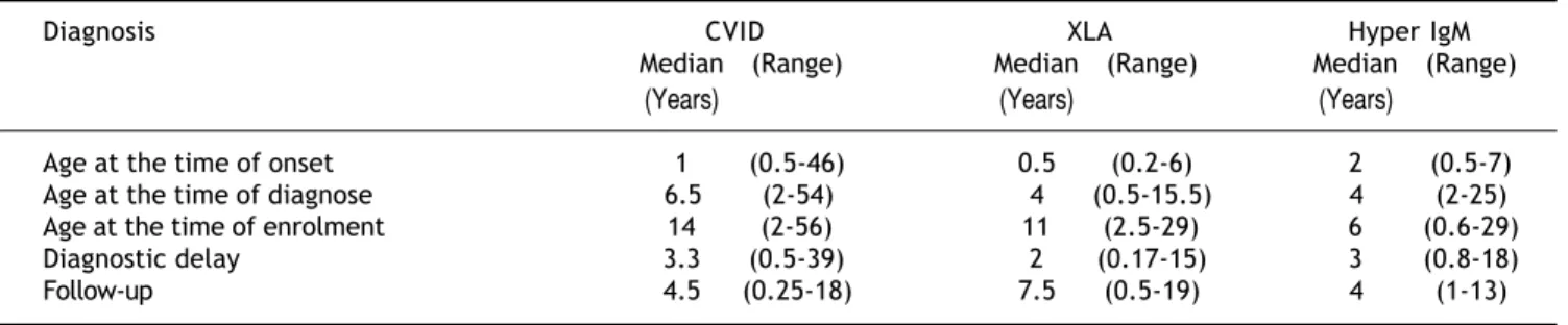
Sixty-two patients with PAD, including: 38 pa-tients with common variable immunodeficiency (CVID), 19 with XLA and five with HIGM, were in-troduced. There were 48 males and 14 females; the median age of patients was 10.3 years (range 2-56). Forty six patients (75%) were under 18 years. Pa-tients were followed-up for a mean of five years (range 0.2-19 years).
Baseline laboratory findings, including serum im-munoglobulin levels and immunophenotyping of pe-ripheral blood lymphocytes, are summarized in the table 1.
(diag-nostic delay) was 39 months for CVID, 24 months for XLA, and 36 months for HIGM. Age at onset of disea-se, diagnostic age, delay in diagnosis, follow up period and current age of patients are recorded in the table 2. Patients received intravenous immunoglobulin for mean period of 5.2 years (range: 2-19 years). Twenty-one patients (34%) had received other pro-ducts of blood such as packed red blood cells, plate-let or fresh frozen plasma. The prevalence of blood product transfusion was not significantly different (P value > 0.05) in each type of PID (Table 3).
Table 1. Laboratory parameters in patients with primary antibody deficiencies.
Laboratory Parameters Normal range CVID XLA Hyper IgM
Median (Range) Median (Range) Median (Range)
Immunoglobulins (mg/dL)
IgG 800-1,800 121 (0-500) 100 (0-700) 135 (0-460)
IgA 90-145 6.5 (0-220) 5 (0-30) 17 (0-420)
IgM 80-300 15 (0-230) 10 (0-76) 625 (540-1,260)
Cellular Count
WBC 4,000-10,000 6,757(1,310-15,100) 8,200 (2,800-22,700) 4,850 (2,500-5,800) Lymphocyte (%) 25-35 36.5 (15.7-96) 42 (4-79) 44 (10-66)
Lymphocyte Markers (%)
CD3 65-95 72.8 (38.42-99) 91.6 (30-97) 61 (43-84)
CD4 31 (4.5-68.37) 44 (16.3-93.62) 30 (20-40)
CD8 35.08 (18-73) 33 (17-75) 40.5 (12-52)
CD19 3-25 10 (2-33) 0.5 (0-2) 14 (8.9-19)
CD4/CD8 ratio 0.9 (0.16-3.47) 1.37 (0.22-3.54) 0.74 (0.38-3.33)
Liver Enzymes(IU/L)
AST 5-42 22 (9-251) 16 (0.12-148) 29 (17-34)
ALT 5-40 22.5 (9-147) 21 (8-138) 31 (12-42)
Table 2. Diagnostic parameters in patients with primary antibody deficiencies.
Diagnosis CVID XLA Hyper IgM
Median (Range) Median (Range) Median (Range)
(Years) (Years) (Years)
Age at the time of onset 1 (0.5-46) 0.5 (0.2-6) 2 (0.5-7) Age at the time of diagnose 6.5 (2-54) 4 (0.5-15.5) 4 (2-25) Age at the time of enrolment 14 (2-56) 11 (2.5-29) 6 (0.6-29)
Diagnostic delay 3.3 (0.5-39) 2 (0.17-15) 3 (0.8-18)
Follow-up 4.5 (0.25-18) 7.5 (0.5-19) 4 (1-13)
Table 3. Liver involvement in antibody deficient patients.
Patients Medical History CVID XLA Hyper IgM Total
(38) (19) (5) (62)
Clinical signs of liver involvement 15 (40%) 2 (10.5%) 5 (100%) 22 (35.5%) Pathologic features of liver disease 6 (15.7%) 1 (5%) 1 (20%) 8 (13%) History of blood product transfusion 13 (34.2%) 7 (36.8%) 1 (20%) 21 (34%)
Liver Disease
Among 62 studied patients, 22 patients (35.5%) had clinical features in favor of chronic liver disea-se, including hepatomegaly. These patients include five HIGM (100%), 15 CVID (40%) and two XLA (10.5%) patients (Table 3). Percutaneous liver biop-sy was performed that revealed abnormalities in eig-ht of them (13%) (Table 4).
bronchiec-tasis (P2) were common in this group of patients, while other patients did not experience such mani-festations (Table 4).
The presences of liver abscess, liver mass or any sign of fatty liver were excluded by abdominal ultra-sonography.
Laboratory Studies
Elevations of amino transaminases (ALT, AST) were found in 10 patients (16%). This rising was transient in four patients (whom were negative for HCV RNA). Two of them were considered as drug-induced hepatitis. The enzyme levels became normal after discontinuation of the drug (clari-thromycin) and no other etiology was detected in them. Six patients had persistent (above six months) elevated levels of serum amino transami-nases (mean for ALT: 159 IU/L and for AST: 109 IU/L) (Table 4).
HCV infection was detected only in one patient who had CVID and suffered from end stage liver di-sease (P2). All patients were negative for HBsAg.
In examination of stool, only one patient with HIGM due to CD40-ligand deficiency was positive for C. parvum. This patient had hydrops of gall bladder in ultrasonography but he was asymptomatic with
normal liver enzymes. Sclerosing cholangitis ruled out using MRCP in this patient.
Further Details
In general, pathologically proved liver disease was found in eight out of 62 patients (13%) with median age of 15 years. All of them had abnormal clinical examination in favor of liver disease, six pa-tients had persistent elevation of amino transamina-ses (ALT, AST) and two patients had normal level of liver enzymes. These later two patients (P2, P6) had history of increased level of amino transaminases (ALT, AST) since some years ago and had palmar erythema, clubbing and ascites due to chronic liver dysfunction in first clinical examination (Table 4).
Among the eight patients with proved liver disea-ses, seven patients underwent percutaneous liver biopsy. The remaining one patient declined li-ver biopsy. The microscopic evaluation of lili-ver biop-sies showed mild to moderate chronic active hepatitis in five patients, which were not compatible with any specific etiology. One patient (P4) had his-tological evidences of sclerosing cholangitis and died due to severe hepatic insufficiency. In remaining pa-tient (P2) with HCV infection, liver biopsy had been performed four years before our study, and showed
Table 4. Clinical features of primary antibody deficient patients with chronic liver disease.
No Diagnosis Age /Sex Physical Examination Histologic Findings Other accompanying
(years) morbidities and current status
P1 CVID 20 / Female Purpura ,Clubbing, Chronic Active Hepatitis, NLH, Anemia, Rise of ESR/
Hepatosplenomegaly Cirhosis Alive
P2 CVID 27 / Male Ascitis Reactive Hepatitis, Cirhosis Positive HCV PCR, Hepatosplenomegaly Bronchiectasia/Alive P3 CVID 18 /Male Clubbing Chronic Active Hepatitis Ulcerative collitis/Died
Hepatosplenomegaly due to hepatic failure P4 CVID 17 / Male Hepatosplenomegaly Sclorising Cholangitis ITP, Neutropenia,
Esophageal Varices grade 2/ Died due to hepatic failure P5 CVID 7 / Male Hepatosplenomegaly Chronic Active Hepatitis Alive
P6 HIM 17 / Female Clubbing Chronic Active Hepatitis Evance Syndrome, IBD/Alive Hepatosplenomegaly
P7 XLA 24 / Male Hepatosplenomegaly Chronic Active Hepatitis Dermatomyosit Like Syndrome/Died due to retroviral encephalitis P8 CVID 6 / Male Hepatosplenomegaly Not Given CVA/ Alive
reactive hepatitis with fibrosis indicating liver cirr-hosis. This patient developed esophageal varices gra-deπ, end stage liver disease, and cachexia.
As it has been shown in table 4, the known cau-ses of liver disease were hepatitis C infection (P2) and sclerosing cholangitis (P4). In remaining six pa-tients, no etiology were found (75%). There was no meaningful relationship between liver involvement and type of primary antibody deficiency.
The cumulative survival within patients with pri-mary antibody deficiency based on liver involvement is shown in figure 1. Among eight patients who com-plicated with liver disease, three (37.5%) died during their follow-up.
DISCUSSION
The incidence of liver involvement in patients with primary immunodeficiency varies from 18% to 55%14,32 and its severity ranging from mild bioche-mical abnormalities to end-stage hepatic failure. 14-16,23,35,36 In our current study, the prevalence of liver
diseases in 62 patients with primary antibody defi-ciency was 13%, which supports previous studies.
Fiore M, et al.15 in 1998 studied 30 Italian chil-dren (< 16 years) with PID (T cell, B cell and com-bined immunodeficiency). Their study showed that 11 patients (36.8%) had liver disease including 10 patients with T-cell and one with B-cell (CVID) defi-ciency. Also in another study which was performed
by an American group,16 among the 147 patients
with PID (< 18 years), 35 patients (26%) were de-tected to have liver involvement. The most common type of PID was combined immunodeficiency
syndro-me (14 out of 35), in the reported study. All these data show that the involvement of liver in patients with T-cell and combined immunodeficiencies is more common and more severe comparing with pri-mary antibody deficiencies.37
HCV infection was a major cause of liver disease in hypogammaglobulinemic patients in the past. Bet-ween February and July of 1994, 112 cases of hepa-titis C infection associated with the use of infected IVIG were reported to the CDC,27 as well as some
re-ports from Europe.25,32 Recent data clearly show
that most immunoglobulin preparations are accepta-bly safe, at least with regard to HIV, HBV and HCV transmission,38 as we found only one patient infec-ted with hepatitis C.
Our data provide evidence that a remarkable number of patients with primary antibody deficiency have liver disease unrelated to the common hepato-tropic pathogens (Table 4).
Previous reports showed that sclerosing cholangi-tis (SC) is a common feature of liver involvement as-sociated with immune disorders.23,24,39,40C. parvum had been implicated in the development of SC in HIV infection and in other immunocompromised patients, in particular, in those with X-linked HIGM syndro-me.35,36 In our study, C. parvum infection was found in one patient. This patient had X-linked HIGM syn-drome, was asymptomatic and had no laboratory and radiological evidence of sclerosing cholangitis and treated with azythromycin. The low incidence of C. parvuminfection in our study could be due to geo-graphical situations and good care of patients.
The rising of serum amino transaminases (ALT, AST) is characteristic of liver involvement. The level of this rising indicates the severity of hepatocellular injury.41 Based on results of our study and those from others,14-16,32,42 persistent elevation of liver en-zymes in patients with primary antibody deficiency, above two times greater than normal, is an indica-tor of liver involvement and can be the first sign of an unknown progressive systemic disease. Chronic active hepatitis with unknown origin was the most common feature of liver injury in our series (75%). Because of high incidence of liver involvement, it is advised to perform a biochemical liver profile, inclu-ding serum concentrations of amino transaminases must be performed at least twice yearly in all pa-tients with primary antibody deficiency.43
The presence of autoantibodies like smooth mus-cle antibody (SMA) is usual in PID specially in T-cell deficiencies that can reflect dysregulation in immune system and may involve the liver.5,14,16,30,44 Triggering of autoimmune inflammatory processes
Figure 1. Cumulative survival within patients with primary
antibody deficiency divided based on liver involvement. Liver abnormal
Liver normal
0 2 4 6 8 10 12 14 16 Follow up (year)
1.2
1.0
0.8
0.6
0.4
0.2
0.0
Cummulative
by hepatotropic viruses is one possible mecha-nism by which such chronic liver disease may develop.14,44 Since in primary antibody deficiencies, production of antibodies is impaired, we cannot de-tect autoantibodies in these patients. High inciden-ce of other co-morbidities such as bone marrow suppression, autoimmune cytopenias, inflamma-tory bowel diseases and dermatomyositis like syn-drome can propose that a systemic autoimmune process is present.
More investigations are needed to determine etio-logy of this high prevalence of unexplained liver di-sease (75%) and unknown progressive systemic disease. Interestingly, the survival of PAD patients with liver involvement seems to be less than that of patients who do not have this complication. Applica-tion of preventive measures and beginning of timely therapy for this complication could lead to increased life span of PAD patients.
Other reports revealed that approximately 10 to 23% of adult patients with some types of primary immunodeficiency have unexplained liver disea-ses.14,15,32 Chronic active hepatitis is the most com-mon pathologic feature of liver injury in our patients with primary antibody deficiency (five from seven patients). Whether this high prevalence of li-ver disease with unknown origin (75%) is the result of an unidentified hepatotropic virus or other me-chanisms such as autoimmunity, is currently diffi-cult to understand.
ACKNOWLEDGMENTS
This study was supported by a grant from Tehran University of Medical Sciences and Immunology, As-thma and Allergy Research Institute.
The authors wish to thank all the patients, their families and the Iranian Primary Immunodeficiency Association (IPIA) for their kind collaboration in this research project. We are also grateful to Dr. Me-mar, Dr. Jannati, Dr. Arshi, Ms. Akbarzadeh, and Ms. Faridoni and the staff of the Department of Allergy and Clinical Immunology, Tehran Universi-ty of Medical Sciences.
REFERENCES
1. Aghamohammadi A, Moein M, Farhoudi A, Pourpak Z, Re-zaei N, Abolmaali K, et al. Primary immunodeficiency in Iran: first report of the National Registry of PID in Chil-dren and Adults. J Clin Immunol 2002; 22: 375-80.
2. Knerr V, Grimbacher B. Primary immunodeficiency regis-tries. Curr Opin Allergy Clin Immunol 2007; 7: 475-80. 3. Rezaei N, Aghamohammadi A, Moin M, Pourpak Z, Movahedi
M, Gharagozlou M, et al. Frequency and clinical
manifesta-tions of patients with primary immunodeficiency disorders in Iran: update from the Iranian Primary Immunodeficien-cy Registry. J Clin Immunol 2006; 26: 519-32.
4. Notarangelo L, Casanova JL, Fischer A, Puck J, Rosen F, Seger R, Geha R. Primary immunodeficiency diseases: An update. Journal of Allergy & Clinical Immunology 2004; 114: 677-87.
5. Cunningham-Rundles C, Bodian C. Common variable immu-nodeficiency: clinical and immunological features of 248 patients. Clin Immunol 1999; 92: 34-48.
6. Lederman HM, Winkelstein JA. X-linked agammaglobuline-mia: an analysis of 96 patients. Medicine (Baltimore) 1985; 64: 145-56.
7. Aghamohammadi A, Farhoudi A, Moin M, Rezaei N, Kouhi A, Pourpak Z, et al. Clinical and immunological features of 65 Iranian patients with common variable immunodeficiency. Clin Diagn Lab Immunol 2005; 12: 825-32.
8. Moin M, Aghamohammadi A, Farhoudi A, Pourpak Z, Rezaei N, Movahedi M, et al. X-linked agammaglobulinemia: a survey of 33 Iranian patients. Immunol Invest 2004; 33: 81-93. 9. Hermaszewski RA, Webster AD. Primary
hypogammaglobu-linaemia: a survey of clinical manifestations and complica-tions. Q J Med 1993; 86: 31-42.
10. Farhoudi A, Aghamohammadi A, Moin M, Rezaei N, Pourpak Z, Movahedi M, et al. Distribution of primary immunodefi-ciency disorders diagnosed in the Childrens Medical Cen-ter in Iran. J Investig Allergol Clin Immunol 2005; 15: 177-82.
11. Aghamohammadi A, Moazzami K, Rezaei N, Karimi A, Mova-hedi M, Gharagozlou M, et al. ENT manifestations in Ira-nian patients with primary antibody deficiencies. J Laryngol Otol 2008; 122: 409-13.
12. Aghamohammadi A, Parvaneh N, Tirgari F, Mahjoob F, Mova-hedi M, Gharagozlou M, et al. Lymphoma of mucosa-associa-ted lymphoid tissue in common variable immunodeficiency. Leuk Lymphoma 2006; 47: 343-6.
13. Aghamohammadi A, Pouladi N, Parvaneh N, Yeganeh M, Mo-vahedi M, Gharagolou M, et al. Mortality and morbidity in common variable immunodeficiency. J Trop Pediatr 2007; 53: 32-8.
14. Bjoro K, Haaland T, Skaug K, Froland SS. The spectrum of hepatobiliary disease in primary hypogammaglobulinaemia. J Intern Med 1999; 245: 517-24.
15. Fiore M, Ammendola R, Gaetaniello L, De Felice C, Iorio R, Vegnente A, et al. Chronic unexplained liver disease in children with primary immunodeficiency syndromes. J Clin Gastroenterol 1998; 26: 187-92.
16. Rodrigues F, Davies EG, Harrison P, McLauchlin J, Karani J, Portmann B, et al. Liver disease in children with primary immunodeficiencies. J Pediatr 2004; 145: 333-9.
17. Seymour B, Miles J, Haeney M. Primary antibody deficien-cy and diagnostic delay. J Clin Pathol 2005; 58: 546-7. 18. Khodadad A, Aghamohammadi A, Parvaneh N, Rezaei N,
Ma-hjoob F, Bashashati M, et al. Gastrointestinal manifesta-tions in patients with common variable immunodeficiency. Dig Dis Sci 2007; 52: 2977-83.
19. Rezaei N, Aghamohammadi A, Siadat SD, Nejati M, Ahmadi H, Moin M, et al. Serum bactericidal antibody response to serogroup C polysaccharide meningococcal vaccination in children with primary antibody deficiencies. Vaccine 2007; 25: 5308-14.
21. Busse Pj, Razvi S, Cunningham-Rundles C, Cunningham-Run-dles C. Efficacy of intravenous immunoglobulin in the pre-vention of pneumonia in patients with common variable immunodeficiency. J Allergy Clin Immunol 2002; 109: 1001-4.
22. Chapel HM. Consensus on diagnosis and management of primary antibody deficiencies. Consensus Panel for the Diagnosis and Management of Primary Antibody Deficien-cies. BMJ 1994; 308(6928): 581-5.
23. Davis JJ, Heyman MB, Ferrell L, Kerner J, Kerlan R, Jr., Thaler MM. Sclerosing cholangitis associated with chronic cryptosporidiosis in a child with a congenital immunodefi-ciency disorder. Am J Gastroenterol 1987; 82: 1196-202. 24. DiPalma JA, Strobel CT, Farrow JG. Primary sclerosing
cho-langitis associated with hyperimmunoglobulin M immunode-ficiency (dysgammaglobulinemia). Gastroenterology 1986; 91: 464-8.
25. Bjoro K, Froland SS, Yun Z, Samdal HH, Haaland T. Hepati-tis C infection in patients with primary hypogammaglobuli-nemia after treatment with contaminated immune globulin. N Engl J Med 1994; 331: 1607-11.
26. Chapel HM, Christie JM, Peach V, Chapman RW. Five-year follow-up of patients with primary antibody deficiencies following an outbreak of acute hepatitis C. Clin Immunol 2001; 99: 320-4.
27. Rossi G, Tucci A, Cariani E, Ravaggi A, Rossini A, Radaeli E. Outbreak of hepatitis C virus infection in patients with hematologic disorders treated with intravenous immunog-lobulins: different prognosis according to the immune sta-tus. Blood 1997; 90: 1309-14.
28. Yap PL, McOmish F, Webster AD, Hammarstrom L, Smith CI, Bjorkander J, et al. Hepatitis C virus transmission by intravenous immunoglobulin. J Hepatol 1994; 21: 455-60. 29. McLauchlin J, Amar CF, Pedraza-Diaz S, Mieli-Vergani G,
Hadzic N, Davies EG. Polymerase chain reaction-based diagnosis of infection with Cryptosporidium in children with primary immunodeficiencies. Pediatr Infect Dis J 2003; 22: 329-35.
30. Knight AK, Cunningham-Rundles C. Inflammatory and au-toimmune complications of common variable immune defi-ciency. Autoimmunity Reviews 2006; 5: 156-9.
31. Zelazko M, Carneiro-Sampaio M, Cornejo de Luigi M, Gar-cia de Olarte D, Porras Madrigal O, Berron Perez R, et al. Primary immunodeficiency diseases in Latin America: first
report from eight countries participating in the LAGID. Latin American Group for Primary Immunodeficiency Di-seases. J Clin Immunol 1998; 18: 161-6.
32. Webster AD, Brown D, Franz A, Dusheiko G. Prevalence of hepatitis C in patients with primary antibody deficiency. Clin Exp Immunol 1996; 103: 5-7.
33. Okamoto H, Sugiyama Y, Okada S, Kurai K, Akahane Y, Su-gai Y, et al. Typing hepatitis C virus by polymerase chain reaction with type-specific primers: application to clinical surveys and tracing infectious sources. J Gen Virol 1992; 73(Pt 3): 673-9.
34. Garson JA, Ring C, Tuke P, Tedder RS. Enhanced detection by PCR of hepatitis C virus RNA. Lancet 1990; 336: 878-9. 35. Naveh Y, Mendelsohn H, Spira G, Auslaender L, Mandel H,
Berant M. Primary sclerosing cholangitis associated with immunodeficiency. Am J Dis Child 1983; 137: 114-7. 36. Levy J, Espanol-Boren T, Thomas C, Fischer A, Tovo P,
Bor-digoni P, et al. Clinical spectrum of X-linked hyper-IgM syndrome. J Pediatr 1997; 131: 47-54.
37. Rodrigues F, Davies EG, Harrison P, McLauchlin J, Karani J, Portmann B, et al. Liver disease in children with primary immunodeficiencies. J Pediatr 2004; 145: 333-9.
38. Quinti I, Pierdominici M, Marziali M, Giovannetti A, Don-nanno S, Chapel H, et al. European surveillance of immuno-globulin safetyresults of initial survey of 1243 patients with primary immunodeficiencies in 16 countries. Clin Im-munol 2002; 104: 231-6.
39. Yoshioka R, Sato Y, Kogure A, Ohira H, Takagi T, Kuroda M, et al. Association of primary sclerosing cholangitis, thymoma and hypogammaglobulinemia. Liver 1995; 15: 53-5.
40. Gremse DA, Bucuvalas JC, Bongiovanni GL. Papillary steno-sis and sclerosing cholangitis in an immunodeficient child. Gastroenterology 1989; 96: 1600-3.
41. Behrman RE, Kliegman R, Jenson HB. Nelson textbook of pe-diatrics. 17th Ed. Philadelphia, PA: Saunders; 2004. 42. Bjoro K, Skaug K, Haaland T, Froland SS. Long-term
outco-me of chronic hepatitis C virus infection in primary hypo-gammaglobulinaemia. Q J Med 1999; 92: 433-41.
43. Stiehm ER. Human intravenous immunoglobulin in primary and secondary antibody deficiencies. Pediatr Infect Dis J 1997; 16: 696-707.