392
www.medigraphic.com
* Cardiology Department. UMAE 1 Bajío, Mexican Institute of Social Security. Leon, Guanajuato, Mexico. ** Unit of Clinical Research, UMAE Bajío 1. Mexican Institute of Social Security. Leon, Guanajuato, Mexico. *** School of Medicine. University of Guanajuato. León, Guanajuato, Mexico.
Correspondence: Sergio Eduardo Solorio Meza. Unit of Clinical Research, UMAE Bajío 1. Mexican Institute of Social Security. Blvd Lopez Mateos esq. Insurgentes SN. Col. Los Paraísos. 37320 Leon, Guanajuato, Mexico. Tel. (52) 477 7174800 ext 31742, E-mail: [email protected]; [email protected]
Recibido: 12 de diciembre de 2007 Aceptado: 15 de abril de 2008
Summary
Objective: To evaluate the diastolic function af-ter regression of left ventricular hypertrophy, in mild to moderate hypertension treated with an-giotensin converting enzyme(ACE) inhibitor and, if necessary, with a diuretic. Methods: Nine-ty-eight hypertensive patients with left ventricu-lar hypertrophy (LVH) and abnormal left ventri-cle diastolic function indexes received captopril (Capotena®) 50 to 200 mg/day plus
chlortali-done during 12 months to reach blood pressure control, defined as a diastolic blood pressure ≤ 90 and systolic blood pressure ≤ 140 mm Hg. Left ventricular (LV) mass index was calculated by M mode and two-dimensional echocardio-graphy, and left ventricular diastolic function was assessed by transmitral pulsed Doppler ultra-sound every 3 months. Results: Sixty-three pa-tients were women and 35 were men, mean age was 53.4 ± 8.4 years (range 34-70). Thirty-six patients had mild (36.7%) and 62 (63.3%) mod-erate hypertension. Treatment reduced signifi-cantly both systolic pressure from 165 ± 13 to 137 ± 12.9 mm Hg (p < 0.05) and diastolic pres-sure from 99 ± 8.6 to 86 ± 6.37 mm Hg (p < 0.05). LV mass index decreased from 155.4 ± 32.9 to 121.7± 29.14 g/m2 (p < 0.05). Late diastolic
fill-ing velocity (A wave) and the ratio of E/A waves improved (p < 0.05), but early diastolic filling velocity (E wave) and isovolumetric relaxation time did not change with treatment. Conclusions:
Some indexes of diastolic function improved af-ter regression of left ventricular hypertrophy and good blood pressure control with captopril and chlortalidone.
(Arch Cardiol Mex 2008; 78: 392-399)
Resumen
MEJORADELAFUNCIÓNDIASTÓLICATRASREGRESIÓNDE LAHIPERTROFIAVENTRICULARIZQUIERDA
Objetivo: Evaluar la función diastólica después de revertir la hipertrofia ventricular izquierda, en hipertensión leve a moderada tratada con inhibidores de la enzima convertidora angio-tensina (ECA) y, si era necesario, con un diuré-tico. Métodos: Noventa y ocho pacientes hiper-tensos con hipertrofia ventricular izquierda e índices de función diastólica anormal del ven-trículo izquierdo recibieron captopril 50 a 200 mg/día (Capotena®) más clortalidona durante
12 meses para lograr el control de la presión arterial, definido como presión diastólica ≤ 90 mm Hg y presión sistólica ≤ 140 mm Hg. El índi-ce de masa ventricular izquierdo fue calculado por ecocardiografía modo M y bidimensional y la función diastólica ventricular izquierda fue determinada por Doppler pulsado transmitral cada 3 meses. Resultados: Sesenta y tres pa-cientes eran del género femenino y 35 del gé-nero masculino, con edad de 53.4 ± 8.4 años (rango 34-70). Treinta y seis pacientes (36.7%) tenían hipertensión leve y 62 (63.3%) hiperten-sión moderada. El tratamiento redujo significa-tivamente tanto la presión sistólica de 165 ± 13 a 137 ± 12.9 mm Hg (p < 0.05) como la presión diastólica de 99 ± 8.6 a 86 ± 6.37 mm Hg (p < 0.05). El índice de masa ventricular izquierda disminuyó de 155.4 ± 32.9 a 121.7 ± 29.14 g/m2
(p < 0.05). La velocidad diastólica de llenado tardío (onda A) y la relación E/A mejoraron (p < 0.05), pero la velocidad diastólica de llenado temprano (onda E) y el tiempo de relajación
Improvement of diastolic function after regression of left
ventricular hypertrophy
www.medigraphic.com
Key words: Diastolic function. Left ventricular hypertrophy. Hypertension. Palabras clave: Función diastólica. Hipertrofia ventricular izquierda. Hipertensión.isovolumétrica no cambiaron con el tratamien-to. Conclusiones: Algunos índices de la función diastólica mejoraron después de la regresión de la hipertrofia ventricular izquierda y del buen control de la presión arterial con el captopril y clortalidona.
Background
eft ventricular hypertrophy (LVH) is often a complication of systemic arteri-al hypertension, especiarteri-ally in uncon-trolled hypertension. LVH increases the risk of coronary artery disease, stroke, ventricular ar-rhythmias, sudden death, and congestive heart failure.1,2 LVH produce, at least in part, left
ven-tricle wall stiffnessand alteration of left ventri-cle diastolic function (LVDF).3-6 Other factors
can alter LVDF, including aging, increased heart rate, high left ventricle filling pressure, and adr-energic hyperactivity.7 Altered LVDF is
associ-ated to LVH, it can be asymptomatic or precipi-tate congestive heart failure (CHF) with normal systolic function. A condition found in 51% of patients with systemic arterial hypertension, in-creasing their morbidity and mortality.8
The regression of LVH has been documented to occur with appropriate antihypertensive drugs, except with vasodilators, such as hydralazine and minoxidil. The most effective drugs to re-verse LVH are angiotensin-converting enzyme (ACE) inhbitors.9,10 However, results from
sever-al studies that evsever-aluated diastolic function after LVH had been corrected are contradictory.11-17
These results could be explained mainly based on differences in the duration of the studies and, in some cases, by the poor hypertension control. Thus, if the systolic overload of the LV is elim-inated through suitable blood pressure control, regression of the LVH could be expected, and therefore the stiffness of the LV wall might be corrected reversing the diastolic dysfunction. The objective of this study was to evaluate the effect of treatment with captopril and whether a diuretic was necessary for LVH and LVDF in patients with mild and moderate high blood pres-sure (HBP).
Patients and methods
Patients were eligible for enrollment when:
1. Mild HBP (arterial diastolic pressure higher than 90 but lower than 99 mm Hg and/or arterial systolic pressure higher than 140 but lower than 160 mm Hg); or moderate HBP (arterial diastolic pressure higher than 99 but lower than 115 mm Hg and/or arterial sys-tolic pressure higher than 160 but lower than 180 mm Hg);
2. LVH determined by echocardiogram (left ventricular mass ≥ 110 g/m2 in women and ≥
134 g/m2 in men);18
3. Altered LVDF (isovolumetric relaxation time higher than 73 ± 12 msec; maximum speed of wave E diminished below 86 ± 16 cm/sec, maximum auricular contraction speed over 56 ± 13 cm/sec, and a relationship E wave/A wave ratio lower than 1.6 ± 0.5);19
4. Normal LV systolic function (ejection frac-tion > 50%) by echocardiogram Doppler (echo-Doppler).
Patients with secondary arterial hypertension, myocardial ischemia (stable or unstable angi-na, acute myocardial infarction, silent is-chemia), resting electrocardiogram (ECG) with ischemia, lesion or necrosis; positive ECG for myocardial ischemia during an exercise tread-mill test; LV wall motion abnormalities in the echocardiogram; valvular heart disease, over-weight higher than 20%, creatinine plasma levels higher than 1.6 mg/dL, abnormal he-patic function tests, and well-known hyper-sensitivity to captopril or chlortalidone were excluded.
www.medigraphic.com
Study designA prospective, longitudinal study was performed in 98 hypertensive patients. Sample size was calculated considering a 15% prevalence rate of hypertension, in the population of the city of León with LVH (30% of them), and expecting an improvement in LVH of at least 50%, a power of 90%, and an α value of 5%. Eligible patients were withdrawn from their antihypertensive medication two weeks before tests to confirm mild or moderate hypertension. A full medical history and physical examination were obtained from each patient. They had a laboratory screen-ing (BH, fastscreen-ing blood glucose, urea,creatinine, electrolytes, total cholesterol and triglycerides), ECG at rest and during a stress test with Bruce protocol and an echo-Doppler. Patients meeting all the selection criteria were invited to partici-pate. All of them were followed-up during 12 months; once a month, we evaluated them with physical examination, laboratory tests (blood glucose, urea, creatinine, potassium), and adjust-ing the antihypertensive medication. An echo-Doppler was recorded every 3 months until com-pleting the 12 months.
Arterial blood pressure measurement and treatment follow-up
Arterial blood pressure was measured indirectly with a standard sphygmomanometer (bladder size, 12.5 cm wide and 48 cm long), with the patient comfortably seated, arms resting at heart level, without having smoked or drunk coffee within the last 30 minutes. Systolic blood pres-sure (SBP) was indicated by phase I of Korotkoff sounds (sudden appearance of sounds); while diastolic BP (DBP) was marked by phase V (all sounds disappear completely). Each measure-ment was made in triplicate after 5 minutes of rest; at least 2 minutes were allowed between successive measurements and the mean value was recorded. The main goal of treatment was to achieve a SBP below 140 and a DBP below 90 mm Hg or to reduce 20 mm Hg the systolic and/ or diastolic BP in relation to the basal figures. Initial captopril dose was 25 or 50 mg (Capote-na®) twice a day for mild or moderate HBP,
re-spectively. This dose was adjusted every month according to the arterial blood pressure measure-ments, up to a maximum of 200 mg/day. When patients did not reach the treatment goal, a di-uretic (Chlortalidone) was added at a dose of 12.5 mg/day that could be increased to 25 mg/
day. To monitor adherence to treatment, patients were asked to bring all their medications to each appointment and tablets were counted. Antihy-pertensive dosage adjustments were required only in the first 4 months of the study.
Echo-Doppler measurements
Echocardiograms were recorded in the M, B/D, and Doppler modes using a Toshiba equipment (Toshiba Sonolayer SSA-270A, Tochigi Ken 329-26, Japan), with a 3.5-MHz transducer. Patients were in the left lateral decubitus position with the transducer placed on the 4th or 5th left inter-costal space. Recordings were made preferably in a long axis view at the level of the tip of the papillary muscles according to the recommenda-tions of the ASE.20 The following measurements
were recorded: interventricular septum thickness (IVS), left ventricular posterior wall (LVPW), end-diastolic and end-systolic diameter of the left ventricle (LVED, LVES). Fractional shortening and ejection fraction (EF) were estimated. LV mass was calculated using the formula proposed by Devereux 21 as follows: LV Mass = 1.04
(LVPW+IVS+LVED)3-(LVED)3-13.6. Results
werecorrected for each patient’s body surface area (BSA) and expressed in g/m2 BSA.
Left ventricular filling was obtained as follows: Mitral velocities were obtained by placement of the Doppler sample volume within the valve leaflets just distal to the annulus. The sample volume was aligned parallel to the interventric-ular septum and then adjusted in the non visual-ized plane until the optimal wave form was found, at the end of non forced expiration. Mea-surements were obtained for LV isovolumetric relaxation time, early diastolic “E” wave associ-ated with the rapid left ventricular filling phase, followed by an “A” wave associated with atrial contraction. The E wave/A wave ratio was cal-culated. All echo-Doppler recordings were made blind in relation to patient HBP control, as well as to previous recordings or results.
Statistical analysis
www.medigraphic.com
ResultsOf the 342 evaluated patients, 98 met the selec-tion criteria and formed the study group. Their mean age was 53.4 ± 8.4 years (range 34 -70). There were 63 women and 35 men (Table I). All of the patients were known to be hypertensive for at least 1 year and all were taking antihyperten-sive drugs different from the medications used in this study. Mild HBP was present in 36 patients and moderated HBP in the remainder. Only 74 patients completed the study; 18 abandoned it, 5 had persistent cough with medication, and 1 pre-sented a significant elevation in blood glucose. Drug therapy: The average initial captopril dose was 82.9 ± 38.9 mg/day and the average final dose was 167.57 ± 46.58 mg/day; 34 patients required Chlortalidone at a dose of 25 mg/day. Blood pressure measurements: All patients had their SBP or DBP or both reduced by at least 20 mm Hg after 12 months of treatment. SBP di-minished from 165 ± 13 to 137 ± 13 mm Hg (p < 0.05); DBP lowered from 100 ± 8.7 to 86.7 ± 6.4 mm Hg (p < 0.05) (Fig. 1). In 26 patients, it was not possible to diminish the SBP below 140 mm Hg and in 5 more it was not possible to diminish DBP to less than 90 mm Hg.
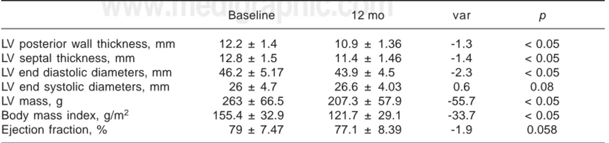
Left ventricular mass: After 12 months of treat-ment, LVPW diminished from 12.2 ± 1.4 to 10.9 ± 1.36 mm (p < 0.05); IVS thickness was reduced from 12.8 ± 1.5 to 11.4 ± 1.46 mm (p < 0.05) and LVED decreased from 46.2 ± 5.17 to 43.9 ± 4.5 mm (p < 0.05). The LVES did not change in value (26 ± 4.7 and 26.6 ± 4.03; p = ns) (Table II). The LV mass diminished significantly from 155.4 ± 32.9 to 121.7 ± 29.14 g/m2 BSA (p <
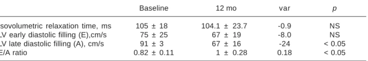
0.05) after treatment, as shown in Figure 2. Diastolic function indexes: After the 12-month period, LV isovolumetric relaxation diminished from 105 ± 18 to 104.1 ± 23.73 msec (p = ns). The maximum wave E speed decreased from 75 ± 0.25 to 67 ± 0.19 cm/sec (p = ns), the maxi-mum wave A speed lowered significantly from
91 ± 0.3 to 67 ± 0.16 cm/sec (p < 0.05) (Fig. 3), and the E wave/A wave ratio decreased from 0.82 ± 0.11 to 1 ± 0.28 (p < 0.05) (Table III and Fig. 4).
Discussion
As in similar studies, we documented a signifi-cant 21% regression of the LV mass, a reduction that is larger than previously reported of about 15%.8,9 The difference could be explained by
the higher captopril dose, use of diuretics, agents known to reduce LV diastolic diameter, and the
Table I. Characteristics of patients included in the study at baseline.
Male/Female 35/63
Age, yr 53.4 ± 8.38 (34-70)
n (%)
30-39 6 (6.18)
40-49 28 (28.57) 50-59 40 (40.82) 60-70 24 (24.49)
Height, cm 157 ± 18
Weight, kg 74.8 ± 10.2
Mild hypertension, n (%) 36 (36.7) Moderated hypertension, n (%) 62 (63.3)
Fig. 1. Blood pressure evolution.
Systolic Diastolic
200
150
100
50
0
mmHg
165
149 144 143 137
87 89 88
91 100
p<0.05
0 3 6 9 12
Months
Table II. Echocardiographic measurements before and after 12 months of treatment.
Baseline 12 mo var p
LV posterior wall thickness, mm 12.2 ± 1.4 10.9 ± 1.36 -1.3 < 0.05
LV septal thickness, mm 12.8 ± 1.5 11.4 ± 1.46 -1.4 < 0.05
LV end diastolic diameters, mm 46.2 ± 5.17 43.9 ± 4.5 -2.3 < 0.05
LV end systolic diameters, mm 26 ± 4.7 26.6 ± 4.03 0.6 0.08
LV mass, g 263 ± 66.5 207.3 ± 57.9 -55.7 < 0.05
Body mass index, g/m2 155.4 ± 32.9 121.7 ± 29.1 -33.7 < 0.05
www.medigraphic.com
longer observation period in our study.8,9 Shahiet al.22 reported a similar reduction in LV mass
(22%) in their 18 month study on 25 hyperten-sive patients. These authors used a similar combination of captopril and a diuretic to con-trol HBP. They did no find changes in LVDF after 9 or 18 months of treatment.
Thus, contradictory results have been found in relation to LVDF and regression of LVH in
hy-pertension. A clear improvement in diastolic function was documented by White et al.14 They
studied nine hypertensive patients with LVH and altered diastolic function. None of the patients had received any drug treatment before the study, and they prescribed metoprolol at an av-erage dose of 167 mg/day during 6 months. They reported a significant 11% decrease of the LV mass (p < 0.05). The rate of LV fast filling (mea-sured by nuclear medicine) improved from 1.89 ± 0.24 to 2.09 ± 0.27 end diastolic volume/sec (p < 0.01). Trimarco et al.12 treated hypertensive
patients with a β-blocker (tertatolol) and showed a reduction in HBP, regression of the LVH, and improvement in diastolic function, although half of the patients had abnormal systolic function at the beginning of the study. The reduction in BP and the sympathetic inhibition could be re-sponsible for the improvement. However, other investigators have not been able to confirm these results with β-blockers.16 Habid et al.23 studied
27 hypertensivepatients with LVH and diastol-ic dysfunction to whom they administered long acting nifedipine or diltiazem for 7 months. They observed a 34% decrease in LV mass and an improvement in diastolic function. All of their results were statistically significant and were not different from those observed in a control group of normotensive subjects. LVH regression in this study is larger than in any other reports8,9
in-cluding ours. On the other hand, Szlachcic et al.24 in a controlled randomized double blind
study on 24 patients with slightly moderate hy-pertension, LVH, and alteration in diastolic func-tion and treated with either diltiazem or a place-bo during 16 weeks, demonstrated a 10% re-gression in ventricular hypertrophy in the active treatment group. However, they did not record indexes of diastolic function. Zakynthinos et al.25 reported that after 6 months of treatment
with losartan, an angiotensin II receptor block-ing agent, LV mass was reduced in 6.2% in a group of patients with mild or moderate HBP but diastolic dysfunction was not modified.
Fig. 2. Left ventricular mass evolution. 200
150
100
50
0 155
136 128 124 122
g/m
2
p<0.05
0 3 6 9 12
Months
Fig. 3. Diastolic function indexes.
Wave E Wave A
* 1
0.8
0.6
0.4
0.2
0
m/s
0.91 0.82
0.8
0.7 0.67*
0.75 0.76 0.7 0.7
0.67
*p<0.05
0 3 6 9 12
Months
Table III. Diastolic function indexes before and after 12 months of treatment.
Baseline 12 mo var p
Isovolumetric relaxation time, ms 105 ± 18 104.1 ± 23.7 -0.9 NS
LV early diastolic filling (E),cm/s 75 ± 25 67 ± 19 -8.0 NS
LV late diastolic filling (A), cm/s 91 ± 3 67 ± 16 -24 < 0.05
E/A ratio 0.82 ± 0.11 1 ± 0.28 0.18 < 0.05
Fig. 4. E wave/A wave ratio. Months
0 3 6 9 12
1
0.5
0 *
*
0.82 0.9
1
*p<0.05 1
www.medigraphic.com
ESTE DOCUMENTO ES ELABORADO POR MEDIGRAPHIC
Although long term treatment of hypertension is known to result in regression of existing left ventricular hypertrophy, the effect of hyperten-sion treatment on myocardial function is less well understood. Solomon et al.25 in the VALIDD
study (a multicentric study) only a small but significant improvement associated with blood pressure lowering was observed, whereas in oth-er studies no changes woth-ere obsoth-erved in diastolic function.26,27
This study was performed in chronic hyperten-sive patients who reached the proposed treat-ment goal for their HBP. Further, our results are in agreement with previous observations that an ACE inhibitor in combination with a diuretic is able to control the hypertension in over 90% of cases with mild to moderate disease. These pa-tients had been diagnosed at least one year be-fore and were receiving some form of drug treat-ment but with poor BP control. In our study, there were more patients with moderated HBP than with mild HBP. All of the patients had im-portant LVH, alteration of the LV diastolic fill-ing pressure, prolonged myocardial relaxation, and a changed compliance (type I of Tajik) usu-ally observed in a hypertrophic heart with nor-mal ventricular size.
Based on the present evidence, an improvement in LVDF would require sustained normalization of the BP level to eliminate the hemodynamic overload. This has been demonstrated in pa-tients with LVH and diastolic dysfunction sec-ondary to aortic stenosis by Villari et al.28 These
latter authors found a larger reduction in LVH followed by recovery in LVDF, which required from months to years for normalization. This find-ing might be due, initially, to improvement in hypertrophic cardiomyocytes and, in the long term, to reduction in collagen content, which plays a role in myocardial stiffness.28
Study limitations: Magnetic resonance imag-ing is considered the ideal method for the deter-mination of LV mass because of its high spatial resolution, generally good image quality, and ability to reconstruct the heart’s shape in three
dimensions. However, it is not widely used clin-ically because of higher costs, reduced avail-ability, and limited access for critically ill pa-tients or papa-tients with implanted electronic de-vices (for example, pacemakers and defibrillators).29 The superiority of 3-D
echocar-diographic LV mass calculations over values calculated from M-mode-derived or 2-D echocar-diography has been convincingly shown. Cur-rent limitations include the requirement of reg-ular rhythm, relatively poorer image quality of real-time 3-D echocardiography as compared to 2-D images, and the time necessary for off-line data analysis. However, the greater number of acquired data points, the lack of geometric as-sumptions, increasingly sophisticated 3-D im-age, and measurements solutions set off these limitations.30 Echocardiographic imaging with
single dimensional (M mode) and two-dimen-sional techniques is the most widely available clinical tool to detect LV hypertrophy, as deter-mined by the calculated LV mass. Echocardio-graphic techniques have the advantage of being widely available, non-invasive, and relatively inexpensive. However, echocardiography is lim-ited by imaging artefacts and by the planar na-ture of the imaging technique when the LV cav-ity is misshapen.Indeed, each step in LV mass measurement is a potential source of variability. In M-mode measurement, differences of approx-imately 5% may translate into differences in LV mass between 8% and 15%, which can represent about 50 g. This variability can be attributed particularly to the measurement of wall thick-nesses and border layer definition.31
Conclusion
Improvement in most indexes of LVDF and re-gression of the LVH were observed when an ap-propriate control of HBP had been achieved with captopril alone or plus chlortalidone in patients with mild or moderate HBP, LVH, and alteration of the diastolic function. The changes are linked to obtaining a proper control of blood pressure and the time of its maintenance.
References
1. LEVY D, GARRISON RJ, SAVAGE DD, KANNEL WB,
CASTELLI WP: Prognostic implications of echocar-diographically determined left ventricular mass
in The Framingahm Heart Study. N Engl J Med
1990; 322: 1561-1566.
2. KOREN MJ, DEVEREUX RB, CASALE PN, SAVAGE
DD, LARAGH JH: Relation of left ventricular mass and geometry to morbidity and mortality in
un-complicated essential hypertension. Ann Intern
www.medigraphic.com
3. HANRATH P, MATHEY DG, SIEGERT R, BLEIFELDW: Left ventricular relaxation and filling pattern
in different forms of left ventricular hypertrophy:
an echocardiographic study. Am J Cardiol 1980;
45: 15-23.
4. FOUAD FM, SLOMINSKI JM, TARAZI RC: Left ven-tricular diastolic function in hypertension: rela-tion to left ventricular mass and systolic funcrela-tion.
J Am Coll Cardiol 1984; 3: 1500-6.
5. DRESLINSKI GR, FROHLICH ED, DUNN FG, MESSERLI
FH, SUAREZ DH, REISIN E: Echocardiographic di-astolic ventricular abnormality in hypertensive
heart disease: atrial emptying index. Am J
Cardi-ol 1981; 47: 1087-1090.
6. INOUYE I, MASSIE B, LOGE D, TOPIC N, SILVERSTEIN
D, SIMPSON P, TUBAU J: Abnormal left ventricular filling: an early finding in mild to moderate
system-ic hypertension. Am J Cardiol 1984; 53: 120-126.
7. HESS OM, SCHNEIDER J, KOCH R, BAMERT C, GRIMM
J, KRAYENBUEHL HP: Diastolic function and myo-cardial structure in patients with myomyo-cardial
hy-pertrophy. Circulation 1981; 63: 360-371.
8. VASAN RS, LARSON MG, BENJAMIN EJ, EVANS JC,
REISS CK, LEVY D: Congestive heart failure in subjects with normal versus reduced left ventricu-lar ejection fraction: prevalence and mortality in
a population-based cohort. J Am Coll Cardiol.
1999; 33: 1948-1955.
9. DAHLOF B, PENNERT K, HANSSON L: Reversal of left ventricular hypertrophy in hypertensive patients:
a meta-analysis of 109 treatment studies. Am J
Hypertens 1992; 5: 95-110.
10. SCHMIEDER R, MARTUS P, KLINGBEIL A: Reversal of left ventricular hypertrophy in essential hyperten-sion: a meta-analysis of randomized double-blind
studies. JAMA 1996: 275: 1507-1513.
11. GOTTDIENER JS: Measuring diastolic function. J
Am Coll Cardiol 1991; 18: 83-84.
12. TRIMARCO B, DELUCA N, ROSIELLO G, RICCIARDELLI
B, BETOCCHI S, FILARDI PP, ETAL: Improvement of diastolic function after reversal of left ventricular hypertrophy induced by long-term
antihyperten-sive treatment with tertalol. Am J Cardiol 1989;
64: 745-751.
13. MUIESAN ML, AGABITI-ROSEI E, ROMANELLI G,
BESCHI M, CASTELLANO M, ALARI G, ETAL: Im-proved left ventricular systolic and diastolic func-tion after regression of cardiac hypertrophy, treat-ment withdrawal, and redeveloptreat-ment of
hyper-tension. J Cardiovasc Pharmacol 1991; 17 (Suppl
2): S179-S181.
14. WHITE WB, SCHULMANP, KARIMEDDINI, MK, SMITH
VE: Regression of left ventricular mass is
accom-panied by improvement in rapid left ventricular filling following antihypertensive therapy with
metoprolol. Am Heart J 1989; 117: 145-150.
15. MOTZ W, STRAUER BE: Left ventricular function and collagen content after regression of
hyperten-sive hypertrophy. Hypertension 1989; 13: 43-50.
16. INOUYE IK, MASSIE BM, LOGE D, SIMPSON P, TU -BAU JF: Failure of antihypertensive therapy with diuretic, beta-blocking and calcium channel-block-ing drugs to consistently reverse left ventricular
diastolic filling abnormalities. Am J Cardiol 1984;
53: 1583-1587.
17. SEN S, BUMPUS FM: Collagen synthesis in devel-opment and reversal of cardiac hypertrophy in
spontaneously hypertensive rats. Am J Cardiol
1979; 44: 954-958.
18. CASALE PN, DEVEREUX RB, MILNER M, ZULLO G,
HARSHFIELD GA, PICKERING TG, ETAL: Value of echocardiographic measurement of left ventricu-lar mass in predicting cardiovascuventricu-lar morbid
events in hypertensive men. Ann Intern Med 1986;
105: 173-178.
19. NISHIMURA RA, ABEL MD, HATLE LK, TAJIK AJ: Assessment of diastolic function of the heart: back-ground and current applications of Doppler
echocardiography. Part II. Clinical studies. Mayo
Clin Proc 1989; 64: 181-204.
20. SAHN DJ, DEMARIA A, KISSLO T: Committee on M-mode standardization of the American Soci-ety of Echocardiography: recommendations re-garding quantitation in M-mode
echocardio-graphic measurements. Circulation 1978; 58:
1072-1083.
21. DEVEREUX RB: Detection of left ventricular hyper-trophy by M-mode echocardiography. Anatomic validation, standardization, and comparison to
other methods. Hypertension 1987; 9 (Suppl II):
II19-II26.
22. SHAHI M, THOM S, POULTIER N, SEVER PS, FOALE
RA: Regression of hypertensive left ventricular
hypertrophy and left ventricular diastolic
func-tion. Lancet 1990; 336: 458-461.
23. HABIB G, MANN DL, ZOGHBI WA: Normalization of cardiac structure and function after regression
of cardiac hypertrophy. Am Heart J 1994; 128:
333-343.
24. SZLACHCIC J, TUBAU JF, VOLLMER C, MASSIE B: Effect of diltiazem on left ventricular mass and diastolic filling in mild to moderate hypertension.
Am J Cardiol 1989; 63: 198-201.
25. SOLOMON SD, JANARDHANAN R, VERMA A, BOUR -GOUN M, DALEY WL, PURKAYASTHA D, ETAL: Effect of angiotensin receptor blockade and an-tihypertensive drugs on diastolic function in patients with hypertension and diastolic
dys-function: a randomized trial. Lancet 2007; 369:
2079-2087.
26. ZAKYNTHINOS E, PIERUTSAKOS CH, KONSTANTINIDIS
K, ZAKYNTHINOS S, PAPADOGIANNIS D: Losartan reduces left ventricular hypertrophy proportion-ally to blood pressure reduction in hypertensives,
but does not affect diastolic cardiac function.
An-giology 2004; 55: 669-678.
27. BARRIOS V, ESCOBAR C, CALDERÓN A, TOMÁS JP,
ventric-www.medigraphic.com
ular hypertrophy by a candesartan-based
regi-men in clinical practice. The VIPE study. J Renin
Angiotensin Aldosterone Syst. 2006; 7: 236-42. 28. VILLARI B, VASSALLI G, MONRAD ES, CHIARIELLO
M, TURINA M, HESS OM: Normalization of dias-tolic dysfunction in aortic stenosis late after valve
replacement. Circulation 1995; 91: 2353-2358.
29. BEZANTE G P, CHEN X, MOLINARI G, VALBUSA A,
DEFERRARI L, SEBASTIANI V, ETAL: Left ventricular myocardial mass determination by contrast
en-hanced colour Doppler compared with magnetic
resonance imaging. Heart 2005; 91: 38-43.
30. FOPP M, DUNCAN BB, ROHDE LEP: Echocardio-graphy-based left ventricular mass estimation.
How should we define hypertrophy?
Cardiovas-cular Ultrasound 2005; 3: 17.
31. LANG RM, BIERIG M, DEVEREUX RB, FLACHSKAMPF
FA, FOSTER E, PELLIKKA PA: Recommendations
for chamber quantification. Eur J