1
Layer-by-layer biofabrication of coronary covered stents with clickable
elastin-like recombinamers
Alicia Fernández-Colinoa†, Frederic Wolfa†, Ricardo Moreiraa, Stephan Rüttenb, Thomas Schmitz-Rodea, J. Carlos Rodríguez-Cabelloc, Stefan Jockenhoevela*, Petra Melaa,d *
a Department of Biohybrid & Medical Textiles (BioTex), AME Institute of Applied Medical Engineering, Helmholtz Institute, RWTH Aachen University, Forckenbeckstr. 55, 52074 Aachen, Germany
b Electron Microscopy Facility, Uniklinik RWTH Aachen, Pauwelsstrasse, 30, D-52074 Aachen, Germany
c Bioforge Lab, University of Valladolid, CIBER-BBN, Paseo de Belen 11, 47011 Valladolid, Spain
d Medical Materials and Implants, Department of Mechanical Engineering, Technical University of Munich, Boltzmannstr. 15, 85748 Garching, Germany
†Equal contribution
* Corresponding authors: [email protected]; [email protected]
Abstract
Coronary artery disease is the leading cause of death around the world. Endovascular stenting
is the preferred treatment option to restore blood flow in the coronary arteries due to the lower
perioperative morbidity when compared with more invasive treatment options. However, stent
failure is still a major clinical problem, and further technological solutions are required to
improve the performance of current stents. Here, we developed coronary stents covered with
elastin-like recombinamers (ELRs) by exploiting a layer-by-layer technique combined with
catalyst free click-chemistry. The resulting ELR-covered stents were intact after an in vitro
simulated implantation procedure by balloon dilatation, which evidence the elastic performance
2
which is in agreement with the covalent and stable nature of the click-chemistry crosslinking
strategy exploited during the ELR-membrane manufacturing and the successful embedding of
the stent. Minimal platelet adhesion was detected after blood exposure in a Chandler loop as
shown by scanning electron microscopy. The seeding of human endothelial progenitor cells
(EPCs) on the ELR-membranes resulted in a confluent endothelial layer. These results prove
the potential of this strategy to develop an advanced generation of coronary stents, with a stable
and bioactive elastin-like membrane to exclude the atherosclerotic plaque from the blood stream
or to seal coronary perforations and aneurysms, while providing a non-thrombogenic luminal
surface and favouring the endothelialization.
Keywords: coronary stents, layer-by-layer, elastin-like recombinamers, biobased covered stents, click-chemistry, hemocompatibility.
1. INTRODUCTION
Coronary artery disease is responsible for approx. 20% of all deaths in Europe [1]. Endovascular
stenting is a minimally invasive procedure to restore blood flow in the coronary arteries, which
offers lower perioperative morbidity when compared with more invasive treatment options.
Despite the great progress made with coronary stenting [2], restenosis and thrombosis after stent
implantation are still major clinical complications that lead to stent failure and often to the need
for reintervention [3]. Coated stents (e.g. drug eluting polymer coated stents and
biofunctionalized endothelial progenitor cell-capturing stents) [4] and covered stents [5] have
been proposed as a solution for these complications. Coronary covered stents feature a
membrane that covers the metallic struts and functions as a physical barrier that can i) exclude
the underlying atherosclerotic plaque from the blood flow, potentially preventing the ingrowth
3
coronary artery perforations [7]. The success of such an approach relies mainly on the properties
of the membrane’s material. Ideally, it should be hemocompatible, mechanically stable and
elastic enough to allow the stent’s configurational changes upon implantation [5].
Unfortunately, the synthetic materials originally used for covering small-caliber stents (Table
1) display limited hemocompatibility, and their use has resulted in restenosis, thrombotic [8, 9]
and inflammatory events [10, 11].
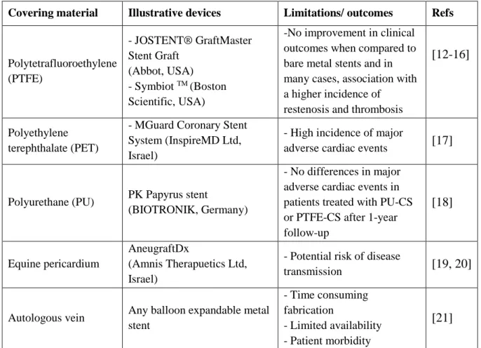
Table 1: Materials employed for the fabrication of covered coronary stents and clinical outcomes.
The need to address these issues has motivated the search for new membranes with
non-thrombogenic properties. In this regard, the use of bio-based materials (i.e. materials that have
either a biological origin or a bio-inspired chemical composition) has been proposed as Covering material Illustrative devices Limitations/ outcomes Refs
Polytetrafluoroethylene (PTFE)
- JOSTENT® GraftMaster Stent Graft
(Abbot, USA) - Symbiot TM (Boston
Scientific, USA)
-No improvement in clinical outcomes when compared to bare metal stents and in many cases, association with a higher incidence of
restenosis and thrombosis
[12-16]
Polyethylene terephthalate (PET)
- MGuard Coronary Stent System (InspireMD Ltd, Israel)
- High incidence of major
adverse cardiac events [17]
Polyurethane (PU) PK Papyrus stent
(BIOTRONIK, Germany)
- No differences in major adverse cardiac events in patients treated with PU-CS or PTFE-CS after 1-year follow-up
[18]
Equine pericardium
AneugraftDx
(Amnis Therapuetics Ltd, Israel)
- Potential risk of disease
transmission [19, 20]
Autologous vein Any balloon expandable metal stent
- Time consuming fabrication
- Limited availability - Patient morbidity
4
alternative to the synthetic ones with the rationale of eliciting a more favourable host response
[22]. Equine pericardium coronary covered stents are currently available on the market (Table
1), but the xenogenic origin may represent a limitation. Autologous veins are also used in the
operating room to cover bare metal coronary stents, but their use is hampered by their limited
availability and the invasiveness of the harvesting procedure. Therefore, advanced materials,
able to provide a non-thrombogenic surface while offering off-the-shelf availability, are
required to address the unmet clinical need of coronary covered stents.
The elastin-like recombinamers (ELRs) are a family of artificial polymers bioinspired by the
pentapeptide VPGVG present in the natural elastin [18, 19]. The ELRs show the advantages of
an engineered material because of the exhaustive control over their composition thanks to their
recombinant nature, while maintaining inherent properties of the natural elastin (i.e. elastic
mechanical behaviour, hemocompatibility and bioactivity) [20]. ELRs are therefore excellent
candidates for the fabrication of devices intended to be in contact with blood [23, 24]. Besides
the membrane material, the fabrication strategy represents a paramount aspect to be considered,
as it must be suitable for covering a device with a very small diameter (e.g. 2-4 mm).
Here, we show the fabrication of covered coronary stents by combining the efficiency, stability
and selectivity of catalyst-free-click chemistry [25, 26], with a layer-by-layer dip-coating
technique, and the hemocompatibility and elasticity of ELRs. We tested the resulting
click-ELR-coronary covered stents for their ability to withstand deployment by balloon catheter and
high shear stress flow. We also evaluated the hemocompatibility of the stents and their
5
2. MATERIALS AND METHODS
2.1. Layer-by-layer fabrication of ELR-covered coronary stents
Two ELRs modified with either cyclooctyne or azide groups (Table 2) [27] were dissolved at
100 mg/mL in PBS at 4 °C. ELR-c is a structural recombinamer while ELR-a contains the RGD
adhesion sequence. For the dip coating process, ELR solutions were kept at 4 °C in separate
cylindrical containers and the coronary stents (custom-made by R.T.M. Rainer Trapp
Medizintechnik GmbH (Germany), L 605 Co-Cr-alloy, polished, with a length of 14.9 mm, an
outer diameter of 1.8 mm and a strut thickness of 90 µm) were sequentially immersed in the
ELR solutions and washed in PBS to remove the excess of polymer. The dipping procedure
was repeated 5 times. The covered stents were stored in ethanol 70%.
Table 2: Amino acid sequences of each ELR and the corresponding reactive groups used for catalyst-free-click chemistry (specifically Huisgen 1,3-dipolar cycloaddition of azides and alkynes). The reactive groups were incorporated by chemical modification of the lysine residues.
ELR Amino acid sequence Reactive group
ELR-c MESLLPVGVPGVG[VPGKG(VPGVG)5]23VPGKGVPGVGV PGVGVPGVGVPGV
Cyclooctyne
ELR-a MGSSHHHHHHSSGLVPRGSHMESLLP[(VPGIG)2(VPGK G)(VPGIG)2]2AVTGRGDSPASS
[(VPGIG)2(VPGKG)(VPGIG)2]2
6
2.2. Mechanical stability of ELR-covered coronary stents
The ELR-coronary covered stents (n=3) were exposed for 24 h to high shear stress flow in a
closed-loop flow system to evaluate the robustness of the polymer covering. The system
consisted of two peristaltic pumps (Ismatec, MCP Process) connected in parallel to a fluid
reservoir through silicone tubes (inner diameter 6.4 mm; Ismatec). The stents were fixed in the
expanded state in a tube with a diameter of 3 mm, and then subjected to the pulsatile arterial
pressure of 80-120 mmHg. The resulting flow rate was set to 300 mL/min resulting in a mean
shear stress of 1.5 Pa, according to the equation [28].
P = (4*η*Q)/(π*r³) Eq. 1
where P is the shear stress (in Pa), η the dynamic viscosity (in Pa*s), r is the inner radius (in m)
of the covered stent and Q the volumetric flow rate (in m³*s-1).
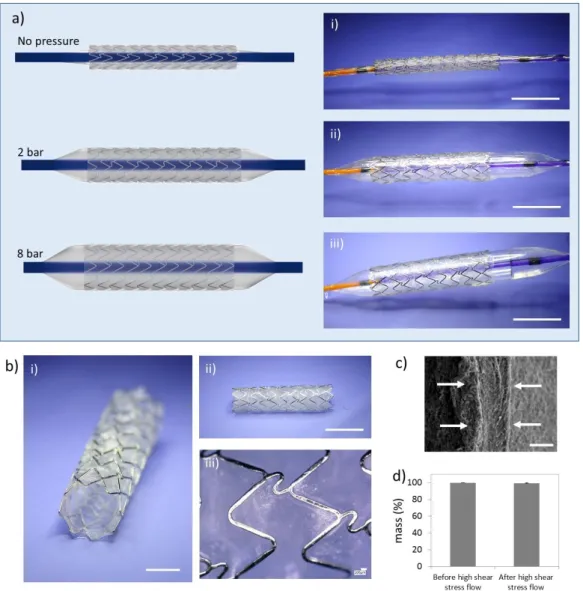
The completeness of the ELR-layer was evaluated by measuring the difference of the dry weight
of the ELR-covered stents before and after exposure to the flow. The dry weight was determined
after storing the ELR-covered stent in a 70 % ethanol solution overnight and subsequently
placing it in an oven (Binder GmbH, Germany) at 60 °C for 90 min. Values are expressed as
mean ± standard deviation (SD). Wilcoxon signed rank test was used for the statistical analysis.
2.3. Balloon expansion of the ELR-covered coronary stents
The stents were positioned on the balloons (TREK coronary dilation catheter, Abbot), and
expanded up to a pressure of 8 atm, corresponding to a stent diameter of 3.4 mm (supplementary
7
2.4. Thrombogenicity assay
Human blood was drawn from healthy volunteers and mixed with 3.2% sodium citrate using an
S-Monovette CPDA1 device (Sarstedt, Germany) at a volumetric ratio of 1:9 to prevent
coagulation. PVC tubes (CODAN pvd Medical GmbH) were filled with 1.5 mL of human
blood. The blood and the ELR-covered stent were placed into a Chandler-loop system, formed
by a closed PVC tube mounted on the rotating head of a roller pump (company) [29, 30]. The
rotational speed of the Chandler loop resulted in a shear rate of 429 s-1 and a shear stress of ~ 1.5 Pa (considering a blood dynamic viscosity of 3.5 x10⁻³ Pa*s), within the range of physiological values [31]. After 1 h of exposure to the blood flow, the stents were washed with
PBS and cut into pieces for SEM visualization. The same procedure was done with the
commercially available ePTFE grafts (GORE-TEX®) as controls. The platelet covered-area
was assessed by image analysis with ImageJ software [32] by analyzing three different regions
per sample, each with an area of 461 µm2. SEM images were coloured with MountainsMap7 SEM software courtesy of Digital Surf, France.
2.5. Scanning electron microscopy
Samples for SEM investigation were fixed in 3% glutaraldehyde in 0.1 M Sorenson’s buffer
(pH 7.4) at room temperature for 1h. They were rinsed with sodium phosphate buffer (0.2 M,
pH 7.39, Merck) and dehydrated consecutively in 30%, 50%, 70% and 90% acetone and then
three times in 100% acetone for 10 minutes. After critical-point-drying in CO2, they were sputter-coated (Leica EM SC D500) with a 20 nm gold-palladium layer. Images were obtained
with an ESEM XL 30 FEG microscope (FEI, Philips, Eindhoven, the Netherlands) with
8
2.6. Cell isolation and culture
Endothelial progenitor cells (EPCs) were isolated from peripheral blood of human adult
volunteers. Anticoagulated blood of healthy donors was carefully added to the separating
solution Histo-Paque-1077 (Sigma-Aldrich, St Louis, Missouri), and centrifuged at 400 g at
room temperature for 30 min. The layer containing mononuclear cells was gently washed twice
with PBS. The cell pellet was resuspended with 15 mL of endothelial cell growth medium
(Endothelial Cell Growth Medium MV2; PromoCell, Heidelberg, Germany) containing
epidermal growth factor (5 ng/mL), basic fibroblast growth factor (10 ng/mL), insulin-like
growth factor (20 ng/mL), vascular endothelial growth factor 165 (0.5 ng/mL), ascorbic acid (1
µg/mL) and hydrocoecisone (0.2 µg/mL), and transferred into T-75 culture flask precoated with
human fibronectin (1 mg/cm2, Sigma-Aldrich, St. Louis, MO). The cells were cultured in 5% CO2 and 95% humidity at 37 °C. EPCs colonies were trypsinized (0.25% trypsin/0.02% ethylenediaminetetraacetic acid solution (Gibco, Karlsruhe, Germany)) and transferred to T-25
culture flask precoated with type I rat-tail collagen (5 mg/cm2, BD Biosciences, San Jose, CA). Cells with 70-80% of confluence were trypsinized and transferred into T-75 culture flasks. Cells
up to passage 9 were used for all experiments.
To study the ability to support endothelialization, the ELR membranes were incubated with a
suspension of EPCs (5x10⁴ cells/mL) for 6 h, after which the medium was exchanged. The scaffolds were then cultured for additional 18 h and subsequently investigated by
9
2.7. Immunohistochemistry
ELR membranes seeded with EPCs were fixed with methanol-free formaldehyde (Roth,
Karlsruhe, Germany) at 4% for 1h at room temperature. Nonspecific sites were blocked and the
cells were permeabilized by incubation in 5% normal goat serum (NGS, Dako) in 0.1%
Triton-PBS. The samples were incubated overnight at 4°C with a 1:100 dilution of the primary mouse
anti-CD31 antibody (P8590, Sigma). After washing three times with PBS, the samples were
incubated for 1 h at room temperature with a 1:400 dilution of AlexaFluor 594 goat anti-mouse
antibody (A11005, Invitrogen). The samples were washed three times with PBS, and then
incubated with Triton X-100 at 0.1 % in PBS for 5 min. The membranes were then incubated
for 45 min at room temperature with Acti-stainTM 488 fluorescent phalloidin (7:1000 in PBS). Samples were counterstained with 4’,6- diamidino- 2-phenylindole (DAPI) nucleic acid stain
(Molecular Probes). Images were acquired using a Zeiss LSM 710 confocal laser scanning
microscope.
3. RESULTS
3.1. Layer-by-layer fabrication of ELR-covered coronary stents
The layer-by-layer fabrication approach (Figure 1 a) resulted in a complete click-ELR
membrane which uniformly covered the whole stent with no defects or voids detected by
10
Figure 1: Fabrication of the click-ELR coronary covered stents. a) i) Schematic of the layer-by-layer technique, in which the stent is sequentially immersed in the solution of ELR-azide and ELR-cyclooctyne to create a crosslinked ELR-covering membrane by means of catalyst-free click-chemistry; ii) Cross-section scheme of the click-ELR coronary covered stent; b) Bare metal coronary stent. i) lateral and ii) frontal view; iii) detailed view of the struts; c) ELR-covered coronary stent i) lateral and ii) frontal view; iii) detailed view of the struts ELR-covered with the click-ELR-membrane. Scale bars: 1 mm.
3.2. Simulated delivery procedure of ELR-covered stent and mechanical stability
The ELR-membrane was able to undergo the increase in diameter from 1.8 mm to 3.4 mm
during balloon expansion without rupture (Figure 2 a and Supplementary video S1). SEM
analysis revealed a thickness of the ELR-layer of approximately 30 µm after expansion (Figure
11
was not significantly different from that before the exposure (Figure 2 d), indicating a stable
covering membrane.
12
3.3. Thrombogenicity evaluation
After stent delivery and exposure to blood under physiological shear stress in the Chandler loop
system, no macroscopic blood clots were found on the surface of the covered stents. Further
microscopic analysis by SEM revealed a complete and homogeneous ELR-membrane, which
completely embedded the struts and bridged the area in between (Figure 3 a-b). The zoom-in
views of the luminal side of the stents showed minimal platelet adhesion on the surface of the
ELR-covered stents, in contrast with the high number of activated platelets present on the
GORE-TEX® surfaces used as control. Further image analysis showed that ELR-stents had a
platelet covered area 5 fold-smaller than GORE-TEX® (Figure 3 g).
13
lumen of the ELR-covered stent; e)-f) Detailed views of the lumen of GORE-TEX® used as control; g) Percentage of area covered by platelets for ELR-covered stent and for GORE-TEX®. Scale bars: a) 500 µm; b) 100 µm; c)-f) 5 µm.
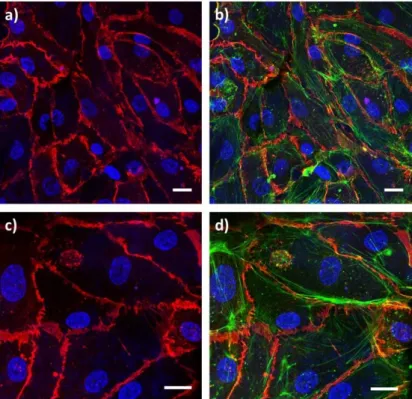
3.4. Endothelialization
The culture of human EPCs on the surface of the click-ELR membranes resulted in a confluent
endothelial layer after 1 day, as shown by the CD31 immunostaining (Figure 4). This bioactive
behaviour is in agreement with the presence of the integrin-mediated adhesion epitope RGD in
the ELR (Figure 4).
14
4. DISCUSSION
The concept of the covered coronary stents relies on a membrane that bridges the area between
the stent struts. The membrane material should ideally be hemocompatible to avoid thrombosis
and be mechanically stable upon implantation to function as physical barrier either to prevent
restenosis or to seal coronary perforations and aneurysms [33]. Thrombosis and restenosis are
still major clinical complications associated with covered stents. Indeed, stents covered with
synthetic traditional polymers (i.e. PET and ePTFE) suffer from the lack of hemocompatibility,
and can elicit severe inflammatory reactions and thrombus formation, leading to low patency
rates [34]. While drug-eluting stents loaded with anti-proliferative and anti-thrombotic drugs
have been suggested as a solution to avoid thrombosis, intimal hyperplasia and subsequent stent
occlusion, they have been also associated with delayed endothelialisation and allergic reactions,
which hampers the expected benefits of the drug-delivery strategy [35, 36].
Alternative solutions that combine advanced biomaterials with suitable fabrication techniques
are, therefore, needed in order to develop covered stents that address the aforementioned current
limitations. This represents an extra challenge for coronary stents, whose small diameter (in the
order of a few millimeters) makes it difficult to use manufacturing techniques that are applicable
to peripheral stents (e.g. injection moulding [24, 37] or suturing of preformed membranes on
bare metal stents [22]). Additionally, the covering-membrane must be elastic to withstand the
change in the diameter during the implantation.
Here, we propose the elastin-like recombinamers, as the biopolymers of choice for covering
coronary stents. The ELRs have shown their potential in a wide range of applications due to
their good biocompatibility [38, 39], low thrombogenicity [23, 24] and their elastic properties
[40]. Specifically, we have used two ELRs that were chemically modified with cyclooctyne and
15
Catalyst-free click-chemistry (and specifically Huisgen 1,3-dipolar cycloadition of azides and
alkynes) outperforms other cross-linking reactions, as it features i) high chemoselectivity, so
that the reactant pairs do not interfere with other biomolecules or cellular reactions [25], ii) high
yields under mild, non-toxic conditions; iii) rapidity; iv) no risk of the release of excess
crosslinking reagents, as the chemical reactive groups are incorporated into the molecules
intended to be crosslinked; v) no need of catalyst, guaranteeing the cytocompatibility of the
reaction [26].
By exploiting the click-chemistry as a reliable cross-linking strategy and the layer-by-layer
dip-coating as a simple and reproducible fabrication approach, we have created ELR covered
coronary stents (Figure 1). The resulting stents were placed on a balloon to undergo a simulated
delivery procedure, in which they were expanded from 1.8 to 3.4 mm (Figure 2), to test their
suitability for percutaneous implantation into coronary arteries. After stent deployment, neither
macroscopic nor microscopic tears were observed when exposed to arterial flow and pressure
conditions (Figure 2). The realization of an intact membrane enables also the use of these stents
to seal aneurysms or arterial perforations, besides restoring the flow through occluded vessels
by excluding the plaque or damaged tissue.
Besides the mechanical elasticity and their potential to act as a physical barrier and to inhibit
intimal hyperplasia, the click-elastin membrane might also provide an antithrombogenic surface
that avoids platelet adhesion and activation. In order to check this, we deployed our
ELR-covered stents in a Chandler loop system and exposed them to whole human blood under
dynamic arterial flow. There were no signs of leukocyte adhesion, and only few platelets were
observed by SEM, in sharp contrast with the high number of platelets adhered and activated on
the GORE-TEX® surface (Figure 3). These results agree with previous studies which proposed
16
devices [23, 24, 41, 42] and support the use of ELRs for the fabrication of covered stents that
will be in direct contact with the blood before endothelialization.
In addition to the intrinsic hemocompatibility shown in the blood-contacting test, the
anti-thrombogenicity of the ELR-covered stents is further guaranteed by their ability to support the
formation of a confluent endothelial layer, as shown by culturing human EPCs (Figure 4).
Circulating EPCs are increasingly recognized as important contributors to vascular prosthesis
endothelialization, making them important mediators of implant compatibility [43, 44]. Besides
its paramount role as anti-thrombogenic surface, the endothelium is involved in several aspects
of vascular biology, e.g. blood vessel tone, hemostasis, neutrophil recruitment and hormone
trafficking [45, 46]. Therefore, the ability of the covered stent to support endothelialization is
of key importance to achieve the success of the implant.
It is important to consider that a stented artery presents a geometry based on the stent strut
configuration, which contrasts with the smoothness of the healthy arteries. This inevitably leads
to a disturbed local hemodynamics (i.e. turbulent flow) [47], which has been related to the
stimulation of vascular smooth muscle cell proliferation [48] and platelet activation [49].
Indeed, the thickness of the stent-struts plays an important role in-stent restenosis, with thinner
struts showing considerably lower restenosis rates [50-52]. Importantly, covering the stent with
the ELR-membrane bridges the stent-struts, with a concomitant reduction in the height changes
in between the stent struts, which may further contribute to the positive outcome of the device.
Overall, we have established a strategy that combines the simplicity of the layer-by-layer
technique with the biocompatibility and specificity of the catalyst-free click-chemistry to
develop a new class of coronary covered stents. The elastic membrane provided physiological
hemocompatibility to the implant, and enabled endothelialization, while representing a physical
17
recombinant nature of the ELR allows for the introduction of further biological cues (e.g.
immunomodulatory cues, antimicrobial peptides) to develop a platform that enables stent
fabrication with tailored biofunctionalities.
Acknowledgements
This work was funded by the Excellence Initiative of the German federal and state governments
in the framework of the START-UP Program (StUpPD_330-18), and by the START-Program
of the Medical Faculty of RWTH Aachen University (60/17). JCRC acknowledges the funding
from the Spanish Government (PCIN-2015-010 (FunBioPlas)) and MAT2016-78903-R), Junta
de Castilla y León (VA317P18) and Centro en Red de Medicina Regenerativa y Terapia Celular
de Castilla y León.
The authors acknowledge the support of Prof. Dr. Müller-Newen, Sabrina Ernst and the
confocal microscopy facility, a core facility of the Interdisciplinary Center for Clinical Research
(IZKF) Aachen within the Faculty of Medicine at RWTH Aachen University. The authors also
thank Andreas Lubig for his assistance with the graphic software.
Data availability
The data associated with this manuscript is available upon request to the corresponding author.
References
1. Townsend, N., et al., Cardiovascular disease in Europe: epidemiological update 2016. European Heart Journal, 2016. 37(42): p. 3232-3245.
2. Byrne, R.A., M. Joner, and A. Kastrati, Stent thrombosis and restenosis: what have we learned and where are we going? The Andreas Gruntzig Lecture ESC 2014. Eur Heart J, 2015. 36(47): p. 3320-31.
3. Collet, C., et al., Coronary stent thrombosis: what have we learned? Journal of thoracic disease, 2016. 8(7): p. 1398-1405.
4. Babapulle, M.N. and M.J. Eisenberg, Coated Stents for the Prevention of Restenosis: Part I. Circulation, 2002. 106(21): p. 2734-2740.
5. Farhatnia, Y., et al., Evolution of covered stents in the contemporary era: clinical application, materials and manufacturing strategies using nanotechnology. Biotechnol Adv, 2013. 31(5): p. 524-42.
18
7. Romaguera, R. and R. Waksman, Covered stents for coronary perforations: is there enough evidence? Catheter Cardiovasc Interv, 2011. 78(2): p. 246-53.
8. Jamshidi, P., K. Mahmoody, and P. Erne, Covered stents: a review. Int J Cardiol, 2008. 130(3): p. 310-8.
9. Takano, M., et al., Delayed endothelialization after polytetrafluoroethylene-covered stent implantation for coronary aneurysm. Circ J, 2009. 73(1): p. 190-3.
10. Nakayama, Y., et al., Fabrication of micropored elastomeric film-covered stents and acute-phase performances. J Biomed Mater Res A, 2003. 64(1): p. 52-61.
11. Sato, S., et al., Development of self-expandable covered stents. J Biomed Mater Res B Appl Biomater, 2007. 83(2): p. 345-53.
12. Kawamoto, H., et al., Short-Term and Long-Term Outcomes After Polytetrafluoroethylene-Covered Stent Implantation for the Treatment of Coronary Perforation. Am J Cardiol, 2015. 116(12): p. 1822-6.
13. Stone, G.W., et al., 5-year follow-up of polytetrafluoroethylene-covered stents compared with bare-metal stents in aortocoronary saphenous vein grafts the randomized BARRICADE (barrier approach to restenosis: restrict intima to curtail adverse events) trial. JACC Cardiovasc Interv, 2011. 4(3): p. 300-9.
14. Schächinger, V., et al., A randomized trial of polytetrafluoroethylene-membrane-covered stents compared with conventional stents in aortocoronary saphenous vein grafts. Journal of the American College of Cardiology, 2003. 42(8): p. 1360-1369.
15. Turco, M.A., et al., Pivotal, randomized U.S. study of the Symbiot™ covered stent system in patients with saphenous vein graft disease: Eight-month angiographic and clinical results from the Symbiot III trial. Catheterization and Cardiovascular Interventions, 2006. 68(3): p. 379-388. 16. Stankovic, G., et al., Randomized evaluation of polytetrafluoroethylene-covered stent in saphenous vein grafts: the Randomized Evaluation of polytetrafluoroethylene COVERed stent in Saphenous vein grafts (RECOVERS) Trial. Circulation, 2003. 108(1): p. 37-42.
17. Dudek, D., et al., Mesh-covered embolic protection stent implantation in ST-segment-elevation myocardial infarction: final 1-year clinical and angiographic results from the MGUARD for acute ST elevation reperfusion trial. Circ Cardiovasc Interv, 2015. 8(2): p. e001484.
18. Hernandez-Enriquez, M., et al., Outcomes after use of covered stents to treat coronary artery perforations. Comparison of old and new-generation covered stents. J Interv Cardiol, 2018. 31(5): p. 617-623.
19. Chen, S., et al., Pericardial covered stent for coronary perforations. Catheter Cardiovasc Interv, 2015. 86(3): p. 400-4.
20. Buchanan, G.L., A. Durante, and A. Chieffo, Pericardium-covered stent: a reality for coronary interventions of the future? . Interventional Cardiology, 2012. 4(4): p. 411-418.
21. Chae, J.K., et al., Successful treatment of coronary artery perforation during angioplasty using autologous vein graft-coated stent. European Heart Journal, 1997. 18(6): p. 1030-1032. 22. Ichihashi, S., et al., Bio-Based Covered Stents: The Potential of Biologically Derived Membranes.
Tissue Eng Part B Rev, 2019. 25(2): p. 135-151.
23. Jordan, S.W., et al., The effect of a recombinant elastin-mimetic coating of an ePTFE prosthesis on acute thrombogenicity in a baboon arteriovenous shunt. Biomaterials, 2007. 28(6): p. 1191-7.
24. de Torre, I.G., et al., Elastin-like recombinamer-covered stents: Towards a fully biocompatible and non-thrombogenic device for cardiovascular diseases. Acta Biomater, 2015. 12: p. 146-155.
25. McKay, C.S. and M.G. Finn, Click chemistry in complex mixtures: bioorthogonal bioconjugation. Chem. Biol., 2014. 21(9): p. 1075-101.
26. Jiang, Y., et al., Click hydrogels, microgels and nanogels: emerging platforms for drug delivery and tissue engineering. Biomaterials, 2014. 35(18): p. 4969-85.
19
28. Darby, R. and R.P. Chhabra, Chemical Engineering Fluid Mechanics. 2001: CRC Press. 559. 29. Chandler, A.B., In vitro thrombotic coagulation of the blood; a method for producing a
thrombus. Lab Invest, 1958. 7(2): p. 110-4.
30. Gong, J., et al., Tubing loops as a model for cardiopulmonary bypass circuits: both the biomaterial and the blood-gas phase interfaces induce complement activation in an in vitro model. J Clin Immunol, 1996. 16(4): p. 222-9.
31. Gaamangwe, T., S.D. Peterson, and M.B. Gorbet, Investigating the Effect of Blood Sample Volume in the Chandler Loop Model: Theoretical and Experimental Analysis. Cardiovascular Engineering and Technology, 2014. 5(2): p. 133-144.
32. Schneider, C.A., W.S. Rasband, and K.W. Eliceiri, NIH Image to ImageJ: 25 years of image analysis. Nature Methods, 2012. 9: p. 671.
33. Al-Mukhaini, M., et al., Coronary perforation and covered stents: an update and review. Heart views : the official journal of the Gulf Heart Association, 2011. 12(2): p. 63-70.
34. Banerjee, S., et al., Femoropopliteal Artery Stent Thrombosis: Report From the Excellence in Peripheral Artery Disease Registry. Circ Cardiovasc Interv, 2016. 9(2): p. e002730.
35. Nebeker, J.R., et al., Hypersensitivity cases associated with drug-eluting coronary stents: a review of available cases from the Research on Adverse Drug Events and Reports (RADAR) project. J Am Coll Cardiol, 2006. 47(1): p. 175-81.
36. Khan, W., S. Farah, and A.J. Domb, Drug eluting stents: Developments and current status. Journal of Controlled Release, 2012. 161(2): p. 703-712.
37. Ichihashi, S., et al., In Vitro Quantification of Luminal Denudation After Crimping and Balloon Dilatation of Endothelialized Covered Stents. CardioVascular and Interventional Radiology, 2017. 40(8): p. 1229-1236.
38. Urry, D.W., et al., Biocompatibility of the Bioelastic Materials, Poly(GVGVP) and Its γ-Irradiation Cross-Linked Matrix: Summary of Generic Biological Test Results. Journal of Bioactive and Compatible Polymers, 1991. 6(3): p. 263-282.
39. Ibanez-Fonseca, A., et al., Biocompatibility of two model elastin-like recombinamer-based hydrogels formed through physical or chemical cross-linking for various applications in tissue engineering and regenerative medicine. J Tissue Eng Regen Med, 2018. 12(3): p. e1450-e1460. 40. Hoeve, C.A. and P.J. Flory, The elastic properties of elastin. Biopolymers, 1974. 13(4): p.
677-86.
41. Woodhouse, K.A., et al., Investigation of recombinant human elastin polypeptides as non-thrombogenic coatings. Biomaterials, 2004. 25(19): p. 4543-53.
42. Ito, S., S. Ishimaru, and S.E. Wilson, Application of coacervated alpha-elastin to arterial prostheses for inhibition of anastomotic intimal hyperplasia. ASAIO J, 1998. 44(5): p. M501-5. 43. Melchiorri, A.J., et al., In Vitro Endothelialization of Biodegradable Vascular Grafts Via
Endothelial Progenitor Cell Seeding and Maturation in a Tubular Perfusion System Bioreactor. Tissue Eng Part C Methods, 2016. 22(7): p. 663-70.
44. Yu, Y., et al., Characterization of Endothelial Progenitor Cell Interactions with Human Tropoelastin. PLoS One, 2015. 10(6): p. e0131101.
45. Rajendran, P., et al., The vascular endothelium and human diseases. Int J Biol Sci, 2013. 9(10): p. 1057-69.
46. Sandoo, A., et al., The endothelium and its role in regulating vascular tone. Open Cardiovasc Med J, 2010. 4: p. 302-12.
47. Murphy, E.A. and F.J. Boyle, Reducing In-Stent Restenosis Through Novel Stent Flow Field Augmentation. Cardiovascular Engineering and Technology, 2012. 3(4): p. 353-373.
48. Shigematsu, K., et al., Direct and indirect effects of pulsatile shear stress on the smooth muscle cell. Int Angiol, 2000. 19(1): p. 39-46.
20
50. Sakamoto, A., et al., Understanding the Impact of Stent and Scaffold Material and Strut Design on Coronary Artery Thrombosis from the Basic and Clinical Points of View. Bioengineering (Basel), 2018. 5(3).
51. Kastrati, A., et al., Intracoronary stenting and angiographic results: strut thickness effect on restenosis outcome (ISAR-STEREO) trial. Circulation, 2001. 103(23): p. 2816-21.