7
7
PAPER
View Article OnlineView Journal | View Issue
Fourier transform microwave spectroscopy of
Ac-Ser-NH
2
: the role of side chain interactions in peptide
folding
Carlos Cabezas,a Martinus A. T. Robben,b Anouk M. Rijs,b Isabel Peñaa and J. L. Alonso*a
Serine capped dipeptide N-acetyl-L-serinamide (Ac-Ser-NH2) has been investigated using Fourier transform microwave
spectroscopic techniques combined with laser ablation sources. Spectral signatures originating from one dominant species have been detected in the supersonic expansion. Rotational and nuclear quadrupole coupling constants of the two 14N nuclei have been used in the characterization of a Ceq/g-turn structure, which is stabilized by a CO· · ·HN
intramolecular hydrogen bond closing a seven-membered ring. Two extra hydrogen bonds involving the polar side chain (–CH2OH) further stabilize the structure. The non-observation of C5 species, attributed to the presence of
the polar side chain, is in contrast with the previous gas phase observation of the related dipeptides containing glycine or alanine residues. The A–E splitting pattern arising from the internal rotation of the methyl group has been analyzed and the internal rotation barrier has been determined.
Introduction
Detailed knowledge of the mechanisms of protein folding is important in order to unravel structure–function relationships and improve our understanding of the numerous processes in
living cell functions.1 In the solution or the solid state, the
protein folding process is a complex interplay of both intra- and intermolecular interactions with the solvent or surrounding amino acids of the protein. Although all these interactions might be present in their biological environment, in order to fully understand the intramolecular folding mechanisms it is also necessary to be able to investigate the intrinsic interactions within the proteins, in the absence of any solvent or crystal phase. In particular, non-covalent interactions such as intra- molecular hydrogen bonds are interesting, since they define the present secondary structure. Gas phase experiments can provide
a Grupo de Espectroscopia Molecular (GEM), Edificio Quifima, Laboratorios de
Espectroscopia y Bioespectroscopia, Unidad Asociada CSIC, Parque Cient´ıfico UVa, Universidad de Valladolid, Paseo de Bel´en 5, 47011 Valladolid, Spain.
E-mail: jlalonso@qf.uva.es; Tel: +34 983186345
b Radboud University, Institute for Molecules and Materials, FELIX Laboratory,
Toernooiveld 7-c, 6525 ED Nijmegen, The Netherlands
conditions required for studying only the inherent properties of the molecules under investigation, free of interactions with
the environment.2,3
The first step towards fully understanding protein folding requires detailed information on the intrinsic conformational preferences of amino acids, small peptides and peptide mimics. In particular, dipeptide mimics (containing two peptide linkages, –CO–NH–) have received much attention because they represent the smallest realistic and representative systems for designing local conformational effects in peptides and proteins. These molecules are also named capped dipeptides. The vast majority of this work has been done in molecular beam IR/UV double
resonance experiments.3,4 A disadvantage of those techniques,
however, is that they require the molecule under study to possess a UV chromophore. Roughly speaking, this restricts this type of spectroscopy to molecules containing one or more aromatic rings, thereby excluding the majority of the amino acids and limiting strongly the different peptides that can be studied.
In contrast, Fourier transform microwave (FTMW) spectro- scopy does not require any chromophore; it only needs the
molecule to be studied to have a permanent dipole moment.5
Therefore, it is particularly well suited for studying dipeptides containing amino acids as relevant as glycine, alanine or proline,
elusive to IR/UV double resonance experiments. Lavrich et al.6
investigated the alanine dipeptide N-acetyl-alanine N0-methylamide
(Ac-Ala-NHMe) and observed only a Ceq conformation using
Paper
7
Scheme 1 Chemical structures of the Ceq (left) and C
5 (right) configura-
was prepared by pressing the compound’s fine powder mixed with a small amount of commercial binders and was placed in the ablation nozzle. A picosecond Nd:YAG laser (10 mJ per pulse, 20 ps pulse width) was used as a vaporization tool. Products of the laser ablation were supersonically expanded using the flow of carrier gas (Ne, 15 bar) and characterized by both chirped pulse
Fourier transform microwave (CP-FTMW)10 and laser ablation
molecular beam Fourier transform microwave (LA-MB-FTMW)20
tions for Ac-XX-NH2 (XX = Gly and Ala) derivatives. Nc and Nt indicate
central and terminal nitrogen atoms, respectively.
with laser ablation methods7–10 has been successfully applied in
the investigation of the conformational preferences of isolated
protected dipeptides such as N-acetyl-glycinamide (Ac-Gly-NH2),11
N-acetyl-alaninamide (Ac-Ala-NH2)12 and N-acetyl-prolinamide
(Ac-Pro-NH2).13 For both Ac-Gly-NH2 and Ac-Ala-NH2, the mole-
cules were found to exist as both the Ceq (g-turn) as well as the C
spectroscopy. N-Acetyl-L-serinamide was first investigated using a
CP-FTMW spectrometer to sample swiftly the rotational spectra of the different conformers present in the gas-phase mixture.
Details of the experimental setup have been given elsewhere.21
Chirped-pulses of 4 ms directly generated by the 24 Gs s-1 AWG
were amplified to about 300 W peak power using a traveling wave tube amplifier. A parabolic reflector system composed of dual ridge horns and two parabolic reflectors in a paraxial beam
configuration21 was used to broadcast the excitation pulse and
7 5
(bL-turn) conformation (see Scheme 1), in which the backbones
are stabilized by a CO· · ·HN intramolecular hydrogen bond
closing a seven- or five-membered ring, respectively. In contrast,
in the investigation of the Ac-Pro-NH2 only the C7 (g-turn)
conformer was detected. The presence of the pyrrolidine ring provides rigidity to the peptide backbone and the formation of other configurations, possible in the other model peptides, is not
observed for the Ac-Pro-NH2 dipeptide.
The present work reports the first conformational study using Fourier transform microwave techniques of a dipeptide analogue
containing an amino acid with a polar side chain, N-acetyl-L-
serinamide (Ac-Ser-NH2). The introduction of a polar side chain is
an important step because amino acids with polar functional
groups are very relevant to the protein function and structure.14 In
fact, serine residues have been found to have detrimental effects on the stability of transmembrane helices since they are involved
in interhelical hydrogen bonds.15 Moreover, the hydrogen bonds
established by the polar group of serine residues have been shown
to be at the origin of the activation processes of b2-adrenergic
receptors.16,17 From the conformational point of view, the
presence of a polar functional group, –CH2OH, in the side chain
of serine allows the establishment of additional intramolecular hydrogen bonds, which dramatically increase the number of
low-energy conformers.18 However, sometimes these extra intra-
molecular interactions are the motif of the selective stabilization of a determined conformation, as it has been shown for the
natural a-amino acid asparagine.19 These additional interactions
do not occur in the aliphatic amino acids whose dipeptides have been previously studied. Consequently, the main goal of our research is to elucidate how this polar side chain of serine affects
the conformational preferences of the dipeptide Ac-Ser-NH2. Will
a rich conformational space be observed? Or will the polar side
receive the broadband molecular emission. At a repetition rate of 2 Hz, a total of 70 000 free induction decays (4 FID emissions per
gas pulse) each with 10 ms length duration were averaged and
digitized using a 50 Gs s-1 digital oscilloscope. The frequency
domain spectrum in the 6–12 GHz frequency range was obtained by taking a fast Fourier transform (FFT) following the application of a Kaiser–Bessel window to improve the baseline resolution.
The sub-Doppler resolution LA-MB-FTMW technique,20 operating
from 4 to 10 GHz, was used to record the rotational spectrum with the resolution necessary to analyze the hyperfine structure
due to the presence of two 14N nuclei in the molecule. A short
microwave radiation pulse of 0.3 ms duration was applied
to polarize all the vaporized molecules. The registered free induction decay was then converted to the frequency domain by Fourier transformation. All the transitions appeared as Doppler doublets due to the parallel configuration of the molecular beam and the microwave radiation. The resonance frequency was determined as the arithmetic mean of the two Doppler components. Frequency accuracy better than 3 kHz and an estimated resolution of 5 kHz are achieved in the experiment. From 50 to 100 averages were phase-coherently coadded to achieve reasonable signal to noise ratios (S/N).
Results and discussion
The recorded broadband rotational spectrum of laser ablated
N-acetyl-L-serinamide from 6 to 12 GHz is shown in Fig. 1a.
Decomposition product lines common to other studies of
biomolecules22,23 attributable to cyano-derivatives, water clusters,
etc. were easily identified. After excluding the aforementioned
signals from the spectral analysis, the identification of rotational
chain favor one of the Ceq or C conformations? transitions belonging to a single species was accomplished. Assign-
7 5
Experimental
A commercial sample of Ac-Ser-NH2 (GeneCust, B99%, m.p.
B183 1C) was used without any further purification. A solid rod
ments were based on the identification of a-type J + 10, J+1 ’ J0, J and
J + 11, J+1 ’ J1, J (with J ranging from 3 to 6) pairs of rotational
progressions which appear to be splitted in two components as
shown in Fig. 1b for 414–313 and 404–303 transitions. We attributed
Fig. 1 (a) Broadband spectrum of Ac-Ser-NH2 in the 6–12 GHz frequency region showing the most intense rotational transitions for the observed
rotamer together with some of the photofragmentation product lines. (b) A section of the broadband spectrum showing the A–E states of the 414–313
and 404–303 rotational transitions. (c) The unperturbed A-state of the 404–303 rotational transition of Ac-Ser-NH2 showing its hyperfine structure
completely resolved using a LA-MB-FTMW spectrometer. Each hyperfine component labeled with the corresponding values of I0 , F0 ’ I00 , and F00
quantum numbers is split by the Doppler effect.
The V3 torsional barriers for these methyl groups are low, as 12 LA-MB-FTMW technique, which provides the high resolution
shown, for example, for the related molecule Ac-Ala-NH2 causing needed to fully resolve this complicated hyperfine structure
the occurrence of the A–E splittings due to the coupling of the torsional vibration to the overall rotational angular momentum.
Once the analysis of the ma-type spectrum was completed, mb-type
transitions were subsequently predicted and measured with no
mc-type spectrum being observed. Although several lines remained
unassigned in the spectrum, the identification of further rotamers could not be achieved.
Both A and E components showed a partially resolved hyper-
(as shown in Fig. 1c). Hence, a total of 89 hyperfine compo-
nents from twelve aR- and three bR-branch transitions for the
unperturbed A state (see Table S2 of the ESI†) were analyzed24
using Watson’s Hamiltonian H = HR + HQ, where HR is the
semirigid rotor Hamiltonian and HQ describes the nuclear
quadrupole coupling interaction.25 The quadrupole coupling
Hamiltonian was set up in the coupled basis set (I1 I2 I J F),
I1 + I2 = I, and I + J = F.26 The energy levels involved in each
fine structure corresponding to a compound with 14N nuclei. transition were thus labelled with the quantum numbers
J, K -1,
This is because the 14N nuclei have a non-zero quadrupole
moment (I = 1) owing to a non-spherical distribution of the
nuclear charge, which interacts with the electric field gradient created by the rest of the molecule at the site of these nuclei. The
nuclear spin of 14N nuclei couples to the rotational angular
momentum resulting in a hyperfine structure in the rotational
spectrum.5 However, the spectral resolution attainable in the
CP-FTMW experiments is not enough to completely resolve these hyperfine effects. For this reason, in a second stage of the
investigation N-acetyl-L-serinamide was probed using our
K+1, I, and F. The analysis rendered the accurate experimental
rotational and nuclear quadrupole coupling constants shown
in the left column of Table 1. The internal rotation barrier V3
has been determined using the internal axis method in the
form given by Woods.27 The A–E splittings (see Table S3 in the
ESI†) were fit using the program XIAM28 yielding the internal
rotation parameters summarized in Table 2.
In order to be able to identify the conformation corresponding to the observed rotamers in the spectrum, the conformational
Paper
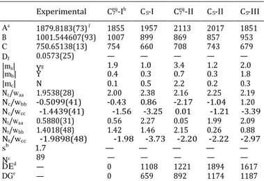
Experimental Ceq-Ih C -I Ceq-II C -II C -III
Aa 1879.8183(73) f 1855 1957 2113 2017 1851
B 1001.544607(93) 1007 899 869 857 953 C 750.65138(13) 754 660 708 743 679 DJ
|ma|
0.0573(25) Yg
—
1.9 — 1.0 — 3.4 — 1.2 — 2.0
89 — — — — —
— 0 1108 1221 1894 1617 — 0 659 892 1174 1187
7
7
7
7
7
Table 1 Experimental and calculated spectroscopic parameters for the
observed rotamer and the five lowest energy conformers of Ac-Ser-NH2.
Ab initio energies are included for the predicted species
7 5 7 5 5
|mb| Y 0.4 0.3 0.7 0.3 1.8
|mc| N 0.1 0.5 2.2 0.2 0.3
Nc/waa 1.9538(28) 2.00 2.38 2.16 2.25 2.19
Nc/wbb -0.5099(41) -0.43 0.86 -2.17 -1.04 1.20
Nc/wcc -1.4439(41) -1.56 -3.25 0.01 -1.21 -3.39
Nt/waa 0.5880(31) 0.56 2.27 0.05 1.99 2.09
Nt/wbb 1.4018(48) 1.42 1.46 2.15 0.26 0.88
Nt/wcc -1.9898(48) -1.98 -3.73 -2.20 -2.22 -2.97
sb 1.7 — — — — —
Nc
D Ed
DGe
a A, B, and C represent the rotational constants (in MHz); D J is the
quartic centrifugal distortion constant (in kHz), and waa, wbb and wcc are
the diagonal elements of the 14N nuclear quadrupole coupling tensor
(in MHz); Nc and Nt correspond to the central and terminal 14N nuclei,
respectively; ma, mb and mc are the electric dipole moment components
(in D). b Rms deviation of the fit (in kHz). c Number of measured
transitions. d Relative energies (in cm-1) with respect to the global
minimum calculated at the MP2/6-311++G(d,p) level of theory. e Gibbs
Fig. 2 The predicted five low-energy conformers of Ac-Ser-NH2.
satisfactorily in previous studies of several biomolecules.7–10,12,13
The results of the calculations at the B3LYP/6-311++G(d,p) level
of theory are shown in Table S1 of the ESI.†
The conformational assignment of the observed rotamer was achieved by comparing the experimental spectroscopic
constants with those predicted ab initio. The experimentally
determined rotational constants are similar to those for the
energies (in cm-1) calculated at 298 K at the MP2/6-311++G(d,p) level of
Ceq 11–13
theory. f Standard error in parentheses in units of the last digit. g Yes or 7 -I conformer. As we have recently shown, nuclear quad-
No to observation of a-, b-, and c-type transitions. h The conformers are
labeled by the size of the ring closed by the CO· · ·NH hydrogen bond
and an index going up with increasing energy.
Table 2 Methyl internal rotation experimental parameters for the identi-
fied rotamer of Ac-Ser-NH2
V3a 97.2 (3)c [127] o(i,a)b 43.40 (2) [46]
o(i,b) 59.68 (2) [59]
o(i,c) 62.23 (2) [60]
rupole coupling constants can be used as fingerprints in con- formational analysis of related dipeptides. These parameters are sensitive to the chemical environment of the nitrogen nuclei and to the orientation of the amino group with respect to the principal inertial axis system. Thus, a final comparison between the experimental and theoretical values for those constants clearly serves to discriminate between all the conformers and allows the unequivocal identification of the observed rotamer as
conformer Ceq-I. The observed rotamer exhibited an intense
ma-type spectrum and weak mb-type transitions, which is also in
a V
3 is the internal rotation barrier in cm
-1. b Experimentally deter- agreement with the identification of the Ceq-I conformer when
mined values for the angles between the top rotational axis and the principal axis system, in degrees. In squared brackets, the theoretical values predicted for conformer Ceq-I are shown. c Standard error in
parentheses in units of the last digit.
considering the respective calculated values of the electric dipole moment components. Additionally, the methyl internal rotation experimental parameters are in good agreement with those
estimated theoretically (Table 2) for conformer Ceq-I, which also
chemical calculations. In a first step, semiempirical calculations29
were performed to search for all possible energetic minima of the
Ac-Ser-NH2 molecular system. The resulting molecular geometries
were further optimized within the Gaussian suite of programs,30
using a computationally effective B3LYP density functional model with Pople’s 6-311++G(d,p) basis set and afterward by second- order Møller–Plesset (MP2) perturbation theory in the frozen core approximation with the same basis set. Frequency calculations were performed using both methods to compute the Gibbs free energies, which should be more representative of the relative populations of each structure. The derived rotational and
14N nuclear quadrupole coupling constants together with the
dipole moment components for the lowest-lying energy con- formers (see Fig. 2) at the MP2/6-311++G(d,p) level of theory are shown in Table 1. This level of theory has been found to behave
supports the achieved assignment.
The experimental determination of the 14N nuclear quadru-
pole coupling constants constitutes an exceptional tool that allows the unequivocal establishment of the orientation of the
side chain –NH2 and –NH groups with respect to the molecular
frame. These constants can be used to deduce the nature of the intramolecular interactions in which this functional group is
involved. Hence, in the Ceq-I conformer structure the acetyl
carbonyl oxygen is hydrogen bonded to one of the terminal
amide hydrogens (CQO· · ·H–Nt), closing a seven-membered
cycle in which the serine side chain is oriented equatorially.
Moreover, the –CH2OH group of the serine side chain participates
in two additional hydrogen bonds: one Nc–H· · ·O–H and one
O–H· · ·OQC. As can be seen in Fig. 3, the estimated distances
of the hydrogen bonds show that the O–H· · ·OQC interaction is
7
7
2 2
7
Fig. 3 3D ab initio structure (Cartesian coordinates in Table S4 of
the ESI†) of the observed conformer of Ac-Ser-NH2 showing the intra-
molecular interactions and the estimated distances which stabilize the structure.
strength, allowing the competition with other types of inter-
actions such as SH–p interactions resulting in the presence of
both g-turn as b-turn conformations. This result confirms our
conclusion that for Ac-Phe-Ser-NH2, which exhibits a strong
hydrogen bond, only one conformer is present, while when weaker H-bond interactions are formed such as for Cys or for
the previously studied Gly11 and Ala12 capped peptides, other
conformers besides this C7 structure will be present.
Conclusions
The present investigation of the Ac-Ser-NH2 system together with
the previous work on Ac-Gly-NH2, Ac-Ala-NH2 and Ac-Pro-NH2
illustrates the capabilities of the Fourier transform microwave techniques to investigate the conformational preferences of biologically relevant peptides isolated in the gas phase. The structural information derived for these aliphatic dipeptides, which are elusive to other high resolution spectroscopic tech-
niques, is of utmost importance not only to gain knowledge
about their intrinsic conformational properties but also to serve the dominant donor character of the OH group over its acceptor
propensity, leading to a quite unbalanced H-bonding network.
The distance for the CQO· · ·H–Nt bond (g-turn) is analogous to
those related for other g-turns of aliphatic dipeptides such as
Ac-Gly-NH211 and Ac-Ala-NH2,12 for which intramolecular bond
distances of 2.03 and 2.07 Å were found, respectively. This fact points to the fact that the intramolecular interactions of the
side chain do not affect the strength of the g-turn bond.
However, the side chain extra interactions, which cannot take
place in any other possible conformation of the Ac-Ser-NH2,
seem to be the factor which accounts for the overstabilization
of this species and, thus, the non-observation of C5 species. In
as a benchmark for theoretical investigations.
Our results for the serine dipeptide Ac-Ser-NH2 show that its
conformational landscape in the gas phase is dominated by a
single Ceq/
g-turn species. Now, the initial research question
about whether the weaker polar side chain favors one of the C7
and C5 conformations through the formation of intramolecular
hydrogen bonds can be answered. The additional intramolecular interactions formed by the presence of a polar group in the side chain of serine have been shown to be at the origin of
the conformational locking to a Ceq species observed for the
dipeptide Ac-Ser-NH2. Hence, it has been demonstrated that
the presence of a polar side chain increases the plausible
contrast, for the related Ac-Gly-NH 11 and Ac-Ala-NH 12 dipeptides number of conformations but in contrast imparts restrictions
both Ceq and C
5 species were detected with the approximate on the amount of conformers observed.
population Ceq/C ratio of around 2 and 3, respectively. Because
7 5
no intramolecular interactions involving the lateral side chain can occur in Gly and Ala dipeptides, the stability/abundance difference
between Ceq and C species is exclusively determined by the strength
Acknowledgements
7 5 This research was supported by MINECO (grant numbers CTQ
of the CQO· · ·H–N interactions (seven- or five-membered ring).
On this basis, we can infer that the presence of polar side chains alters significantly the conformational preferences of dipeptides containing aliphatic amino acids.
The results obtained here for Ac-Ser-NH2 can be compared
with those reported previously for the analogous tripeptide
Ac-Phe-Ser-NH2 using IR-UV ion dip spectroscopy.31,32 For the
Ac-Phe-Ser-NH2 molecule only the C7/g-turn conformer, where the
C7 ring established by the serine residue is structurally similar to
that of the detected conformer of Ac-Ser-NH2, was observed.
Additionally, Yan et al.31 found the same C7 structure to be the
most stable for the tripeptide Ac-Phe-Cys-NH2. This peptide only
differs by one atom with respect to Ac-Phe-Ser-NH2, where the –OH
side chain of serine is replaced by the –SH group in cysteine.
For the Ac-Phe-Cys-NH2 an additional structure which exhibits
C10/b-turn geometry was observed. The presence of the second
structure was attributed to the weaker SH· · ·O hydrogen bond
2010-19008, CTQ 2013-40717-P and Consolider Ingenio 2010 CSD 2009-00038), Junta de Castilla y Leo´n (grant number VA175U13) and ERC (grant number 610256 ‘‘Nanocosmos’’).
C.C.thanks the Junta de Castilla y Leo´n for the postdoctoral
contract (grant number CIP13/01).
Notes and references
1 Y. Park and V. Helms, On the Derivation of Propensity
Scales for Predicting Exposed Transmembrane Residues
of Helical Membrane Proteins, Bioinformatics, 2007, 23, 701–
708.
2 E. G. Robertson and J. P. Simons, Getting into Shape:
Conformational and Supramolecular Landscapes in Small
Biomolecules and their Hydrated Clusters, Phys. Chem.
Paper
7
Ceq
3 M. S. de Vries and P. Hobza, Gas-phase Spectroscopy of
Biomolecular Building Blocks, Annu. Rev. Phys. Chem., 2007,
58, 585–612.
4 (a) W. Chin, F. Piuzzi, I. Dimicoli and M. Mons, Probing
the competition between secondary structures and local
preferences in gas phase isolated peptide backbones, Phys.
Chem. Chem. Phys., 2006, 8, 1033–1048; (b) S. Jaeqx, W. Du,
E. J. Meijer, J. Oomens and A. M. Rijs, Conformational Study
of Z-Glu-OH and Z-Arg-OH: Dispersion Interactions versus
Conventional Hydrogen Bonding, J. Phys. Chem. A, 2012,
117, 1216–1227; (c) S. Jaeqx, J. Oomens and A. M. Rijs,
Gas-phase Salt Bridge Interactions between Glutamic Acid and
Arginine, Phys. Chem. Chem. Phys., 2013, 15, 16341–16352;
(d) S. Jaeqx, J. Oomens, A. Cimas, M.-P. Gaigeot and
A. M. Rijs, Gas-Phase Peptide Structures Unraveled by Far-IR Spectroscopy: Combining IR-UV Ion-Dip Experiments with
Born–Oppenheimer Molecular Dynamics Simulations, Angew.
Chem., Int. Ed., 2014, 53, 3663–3666; (e) E. Gloaguen and
Michel Mons, in Gas-Phase IR Spectroscopy and Structure of
Biological Molecules, ed. A. M. Rijs and J. Oomens, Topics in
Current Chemistry, 2015, vol. 364, pp. 225–270.
5 W. Gordy and R. L. Cook, Microwave Molecular Spectra,
Wiley, New York, 1984.
6 R. J. Lavrich, D. F. Plusquellic, R. D. Suenram, G. T. Fraser,
A. R. Hight Walker and M. J. Tubergen, Experimental
studies of peptide bonds: identification of the Ceq confor-
mation of the alanine dipeptide analog N-acetyl-alanine
N0-methylamide from torsion-rotation interactions, J. Chem.
Phys., 2003, 118, 1253–1265.
7 J. L. Alonso and J. C. Lop´ez, in Gas-Phase IR Spectroscopy and
Structure of Biological Molecules, ed. A. M Rijs and J. Oomens,
Topics in Current Chemistry, 2015, vol. 364, pp. 335–401.
8 J. L. Alonso, C. P´erez, M. E. Sanz, J. C. Lop´ez and S. Blanco,
Seven Conformers of L-Threonine in the Gas Phase: a
LA-MB-FTMW Study, Phys. Chem. Chem. Phys., 2009, 11, 617–627.
9 I. Pen˜a, M. E. Sanz, J. C. Lo´pez and J. L. Alonso, Preferred
Conformers of Proteinogenic Glutamic Acid, J. Am. Chem.
Soc., 2011, 134, 2305–2312.
10 S. Mata, I. Pen˜a, C. Cabezas, J. C. Lo´pez and J. L. Alonso, A
Broadband Fourier-transform Microwave Spectrometer with Laser Ablation Source: The Rotational Spectrum of Nicotinic
Acid, J. Mol. Spectrosc., 2012, 280, 91–96.
11 C. Puzzarini, M. Biczysko, V. Barone, L. Largo, I. Pen˜a,
C. Cabezas and J. L. Alonso, Accurate Characterization of the Peptide Linkage in the Gas Phase: A Joint Quantum- Chemical and Rotational Spectroscopy Study of the Glycine
Dipeptide Analogue, J. Phys. Chem. Lett., 2014, 5, 534–540.
12 C. Cabezas, M. Varela, V. Cortijo, A. I. Jimenez, I. Pena,
A. M. Daly, J. C. Lopez, C. Cativiela and J. L. Alonso, The
Alanine Model Dipeptide Ac-Ala-NH2 Exists as a Mixture of
7 and C5 Conformers, Phys. Chem. Chem. Phys., 2013, 15,
2580–2585.
13 C. Cabezas, M. Varela and J. L. Alonso, Probing the g-Turn
in a Short Proline Dipeptide Chain, ChemPhysChem, 2013,
14, 2539–2543.
14 C. Budiman, T. Tadokoro, C. Angkawidjaja, Y. Koga and
S. Kanaya, Role of Polar and Nonpolar Residues at the Active
Site for PPIase Activity of FKBP22 from Shewanella sp. SIB1,
FEBS J., 2012, 279, 976–986.
15 A. Senes, I. Ubarretxena-Belandia and D. M. Engelman, The
Ca–H· · ·O Hydrogen Bond: A Determinant of Stability and
Specificity in Transmembrane Helix Interactions, Proc. Natl.
Acad. Sci. U. S. A., 2001, 98, 9056–9061.
16 K. Wieland, H. M. Zuurmond, C. Krasel, A. P. Ijzerman and
M. J. Lohse, Involvement of Asn-293 in Stereospecific Agonist
Recognition and in Activation of the b2-Adrenergic Receptor,
Proc. Natl. Acad. Sci. U. S. A., 1996, 93, 9276–9281.
17 G. Liapakis, J. A. Ballesteros, S. Papachristou, W. C. Chan,
X. Chen and J. A. Javitch, The Forgotten Serine, J. Biol.
Chem., 2000, 275, 37779–37788.
18 S. Blanco, M. E. Sanz, J. C. Lo´pez and J. L. Alonso, Revealing
the Multiples Structures of Serine, Proc. Natl. Acad. Sci.
U. S. A., 2007, 104, 20183–20188.
19 C. Cabezas, M. Varela, S. Mata, I. Pen˜a, J. C. Lo´pez and
J. L. Alonso, The Conformational Locking of Asparagine,
Chem. Commun., 2012, 48, 5934–5936.
20 C. Bermu´dez, S. Mata, C. Cabezas and J. L. Alonso, Tauto-
merism in Neutral Histidine, Angew. Chem., Int. Ed., 2014,
53, 11015–11018.
21 I. Pen˜a, S. Mata, A. Mart´ın, C. Cabezas, A. M. Daly and
J. L. Alonso, Conformations of D-xylose: The Pivotal Role of
the Intramolecular Hydrogen-Bonding, Phys. Chem. Chem.
Phys., 2013, 15, 18243–18248.
22 I. Pen˜a, L. Kolesnikov´a, C. Cabezas, C. Bermu´dez, M. Berdakin,
A.Sim˜ao and J. L. Alonso, The shape of D-glucosamine, Phys.
Chem. Chem. Phys., 2014, 16, 23244–23250.
23 I. Pen˜a, C. Cabezas and J. L. Alonso, The Nucleoside Uridine
Isolated in the Gas Phase, Angew. Chem., Int. Ed., 2015, 54,
2991–2994.
24 H. M. Pickett, J. Mol. Spectrosc., 1991, 148, 371–377.
25 J. K. G. Watson, Vibrational Spectra and Structure, Elsevier,
Amsterdam, 1977, vol. 6.
26 H. M. Foley, Phys. Rev., 1947, 71, 747–751.
27 (a) R. C. Woods, J. Mol. Spectrosc., 1996, 21, 4–24;
(b) R. C. Woods, J. Mol. Spectrosc., 1997, 22, 49–59.
28 H. Hartwig and H. Dreizler, Z. Naturforsch., 1996, 51a,
923–932.
29 J. J. P. Stewart, Optimization of Parameters for Semiempirical
Methods I. Method, J. Comput. Chem., 1989, 10, 209–220.
30 G. W. T. M. J. Frisch, H. B. Schlegel, G. E. Scuseria,
R. Gomperts, R. E. Stratmann, O. Yazyev, A. J. Austin, R. Cammi, C. Pomelli, J. W. Ochterski, R. L. Martin, K. Morokuma, V. G. Zakrzewski, G. A. Voth, P. Salvador, J. J. Dannenberg, S. Dapprich, A. D. Daniels, O. Farkas,
J. B. Foresman, J. V. Ortiz, J. Cioslowski, D. J. Fox, Gaussian
09, Revision B.01, Gaussian, Inc., Wallingford, CT, 2010.
31 B. Yan, S. Jaeqx, W. J. van der Zande and A. M. Rijs,
A Conformation-Selective IR-UV Study of the Dipeptides
Ac-Phe-Ser-NH2 and Ac-Phe-Cys-NH2: Probing the SH· · ·O
and OH· · ·O Hydrogen Bond Interactions, Phys. Chem.
Chem. Phys., 2014, 16, 10770–10778.
32 M. Alauddin, H. S. Biswal, E. Gloaguen and M. Mons, Intra-
Residue Interactions in Proteins: Interplay between Serine or Cysteine Side Chains and Backbone Conformations, Revealed by Laser Spectroscopy of Isolated Model Peptides,