Objectives: Hyponatremic encephalopathy, symptomatic cerebral edema due to a low osmolar state, is a medical emergency and often encountered in the ICU setting. This article provides a criti-cal appraisal and review of the literature on identification of high-risk patients and the treatment of this life-threatening disorder. Data Sources, Study Selection, and Data Extraction: Online search of the PubMed database and manual review of articles involving risk factors for hyponatremic encephalopathy and treat-ment of hyponatremic encephalopathy in critical illness.
Data Synthesis: Hyponatremic encephalopathy is a frequently encountered problem in the ICU. Prompt recognition of hypona-tremic encephalopathy and early treatment with hypertonic saline are critical for successful outcomes. Manifestations are varied, depending on the extent of CNS’s adaptation to the hypoosmo-lar state. The absolute change in serum sodium alone is a poor predictor of clinical symptoms. However, certain patient specific risks factors are predictive of a poor outcome and are important to identify. Gender (premenopausal and postmenopausal females), age (prepubertal children), and the presence of hypoxia are the three main clinical risk factors and are more predictive of poor outcomes than the rate of development of hyponatremia or the absolute decrease in the serum sodium.
Conclusions: In patients with hyponatremic encephalopathy exhib-iting neurologic manifestations, a bolus of 100 mL of 3% saline, given over 10 minutes, should be promptly administered. The goal of this initial bolus is to quickly treat cerebral edema. If signs persist, the bolus should be repeated in order to achieve clinical remission. However, the total change in serum sodium should not exceed 5 mEq/L in the initial 1–2 hours and 15–20 mEq/L in the first 48 hours of treatment. It has recently been demonstrated in a prospective fashion that 500 mL of 3% saline at an infusion rate of
100 mL per hour can be given safely. It is critical to recognize the early signs of cerebral edema (nausea, vomiting, and headache) and intervene with IV 3% sodium chloride as this is the time to intervene rather than waiting until more severe symptoms develop. Cerebral demyelination is a rare complication of overly rapid cor-rection of hyponatremia. The principal risk factors for cerebral demyelination are correction of the serum sodium more than 25 mEq/L in the first 48 hours of therapy, correction past the point of 140 mEq/L, chronic liver disease, and hypoxic/anoxic episode. (Crit Care Med 2017; XX:00–00)
Key Words: central pontine myelinolysis; DDAVP; hypertonic saline; hyponatremia
H
yponatremia is a frequently encountered problem in the ICU. Hyponatremic encephalopathy, which can occur in either acute or chronic hyponatremia, is defined as neurologic symptoms due to hypoosmolar-induced cerebral edema (1, 2). The presenting symptoms may range from minimal to severe. This is a medical emergency that must be recognized promptly, and treatment initiated expedi-tiously to avoid devastating neurologic complications, which may ultimately result in death (3). Although the chronicity of the illness (the duration of time between a normal serum sodium and the presentation to the ICU) can affect the clinical presentation and the risks of treatment-related injury due to overcorrection, the decision to treat with hypertonic saline is determined by clinical symptoms not the duration of hypo-natremia or the absolute decrease in the serum sodium (1, 2). This is in recognition that chronic hyponatremia of even modest degrees can lead to poor outcomes (2). This review will focus on the treatment of hyponatremic encephalopathy in the ICU setting and not on hyponatremia without symp-toms of cerebral edema.PATHOGENESIS OF HYPONATREMIA
Hyponatremia, a serum sodium of less than 135 mEq/L, is a reflection of a systemic hypoosmolar state and develops when water intake exceeds water excretion. This typically occurs in the setting of impaired free water excretion by the kidneys or less commonly psychogenic polydipsia resulting in an excess of
1Department of Nephrology, Watson Clinic LLP, Lakeland, FL. 2Renal Consultants of Houston, Department of Research, Houston, TX. 3Department of Nephrology, Hospital Italiano, Buenos Aires, Argentina. 4Department of Nephrology, Hospital Austral, Austral University, Buenos
Argentina, Argentina.
5Department of Nephrology, University of California, Irvine, CA.
Dr. Ayus received funding from OTSUKA Laboratories. Dr. Achinger has disclosed that he does not have any potential conflicts of interest. For information regarding this article, E-mail: carlosayus@yahoo.com Copyright © 2017 by the Society of Critical Care Medicine and Wolters Kluwer Health, Inc. All Rights Reserved.
DOI: 10.1097/CCM.0000000000002595
Treatment of Hyponatremic Encephalopathy in the
Critically Ill
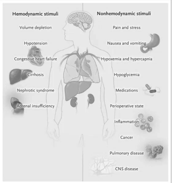
total body water relative to total body sodium and potassium (Fig. 1). Conditions that impair free water excretion are associ-ated with both physiologic and pathologic release of antidi-uretic hormone (ADH) (4) and have been well established (5) (Table 1). These conditions impair the kidneys’ primary defense against the development of hyponatremia, the urinary diluting capacity.
During hyponatremic states, an osmotic gradient devel-ops between the circulation and the brain, resulting in water movement into the brain through aquaporin-4 channels located in astrocytes, one of the types of neuroglial cells (6). Astrocytes, located on the brain side of the microcirculation, help to regulate the movement of fluids and molecules across the blood brain barrier by the cells’ foot processes that contact brain capillaries. This fluid movement results in cell expan-sion and an increase in brain volume. The resultant increase in intraparenchymal brain volume is immediately offset by the shunting of cerebrospinal fluid from the intracranial vault, which accommodates some of the volume perturbation, but this mechanism has limited capacitance (7). The main accom-modation comes through energy-dependent extrusion of
solutes from the neuroglial cell via the Na+/K+ ATPase pump (8). By reducing the neuroglial cell’s intracellular osmolality, water leaves the cell down the osmotic gradient resulting in an overall reduction in brain volume, a process known as “regu-latory brain volume decrease” (9). Neurologic injury occurs when these mechanisms are unable to fully compensate for the increased intracranial pressure due to the cerebral edema (10). Several clinical factors have been shown to impair the neuro-glial cell’s adaptive responses, and these clinical factors predict poor patient outcomes (1).
CLINICAL MANIFESTATIONS
Manifestations are varied, depending on the extent of CNS’s adaptation to the hypoosmolar state. The absolute change in serum sodium alone is a poor predictor of clinical symptoms (11). On a practical level, clinical presentations can be classi-fied as acute (where the duration of hyponatremia is known to be < 48 hr). When this occurs, it is often hospital acquired (postoperative or due to inappropriate use of hypotonic flu-ids in other settings). Acute hyponatremia does also occur in settings such as exercise-induced hyponatremia or methylene-dioxymethamphetamine-associated hyponatremia. Chronicity uncertain is the most common presentation and includes most ICU hyponatremia admissions where the time development is uncertain (diuretic related, antidepressant related, hypo-volemia, syndrome of inappropriate ADH (SIADH), adre-nal insufficiency, etc,). Chronic hyponatremia occurs where the duration of hyponatremia is known to be greater than 48 hours (e.g., a patient with recurrent congestive heart fail-ure admissions and documented laboratory studies showing hyponatremia). Although the chronicity can affect the degree of symptoms (a patient who develops a serum sodium of 122 in 48 hr following surgery is far more likely to develop hypo-natremic encephalopathy than a patient who develops a serum sodium of 122 3 wk after initiation of a thiazide diuretic), it is important to understand that all patients with hyponatremia,
Figure 1. Mechanisms for the maintenance of water balance. ADH = antidiuretic hormone.
TABLE 1.
Major Etiologies of Hyponatremia in the ICU
Hypovolemic Hyponatremia Euvolemic Hyponatremia Hypervolemic Hyponatremia
Renal losses Postoperative state Congestive heart failure
Diuretics (especially thiazide type) Mineralocorticoid deficiency Renal tubular acidosis Cerebral salt wasting
Exercise-induced hyponatremia Drugs
Ecstasy (methylenedioxymethamphetamine) Anticonvulsants
Polydipsia
Cirrhosis
Acute and chronic renal failure
Extrarenal losses Vomiting Diarrhea Pancreatitis Burns
Syndrome of inappropriate antidiuretic hormone secretion
Hypothyroidism
regardless of the chronicity of the development of hyponatre-mia, can develop hyponatremic encephalopathy, especially in high-risk groups such as premenopausal and postmenopausal females and when hypoxia is present (1, 2). Early signs of cere-bral edema such as nausea, vomiting, and headaches often go unrecognized (12). As intracranial pressure continues to increase, brain dysfunction, exhibited as altered mentation and seizures, occurs. If left uncorrected, hypercapnic respiratory failure, a sign of brainstem involvement with the possibility of herniation, and death may follow (1, 2, 12, 13).
Although there are many causes of hyponatremia in the ICU, there are two entities that deserve special attention, noncardio-genic pulmonary edema and hyponatremia as well as cerebral/ renal salt wasting. Noncardiogenic pulmonary edema associ-ated with the presence of cerebral edema (also known as “the Ayus-Arieff syndrome”) (Fig. 2) could be the initial presenta-tion of a patient with hyponatremic encephalopathy (14). The two most common causes of hyponatremia and pulmonary edema are exercise-induced hyponatremia and hyponatremia due to ingestion of the recreational drug methylenedioxy-methamphetamine, commonly known as “ecstasy” (14, 15) but can be seen in any cause of hyponatremia. The main mecha-nism for hyponatremic encephalopathy to produce neurogenic pulmonary edema is the increase in intracranial pressure due to hyponatremia (16). The patients described in these reports
had normal troponin levels, normal echocardiograms, and normal pulmonary artery occlusion pressures demonstrating that the pulmonary edema seen in these cases was not due to a cardiogenic etiology (14, 16). Hyponatremia produces cerebral edema, which in turn leads to neurogenic pulmonary edema (14). Pulmonary edema then leads to hypoxia which impairs brain volume regulation (6, 17, 18), resulting in a vicious cycle of worsening cerebral edema and hypoxia. This syndrome is treated with a bolus of hypertonic saline (14).
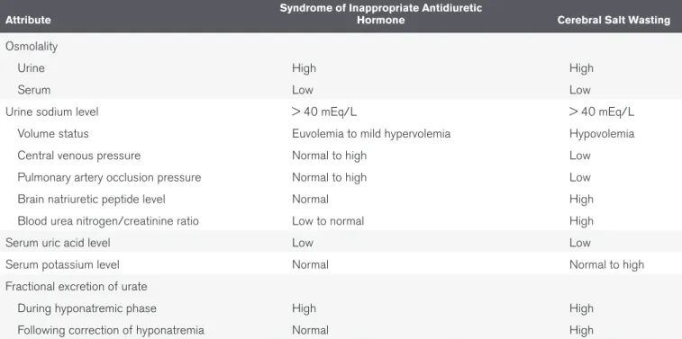
Cerebral/renal salt wasting occurs most commonly in neu-rosurgical patients (19). This condition is characterized by a neurogenic natriuresis by an unknown mechanism that leads to volume depletion and secondary ADH secretion from the pituitary that results in hyponatremia. In this way, the cardinal distinguishing factor between the SIADH, where nonosmotic ADH secretion is the key pathologic feature, and cerebral salt wasting is the presence or absence of a low circulating effective arterial blood volume (Table 2). In the past, this differentiation was difficult as the effective arterial blood volume could not be measured directly; however, pulmonary artery occlusion pres-sure (20, 21) (in the absence of diastolic cardiac dysfunction) or right atrial pressure (22) (in the absence of pulmonary hyper-tension) could be used to estimate this. Natriuretic peptides (23) hold promise as a tool to estimate effective arterial blood vol-ume as they are directly related to vascular blood volvol-ume and can help in determining the vascular status of patient. An addi-tional tool in differentiating SIADH and cerebral/renal salt wast-ing is the fractional excretion of urate as the excretion of urate is usually reduced in patients with hypovolemia (24). In many cases, patients who have SIADH due to the cerebral trauma dur-ing neurosurgery are misclassified as havdur-ing cerebral salt wast-ing. This distinction is important to make as the treatment of cerebral salt wasting, when hyponatremic encephalopathy is not present, is volume expanded with 0.9% saline, whereas in a patient with SIADH, 0.9% saline is futile therapy and can lead to iatrogenic fluid overload. When a patient with cerebral salt wasting is aggressively volume expanded with 0.9% saline and there is no improvement in the condition, the diagnosis of needs to be revisited. Following serum sodium, urine osmolality, and serum brain, natriuretic peptide can be helpful in assessing such patients.
RISK FACTORS FOR HYPONATREMIC ENCEPHALOPATHY
Gender (1), age (25), and the presence of hypoxia (1, 14, 16) are the three main clinical risk factors and are more predic-tive of poor outcomes than the rate of development of hypo-natremia or the absolute decrease in the serum sodium (13) (Table 3). These are equivalent risk factors with summative effects (except that in children where gender does not play a role). Children are at increased risk due to a high brain-to-cranial vault size ratio as there is less space for accommodation as brain volume increases (25). Among postsurgical patients who develop hyponatremia, females of premenopausal age are also at increased risk for development of cerebral edema (1).
This increased risk of premenopausal females has been seen in several clinical scenarios associated with hyponatremia: the use of ecstasy (15, 26), use of diuretics (13), exercise (14), and use of desmopressin (DDAVP; Ferring Pharmaceuticals, Saint-Prex, Switzerland) (27). Estrogens are believed to inhibit the catalytic activity of the astrocyte’s Na+/K+ ATPase resulting in impairment of regulatory volume decrease. This has been demonstrated in rat astrocytes treated in vitro (28). Finally, hypoxia has been shown to predict poor outcomes in patients with hyponatremic encephalopathy (1), as it also impairs the Na+/K+ ATPase from extruding solutes (17, 18). Hypoxia can develop in encephalopathic hyponatremic patients through two mechanisms: hypercapnic respiratory failure (16) and neurogenic pulmonary edema (14).
PREVENTION OF ICU-ACQUIRED HYPONATREMIA
Until proven otherwise, a patient in a critical care setting should be assumed to have an impairment in free water excre-tion. In patients with intact kidney function, ADH levels are likely to be high. In patients with renal failure, free water excre-tion is also impaired. Common situaexcre-tions where water balance is impaired include the following: cases of effective circulating volume depletion (cirrhosis, heart failure, and third spacing of fluid), gastrointestinal fluid losses, diuretic use (especially thiazides), renal failure (acute and chronic), SIADH, corti-sol deficiency, and hypothyroidism (Fig. 3). As noted above, hypotonic IV fluids should not be used except in the setting of replacement of a water deficit (i.e., hypernatremia). Isotonic
TABLE 2.
Comparison of Syndrome of Inappropriate Antidiuretic Hormone and Cerebral
Salt Wasting
Attribute Syndrome of Inappropriate Antidiuretic Hormone Cerebral Salt Wasting
Osmolality
Urine High High
Serum Low Low
Urine sodium level > 40 mEq/L > 40 mEq/L
Volume status Euvolemia to mild hypervolemia Hypovolemia
Central venous pressure Normal to high Low
Pulmonary artery occlusion pressure Normal to high Low
Brain natriuretic peptide level Normal High
Blood urea nitrogen/creatinine ratio Low to normal High
Serum uric acid level Low Low
Serum potassium level Normal Normal to high
Fractional excretion of urate
During hyponatremic phase High High
Following correction of hyponatremia Normal High
TABLE 3.
Risk Factors for Hyponatremic Encephalopathy
Risk Factors Mechanism
Postsurgical females of
premenopausal agea (1) Impaired regulatory brain volume decrease
b: Estrogens decrease the catalytic activity of astrocyte
Na+/K+, ATPase preventing solute extrusion
Age < 16 (25) High brain volume-to-cranial vault size ratio resulting in less space to accommodate brain volume increases
Hypoxia (1, 14, 16) Impaired regulatory brain volume decrease: Hypoxia decreases the catalytic activity of astrocyte Na+/
K+, ATPase preventing solute extrusion
Brain injury Vasogenic cerebral edema Cytotoxic cerebral edema
Acute hyponatremia Decreased time for brain adaptation
a When compared with postsurgical males, this subgroup is 25 times more likely to develop permanent brain damage or death (1).
normal saline (0.9% NaCl) containing 154 mEq/L of Na+ and Cl- is the most appropriate parenteral fluid when IV fluids are indicated for the maintenance of intravascular volume in the postoperative period (29). Also, in the ICU setting, it is the most appropriate fluid choice in almost all circumstances unless replacing a water deficit and hypotonic fluids are clearly needed (29). Also, any patient receiving fluid therapy should have the serum sodium measured at least daily (29).
Hospital-acquired hyponatremic encephalopathy occurs most commonly when hypotonic fluids are administered to a patient with an impairment of free water excretion (29). A clinical setting that merits specific discussion is the postopera-tive state (1). Approximately 1% of patients develop a serum sodium of less than 130 mEq/L following surgery, and clinically important hyponatremia complicates 20% of these cases (1). The postoperative state commonly includes multiple stimuli for ADH release including pain, stress, nausea, vomiting, nar-cotic medications, and volume depletion (29). Administration of a hypotonic IV fluid in the postoperative state or in other
clinical settings characterized by impaired free water excre-tion (29) can have disastrous consequences (1). The use of hypotonic fluids is reserved for treatment of a free water defi-cit, such as exists in the setting of hypernatremia (29).
THE HYPERTONIC SALINE BOLUS: A SAFE AND EFFECTIVE APPROACH TO TREAT HYPONATREMIC ENCEPHALOPATHY Hypertonic saline is the treat-ment of choice for hypona-tremic encephalopathy (2, 11, 30). Our group has used hypertonic saline to treat over 150 patients (2, 11, 14, 25, 27, 30–33) with hypona-tremic encephalopathy. We have used this in both children and adults, in patients with acute and chronic hyponatre-mia, in patients presenting at a medical tent or emergency department, and in patients with hospital-acquired hypo-natremia. In these groups of patients, brain imaging and long-term neurologic follow-up was performed, and we have not encountered cerebral demyelination as a complica-tion of hypertonic saline in any of these cases. Our findings of the efficacy of hypertonic saline in the treatment of hypona-tremic encephalopathy have been confirmed by other investi-gators (34–36).
However, despite the efficacy of this therapeutic approach, we recognize that the indiscriminate use of prolonged infu-sions of hypertonic saline without appropriate monitoring can produce neurologic injury from an excessive correction of hyponatremia. Because of this concern, and to minimize the risk of hypertonic saline therapy, in 2005, we introduced a simple approach of using small repeated intermittent boluses of hypertonic saline (37) in order to achieve a rapid and controlled increase in serum sodium to treat hyponatremic encephalopathy, that conformed to the various opinions on the safe limits of correcting hyponatremia (38).
Our approach is to treat any patient with suspected hypo-natremic encephalopathy (whether acute or chronic), with either early (nausea, vomiting, headache) or advanced symp-toms (seizures, respiratory arrest), child or adult, and with a
2 mL/kg bolus of 3% NaCl. The time course over which the hyponatremia has developed is not a determinant of who should receive therapy for suspected hyponatremic encepha-lopathy, this is because chronicity is not always known and hypertonic saline therapy is known to be safe, even when the duration of hyponatremia is clearly chronic (known to be > 48 hr in duration). Early symptoms (nausea, vomiting, head-ache, and ataxia) while they are milder in severity should still be treated with hypertonic saline as they may be a precursor to more severe manifestations (such as seizures and respira-tory arrest). Even though the risk of imminent decompensa-tion may be low in many cases (e.g., a patient with cirrhosis, ataxia, and a serum sodium of 115), given the safety of hyper-tonic saline when used appropriately (30) and the superiority of hypertonic saline over fluid restriction alone in manage-ment of chronic hyponatremic encephalopathy (2), we feel that the use of hypertonic saline, especially in an ICU setting, rather than fluid restriction alone is warranted. Early IV saline therapy in symptomatic patients with chronic hyponatremic encephalopathy is associated with 0% morbidity and mortal-ity, whereas delay of therapy with IV saline until respiratory insufficiency develops is associated with 64% morbidity and
mortality (either death or neurologic injury leading to perma-nent institutionalization) (2). We advocate for earlier inter-vention, rather than waiting for more catastrophic symptoms to develop prior to initiating therapy. As long as precautions regarding hypertonic saline therapy we discuss elsewhere are adhered to, concerns about osmotic demyelination syndrome should not prevent appropriate treatment of patients with chronic hyponatremic encephalopathy with hypertonic saline. The bolus could be repeated 1–2 times in sequential fashion if symptoms persisted. A single bolus would result in, at most, a 2 mEq/L acute rise in serum sodium, which could quickly reduce brain edema. In most cases, a 4–6 mEq/L acute rise in plasma sodium will begin to reverse the neurologic symp-toms, and failure to show some clinical improvement follow-ing an acute elevation in serum sodium would suggest that the patient is not suffering from hyponatremic encephalopathy. The bolus approach can be given safely through a peripheral IV and can be used in a non-ICU setting (30). It would be effective regardless of the etiology of hyponatremia, serving as a volume expander in hypovolemic hyponatremia and is suffi-ciently hypertonic to increase the serum sodium in euvolemic hyponatremia from SIADH. It also does not require the use of complicated formulas, which have been demonstrated to be inaccurate as they assume a closed system and do not account for the renal response to therapy (39). Other therapies such as 0.9% NaCl, 1.8% NaCl, mannitol, urea, oral sodium, and vaptans cannot be recom-mended as first-line therapies to treat hyponatremic encepha-lopathy as they do not reliably increase the serum sodium or reverse the neurologic symp-toms (38).
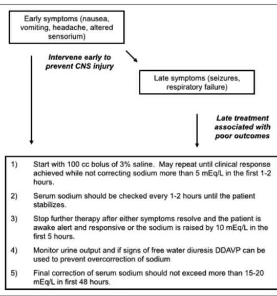
This approach has already been successfully used in critical care patients (36) and has been adopted as the standard of care by the European Societies of Nephrology, Endocrinology and Critical Care (38) and sport med-icines societies (40). The goal of this initial bolus is to quickly treat severe cerebral edema. The sodium should not be corrected more than 5 mEq/L in the first 1–2 hours of treatment and not to exceed 15–20 mEq in the first 48 hours of treatment. Prompt recognition of the problem and early initiation of therapy are the most important factors associ-ated with successful intervention
and good neurologic outcomes. It is critical to recognize the early signs of cerebral edema (nausea, vomiting, and head-ache) and intervene with IV 3% sodium chloride as this is the time to intervene rather than waiting until more severe symp-toms develop (2, 41). When therapy is delayed until after late symptoms (altered sensorium, seizure, and respiratory arrest) develop, the prognosis is much worse (41). The goals of ther-apy with hypertonic saline (Fig. 4) can be summarized as fol-lows: 1) remove patients with severe manifestations of cerebral edema from immediate danger; 2) correct serum sodium to a mildly hyponatremic levels; and 3) maintain this level of serum sodium to allow for the brain to adapt to the change in serum osmolality.
A recent case series has demonstrated that 500 mL of 3% saline over 6 hours can be given safely (30). Any patient receiving bolus therapy of 3% saline should have the serum sodium checked every 1–2 hours until the patient has clinically
stabilized. The rationale for close monitoring is that ongoing water losses cannot always be predicted. Therefore, urine out-put should be followed closely to assess for a water diuresis that is indicative of free water losses that can cause correction of the sodium in addition to the increase in serum sodium provided by hypertonic saline. Finally, the serum sodium should not be corrected past normonatremic levels during the first 48 hours of therapy as this is a further risk factor for the development of cerebral demyelination (11).
No head-to-head studies of continuous IV infusion of 3% saline versus bolus IV infusion have been performed, so this must appropriately temper our conclusions, but given the observation studies demonstrating the efficacy and safety of a bolus approach and the potential for overcorrection with a longer, continuous infusion, we feel that bolus IV infusion therapy is a viable and safer alternative to continuous IV infu-sion of 3% saline and may be the preferred approach.
RISK FACTORS FOR THE DEVELOPMENT OF CEREBRAL DEMYELINATION
Cerebral demyelination is a potential complication from the overcorrection of severe and chronic hyponatremia. It is primarily seen in patients with hyponatremia of greater than 48 hours duration and a plasma sodium less than 115 mEq/L (38, 42–45). It is infrequently seen in patients with acute hyponatre-mia or plasma sodium values greater than 120 mEq/L (34, 35, 38, 45, 46). Although there are multiple isolated reports of cere-bral demyelination occurring in patients with hyponatremia (38) when examined in large series of patients with severe hyponatre-mia, cerebral demyelination is extremely rare (46–48). Animal studies and clinical observations in humans have demonstrated that cerebral demyelination is a complication from an exces-sive magnitude of correction of hyponatremia of greater than 25 mEq/L over a 24–48 hours period (11, 49, 50). Cerebral demyelination does not appear to be related to an excessive hourly rate of correction as long as the overall magnitude is kept less than 20 mEq/L in the first
48 hours of therapy (11). The main risk for an overcorrection of hyponatremia is not from the use of hypertonic saline but rather from renal response to fluid therapy and a spontaneous free water diuresis that occur when the stimulus for arginine vasopressin release abates. The primary conditions where a brisk free water diuresis can occur are thiazide-induced hyponatremia, water intoxication, volume depletion, adrenal insufficiency following replacement therapy, and DDAVP-induced hyponatremia fol-lowing DDAVP withdrawal (13, 27). Cerebral demyelination can occur with any therapy used to correct severe and chronic hypo-natremia, including normal saline and V2 antagonists, if there is an excessive correction of hyponatremia (13).
There are risk factors other than hyponatremia that can place a patient at risk for developing cerebral demyelination such as severe liver disease (11), hypokalemia (44), thiazide diuretic use (13), alcoholism (11, 13), malnutrition (51), hypo-phosphatemia (13), and hypoxia (13). Most reported patient with cerebral demyelination have had one or more of these risk factors. Patients with these risk factors have been reported to have cerebral demyelination in the absence of hyponatremia (13). When symptoms of cerebral demyelination manifest, it is typically a delayed phenomenon, occurring days to weeks following the correction of hyponatremia (11). Patients may present with a continuum of symptoms, ranging from asymp-tomatic to those with extremely altered mentation, which may manifest as a pseudocoma with a locked-in stare (11). MRI of the brain, which is sensitive for the detection of demyelinating lesions, is often necessary to establish the diagnosis (11).
PREVENTION OF OVERCORRECTION OF HYPONATREMIA WITH USE OF DESMOPRESSIN (DDAVP)
Our group in 1993 (52) advanced the concept that DDAVP could be used to minimize water excretion during correction
of hyponatremia in cases that involve a risk of water diuresis (caused by a drop in urine osmolality termed a “reverse urine osmolality”), for example, compulsive water drinking, cor-tisol deficiency, thyroid deficiency, and medication-induced hyponatremia (e.g., thiazide diuretics) (52). This concept has been validated by other groups, showing the clinical usage of this therapeutic maneuver (53–58). During the treatment of hyponatremia, the clinician needs to be vigilant to be sure that a free water diuresis does not occur. In order to prevent ongoing water losses in the urine and “autocorrection” of the serum sodium, DDAVP can be given to increase urinary con-centration and reduce free water losses. However, this must be done carefully, with the patient strictly fluid restricted or kept with no enteral intake in an ICU setting. If unrestricted fluid intake occurs during DDAVP administration, signifi-cant hyponatremia can develop. An increase in urine output is the first sign that a water diuresis is ensuing, and therefore hourly urine output needs to be followed in all patients with hyponatremic encephalopathy, especially those with drug-induced hyponatremia. DDAVP administration leads to a prompt reduction in urine and increase in urine osmolality as seen in (Fig. 5), where a representative case is presented of a patient with alcoholism and thiazide-induced hypona-tremia presents with seizures and an initial serum sodium of 103 mEq/L who developed a water diuresis following cor-rection of her hyponatremia with IV 3% sodium chloride. Keeping the serum sodium from increasing by more than 15 mEq/L in the first 48 hours is the goal during the treatment of hyponatremic encephalopathy. Use of DDAVP during this initial period may require the patient to stay in the ICU in order to monitor intake and output strictly. After 48 hours, the DDAVP can usually be stopped and the patient observed while the sodium continues to correct. This general approach is outlined in Figure 6.
SUMMARY
Hyponatremia is a common clinical disorder in the ICU set-ting, and when it is complicated by hyponatremic encephalop-athy, it is associated with poor outcomes. Prompt recognition of patients with hyponatremic encephalopathy is critical in order to provide immediate therapy. Treatment with bolus therapy of 3% IV sodium chloride is recommended when early symptoms occur rather than waiting until late symptoms develop as delay in therapy is associated with poor neurologic outcomes. We recommend patients be treated by correcting the serum sodium with hypertonic saline beginning with an IV bolus of 100 mL of 3% saline, which may be repeated if needed. The goal of therapy is to adequately treat cerebral edema while avoiding correction of the serum sodium by more than 15–20 mEq/L in the first 48 hours of therapy, thereby preventing the development of cerebral demyelination.
ACKNOWLEDGMENT
We like to acknowledge Elizabeth Ziner, MD, for her assistance in article preparation and preparation of figures.
REFERENCES
1. Ayus JC, Wheeler JM, Arieff AI: Postoperative hyponatremic encepha-lopathy in menstruant women. Ann Intern Med 1992; 117:891–897 2. Ayus JC, Arieff AI: Chronic hyponatremic encephalopathy in
post-menopausal women: Association of therapies with morbidity and mortality. JAMA 1999; 281:2299–2304
3. Ayus JC, Krothapalli RK, Arieff AI: Changing concepts in treatment of severe symptomatic hyponatremia. Rapid correction and possible rela-tion to central pontine myelinolysis. Am J Med 1985; 78(6Pt 1):897–902 4. Dunn FL, Brennan TJ, Nelson AE, et al: The role of blood osmolality
and volume in regulating vasopressin secretion in the rat. J Clin Invest
1973; 52:3212–3219
5. Schrier RW: Body water homeostasis: Clinical disorders of urinary dilution and concentration. J Am Soc Nephrol 2006; 17:1820–1832 6. Ayus JC, Achinger SG, Arieff A: Brain cell volume regulation in hypo-natremia: role of sex, age, vasopressin, and hypoxia. Am J Physiol Renal Physiol 2008; 295:F619–F624
7. Reulen HJ, Tsuyumu M, Tack A, et al: Clearance of edema fluid into cerebrospinal fluid. A mechanism for resolution of vasogenic brain edema. J Neurosurg 1978; 48:754–764
8. Olson JE, Sankar R, Holtzman D, et al: Energy-dependent volume reg-ulation in primary cultured cerebral astrocytes. J Cell Physiol 1986; 128:209–215
9. Melton JE, Patlak CS, Pettigrew KD, et al: Volume regulatory loss of Na, Cl, and K from rat brain during acute hyponatremia. Am J Physiol
1987; 252(4 Pt 2):F661–F669
10. Arieff AI, Ayus JC: Pathogenesis of hyponatremic encephalopathy. Current concepts. Chest 1993; 103:607–610
11. Ayus JC, Krothapalli RK, Arieff AI: Treatment of symptomatic hypona-tremia and its relation to brain damage. A prospective study. N Engl J Med 1987; 317:1190–1195
12. Achinger SG, Moritz ML, Ayus JC: Dysnatremias: Why are patients still dying? South Med J 2006; 99:353–362; quiz 363
13. Moritz ML, Ayus JC: The pathophysiology and treatment of hypona-traemic encephalopathy: An update. Nephrol Dial Transplant 2003; 18:2486–2491
14. Ayus JC, Varon J, Arieff AI: Hyponatremia, cerebral edema, and non-cardiogenic pulmonary edema in marathon runners. Ann Intern Med
2000; 132:711–714
15. Moritz ML, Kalantar-Zadeh K, Ayus JC: Ecstacy-associated hypo-natremia: Why are women at risk? Nephrol Dial Transplant 2013; 28:2206–2209
16. Ayus JC, Arieff AI: Pulmonary complications of hyponatremic enceph-alopathy. Noncardiogenic pulmonary edema and hypercapnic respira-tory failure. Chest 1995; 107:517–521
17. Vexler ZS, Ayus JC, Roberts TP, et al: Hypoxic and ischemic hypoxia exacerbate brain injury associated with metabolic encephalopathy in laboratory animals. J Clin Invest 1994; 93:256–264
18. Ayus JC, Armstrong D, Arieff AI: Hyponatremia with hypoxia: Effects on brain adaptation, perfusion, and histology in rodents. Kidney Int
2006; 69:1319–1325
19. Bitew S, Imbriano L, Miyawaki N, et al: More on renal salt wasting without cerebral disease: Response to saline infusion. Clin J Am Soc Nephrol 2009; 4:309–315
20. Palmer BF: Hyponatremia in patients with central nervous system disease: SIADH versus CSW. Trends Endocrinol Metab 2003; 14:182–187
21. Yee AH, Burns JD, Wijdicks EF: Cerebral salt wasting: Pathophysiology, diagnosis, and treatment. Neurosurg Clin N Am
2010; 21:339–352
22. Damaraju SC, Rajshekhar V, Chandy MJ: Validation study of a central venous pressure-based protocol for the management of neurosurgi-cal patients with hyponatremia and natriuresis. Neurosurgery 1997; 40:312–316; discussion 316–317
23. Marin-Grez M, Fleming JT, Steinhausen M: Atrial natriuretic peptide causes pre-glomerular vasodilatation and post-glomerular vasocon-striction in rat kidney. Nature 1986; 324:473–476
24. Maesaka JK, Miyawaki N, Palaia T, et al: Renal salt wasting without cerebral disease: Diagnostic value of urate determinations in hypona-tremia. Kidney Int 2007; 71:822–826
25. Arieff AI, Ayus JC, Fraser CL: Hyponatraemia and death or permanent brain damage in healthy children. BMJ 1992; 304:1218–1222 26. van Dijken GD, Blom RE, Hené RJ, et al: High incidence of mild
hyponatraemia in females using ecstasy at a rave party. Nephrol Dial Transplant 2013; 28:2277–2283
27. Achinger SG, Arieff AI, Kalantar-Zadeh K, et al: Desmopressin ace-tate (DDAVP)-associated hyponatremia and brain damage: A case series. Nephrol Dial Transplant 2014; 29:2310–2315
28. Fraser CL, Swanson RA: Female sex hormones inhibit volume regu-lation in rat brain astrocyte culture. Am J Physiol 1994; 267(4 Pt 1):C909–C914
29. Moritz ML, Ayus JC: Maintenance intravenous fluids in acutely ill patients. N Engl J Med 2015; 373:1350–1360
30. Ayus JC, Caputo D, Bazerque F, et al: Treatment of hyponatremic encephalopathy with a 3% sodium chloride protocol: A case series.
Am J Kidney Dis 2015; 65:435–442
31. Ayus JC, Olivero JJ, Frommer JP: Rapid correction of severe hypona-tremia with intravenous hypertonic saline solution. Am J Med 1982; 72:43–48
32. Arieff AI, Ayus JC: Endometrial ablation complicated by fatal hypona-tremic encephalopathy. JAMA 1993; 270:1230–1232
33. Ayus JC, Arieff AI: Glycine-induced hypo-osmolar hyponatremia. Arch Intern Med 1997; 157:223–226
34. Worthley LI, Thomas PD: Treatment of hyponatraemic seizures with intravenous 29.2% saline. Br Med J (Clin Res Ed) 1986; 292:168–170
35. Sarnaik AP, Meert K, Hackbarth R, et al: Management of hypona-tremic seizures in children with hypertonic saline: A safe and effective strategy. Crit Care Med 1991; 19:758–762
36. Bhaskar E, Kumar B, Ramalakshmi S: Evaluation of a protocol for hypertonic saline administration in acute euvolemic symptomatic hyponatremia: A prospective observational trial. Indian J Crit Care Med 2010; 14:170–174
37. Ayus JC, Arieff A, Moritz ML: Hyponatremia in marathon runners. N Engl J Med 2005; 353:427–428; author reply 427
38. Spasovski G, Vanholder R, Allolio B, et al; Hyponatraemia Guideline Development Group: Clinical practice guideline on diagnosis and treatment of hyponatraemia. Eur J Endocrinol
2014; 170:G1–G47
40. Hew-Butler T, Rosner MH, Fowkes-Godek S,et al: Statement of the Third International Exercise-Associated Hyponatremia Consensus Development Conference, Carlsbad, California, 2015. Clin J Sport Med 2015; 25:303–320
41. Moritz ML, Ayus JC: Management of hyponatremia in various clinical situations. Curr Treat Options Neurol 2014; 16:310
42. Norenberg MD, Papendick RE: Chronicity of hyponatremia as a factor in experimental myelinolysis. Ann Neurol 1984; 15:544–547 43. Brunner JE, Redmond JM, Haggar AM, et al: Central pontine myelinolysis
and pontine lesions after rapid correction of hyponatremia: A prospective magnetic resonance imaging study. Ann Neurol 1990; 27:61–66 44. Lohr JW: Osmotic demyelination syndrome following correction
of hyponatremia: Association with hypokalemia. Am J Med 1994; 96:408–413
45. Kallakatta RN, Radhakrishnan A, Fayaz RK, et al: Clinical and func-tional outcome and factors predicting prognosis in osmotic demye-lination syndrome (central pontine and/or extrapontine myelinolysis) in 25 patients. J Neurol Neurosurg Psychiatry 2011; 82:326–331 46. Nzerue CM, Baffoe-Bonnie H, You W, et al: Predictors of outcome
in hospitalized patients with severe hyponatremia. J Natl Med Assoc
2003; 95:335–343
47. Geoghegan P, Harrison AM, Thongprayoon C, et al: Sodium correc-tion practice and clinical outcomes in profound hyponatremia. Mayo Clin Proc 2015; 90:1348–1355
48. Greenberg A, Verbalis JG, Amin AN, et al: Current treatment practice and outcomes. Report of the hyponatremia registry. Kidney Int 2015; 88:167–177
49. Ayus JC, Krothapalli RK, Armstrong DL: Rapid correction of severe hyponatremia in the rat: Histopathological changes in the brain. Am J Physiol 1985; 248(5 Pt 2):F711–F719
50. Verbalis JG, Martinez AJ: Neurological and neuropathological sequelae of correction of chronic hyponatremia. Kidney Int 1991; 39:1274–1282
51. Goebel HH, Zur PH: Central pontine myelinolysis. A clinical and path-ological study of 10 cases. Brain 1972; 95:495–504
52. Ayus JC, Arieff AI: Pathogenesis and prevention of hypona-tremic encephalopathy. Endocrinol Metab Clin North Am 1993; 22:425–446
53. Quinn CJ, Iyegha UP, Beilman GJ, et al: Acute correction of hypona-tremia secondary to psychogenic polydipsia. Am J Case Rep 2012; 13:69–71
54. Tomlin SC, Williams R, Riley S: Preventing overcorrection of hypona-traemia with desmopressin. BMJ Case Rep 2011; 2011
55. Lum G: Severe hyponatremia in a schizophrenic patient. Clin Chem
2013; 59:887–889
56. Rafat C, Schortgen F, Gaudry S, et al: Use of desmopressin acetate in severe hyponatremia in the intensive care unit. Clin J Am Soc Nephrol 2014; 9:229–237
57. Gharaibeh KA, Craig MJ, Koch CA, et al: Desmopression is an effec-tive adjunct treatment for reversing excessive hyponatremia overcor-rection. World J Clin Cases 2013; 1:155–158