Noninvasive chromosome screening of human embryos
by genome sequencing of embryo culture medium
for in vitro fertilization
Juanjuan Xua,1, Rui Fangb,1, Li Chena,1, Daozhen Chenb, Jian-Ping Xiaob, Weimin Yangb, Honghua Wangb, Xiaoqing Songb, Ting Mac, Shiping Boc, Chong Shic, Jun Renc, Lei Huangd,e,f,g, Li-Yi Caib,2, Bing Yaoa,2, X. Sunney Xied,g,h,2, and Sijia Luc,2
aReproductive Medical Center of Nanjing Jinling Hospital and the Collaborative Innovation Platform for Reproductive Biology and Technology, Nanjing University School of Medicine, Nanjing, Jiangsu 210002, China;bReproductive Medicine Center, Wuxi Maternity and Child Health Hospital Affiliated to Nanjing Medical University, Wuxi, Jiangsu 214002, China;cDepartment of Clinical Research, Yikon Genomics Company, Ltd., Shanghai 201499, China; dBiodynamic Optical Imaging Center (BIOPIC), School of Life Sciences, Peking University, Beijing 100871, China;eDepartment of Obstetrics, Gynecology, and Reproductive Biology, Brigham and Women’s Hospital, Boston, MA 02115;fHarvard Medical School, Boston, MA 02115;gBeijing Advanced Innovation Center for Genomics, Peking University, Beijing 100871, China; andhDepartment of Chemistry and Chemical Biology, Harvard University, Cambridge, MA 01238
Contributed by X. Sunney Xie, August 10, 2016 (sent for review April 28, 2016; reviewed by Eva Hoffmann and John Rasko)
Preimplantation genetic screening (PGS) is widely used to select in vitro-fertilized embryos free of chromosomal abnormalities and to improve the clinical outcome of in vitro fertilization (IVF). A disadvantage of PGS is that it requires biopsy of the preimplantation human embryo, which can limit the clinical applicability of PGS due to the invasiveness and complexity of the process. Here, we present and validate a noninvasive chromosome screening (NICS) method based on sequencing the genomic DNA secreted into the culture medium from the human blastocyst. By using multiple annealing and looping-based amplification cycles (MALBAC) for whole-genome amplification (WGA), we performed next-generation sequencing (NGS) on the spent culture medium used to culture human blastocysts (n=42) and obtained the ploidy information of all 24 chromosomes. We validated these results by comparing each with their corresponding whole donated embryo and obtained a high correlation for identification of chromosomal abnormalities (sensi-tivity, 0.882, and specificity, 0.840). With this validated NICS method, we performed chromosome screening on IVF embryos from seven couples with balanced translocation, azoospermia, or recurrent pregnancy loss. Six of them achieved successful clinical pregnancies, and five have already achieved healthy live births thus far. The NICS method avoids the need for embryo biopsy and therefore substan-tially increases the safety of its use. The method has the potential of much wider chromosome screening applicability in clinical IVF, due to its high accuracy and noninvasiveness.
chromosomal abnormalities
|
PGS|
IVF|
WGA|
MALBACH
uman embryos are prone to chromosomal abnormalities, mainly due to age-dependent chromosome segregation errors during meiosis I (1). Chromosomal abnormalities could cause early pregnancy loss or severe chromosomal diseases such as Down and Patau syndrome among many others (2, 3). The occurrence of chromosomal abnormalities is substantially higher in patients of advanced maternal age, patients with recurrent pregnancy loss, or those who carry chromosomal aberrations such as translocations, all of which result in poor clinical outcome for reproduction.Chromosomal abnormalities can be prevented in in vitro fertil-ization (IVF) by performing preimplantation genetic screening (PGS) of all 24 chromosomes. There are various PGS methods for comprehensive chromosome screening currently in clinical use, including comparative genomic hybridization (array-CGH) (4, 5), single-nucleotide polymorphism (SNP) arrays (6–9), multiplex quan-titative PCR (10), and next-generation sequencing (NGS) (11, 12). Multiple clinical trials have confirmed the clinical efficacy of PGS, including increasing implantation and clinical pregnancy rates, as well as decreasing miscarriage rates (13–16). However, the applicability of PGS has been limited for a number of reasons:
(i) PGS requires invasive embryo biopsy, which has been shown to decrease embryo quality after cleavage-stage biopsy (17); (ii) long-term biosafety of embryo biopsy in humans has not been evaluated, whereas animal studies have shown negative influences on neural and adrenal development (18–20); (iii) it involves technical ex-pertise, requiring special training and experienced embryologists to perform the biopsy, which significantly increase the overall costs of clinical PGS cycles. Therefore, a noninvasive and easy-to-perform screening tool would greatly facilitate the widespread performing of chromosome screening before embryo implantation, thereby improving success rates.
Efforts have been made to develop noninvasive approaches for PGS (21, 22). Palini et al. (23) reported the observation of the ex-istence of DNA in the blastocoele fluid, Gianaroli et al. (24) per-formed a pilot study on chromosome screening using blastocentesis, and Stigliani et al. (25, 26) observed genomic and mitochondria DNA contents in the culture medium, which were correlated with embryo quality. Wu et al. (27) reported the PCR detection of the secreted genomic DNA in the culture medium for preimplantation
Significance
In in vitro fertilization (IVF), current methods of diagnosing chromosome abnormality and screening for viability of transfer require biopsy of embryos, which affects embryo quality, awaits long-term biosafety test, and requires specialized skills. We demonstrate the principle of noninvasive chromosome screening (NICS), which is based on sequencing the genomic DNA secreted into the culture medium from the embryo, avoiding the need for embryo biopsy and substantially increasing the safety. By char-acterizing its precision and demonstrating successful live births, we validate that NICS offers the potential of significantly im-proving the clinical outcome of IVF.
Author contributions: J.X., R.F., L.C., D.C., J.-P.X., L.-Y.C., B.Y., X.S.X., and S.L. designed research; J.X., R.F., L.C., J.-P.X., W.Y., H.W., X.S., T.M., S.B., J.R., L.H., L.-Y.C., B.Y., and S.L. performed research; R.F., L.C., T.M., S.B., C.S., J.R., L.H., L.-Y.C., X.S.X., and S.L. analyzed data; and J.X., R.F., S.B., C.S., L.H., L.-Y.C., B.Y., X.S.X., and S.L. wrote the paper.
Reviewers: E.H., University of Copenhagen; and J.R., Royal Prince Alfred Hospital, Sydney Local Health District.
Conflict of interest statement: X.S.X. and S.L. are cofounders of Yikon Genomics Company, Ltd.
Data deposition: The data reported in this paper have been deposited in the Gene Expres-sion Omnibus (GEO) database,www.ncbi.nlm.nih.gov/geo(accession no.SRP089980).
1J.X., R.F., and L.C. contributed equally to this work.
2To whom correspondence may be addressed. Email: [email protected], yaobing@nju.
edu.cn, [email protected], or [email protected].
This article contains supporting information online atwww.pnas.org/lookup/suppl/doi:10. 1073/pnas.1613294113/-/DCSupplemental.
GENET
genetic diagnosis (PGD) of α-thalassemia cases. Although the mechanism of secretion is not known, it is most likely that the DNA in the media results from the apoptotic cells of the growing embryo. The genome-wide analysis of the secreted DNA in the culture medium has not been previously reported, and would provide the basis for PGS, assuming that the detected DNA arises from cells of the growing embryo. From the perspective of noninvasiveness and ease in handling the embryos, blasto-cyst culture medium would be an ideal source for chromosome screening. However, such an approach would require high sensitivity and reproducibility of the whole-genome amplifica-tion (WGA) of the DNA from the blastocyst culture medium. To the best of our knowledge, no previous studies have reported validation studies using blastocyst culture medium for noninvasive comprehensive screening of chromosomes to improve the clinical outcome of in vitro fertilization (IVF)/intracytoplasmic sperm injection (ICSI) patients.
In this study, we performed noninvasive chromosome screening (NICS) on spent culture medium samples used for growing human embryos from days 3–5 (D3–D5). The NICS assay was validated by comparing results obtained from the culture medium with the chromosome ploidy information obtained directly from the corre-sponding D5 whole embryos. We then calculated the sensitivity and specificity of the NICS assay in screening chromosomal abnor-malities. After the NICS assay had been validated, we performed this assay on seven couples with balanced chromosomal trans-location or recurrent pregnancy loss to select embryos with normal chromosomal ploidy. Six of the seven couples obtained successful pregnancies, and five have already achieved healthy live births.
In brief, we performed validation experiments on 42 embryos. The donated and institutional review board (IRB)-approved em-bryos were created using ICSI and were vitrified on D3 and sub-sequently warmed and placed in blastocyst culture medium. We collected the spent culture media samples on D5 and performed WGA and sequencing by multiple annealing and looping-based amplification cycles (MALBAC)–NGS (28) (Fig. 1). The MALBAC-NGS protocol has been previously validated in performing PGS with cleavage-stage and blastocyst-stage biopsies (29–31), and is increas-ingly used for single-gene PGD combined with chromosomal PGS (30, 32). Similarly, we performed MALBAC-NGS on all 24 chro-mosomes from the corresponding D5 whole embryos, which we used as the gold standard to evaluate the chromosome screening results from the culture media.
Results
NICS Using Blastocyst Culture Medium.We sequenced∼2 million reads on each culture medium sample using an Illumina HiSeq 2500 platform. The read numbers were counted along the 24 chromosomes with a bin size of 1 Mb, normalized by the mean of
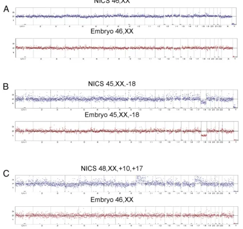
the corresponding bin in all samples. As shown in Fig. 2A, even distribution of reads along the chromosomes represents balanced chromosomal contents and thus a normal karyotype of the em-bryo. A chromosome loss in chromosome 18 results in a 50% decrease of the average read counts mapped to chr18 and was identified by the algorithm, as shown in Fig. 2B. Results for culture medium (Fig. 2, shown in blue) are displayed side by side with the results of the corresponding embryos (Fig. 2, shown in red). Fig. 2
AandBshow two examples of matching results obtained from the NICS assay when directly compared with their corresponding embryos, from a normal/transferrable embryo (Fig. 2A) and an embryo with a chromosome loss at chr18 (Fig. 2B), respectively. Fig. 2C shows an example of a false positive, with the embryo showing a normal karyotype and the NICS assay identifying a chromosome gain at chr10 and chr17.
Comparison of the Results from NICS and Their Corresponding Blastocyst Embryos. To validate our results, we performed com-parisons between 42 blastocyst culture medium samples and the NGS results of the corresponding blastocyst stage embryos. IRB approvals (Nanjing Jinlin: 2014NZKY-005; Wuxi Maternity: 2014-04-0515-02) were obtained. All of these embryos were voluntarily donated by patients with informed consent obtained before per-forming the experiments on each embryo. Results are summarized in Table 1.
By performing MALBAC-NGS, we successfully obtained in-formation of 24-chromosome ploidy from all 42 samples (100%) using the D3–D5 culture medium as well as their corresponding blastocyst-stage embryos. By profiling whole embryos, 25 samples (59.5%) were identified as normal, in which 21 of them showed concordance with the NICS assay, and the remaining four samples showed chromosomal abnormalities (false positives), converting to a specificity of 84.0%. Out of the 17 embryos that were identified with chromosomal abnormalities under the blastocyst embryo as-say, 15 of them showed chromosomal abnormalities with the NICS assay as well, but the remaining two were identified with a normal karyotype with the NICS assay (false negatives), resulting in a sensitivity of 88.2% (Table 1). The positive predictive value (PPV) and negative predictive value (NPV) of NICS in identification of chromosomal abnormalities are 78.9% and 91.3%, respectively. From the four embryos that were identified with chromosomal mosaicism (EM13, EM33, EM11, and EM41), all four were positive for chromosomal imbalance with NICS.
Coverages of 24%, 33%, and 65% were observed with NICS data of three spent cultures. The copy number variation (CNV) results for embryos EM23, EM42, and EM38, by high-depth se-quencing (30×) are shown inFig. S1.
Embryo Selection by NICS on the First Patient with Balanced Translocation.With the NICS assay validated by comparison with the voluntarily donated and IRB-approved embryos, we first ap-plied our NICS method on a patient with a balanced translocation. IRB approval (Wuxi Maternity: 2014-04-0515-02) and informed consent were obtained before applying the NICS assay on the embryos. Karyotype analysis of the patient showed a balanced translocation t(14;15)(q22;q24). We obtained a total of three blastocysts from this patient, and we performed NICS on D3–D5 culture medium of all three embryos. Chromosomal abnormalities were detected with the NICS assay in two of them, and therefore those could not be used for transfer (Fig. 3AandB). To confirm these results, the two embryos were collected and lysed for chro-mosome screening using the whole embryo, and the same results were obtained, confirming the NICS analyses (Fig. S2). Only one out of the three blastocysts showed a normal karyotype with NICS, and therefore that one was selected for transfer (Fig. 3C). A successful pregnancy resulted from this selected embryo, and a karyotype of the developing embryo was obtained by performing amniocentesis at 19 wk of gestation, confirming the karyotype results previously obtained with the NICS assay. The patient’s pregnancy resulted in the live birth of a chromosomally normal and healthy baby boy on March 5, 2016.
NICS Clinical Application Results in Successful Pregnancies. After careful and systematic validation of the NICS assay, we have performed NICS on six more patients, in addition to the trans-location patient described above. Single-blastocyst transfer was performed on all six patients, and five of these patients achieved successful pregnancies; five of these have already delivered chro-mosomally normal, healthy newborns, and we continue to follow up on the last currently ongoing pregnancy. Only one patient
failed to implant. The clinical indications and outcomes are summarized in Table 2.
Discussion
Blastocyst Biopsy and the Controversy of Extensive PGS for Patients Under IVF/ICSI Treatment. Blastocyst trophectoderm biopsy has been increasingly used and widely accepted in the PGS field due to its relatively low invasiveness, compared with performing blasto-mere biopsy at the cleavage stage. Chromosome screening on all 24 chromosomes has been mostly used on patients with advanced maternal age, recurrent pregnancy loss, repeated implantation failure, as well as on patients with abnormal karyotype such as balanced translocation and Robertsonian translocation (32–37). Extensive use of PGS on all IVF/ICSI cycles has been hotly de-bated in the past few years (38–41) due to the invasiveness of the biopsy procedure itself, particularly regarding the potential harm on the trophectoderm and possible compromise of implantation potential, as well as potential concerns on long-term effects on the offspring, which are very difficult to assess. In addition, the pro-cedure of performing blastocyst-stage biopsy requires considerable training and expertise to perform the sophisticated embryo ma-nipulation, increasing the costs of performing PGS. Of note, the procedure we report here, which simply involves collecting embryo culture medium, requires no special expertise in embryo manip-ulation and therefore can be potentially used on all IVF/ICSI cycles, thereby holding promise to improve overall clinical success rates.
False Positives and False Negatives in NICS.We observed two false negatives (2 of 17) and four false positives (4 of 25) with the NICS assay, converting to a false-negative rate and a false-positive rate of
Fig. 2. Examples of validation of results from the comparison of NICS versus the whole-blastocyst embryos. (AandB) Equivalent karyotype results obtained from NICS and the corresponding blastocyst embryo. Fig. 2Ashows consistent results from a normal/transferrable embryo, and Fig. 2Bshows consistent results from an embryo with a chromosome loss in chr18. (C) An example of inconsistent results obtained from NICS and the blastocyst embryo, with the embryo showing balanced chromosomal composition and the NICS assay identifying chromosome gains of chr10 and chr17.
GENET
chromosomal abnormalities identification of 11.8% and 16.0%, respectively. The two false negatives might have resulted from contamination from the cumulus cells, which are maternal in ori-gin and normally have a balanced chromosomal content (Table 1). In the future, the false-negative rate could be further minimized by carefully and thoroughly removing all cumulus cells before embryo culture.
The false positives most likely arose from mosaicism. It was previously hypothesized that, during embryo development, em-bryos tend to exclude those cells with mitotic errors to the exterior of the embryo (42). It is therefore possible that the false-positive rate of 16.0% arose from the debris in the culture medium, originating primarily from cells eliminated from the embryo.
Although the false-positives and false-negatives could come from measurements, we note that MALBAC provides better WGA evenness and hence higher precision for CNV determination com-pared with degenerate oligonucleotide-primed PCR and multiple displacement amplification methods (43).
The NPV of chromosomal abnormalities with the NICS assay is 91.3%, which is substantially higher than the PPV (78.9%) of the assay. The high NPV suggests that the assay is more effective in selecting normal and transferrable embryos than identifying em-bryos with chromosomal abnormalities. Single-embryo transfer has been increasingly used due to its effectiveness in decreasing multiple pregnancy and miscarriage rates (15, 44). As such, we propose NICS as an additional, and risk-free, procedure for single-embryo transfer.
Regarding the clinical implications of the false positives and false negatives, we note that, in the case of all embryos detected to exhibit aneuploidy by NICS, false positives can in principle be verified by further sequencing a blastocyst biopsy on the sixth day. We are currently developing a system in which NICS can be done rapidly and results can be obtained before embryos are frozen so that a cell or two can be extracted for verification. False negatives, on the other hand, usually do not result in successful pregnancies, and hence are less problematic. Even in the case of leading to pregnancy, they can be detected and avoided by noninvasive prenatal test.
Genome Coverage and Estimated DNA Amount in Spent Culture.The genome coverages of the spent culture media for the three samples were determined to be 24%, 33%, and 65% by high-sequencing depth of 30×reads. It has been shown that the normal genome coverage for single diploid human cells is about∼72% with high-sequencing depth (30×) (43). Despite of our low coverage, the CNV results matched exactly their corresponding blastocyst biopsies (a few cells) on the fifth day, in either the normal or aneuploid samples (Fig. S1). Larger cell numbers usually result in saturation of the genome coverage, approaching unity. Our coverage results suggest that the loss or degradation of DNA fragments in the spent culture must occur randomly along the genome and does not affect the inference of the copy number pattern in the embryo. The fact that we observed identical copy number patterns in the spent culture samples and their corresponding embryo biopsies suggests that, under our experimental conditions, the total amount of DNA in each spent culture is equivalent to that of a fraction of a single cell.
Table 1. Summary of the 42 samples profiled by NICS versus their corresponding blastocysts
21 Normal embryos (NICS and biopsy
consistent) 15 Abnormal embryos (NICS and biopsy consistent)
Sample ID NICS Biopsy Sample ID NICS Biopsy
EM02 46,XX 46,XX EM01 50,XX,+1,+5,+10,+13 47,XX,+4
EM04 46,XX 46,XX EM10 45,XX,−18 45,XX,−18
EM06 46,XY 46,XY EM11 46,XY,+5q 46,XY,−5(p12→qter,∼135M,mos∼30%)
EM07 46,XY 46,XY EM13 46,XX,−1p(pter→p21.1) 46,XX,−1p(pter→p21.1,∼103M),
+18q(q12.3→qter,∼31M, mos)
EM09 46,XY 46,XY EM14 45,XY,−18 46,XY,+1(p21.1→qter,∼142M),
−18(pter→q21.31,∼56M)
EM16 46,XX 46,XX EM15 46,XX,+1p(pter→p21.2),
−18(q21.32→qter)
46,XX,+1p(pter→p21.2,∼97M), −18q(q21.32→qter,∼21M)
EM21 46,XY 46,XY EM17 55,XY,+5,+6,+8,+11,+17,
+19,+20,+21,+22
46,XY,+1(p21.1→qter,∼143M), −18(pter→q21.31,∼58M)
EM22 46,XY 46,XY EM18 46,XX,+1p(pter→p21.1),
−18(q21.32→qter)
46,XX,+1p(pter→p21.3,∼100M), −18q(q21.31→qter,∼22M)
EM23 46,XY 46,XY EM19 46,XX,+14q(q23.3→qter),
−15q(q26.1→qte)
46,XX,+14q(q23.1→qter,∼45M), −15q(q26.1→qter,∼12M)
EM24 46,XY 46,XY EM20 46,XY,−1,+15 46,XY,−14,+15
EM25 46,XX 46,XX EM33 52,XX,+4,+6,+9,+10,+14,+17 44,XX,−4,−14,+15(mos),−18(mos)
EM26 46,XY 46,XY EM35 45,XX,−16 45,XX,+14(q32.12→qter,∼13M),−16
EM27 46,XY 46,XY EM37 45,XY,−22 45,XY,−22
EM28 46,XY 46,XY EM41 50,XX,+15,+17,+18,+20 46,XX,−X(mos),+5(p12→q13.1,∼23M,mos),
−5q(q13.1→qter,∼112M,mos)
EM29 46,XX 46,XX EM42 46,XX,−1p,+18q 46,XX,−1p,+18q
EM30 46,XY 46,XY 4 False-positive embryos (NICS abnormal, biopsy normal)
EM31 46,XX 46,XX
EM32 46,XX 46,XX EM03 48,XX,+10,+17 46,XX
EM34 46,XY 46,XY EM08 48,XY,+6,+18 46,XY
EM36 46,XY 46,XY EM12 45,XY,−18 46,XY
EM38 46,XX 46,XX EM39 47,XY,+22 46,XY
2 False-negative embryos (NICS normal, biopsy abnormal)
EM05 46,XX 45,XO
Limitations of the NICS Assay for Extensive Chromosome Screening in IVF/ICSI Patients.We think the limitations of NICS could primarily derive from two aspects: (i) the requirement to be scrupulous in the removal of all cumulus-corona radiata cells (which are of ma-ternal origin and usually with normal chromosomal composition) before performing ICSI. If such removal is not complete, residual cells may release DNA during embryo development, thereby po-tentially being the cause of false-negative detection. (ii) Similar to PGS, the NICS procedure would be highly recommended to be performed in conjunction with ICSI due to the difficulty of en-suring removal of any supernumerary sperm attached to the zona pellucida. Although culture medium is replaced on D3, which may decrease the likelihood of contamination due to residual cumulus cells and supernumerary sperm, all precautions should be made to reduce such contamination to a minimum if NICS is used routinely in clinical IVF. Of note, in the current stage of technological de-velopment, we consider NICS to be a screening assay for chro-mosomal-level abnormalities instead of being a diagnostic assay for segmental aneuploidies. More validation research is needed to identify segmental aneuploidies with the NICS assay.
In summary, our validation data and initial clinical applications strongly suggest that the NICS assay could help to improve the clinical outcome of IVF embryo selection with ploidy information, in a noninvasive manner. We envision that randomized clinical trials will be designed and performed in the near future, using the NICS assay with single-embryo transfer to evaluate the clinical effectiveness of the assay in different patient groups.
Materials and Methods
Embryo Preparation for the NICS Assay.We recruited 17 patients from the Reproductive Medicine Centre of Wuxi Maternity and Child Health Hospital and the Reproductive Medical Center of Nanjing Jinling Hospital. IRB approvals (Nanjing Jinlin: 2014NZKY-005; Wuxi Maternity: 2014-04-0515-02) and in-formed consent were obtained before applying the NICS assay on the embryos. All donated embryos were in excess of clinical needs, and consents on donated D3 embryos were obtained for use in the comparison study. All embryos were fertilized by (ICSI. Donated D3 embryos were previously frozen by vitrification (Cryotop Safety Kit; KITAZATO BioPharma) according to the manufacturer’s instructions and stored in liquid nitrogen. Vitrified embryos were warmed using warming media (Vitrification Thaw Solution; KITAZATO BioPharma). Briefly, the Cryotop strip was quickly immersed into Thawing Solution (con-taining 1 M sucrose) for 1 min at 37 °C. Embryos were then transferred into dilution solution (containing 0.5 M sucrose) and incubated for 3 min, followed by incubation in two 80-μL droplets of Washing Solution for 3 min each at room temperature (25 °C).
Blastocyst Culture and Transfer.Warmed D3 embryos were placed in 30-μL droplets of Quinn’s Advantage Protein Plus blastocyst medium (SAGE) con-taining washed and pregassed mineral oil (SAGE), and they were then further cultured to the blastocyst stage under 5.5% CO2, 5% O2, and balance N2at 37 °C in Labotect C16 incubators (Labotect). After 2 d in culture, the devel-opment and quality of the blastocysts were evaluated according to the blas-tocyst scoring system. A single blasblas-tocyst was selected for transfer to each patient based on the NICS results.
Sample Collection.To prevent media cross-contamination, different Pasteur pipettes were used for each embryo. Five to 20μL of blastocyst medium from each embryo was transferred into RNase–DNase-free PCR tubes containing
Table 2. Clinical outcome of the first seven patients subjected to NICS
Patient no. Maternal age Clinical indications Transfer cycles Clinical outcome
P01 30 Reciprocal translocation 46,XY,t(14;15) 1 Singleton pregnancy—live birth
P02 28 Azoospermia 1 Singleton pregnancy—live birth
P03 34 Inversion 46,XY,inv(9) 1 Singleton pregnancy—live birth
P04 32 Reciprocal translocation 46,XX,t(1;18) 2 Implantation failure
P05 26 Recurrent pregnancy loss 1 Singleton pregnancy—live birth
P06 32 47,XYY 2 Singleton pregnancy—live birth
P07 29 Recurrent implantation failure 1 Singleton pregnancy—following up
Fig. 3. Embryo screening and selection using NICS from a patient carrying a balanced translocation of chr14/15. A total of three embryos successfully de-veloped to the blastocyst stage, and D3–D5 culture medium from each embryo was collected for the NICS assay to screen for chromosomal abnormalities. (AandB) Embryos showed chromosomal abnormalities with chr14/15 and therefore could not be transferred. (C) An embryo showed balanced chromosomal composition and was therefore transferred into the uterus of the patient, resulting in a successful pregnancy and a healthy live birth.
GENET
5μL of cell lysis buffer (Yikon Genomics). As a negative control, we collected the same amount of blastocyst culture medium but without its being used for embryo culture. All collected samples were frozen immediately in liquid ni-trogen and stored at−80 °C until subjected to the NICS assay.
Embryo Selection by NICS on a Patient with Balanced Translocation.The patient couple for NICS treatment in Wuxi Maternity reproduction center had 3 y of infertility history. Their chromosome examination showed that the male partner carries a t(14;15)(q22;q24) translocation, and the female has a normal karyo-type. We collected five eggs in the IVF cycle (SI Materials and Methods); four MII oocytes were fertilized by ICSI, and three of them developed to the blastocyst stage. All blastocysts were frozen with vitrification freezing protocol after performing NICS (SI Materials and Methods). We obtained normal karyotype from one embryo with the NICS assay (Fig. 3C). We therefore transferred this normal embryo and confirmed clinical pregnancy with enhancedβ-HCG fol-lowed up by ultrasound. The result of fetal chromosome examination in the amniotic fluid at 19 wk of gestation confirmed the balanced chromosome results previously obtained by NICS. After obtaining informed consent from the patients, the two embryos with chromosome imbalance, according to our NICS results, were lysed and sequenced for validation purposes (Fig. 1).
WGA and DNA Sequencing from Culture Medium of the Embryos.The MALBAC single-cell WGA method was used to amplify DNA from the culture medium, as well as from the embryos, following the manufacturer’s protocol (catalog no. YK001B; Yikon Genomics), and used to construct the sequencing libraries (NEBNext Ultra DNA Kit; New England Biolabs) for sequencing on an Illumina HiSeq 2500 platform, yielding∼2 million sequencing reads on each sample. The read numbers were counted along the whole genome with a bin size of 1 Mb. A copy number gain from two to three copies results in a 50% increase in read counts, whereas a copy number loss from two copies to one copy re-sults in a 50% decrease in read counts.
ACKNOWLEDGMENTS.We thank Drs. Patty Purcell and Catherine Racowsky for their critical reading and editing of the manuscript and Dr. Luoxing Xiong for her assistance with data analyses. This work was supported by National Basic Research Program of China Grant 2015AA020407; Natural Science Foundation of Jiangsu Province, China, Grant BK20131094; funding from the Beijing Advanced Innovation Center for Genomics at Peking University, Joint Research Program of Medical Science and Technology Development Fund of the Medical Control Center in Wuxi City (Grant YGZX1204); National Natural Science Foundation of China Grant 81503655; and State Key Development Program for Basic Research of China Grant 2013CB945200.
1. Battaglia DE, Goodwin P, Klein NA, Soules MR (1996) Influence of maternal age on meiotic spindle assembly in oocytes from naturally cycling women.Hum Reprod11(10):2217–2222. 2. Munné S (2006) Chromosome abnormalities and their relationship to morphology
and development of human embryos.Reprod Biomed Online12(2):234–253. 3. Korenberg JR, et al. (1994) Down syndrome phenotypes: The consequences of
chro-mosomal imbalance.Proc Natl Acad Sci USA91(11):4997–5001.
4. Hellani A, Abu-Amero K, Azouri J, El-Akoum S (2008) Successful pregnancies after application of array-comparative genomic hybridization in PGS-aneuploidy screening.
Reprod Biomed Online17(6):841–847.
5. Gutiérrez-Mateo C, et al. (2011) Validation of microarray comparative genomic hybrid-ization for comprehensive chromosome analysis of embryos.Fertil Steril95(3):953–958. 6. Thornhill AR, et al. (2015) Karyomapping—a comprehensive means of simultaneous monogenic and cytogenetic PGD: Comparison with standard approaches in real time for Marfan syndrome.J Assist Reprod Genet32(3):347–356.
7. Natesan SA, et al. (2014) Live birth after PGD with confirmation by a comprehensive approach (karyomapping) for simultaneous detection of monogenic and chromo-somal disorders.Reprod Biomed Online29(5):600–605.
8. Treff NR, et al. (2011) Single nucleotide polymorphism microarray-based concurrent screening of 24-chromosome aneuploidy and unbalanced translocations in pre-implantation human embryos.Fertil Steril95(5):1606–1612.e1,2.
9. Natesan SA, et al. (2014) Genome-wide karyomapping accurately identifies the in-heritance of single-gene defects in human preimplantation embryos in vitro.Genet Med16(11):838–845.
10. Treff NR, et al. (2012) Development and validation of an accurate quantitative real-time polymerase chain reaction-based assay for human blastocyst comprehensive chromosomal aneuploidy screening.Fertil Steril97(4):819–824.
11. Martín J, et al. (2013) The impact of next-generation sequencing technology on preimplantation genetic diagnosis and screening.Fertil Steril99(4):1054–1061.e3. 12. Hou Y, et al. (2013) Genome analyses of single human oocytes.Cell155(7):1492–1506. 13. Scott RT, Jr, et al. (2013) Blastocyst biopsy with comprehensive chromosome screening and fresh embryo transfer significantly increases in vitro fertilization implantation and delivery rates: A randomized controlled trial.Fertil Steril100(3):697–703. 14. Forman EJ, et al. (2013) In vitro fertilization with single euploid blastocyst transfer: A
randomized controlled trial.Fertil Steril100(1):100–107.e1.
15. Yang Z, et al. (2012) Selection of single blastocysts for fresh transfer via standard morphology assessment alone and with array CGH for good prognosis IVF patients: Results from a randomized pilot study.Mol Cytogenet5(1):24.
16. Keltz MD, et al. (2013) Preimplantation genetic screening (PGS) with comparative genomic hybridization (CGH) following day 3 single cell blastomere biopsy markedly improves IVF outcomes while lowering multiple pregnancies and miscarriages.J Assist Reprod Genet30(10):1333–1339.
17. Cimadomo D, et al. (2016) The impact of biopsy on human embryo developmental potential during preimplantation genetic diagnosis.BioMed Res Int2016:7193075. 18. Wu Y, et al. (2014) Blastomere biopsy influences epigenetic reprogramming during
early embryo development, which impacts neural development and function in re-sulting mice.Cell Mol Life Sci71(9):1761–1774.
19. Zhao H-C, et al. (2013) Aberrant epigenetic modification in murine brain tissues of off-spring from preimplantation genetic diagnosis blastomere biopsies.Biol Reprod89(5):117. 20. Zeng Y, et al. (2013) Preimplantation genetic diagnosis (PGD) influences adrenal de-velopment and response to cold stress in resulting mice.Cell Tissue Res354(3):729–741. 21. Assou S, Aït-Ahmed O, El Messaoudi S, Thierry AR, Hamamah S (2014) Non-invasive pre-implantation genetic diagnosis of X-linked disorders.Med Hypotheses83(4):506–508. 22. Galluzzi L, et al. (2015) Extracellular embryo genomic DNA and its potential for
genotyping applications.Future Science OA1(4):FSO62.
23. Palini S, et al. (2013) Genomic DNA in human blastocoele fluid.Reprod Biomed Online
26(6):603–610.
24. Gianaroli L, et al. (2014) Blastocentesis: A source of DNA for preimplantation genetic testing. Results from a pilot study.Fertil Steril102(6):1692–1699.e6.
25. Stigliani S, Anserini P, Venturini PL, Scaruffi P (2013) Mitochondrial DNA content in embryo culture medium is significantly associated with human embryo fragmenta-tion.Hum Reprod28(10):2652–2660.
26. Stigliani S, et al. (2014) Mitochondrial DNA in day 3 embryo culture medium is a novel, non-invasive biomarker of blastocyst potential and implantation outcome.Mol Hum Reprod20(12):1238–1246.
27. Wu H, et al. (2015) Medium-based noninvasive preimplantation genetic diagnosis for humanα-thalassemias-SEA.Medicine (Baltimore)94(12):e669.
28. Zong C, Lu S, Chapman AR, Xie XS (2012) Genome-wide detection of single-nucleotide and copy-number variations of a single human cell.Science338(6114):1622–1626. 29. Huang J, et al. (2014) Validation of multiple annealing and looping-based
amplifi-cation cycle sequencing for 24-chromosome aneuploidy screening of cleavage-stage embryos.Fertil Steril102(6):1685–1691.
30. Yan L, et al. (2015) Live births after simultaneous avoidance of monogenic diseases and chromosome abnormality by next-generation sequencing with linkage analyses.
Proc Natl Acad Sci USA112(52):15964–15969.
31. Huang J, et al. (2016) Validation of a next-generation sequencing–based protocol for 24-chromosome aneuploidy screening of blastocysts.Fertil Steril105(6):1532–1536. 32. Gui B, et al. (2016) Chromosomal analysis of blastocysts from balanced chromosomal
rearrangement carriers.Reproduction151(4):455–464.
33. Schoolcraft W, et al. (2012) Comprehensive chromosome screening (CCS) with vitri-fication results in improved clinical outcome in women>35 years: A randomized control trial.Fertil Steril98(3):S1.
34. Hodes-Wertz B, et al. (2012) Idiopathic recurrent miscarriage is caused mostly by aneuploid embryos.Fertil Steril98(3):675–680.
35. Margalioth EJ, Ben-Chetrit A, Gal M, Eldar-Geva T (2006) Investigation and treatment of repeated implantation failure following IVF-ET.Hum Reprod21(12):3036–3043. 36. Frydman N, et al. (2001) Assisting reproduction of infertile men carrying a
Rob-ertsonian translocation.Hum Reprod16(11):2274–2277.
37. Fragouli E, et al. (2010) Comprehensive chromosome screening of polar bodies and blastocysts from couples experiencing repeated implantation failure.Fertil Steril
94(3):875–887.
38. Chen M, Wei S, Hu J, Quan S (2015) Can comprehensive chromosome screening technology improve IVF/ICSI outcomes? A meta-analysis.PLoS One10(10):e0140779. 39. Twisk M, et al. (2008) No beneficial effect of preimplantation genetic screening in women of advanced maternal age with a high risk for embryonic aneuploidy.Hum Reprod23(12):2813–2817.
40. Hardarson T, et al. (2008) Preimplantation genetic screening in women of advanced maternal age caused a decrease in clinical pregnancy rate: A randomized controlled trial.Hum Reprod23(12):2806–2812.
41. Schoolcraft WB, Katz-Jaffe MG, Stevens J, Rawlins M, Munne S (2009) Preimplantation aneuploidy testing for infertile patients of advanced maternal age: A randomized prospective trial.Fertil Steril92(1):157–162.
42. Taylor TH, et al. (2014) The origin, mechanisms, incidence and clinical consequences of chromosomal mosaicism in humans.Hum Reprod Update20(4):571–581. 43. Huang L, Ma F, Chapman A, Lu S, Xie XS (2015) Single-cell whole-genome
amplifi-cation and sequencing: Methodology and appliamplifi-cations.Annu Rev Genomics Hum Genet16:79–102.
44. Ryan GL, et al. (2007) A mandatory single blastocyst transfer policy with educational campaign in a United States IVF program reduces multiple gestation rates without sacrificing pregnancy rates.Fertil Steril88(2):354–360.
45. Bolger AM, Lohse M, Usadel B (2014) Trimmomatic: A flexible trimmer for Illumina sequence data.Bioinformatics30(15):2114–2120.