Año 9, Vol. IX, Nº 2, pp. 1-5
ISSN 1668-5466
CELL PROLIFERATION OF THE M OUSE SEBOCYTES
IN SKIN AND PREPUTIAL GLAND
1,2 1 3
Claudio Gustavo BARBEITO ; Vicente Alberto CATALANO ; Norma Viviana GONZALEZ 1
Cátedra de Histología y Embriología, Facultad de Ciencias Veterinarias, Universidad Nacional de La Plata. Calles 60 y 118, La Plata, Argentina.
2
Instituto de Patología, Facultad de Ciencias Veterinarias, Universidad Nacional de La Plata. Calles 60 y 118, La Plata, Argentina.
3
Cátedra de Histología y Embriología Animal, Facultad de Ciencias Naturales y Museo, Universidad Nacional de La Plata. Calles 60 y 122, La Plata, Argentina.
Correspondence to: Barbeito, Claudio Gustavo.
Cátedra de Histología y Embriología. Facultad de Ciencias Veterinarias. UNLP. 60 y 118. La Plata (1900). Buenos Aires. Argentina.
E-mail:
Abstract.Preputial glands are specialized sebaceous gland of some rodent as mouse and rat. This gland is regulated by androgens and other hormones. In this work we analysed the mitotic activity of the alveolar and ductal cells of the mice preputial gland throughout a circadian period. The results were compared to data obtained from the skin sebaceous gland. The mitotic activity in the preputial sebocytes displayed a bimodal curve with the highest mitotic indices at 12:00 and 00:00 h while the lower values were reached at 20:00 and 08:00h. In the ductal cells, the mitotic activity showed a single mitotic peak at 12:00h and a trough at 20:00 h. In the sebocytes from skin glands, the acrophase was detected at 04:00 h. The average cell proliferation daily value was significantly higher in the preputial sebocytes when compared to cutaneous sebocytes. These results demonstrated the existence of quantitative and qualitative differences in the mitotic activity of the preputial gland sebocytes in relation to the skin sebocytes.
Keywords: cell proliferation, circadian rhythms, preputial glands, sebocytes. [email protected]
INTRODUCTION
Preputial gland s are alveo lar co mplex holocrine glands; they are accessory to the male reproductive tract of some rodents, as the rat and the mouse. Due to its pheromones content, their products have been assigned functions related to territorial and sexual behavioral activities (11, 32). These glands' size and activities are larger in d o mineering aggressiv e males and their secretion is attractive to females (10, 11, 31). In addition, Marchlewska-Koj et al. (1990) found that the presence of these glands' extract induces estrous in virgin female mice.
Their main cellular type is the alveolar cell, mostly referred as sebocyte. Electron microscopy shows that the rat preputial gland resembles the human sebaceous gland, not only in terms of containing a sebocyte-like population of cells in an acinar arrangement at different maturational
stages, but also in the morphology of its organelles (17). In these sense, preputial glands resemble the sebaceous skin glands and can be co nsidered a specializatio n o f these last. Accordingly, the preputial glands have been employed as an experimental model to study sebocytes growth and differentiation (27, 30).
Several hormones are known to influence these glands' activities (9). Insulin-like growth factor 1(IGF-I) and insulin, but not growth ho rm o ne ( G H ) , stim u late in v it r o c ell proliferation of the glands. GH enhances preputial sebocytes differentiation (6). On the other hand, androgens have been found to stimulate preputial gland sebocytes as well as skin sebocytes (7, 13, 21, 24, 30).
2
populations in the adult mouse exhibit a daily proliferation rhythm (2, 8, 25).
For a better understanding of the preputial cell proliferation, the following study was carried out to: 1) characterize its temporal structure in adult mice, and 2) to compare preputial and sebaceous skin glands cell proliferation.
M ATERIALS AND M ETHODS
Forty-two male C3H/ Avy strain mice, five month-old were used. Animals were housed under standardized conditions for a 24 h periodicity analysis in an ad hoc room with a 22 ± 2ºC temperature and lighting regimen of 12 h light - 12 h darkness (lights on 06.00-18.00 h). Pelleted food and water were provided ad libitum. Mice were divided into 6 lots (n: 6-8) for killing by decapitation and exsanguination at the following times of day: 00:00, 04:00, 08:00, 12:00, 16:00, and 20:00 h. Each mouse received an intraperitoneal colchicine dose of 2 µg/ g body weight 4 h before sacrifice.
Preputial glands and a sample of auricular skin from each animal were fixed in 10% buffered formalin. The embedding was performed in paraffin. Five µm-thick sections were stained with H&E.
The observation was carried out under immersion oil objective (1000x). In the preputial glands, two cell-populations were monitored: alveolar cells (preputial sebocytes, PS) from the basal and parabasal layers and ductal cells (DC). No less than 3000 PS and 1500 DC per individual were monitored scoring mitosis along with the total number of cell nuclei every tenth field. In the cutaneous glands, the same procedure was employed for 3000 alveolar cells (cutaneous sebocytes, CS).
T h e m i t o t i c i n d e x ( c o l c h i c i n e metaphase/ 1000 nuclei) (MI) for each animal and cell-type was calculated from data collected. Using these individual data, the arithmetic mean ± standard error from each lot and group was assessed. The significance of the differences
between organs, lots and groups was analyzed by Anova and Student's t- test.
RESULTS
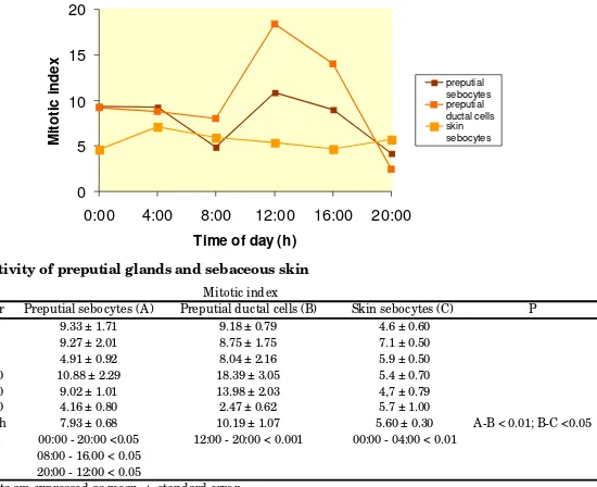
Table 1 and Figure 1 summarize the results on mitotic activity in PS, DC and CS. The mitotic activity in the PS presented important variations in the different time-points, displaying a bimodal curve (Figure 1) with the highest mitotic indices at 12:00 and 00:00 h while the lower values are reached at 20:00 and 08:00h (Table 1). In DC, the mitotic activity showed a single mitotic peak at 12:00 h and a trough at 20:00 h (Figure 1 and Table 1). For CS the acrophase was detected at 04:00 h (Figure 1 and Table 1).
The average cell proliferation daily value was significantly higher in PS (7.93±0.68) when compared to CS (5.6±0.3) (Table 1).
DISCUSSION
Bo th cell-po pulatio ns fro m the mo use preputial glands under study presented mitotic daily variations. Our results demonstrated clear-cut qualitative and quantitative differences in the proliferation of PS and DC.
DC showed a higher mitotic activity than PS. This finding is in agreement w ith higher proliferation indices reported for ductal cells in other exocrine glands as the submaxillary gland (1). Moreover, whereas DC mitotic activity presented a single peak curve, PS had a different rhythmicity for the mitotic daily curve showed a bimodal characteristic. Similar circadian bimodal rhythms have been found in other mouse cell-populations (2, 25). For instance, the pars intermedia in female mice displays a comparable temporal structure at 28 d of age for two mitotic peaks are found at midnight and noon, whereas in adult females the mitotic daily curve is still bimodal but peaks are found at 04:00 and 16.00 h (2).
mitotic curve's structure for PS revealed a bimodal pattern whereas CS curve displayed a single peak.
Socially dominant male mice have larger preputial glands than do subordinate males (4). A similar situation has been observed in rats for dominant individuals that had heavier preputial g land s c o m p ared to su b d o m inant and subordinate rats (20). Accordingly to their sexually-related role, PS are stimulated to proliferate and differentiate by androgen (13, 14, 18) as in other sebaceous glands, namely CS. As both PS and CS posses receptors for androgen and other hormones as the melanocortin 5 (26), the effect of these signals should not be considered as a cause to explain differences concerning PS and CS mitotic activity. Results from the present study suggest a paracrine and autocrine control hypothesis on the preputial gland proliferation and also the existence of differences in regard to skin sebaceous glands.
Differential organ response to the androgens' effects on cell- cycle were previously reported (28); these variations could be related to the presence of different enzymes isotypes that catalyze the conversion of sexual steroids in the PS and in CS (5). Another plausible explanation would be a differential enzymatic activity (3, 19). The preputial glands of several rodents' species have been long employed as a model to investigate a wide spectrum of topics e.g. the rat preputial system is used as an experimental model in sebaceous gland physiology (26). More recently, preputial glands have been used in numerous assessments on toxicological aspects (12, 22), and sex, individuality, and/ or the genetic background differences between mice strains (32). Our work strongly suggests that the extrapolation of PS findings to CS may lead to erroneous interpretations. The existence of circadian rhythms in these glands could be extended to other aspects of the organs`
0 5 10 15 20
0:00 4:00 8:00 12:00 16:00 20:00
Time of day (h)
M
it
o
ti
c
in
d
e
x
preputial sebocytes preputial ductal cells skin sebocytes
Figure 1
Hour Preputial sebocytes (A) Preputial ductal cells (B) Skin sebocytes (C)
0:00 9.33 ± 1.71 9.18 ± 0.79 4.6 ± 0.60
4:00 9.27 ± 2.01 8.75 ± 1.75 7.1 ± 0.50
8:00 4.91 ± 0.92 8.04 ± 2.16 5.9 ± 0.50
12:00 10.88 ± 2.29 18.39 ± 3.05 5.4 ± 0.70
16:00 9.02 ± 1.01 13.98 ± 2.03 4,7 ± 0.79
20:00 4.16 ± 0.80 2.47 ± 0.62 5.7 ± 1.00
X 24 h 7.93 ± 0.68 10.19 ± 1.07 5.60 ± 0.30
P 00:00 - 20:00 <0.05 12:00 - 20:00 < 0.001 00:00 - 04:00 < 0.01
08:00 - 16.00 < 0.05 20:00 - 12:00 < 0.05
a Results are expressed as mean ± standard error
X 24 h: mean of the whole sample period; P: significance of differences in mean values.
A-B < 0.01; B-C <0.05 Mitotic index
P Table 1. M itotic activity of preputial glands and sebaceous skin
[image:3.425.105.380.300.524.2]4
REFERENCES
1. Barbeito CG, García MN, Savignone CA, Catalano VA, Flamini MA, Badrán AF, Moreno FR. (1997). Actividad mitótica de los epitelios de epidermis, lengua, túbulos contorneados renales y conductos de la glándula submaxilar del ratón portador del tumor SS1K. Ciencias Morfológicas; 3: 9-18.
2. Barbeito CG, Surur JM, Badrán AF. (2000). Mitotic activity of the pars intermedia in the female mouse: age-associated variations in proliferation rate and circadian periodicity. Chronobiol Int; 17:751-756.
3. Blanchard PG, Luu-The V. (2007). Differential androgen and estrogen substrates specificity in the mouse and primates type 12 17 beta-hydroxysteroid dehydrogenase. J Endocrinol; 194:449-455.
4. Bronson FH, Marsden, HM. (1973). The preputial gland as an indicator of social dominance in male mice. Behav Biol; 9:625-628.
5. Deplewski D, Liao S, Rosenfield RL. (1997). Preputial sebocyte 5alpha-reductase isoform specificity. Endocrinol; 138:4416-4420.
6. Deplewski D, Rosenfield R. (1999). Growth hormone and insulin-like growth factors have different effects on sebaceous cell growth and differentiation. Endocrinol; 140:4089-4094.
7. Ebling FJ, Ebling E, Randall V, Skinner J. (1975). The effects of hypophysectomy and of bovine growth hormone on the responses to testosterone of prostate, preputial, Harderian and lachrymal glands and of brown adipose tissue in the rat. J Endocrinol; 66:401-406.
8. García MN, Barbeito CG, Andrini LB, Badrán AF. (2001). Circadian rhythm of the DNA synthesis and the mitotic activity of tongue keratinocytes. Cell Biol Int; 25:179-183.
9. Hay JB, Meddis D, Thody AJ, Shuster S. (1982). Mechanism of action of alpha-melanocyte-stimulating hormone in rat preputial glands: the role of androgen metabolism. J Endocrinol; 94:289-294.
10. Hayashi S. (1989). Male mice: Social Dominance Influenced by Strange Male Odors. Aggress Behav; 15:1-3. 11. Hayashi S. (1990). Social Condition Influences Sexual Attractiveness of Dominant Male Mice. Zool. Sci, 7: 889-894. 12. Homady M, Hussein H, Jiries A, Mahasneh A, Al-Nasir F, Khleifat K. (2002). Survey of Some Heavy Metals in Sediments from Vehicular Service Stations in Jordan and Their Effects on Social Aggression in Prepubertal Male Mice. Environmental Res; 89:43-49.
13. Huggins C, Parson FM, Jensen EV. (1955). Promotion and growth of preputial glands by steroids and the pituitary growth hormone. Endocrinol; 57:25-52.
14. Labrie F, Luu-The V, Martel C, Chernomoretz A, Calvo E, Morissette J, Labrie C. (2006). Dehydroepiandrosterone (DHEA) is an anabolic steroid like dihydrotestosterone (DHT), the most potent natural androgen, and tetrahydrogestrinone (THG). J Steroid Biochem Mol Biol; 100:52-58.
15. Lucas LA, Eleftheriou BE. (1980). Circadian variation in concentrations of testosterone in the plasma of male mice: a difference between BALB/ cBy and C57BL/ 6By inbred strains. J Endocrinol; 87:37-46.
16. Marchlewska-Koj A, Pochron E, Sliwowska A (1990). Salivary glands and preputial glands of males as source of estrus-stimulating pheromone in female mice. J Chem Ecol, 16: 2817-2822.
17. Mednieks MI, Laurent SJ, Hand AR, Rosenfield RL. (1991). Cyclic AMP-receptor protein activity in rat preputial cells. J Inv Dermat; 97: 517523.
18. Miyake K, Ciletti N, Liao S, Rosenfield RL. (1994). Androgen receptor expression in the preputial gland and its sebocytes. J Inv Derm; 103, 721725.
19. Pelletier G, Luu-The V, Li S, Labrie F. 2005. Localization of type 8 17beta-hydroxysteroid dehydrogenase mRNA in mouse tissues as studied by in situ hybridization. J Histochem Cytochem; 53:1257-1271.
20. Pohorecky LA, Blakley GG, Ma EW, Soini HA, Wiesler D, Bruce KE, Novotny MV. (2008). Social housing influences the composition of volatile compounds in the preputial glands of male rats. Hormones Behav; 53:536-545. 21. Potter JE, Prutkin L, Wheatley VR. (1979). Sebaceous gland differentiation. I. Separation, morphology and lipogenesis of isolated cells from the mouse preputial gland tumor. J Invest Dermatol; 72:120-127.
22. Rawdah K Al-Thani, Aisha S. Al-Thani, Elbetieha, A, Darmani H. (2003). Assessment of reproductive and fertility effects of amitraz pesticide in male mice. Toxicol Letters;138:253-260.
23. Sayegh JF, Kobor G, Lajtha A, Vadasz C. (1990). Effects of social isolation and the time of day on testosterone levels in plasma of C57BL/ 6By and BALB/ cBy mice. Steroids; 55:79-82.
25. Surur JM, Catalano VA, Flamini MA, Barbeito CG. (2005). Effects of tumors on the daily mitotic activity of mouse pars intermedia. Cell Biol Int; 29: 173-155.
26. Thiboutot D, Sivarajah A, Gilliland K, Cong Z, Clawson GJ. (2000). The melanocortin 5 receptor is expressed in human sebaceous glands and rat preputial cells. Invest Dermatol; 115:614-619.
27. Thody AJ, Shuster S. (1973). Possible role of MSH in the mammal. Nature; 245: 207-208,
28. Tsai TH, Scheving LE, Scheving LA, Pauly JE. (1985). Sex differences in circadian rhythms of several variables in lymphoreticular organs, liver, kidney, and corneal epithelium in adult CD2F1 mice. Anat Rec; 211:263-270.
29. Weinert D, Ulrich FE, Schuh J. (1987). Ontogenetic changes in the circadian rhythm of plasma insulin and its correlation to food intake. Biomed Biochim Acta; 46:387-395.
30. Wheatley VR, Potter JE, Lew G. (1979). Sebaceous gland differentiation: II. The isolation, separation and characterization of cells from the mouse preputial gland. J Invest Dermatol; 73:291-296.
31. Yamashita J, Hayashi S, Hirata Y. (1989). Reduced size of preputial glands and absence of aggressive behavior in the genetically obese (Ob/ Ob) mouse. Zool Sci; 6:1033-1036.
32. Zhang JX, Rao XP, Sun L, Zhao CH, Qin XW. (2007). Putative chemical signals about sex, individuality, and genetic background in the preputial gland and urine of the house mouse (Mus musculus). Chem Senses; 32:293-303.
ACKNOW LEDGEM ENTS
The authors wish to thank PhD Eduardo Gimeno for the critical reading of the manuscript. This work was supported with grants from National University of La Plata. Thanks are due to Mr. Ruben Mario and Ms. Paula Dumont for their technical assistance.