Keywords:
• biomaterials
• calcium titanate coating • tribology
• pin-on-disk • Hank’s solution reevaluated: March 2014, accepted: March 2014
Tribological Behavior of Bone Against Calcium Titanate
Coating in Simulated Body Fluid
Comportamiento tribológico de hueso contra recubrimiento
de titanato de calcio en fluido corporal simulado
Esguerra-Arce Johanna
Escuela de Ingeniería de Materiales Universidad del Valle, Colombia E-mail: johanna.esguerra@correounivalle.edu.co
Aguilar-Castro Yesid
Escuela de Ingeniería de Materiales Universidad del Valle, Colombia E-mail: yesid.aguilar@correounivalle.edu.co
Aperador-Chaparro William
Facultad de Ingeniería
Universidad Militar Nueva Granada, Bogotá, Colombia E-mail: post.ingenieria@unimilitar.edu.co
Ipaz-Cuastumal Leonid
Escuela de Ingeniería de Materiales Universidad del Valle, Colombia
E-mail: leoipazc@gmail.com
Bolaños-Pantoja Gilberto
Laboratorio de Física de Bajas Temperaturas Universidad del Cauca, Colombia E-mail: gbolanos@unicauca.edu.co
Rincón-López Carlos Alberto
Laboratorio de Física de Bajas Temperaturas Universidad del Cauca, Colombia
E-mail: crincon@unicauca.edu.co
Information on the article: received: February 2014,
Abstract
Although calcium titanate has been proposed as a coating for biomedical applications, a characterization of tribological properties simulating hu-man conditions has not been reported. In this work we studied friction and wear mechanism of calcium titanate coating growth onto AISI 304 steel
(750 nm thickness) deposited by r.f. magnetron sputtering. It was found
that the wear mechanisms of the system is bone adhesion to the coating without detachment of the coating, both dry and in Hank’s solution, with
a friction coefficient of 0.84 ± 0.13 and 0.65 ± 0.13, respectively. The wear of the bone was more severe when using a simulated body fluid at 37°C in the
Introduction
Hip prosthesis stems may be cemented or uncemented,
the uncemented have four options for achieving adhe -sion with the surrounding tissue: by pressure, porous surface, screws or with coated surface. Joint prosthesis
with hydroxyapatite coated stems have presented a big
clinical success, as they increase the rate of osseointe-gration regarding uncemented prosthesis (Faig and Gil,
2008; Coathup et al., 200; Rokkum and Reigstad, 1999). However, in the long run this coating dissolves and de -taches exposing the base metal (Porter et al., 2004), which can lead to the loosening of the prosthesis, pain in the patient, wear on the rest of the prosthesis or, in
the worst case, to extensive destruction of bone tissue.
Although the calcium titanate has been studied
ex-tensively in regard to optical and luminescent proper -ties (Moreira et al, 2009; Wang et al., 2009; Yang et al., 2009), an interface layer containing calcium titanate on biomedical titanium alloys coated with hydroxyapatite has been found (Ergun et al., 2003; Kokubo et al., 2003),
so that some authors have reported to have obtained calcium titanate layers by various methods on titanium
and titanium alloys, such as sol-gel (Holliday and Sta-nishevsky, 2004), magnetron sputtering (Asami et al., 2005), ion-beam assisted deposition (Ohtsu et al., 2006), pulsed laser deposition (Ohtsu et al., 2007),
hy-drothermal–electrochemical method (Wiff et al., 2007), thick alkalized calcium oxidation (Ohtsu et al., 2007),
and hydrothermal treatment (Park et al., 2011) for bio -medical applications.
Despite the fact that most commercial prostheses are made of titanium or titanium alloys, because the
material replaced for bone is expected to have a modu
-lus equivalent to that of bone (Geetha et al., 2009), and
due to their high cost and their poor tribological pro-perties, stainless steels is still used in hip prosthesis and
remains under investigation for biomedical purposes
(Lundin et al., 2012; Nie, 2011; Barragán et al., 2010).
Although type 304 is seldom used in biomedical
im-plants or devices and its sensitivity to any sign of corro
-sion is more detectable than in 316L (Tang et al., 2006),
it is used in implants such as brackets and screws. This steel also meets ASTM F138 y ASTM F139 (ASTM F138–08, 2008; ASTM F139–08, 2008) standards for steels used in biomedical applications and it is cheaper
than AISI 316L which would represent a reduction in hip prosthesis cost. This is the reason for proposing here the evluation of tribological properties of AISI 304
stainless steel coated with calcium titanate for prosthe-sis stem applications.
It is known that due to the continuous micro-move -ments between the bone and the implant (Fu et al., 1998) wear femoral stem occurs (Howell et al., 1999),
so it is important to know the tribological properties of the coating with respect to the bone, such as friction and wear in conditions which approach those of the
hu-man body. Therefore, this work aims to evaluate cal -cium titanate coated AISI 304 stainless steel by r.f.
magnetron sputtering, evaluating mechanical proper -ties of hardness and elastic modulus by
nanoindenta-tion; coefficient of friction and wear by pin-on-disc test
in dry conditions at room temperature and in Hank’s
solution at 37 °C with an animal bone pin as a counter -part.
Materials and methods
The CaTiO3 cathode used for the sputtering technique
were from CaTiO3 powder with a purification of 99.9%
from Super Conductor Materials, Inc. Silicon (100) and 304 stainless steel disks (diameter = 1.27 cm and thick
-ness = 3 mm) were used as substrates. The steel was
polished with sandpaper passing numbers 200 to 2000,
and finally with 1 and 0.3 μm alumina solution. Prior to
deposition of calcium titanate an approximately 200 nm
Descriptores:
• biomateriales
• recubrimiento de titanato de calcio
• tribología
• pin-disco
• solución de Hank Resumen
Aunque el titanato de calcio ha sido propuesto como recubrimiento para aplicaciones biomédicas, no se han reportado caracterizaciones tribológicas en condiciones que simulan las condiciones del interior del cuerpo humano. En este trabajo se evalúan propiedades de fricción y mecanismos de desgaste de estos recubrimientos (de 750
nm de espesor) depositados mediante r.f. magnetrón sputtering. Se encontró que el
mecanismo de desgaste del sistema es adhesión de hueso al recubrimiento sin
des-prendimiento de la capa, tanto en seco como en solución de Hank, con coeficientes de
fricción de 0.84 ± 0.13 y 0.65 ± 0.13, respectivamente. El desgaste del hueso fue más
titanium layer was deposited onto steel substrate by
magnetron sputtering, during the 3 hours at a tempera
-ture of 250°C, with a power density of the cathode fixed at 350 W.
Calcium titanate was deposited via magnetron sputtering in Ar atmosphere. Prior to the deposition, the deposition chamber was evacuated to a base pres
-sure of 2 × 10⁻⁶ mbar and the target was sputter-cleaned for 20 min, before film growth. During the 4 hours de
-position process at a temperature of 500°C, the sputte
-ring power density deposition of the CaTiO3 cathode
was fixed at 2.47 W/cm2, the pressure in the chamber
was fixed at 2 × 10⁻² mbar, and the target was fixed 4.3
cm under the substrate.
The crystal structure of the films was determined by using glancing incident X-ray diffraction (GIXRD) at 2° incidence angle with a RIGAKU (Dmax2100) difractro
-meter using Co Kα radiation (λ = 1.78899 Å, 30 kV and
16 mA). The chemical composition of deposited films was performed by energy dispersive X-ray spectrosco -py (EDX) in the scanning electron microscope (JEOL
JSM-649 OLV SEM). The thickness of the films was
measured by Scanning Electron Microscope (JEOL
JSM-649 OLV). The mechanical analysis was performed via nanoindentation (ASTM E2546-07; 2007) by means
of anUBIL - HYSITRON device and a diamond Berko
-vich tip. For each sample several series of ten indents were made and the results were averaged, since the maximal nanoindentation depth was about 10% of coa -ting thickness.
Taking into ac -count that these coa-tings are proposed for use in hip prosthe-sis stems a
pin-on-disk test (ASTM G99-05, 2010) with
animal bone ball as a counterpart was used to measure the wear resistance and the
friction coefficient using a MT60 tribo
-meter from NANO
-VEA™. The bone pin
was prepared from a
bovine metacarpal
diaphysis, which was immersed in boiling
water avoiding tou
-ching the bottom of the vessel to not de
-grade its mechanical properties. Then the soft tissue was removed mechanically by hand and the bone was im
-mersed in a bleach-water mixture (1:5 volume) for 53 hours for further machining to a spherical geometry of 6 mm diameter. For mechanical measurement via nanoin -dentation specimens were embedded in plastic (Lucite,
Buehler) to protect them from damage and to provide a uniform format for preparation. The sample was polis
-hed using a sequence of abrasives silicon carbide grit pa -per ending with 0.3 micron alumina suspension, using water as a lubricant. Each sample was indented 24 times
using a Berkovich pyramidal indenter, varying the maxi
-mum load applied from 479.6 to 5995.8 μN (10 seconds of loading and 10 seconds of unloading).
Tribological test was carried out in dry and wet con -ditions (three bare steel samples and three coated
sam-ples in every condition). Dry tests were carried out at room temperature and relatively humidity of 65% ± 4, wet tests, simulating body conditions (SBC), were performed using Hank’s solution (Table 1) at 37°C ± 1. The test is carried out with Hank’s solution because this is, of all the available solutions, the one that most clo -sely approximates to the composition of blood plasma (Zhanga et al., 2013). The applied load was 3 N, the dis
-tance was 200 meters at 100 rpm (test duration: 1hour, 46 minutes). A detailed characterization of the worn
surfaces was performed using a Scanning Electron
Mi-croscope (JEOL JSM-649 OLV) after cleaning with ace
-tone and water at 50°C.
Table 1. Chemical composition of Hank’s solution, concentration [g/L]
NaCl KCl MgSO4.7H2O CaCl2.H2O Na2HPO4 KH2PO4 NaHCO3 Glucosa
8.0 0.4 0.2 0.185 0.046 0.06 0.35 1.0
Table 2. Elemental chemical composition in calcium titanate coating Elemento % atómico
Ca 13,82
Ti 14,15
O 72,02
Results and discussion
Chemical composition, morphology and phase composition of the coatings
The EDX spectrum of elemental composition of the coa
-ting surface, Figure 1: a) revealed that the coa-ting con
-tains Ca, Ti and O atoms (Si belongs to the substrate) with a Ca-Ti ratio of 0.98. Table 2 shows the elemental
chemical composition. As can be seen in b), the coating obtained is homogeneous without micro-pores or parti-cles on the surface. Finally, a thickness of 750 nm is
shown in a cross sectional view c) on calcium titanate
coating.
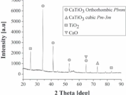
The grazing incident XRD pattern of CaTiO3 film is
shown in Figure 2. This indicates that the coating exhi -bits polycrystalline structure with a (022) preferential orientation corresponding to Pbnm orthorhombic Ca
-TiO3 phase at 33.293°. The shift toward high angles
(0.69 grades) at positions 33,293° and 40,673° of JCPDF 01-088-0790 card is in relation with compressive resi
-dual stress characteristics. The positions 52.827°, 52.931°, 64.747° and 64.866° are in agreement with JCPDF 01-082-0231 card which also corresponds to an
orthorhombic Pbnm calcium titanate. The peak located at 72.779° is in agreement with JCPDF 00-043-0226 card,
corresponding to a cubic Pm-3m calcium titanate, which
is a phase that normally occur above 1300°C ( Rous-hown and Masatomo, 2005), and it is known that
me-tastable phases can be formed in as-deposited thin films by physical vapor deposition whereas they are hardly
seen in bulk counterparts (Krzanowski, 2004). Finally,
anatase phase with (101) and (224) reflections of TiO2
was observed, in agreement with JCPDF 01-083-2243 card; and was also found a coincidence with 01-074-1226 card, in 64.647° position, which correspond to (311) reflection of CaO; that is because the deposition
process is assumed to atomically disassemble the
com-pound AXY, directing A, X and Y atoms toward the
substrate, where they can be reassembled in multiple phases (Krzanowski, 2004), it means that a part of the
material reaching the substrate is suffering the next
change:
3 2
CaTiO →TiO CaO+
The crystallinity of the coatings indicates that hea-ting the substrate at 500°C is appropriate for obtai-ning calcium titanate crystalline coatings. This is better than that reported by Naofumi Ohtsu et al. (2007) who obtained crystalline films at 600°C.
Mechanical properties
Hardness
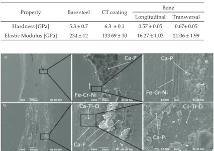
Table 3 shows materials hardness and elastic modulus
measured by nanoindentation. In all three cases the
averaged uniaxial Young’s moduli and hardness (in the axial and transverse directions for bone) values were computed from the experimental curves using the Oli
-ver-Pharr theory (Oliver and Pharr, 1992). The mecha
-nical differences in bone values (longitudinal and transversal directions) are due to structural anisotropy and are also linked to a variation in chemical composi
-tion (Rho et al., 1998).
Tribological properties
In part a) of Figure 3 it can be seen the wear mecha-nisms on bare steel and the spherical counterpart bone.
There are two wear mechanisms: abrasive wear and ad
-hesive wear. Second zoom shows plow lines indicating abrasive wear likely because of some steel particle came off due to fatigue and strain hardened generated abra
-sion to disk. The zoom shows bone adhe-sion to the disk; it is also observed that adhered material fractures
because of fatigue forming wear debris acting as a third
body. In part b) we observe wear mechanisms of coated steel. Abrasive wear is not observed and EDX probe re
-vealed no bare steel, so it follows that the coating main
-tained its integrity after testing. The only wear mecha-nism observed is adhesive and fatigue fracture of bon -ded bone forming wear particles. Figure 4 shows the wear mechanisms of bare steel and coated steel in Hank’s solution. It can be seen that the only wear me-chanisms both cases is bone adhesion, there is no
abra-sive wear in neither case, there is no detachment of the
coating, and there is salts precipitations.
Figure 5 parts a) and b) show bone pin acting as
counterpart of bare steel and coated steel, respectively,
Figure 3. Wear mechanism in a) the AISI 304 steel and, b) calcium titanate coated AISI 304 steel in pin on disc test using a bovine pin as a counterpart in
dry conditions
Figure 4. Wear mechanism in a) the AISI 304 steel and, b) calcium titanate coated AISI 304 steel in pin on disc test using a bovine pin as a counterpart in
Hank’s solution at 37 °C
Figure 5. Wear in pin acting like counterpart to a) bare steel in dry conditions, b) coated steel dry conditions, c) wear zone in pin, d) bare steel in Hank’s solution, e) coated steel in Hank’s solution f) debris from dry test Table 3. Hardness and elastic modulus of bare steel, calcium titanate coated steel and bone
Property Bare steel CT coating Bone
Longitudinal Transversal Hardness [GPa] 5.3 ± 0.7 6.3 ± 0.1 0.57 ± 0.05 0.67± 0.05 Elastic Modulus [GPa] 234 ± 12 133.69 ± 10 16.27 ± 1.03 21.06 ± 1.99
in dry test. Part c) presents how bone layers are formed due to fatigue, this layers subse-quently will be bonded to the steel samples. Parts d) and e) show bone pin acting as counterpart of bare steel and coated steel,
respectively, in Hank’s solution test. Part f)
shows the geometry of wear debris from
dry test, which exhibit brittle fracture and an average size of 8 μm larger ones.
Figure 6 shows friction coefficient in
function of sliding distance in pin-on-disk
test. In part a) stages 1 and 2 refer to aspe -rities deformation and contaminant
remo-val mentioned by Holmberg and Matthews, (2009) appear in small proportion in bare steel and are almost nonexistent in coated steel due to their low initial roughness and high cleanness. Stage 3 where the friction
coefficient increases due to the rapid in -crease of wear particles trapped between the sliding surfaces is larger in bare steel
than in coated steel. This could be because the coating removes more bone wear de -bris due to its higher hardness, as can be
seen in Table 3, which rapidly increases the friction coefficient. Relative to stage 4, one
of the bare steel samples does show a short
interval with a friction coefficient of about 1.7 before reaching the steady state, which may be related to greater wear bone, 1.59 × 10²
mm3, compared with the wear in the bone
provided because of the other two sam
-ples, 0.79 × 102 mm3 and 0.76 × 102 mm3.
The reason for this differences in bone
wear is the bone anisotropy: its hardness
in the longitudinal direction is different in the transversal direction (Rho 1998); as this
orientation could not be controlled in the
pins preparation of the current investiga
-tion, these may exhibit different hardness
according to the area that is in contact with the disk, in this case the hardness of the pin which acted as counter-part of the steel
sample in question is 50.83 ± 3,59 HV, com -pared with the hardness of the other two
samples, 54.23 ± 2.66 HV and 69.04 ± 1.94 HV, respectively. It can also make a correlation
between bone pin wear and dynamic
fric-tion coefficient of coated steel: high to low friction coefficient pin wear were 1.04 × 102 mm3, 2.53 × 102 mm3 and 2.85 × 10²
Figure 6. Friction coefficient for bare steel and calcium titanate coated steel in pin-on-disk test in a) dry conditions and b) simulated body conditions
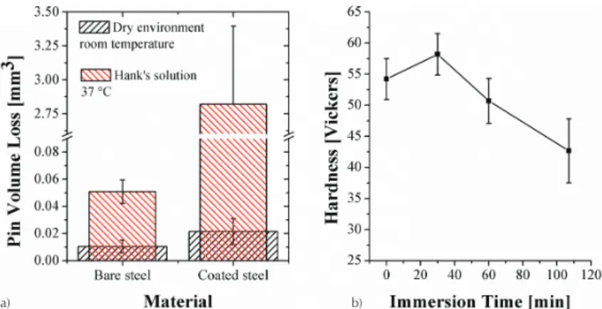
Figure 7. a) Volume loss of bone ball counterpart in pin on disk test in dry and SBF conditions, b) bone hardness in function of immersion time in Hank’s solution
a) b)
a) b)
59.77 ± 2.13 HV, 54.21 ± 3.28 HV and 43.74 ± 1.97 HV. Namely in dry conditions, volume loss of bone decreases with increasing bone hardness. At steady state it can be seen that friction coeffi
-cient of coated steel (0.84 ± 13) is on average greater than the coefficient of friction of bare steel (0.77 ± 0.11), which is related, as shown in Figure 7, with bone volume loss, 0.021 mm3 ±
0.009 mm3 for coated steel and 0.010 mm3 ± 0,004 mm3 for bare
steel. It means a higher friction coefficient goes together with a higher volume loss.
Under conditions which simulate the human environment (Hank’s solution at 37°C), on the contrary, part b) of Figure 6 shows that at steady state friction coefficient of bare steel (0.89 ± 0.12) is on average greater than the friction coefficient of coated steel (0.65 ± 0.12) related with bone volume loss of 0.051 mm3 ± 0,009
mm3 and 2.82 mm3 ± 0.58 mm3, respectively, as can be seeing in
Figure 7. It means, in Hank’s solution, a higher friction coefficient goes together with a lower volume loss.
Part a) of Figure 7 shows the loss in volume
of the bone spherical counterpart in both
test conditions. It can be observed that in
dry conditions and in Hank’s solution at
37°C, coated steel samples cause more wear
in bone pin because of its greater hardness compared to bare steel. Analyzing each
ma-terial is observed that wear was more severe
when test was performed in Hank’s solution
at 37°C than when it was in dry conditions. This can be attributed to a decrease in me -chanical properties of the bone immerse in a
liquid environment which simulates blood plasma, as evidenced in part b) of Figure 7,
which shows that bone loses hardness if it is
immersed in the fluid, and lower hardness
leads to increased wear.
Conclusions
• As depositing calcium titanate by magne
-tron sputtering onto AISI 304 stainless steel using a CaTiO3 as cathode obtained
by powder technology, a coating was ob-tained consisting of Pbnm orthorrombic calcium titanate, Pm–3m cubic calcium tita-nate, titanium oxide (anatase) and calcium oxide. The thickness of the coating was about 750 nm, with a hardness of 6.3 ± 0.1 and an elastic modulus of 133.69 ± 10. • When in contact, polished steel AISI 304
with spherical bovine bone in pin-on-disc tested with a load of 3 N, the wear mecha
-nisms that occur are pin volume loss,
bone adhesion to the steel and steel
abra-sion. Under the same conditions but with calcium titanate coated steel as a flat
counter-part, wear bone also occurs and bone adhesion to the disc without
abra-sion or detachment of the coating. The friction coefficient in each case was 0.77 ± 0.11 and 0.84 ± 0.13.
• In Hank’s solution, when in contact with
both AISI 304 and calcium titanate
coa-ted, AISI 304 with spherical bovine bone in pin-on-disc tested with a load of 3 N,
the wear mechanisms that occur are pin
volume loss and bone adhesion to the disks. Friction coefficients were 0.89 ± 0.12 and 0.65 ± 0.12.
-logical medium, the bone damage is more severe if steel is coated with calcium titanate. Because of that
it is important to measure the adherence of the
coa-ting to the bone, in order to analyze its viability as a
coating in hip stems.
References
Asami K., Ohtsu N., Saito K., Hanawa T. CaTiO3 films sputter-deposited under simultaneous ion implantation on
Ti-substrate. Surface & Coatings Technology, volume 200, 2005: 1005-1008.
ASTM E2546–07: Standard Practice for Instrumented Indentation
Testing, West Conshohocken, PA, American Society for Tes
-ting and Materials, 2007.
ASTM G99–05 (Reapproved 2010): Standard Test Method for
Wear Testing with a Pin-on-Disk Apparatus, West Consho
-hocken, PA, American Society for Testing and Materials, 2010.
ASTM F138 – 08: Standard Specification for Wrought 18Chro
-mium-14Nickel-2.5Molybdenum Stainless Steel Bar and Wire for Surgical Implants (UNS S31673), West Conshohocken, PA, American Society for Testing and Materials, 2008.
ASTM F139 – 08: Standard Specification for Wrought 18Chro
-mium-14Nickel-2.5Molybdenum Stainless Steel Sheet and
Strip for Surgical Implants (UNS S31673), West Conshohoc
-ken, PA, American Society for Testing and Materials, 2008. Barragán F., Guardián R., Menchaca C., Rosales I., Uruchurtu J.
Electrochemical Corrosion of Hot Pressing Titanium Coated
Steels for Biomaterial Applications. International Journal of
Electrochemical Science, volume 5, 2010: 1799-1809.
Coathup M. J., Blunn G. W., Flynn N., Williams C., Thomas N.P. A
comparison of bone remodeling around hydroxyapatite-coa-ted, porous-coated and grit-blasted hip replacements
retrie-ved at post-mortem. The Journal of Bone and Joint Surgery [Br],
volume 82-B, 2000: 118-123.
Ergun C., Doremus R., Lanford W.J. Hydroxyapatite and tita
-nium–interfacial reactions. Journal of Biomedicals Materials Re-search, Part A, volume 65, 2003: 336.
Faig-Martí J. and Gil-Mur F.J. Los recubrimientos de
hidroxiapati-ta en las prótesis articulares. Revista española de cirugía
ortopé-dica y traumatología, volume 52, 2008: 113-120.
Fu Y., Batchelor A.W., Wang Y., Khor K.A. Fretting wear beha
-viors of thermal sprayed hydroxyapatite (HA) coating under
unlubricated conditions. Wear, volume 217, 1998: 32-139.
Geetha M., Singh A.K., Asokamani R., Gogia A.K. Ti based bioma
-terials, the ultimate choice for orthopaedic implants –A
re-view. Progress in Materials Science, volume 54, 2009: 397-425.
Holliday S. and Andrei S. Crystallization of CaTiO3 by sol–gel
synthesis and rapid thermal processing. Surface & Coatings Te -chnology, volume 188 and 189, 2004: 741-744.
Holmberg K. and Matthews A. Coatings tribology: Properties,
me-chanisms, techniques and applications in surface engineering, volu
-me 56, 2nd ed., United Kingdom, Tribology and Interface Engineering Series, 2009, 48p.
Howell J.R., Blunt L.A., Ling R.S.M. An analysis of fretting dama
-ge seen on explanted femoral stems. The journal of bone and joint surgical, volume 81B (Suppl. 3), 1999: 163.
Kokubo T., Kim H.M., Kawashita M. Novel bioactive materials with different mechanical properties. Biomaterials, 24: 2161, 2003.
Krzanowski J.E. Phase formation and phase separation in
mul-tiphase thin film hard coatings. Surface & Coatings Technology,
volume 188 and 189, 2004: 376-383.
Lundin M., Hedberg Y., Jiang T., Herting G., Wang X., Thormann
E., Blomberg E., Odnevall I.W. Adsorption and protein-indu
-ced metal release from chromium metal and stainless Steel.
Journal of Colloid and Interface Science, volume 366, 2012: 155-164.
Moreira M.L., Paris E.C., Do Nascimento G.S., Longo V. M., Sam
-brano J.R., Mastelaro-Valmor R.,. Bernardi Maria I.B, Varela
Juan Andrés José A., Longo Elson. Structural and optical
pro-perties of CaTiO3 perovskite-based materials obtained by
microwave-assisted hydrothermal synthesis: An experimen
-tal and theoretical insight. Acta Materialia, volume 57, August, 2009: 5174-5185.
Nie F.L., Wang S.G., Wang Y.B., Wei S.C., Zheng Y.F. Comparative study on corrosion resistance and in vitro biocompatibility of
bulk nanocrystalline and microcrystalline biomedical 304 stainless Steel. Dental materials, volume 27, 2011: 677-683. Ohtsu N., Kesami S., Katsuhiko A., Takao H. Characterization of
CaTiO3 thin film prepared by ion-beam assisted deposition. Surface & Coatings Technology, volume 200, 2006: 5455-5461.
Ohtsu N., Akihiko I., Kesami S., Takao H. Characterization of cal
-cium titanate thin films deposited on titanium with reactive
sputtering and pulsed laser depositions. Surface & Coatings
Technology, volume 201, 2007: 7686-7691.
Ohtsu N., Chikage A., Tetsuya A., Satoshi S., Kazuaki W. Calcium-hydroxide slurry processing for bioactive calcium-titanate
coating on titanium. Surface & Coatings Technology, volume 202, 2008: 5110-5115.
Oliver W.C., Pharr G.M. An improved technique for determining
hardness and elastic modulus using load and displacement sensing indentation experiment. Journal of Materials Research,
volume 7, 1992: 1564-1583.
Park-JinW., Yusuke T., Chong-Soo L., Chan-Hee P., Youn-Jeong K., Je-Hee J., Dongwoo K., Yeon-Min I., Hisashi D., Naoyuki
N., Takao H. Surface structures and osteoblast response of hy
-drothermally produced CaTiO3 thin film on Ti–13Nb–13Zr
alloy. Applied Surface Science, volume 257, 2011: 7856-7863. Porter A. E., Punam T., Hobbs-Linn W., Coathup M.J., Gordon
W.B., Spector M. Bone bonding to hydroxyapatite and tita
-nium surfaces on femoral stems retrieved from human sub
-jects at autopsy. Biomaterials, volume 25, 2004: 5199-5208. Rho Y.J., Kuhn-Spearing L., Zioupos P. Mechanical properties and
the hierarchical structure of bone. Medical Engineering & Phy
Rokkum M., Reigstad A. Total hip replacement with an entirely hydroxyapatite-coated prosthesis: 5 years’ follow-up of 94
consecutive hips. The Journal of Arthroplasty, volume 14 (issue
6), September, 1999: 689-700.
Roushown A. and Masatomo Y. Space group and crystal structure
of the Perovskite CaTiO3 from 296 to 1720K. Journal of Solid
State Chemistry, volume 178, 2005: 2867-2872.
Tang Y.C., Katsuma S., Fujimoto S., Hiromoto S. Electrochemical stu
-dy of Type 304 and 316L stainless steels in simulated bo-dy fluids
and cell cultures. Acta Biomaterialia, volume 2, 2006: 709-715. Wang W., Mingrong S., Li Y., Liang F., Fengang Z., Xinglong W.
Enhanced photoluminescence of CaTiO3:Pr3+ phosphor films
deposited on SiO2 buffered Si substrates. Thin Solid Films, vo
-lume 517, 2009: 3398-3401.
Wiff J.P., Fuenzalida V.M., Arias J.L., Fernandez M.S.. Hydrother
-mal–electrochemical CaTiO3 coatings as precursor of a biomi
-metic calcium phosphate layer. Materials Letters, volume 61, 2007: 2739-2743.
Yang H.K., Jong-Won C., G.-Seeta R.R., Kee M.,
Byung-Chun C., Jung-Hyun J., Jung-Hwan K. Luminescent characte
-ristics of CaTiO3:Pr3+ thin films prepared by pulsed laser
Citation for this article:
Chicago style citation
Esguerra-Arce, Johanna, Yesid Aguilar-Castro, William Aperador-Chaparro, Leonid Ipaz-Cuastumal, Gilberto Bolaños-Pantoja, Car-los Alberto Rincón-López. Tribological Behavior of Bone Against Calcium Titanate Coating in Simulated Body Fluid. Ingeniería
In-vestigación y Tecnología, XVI, 02 (2015): 279-286.
ISO 690 citation style:
Esguerra-Arce J., Aguilar-Castro Y., Aperador-Chaparro W., Ipaz-Cuastumal L., Bolaños-Pantoja G.B., Rincón-López C.A. Tribologi-cal Behavior of Bone Against Calcium Titanate Coating in Simulated Body Fluid. Ingeniería Investigación y Tecnología, volu-me XVI (issue 2), April-June 2015: 279-286.
About the authors
Johanna Esguerra-Arce. She obtained his bachelor’s degree in Materials Engineering at The Universidad del Valle, Colombia, and is a Ph.D. candidate in Engineering Doctorate with major in Materials Engineering. Her work has been presented in various national and international forums and is aimed at biomaterials and coatings obtained by physical vapor deposition methods. She has been occasional professor in Department of Material Engineering at The Universidad del Valle.
Yesid Aguilar-Castro. He holds a master’s in Metallurgy and Materials Science and a Ph. D in Materials and its New
Technologies from Universidad Politécnica de Valencia. He has more than twelve years of professional expe
-rience as a professor at the Universidad del Valle and is appointed as a full time professor at the school of Materials Engineering. He has dedicated his work to tribology and supervises thesis work at the bachelor, master and doctoral levels.
William Aperador-Chaparro. Research Professor of Universidad Militar Nueva Granada, PhD in Materials Enginee
-ring from the Universidad del Valle. He obtained the degree of Master of Metallurgy and Materials Science from Universidad Pedagógica y Tecnológica de Colombia (UPTC-Tunja), he also has a degree in physicist from the same university. He is a researcher of the Volta group in Mechatronics Engineering program.
Leonid Ipaz-Cuastumal. Professor at Universidad Autónoma de Occidente. PhD in Materials Engineering from the Universidad del Valle, also has a degree in Materials Engineer from the same university. His main research interests are synthesis and characterization of hard coatings. He is a researcher of the Thin Films Group in the Physics Department of The Universidad del Valle.
Gilberto Bolanos-Pantoja. He obtained his bachelor’s degree in physics at the Universidad del Valle, Master and Doctoral studies of science (physics) at the same university. He is a research professor at The Universidad del
Cauca. The research is aimed at manufacturing of multilayer hard coatings and phase transitions. He has di
-rected 14 B.Ing. theses, and co-di-rected 2 doctoral theses.
Carlos Alberto Rincón-López. His doctoral degree is from The Universidad del Valle (science-physics) and holds a master in physics from Universidad Simón Bolívar. At present he is an associate professor at The Universidad
del Cauca. His research work has centered on plasma physics and coatings obtained by physical vapor depo
-sition. He has directed various B.Ing. theses and has participate as a referee in 10 master and doctoral theses.
deposition method with various substrates. Applied Surface
Science: volume 255, 2009: 5062-5066.
Zhanga F., Maa A., Jianga J., Xua H., Songa D., Lua F., Nishida Y. Enhanced biodegradation behavior of ultrafine-grained ZE41A
magnesium alloy in Hank’s solution. Progress in Natural Scien
![Table 1. Chemical composition of Hank’s solution, concentration [g/L]](https://thumb-us.123doks.com/thumbv2/123dok_es/6601261.237866/3.918.276.764.691.731/table-chemical-composition-of-hank-solution-concentration-l.webp)